Université de Montréal
Conception et Caractérisation de Nanoparticules Polymères
Théragnostiques Destinées au Traitement des Tumeurs Cérébrales
par Hoda Besheir
Faculté de Pharmacie
Thèse présentée à la Faculté des études supérieures en vue de l’obtention du grade de Maîtrise (M.Sc.) en Sciences Pharmaceutiques option Technologie Pharmaceutique
Août, 2016
Résumé
L‘intérêt de développer de nouvelles applications de la nanotechnologie pharmaceutique dans les soins de santé augmente année après année. Le rôle des nanosystèmes est devenu évident, surtout après que certains d'entre eux aient contribué à des solutions révolutionnaires dans des maladies graves. Dans notre projet, nous avons cherché à synthétiser des nanoparticules (NPs) intelligentes capables de livrer, de façon sélective, des agents anticancéreux. Les NPs ont été synthétisées afin de cibler des cellules cérébrales atteintes par le glioblastome multiforme (GBM), un cancer du cerveau présentant un mauvais pronostic et un taux de survie médian très faible. À cet effet, nous avons planifié de synthétiser et analyser ces nanoparticules et également d’étudier les preuves de concept de leur efficacité.
Tout d'abord, nous avons sélectionné avec soin, la procédure de formulation ainsi que les polymères avant d’optimiser les caractéristiques physico-chimiques des nanogels (NGs) formulés à base de chitosane.
Après l'optimisation de la taille, du PdI et du potentiel de surface des NGs, nous avons synthétisés des NGs chargés en substance active. Deux molécules thérapeutiques ont été retenues La première était la doxorubicine HCl (DOX) qui est trop hydrophile pour passer la BHE, bien qu’ayant démontré une efficacité in vitro contre le GBM. Le deuxième médicament était le témozolomide (TMZ) qui est déjà utilisé dans le traitement de GBM, comme adjuvant à la radiothérapie, après l’intervention chirurgicale. Les méthodes d'analyses ont ensuite été développées pour déterminer l’efficacité du taux d'encapsulation (EE%) et l'efficacité de chargement de médicament (DLE%) des deux médicaments.
Pour la préparation des nanogels théranostiques, nous avons suivi les mêmes procédures après l’addition de l’agent de contraste USPIO, durant la synthèse des NGs.
Ensuite, nous avions besoin d’assurer la qualité de nos NGs lors du stockage à long terme. Pour atteindre cet objectif, nous avons développé une procédure de lyophilisation en utilisant différents sucres de nature et de concentrations variables. Les sucres ont été ajoutés pour cryoprotéger les NGs contre les contraintes mécaniques et physiques mises en jeu lors de la lyophilisation. Les sucres qui ont démontré des résultats satisfaisants avec NGs vides ont été utilisés, par la suite, dans la cryoprotection des NGs chargées de médicament au cours de leur lyophilisation.
Finalement, nous avons étudié la libération de la DOX à partir des NGs chargées avant et après lyophilisation. Cette étude, en particulier avait deux objectifs. Le premier était de comparer l'effet de la lyophilisation sur le comportement de la libération de DOX des NGs, en observant l’impact de cette procédure sur la cinétique de libération Le deuxième but de l'étude de relargage était de tester la capacité des NGs à libérer leur contenu à trois pH différents : 5.8 (pH intracellulaire des cellules tumorales), 6.8 (espace interstitiel de la tumeur) et 7.4 (plasma sanguin).
Mots clés: GBM, doxorubicine, nanoparticules, nanogels, chitosane, lyophilisation, cryoprotecteurs.
Abstract
The interest in developing new applications of nanotechnology in the pharmaceutical health care increases year after year. The role of nanosystems became clearer, especially after some of them contributed to revolutionary solutions in serious diseases. In our project, we sought to synthesize ‘intelligent’ nanoparticles (NPs) capable of selectively delivering anticancer agents. NPs were synthesized to target brain cells affected by glioblastoma multiforme (GBM), a brain cancer with a poor prognosis and a very low rate of median survival. To this end, we planned to synthesize and analyze these nanoparticles and also to study the proof-of-concept of their efficiency.
First, we carefully selected the formulation process and the polymers prior to optimize physicochemical characteristics of the nanogels (NGs) formulated with chitosan.
After optimization of the NGs size, PDI and surface potential, we synthesized NGs loaded with active substances. Two therapeutic molecules were selected. First we chose doxorubicin HCl (DOX) which is too hydrophilic to cross the BBB, whereas demonstrating in vitro efficacy against GBM. The second drug was temozolomide (TMZ), already used in the treatment of GBM as an adjuvant to radiotherapy after surgery. Analytical methods were then developed to determine the encapsulation efficiency (EE %) and drug loading efficiency (DLE %) of both drugs.
For the preparation of theranostic nanogels, we followed the same procedures after the addition of the USPIO contrast agent during the NGs synthesis.
Next, we needed to ensure the quality of our NGs during long-term storage. To achieve this goal, we developed a freeze-drying process using different kind of sugar cryoprotectants at varying concentrations, in order to protect NGs against mechanical and physical stresses at play during freeze-drying. Most promising sugars used with unloaded NGs were subsequently used to cryoprotect DOX-loaded NGs.
Finally, we studied the release of DOX from DOX-loaded NGs before and after freeze-drying. This study in particular had two objectives. The first was to compare the effect of the freeze-drying process on the behavior of the DOX-loaded NGs, observing the impact of this procedure on the release kinetics. The second purpose of the release study was to test the ability of NGs to release their contents at three different pH: 5.8 (intracellular pH of tumor cells), 6.8 (interstitial space of the tumor) and 7.4 (blood plasma).
Contents
Résumé ... ii
Abstract ... iii
List of tables ... vii
List of figures ... viii
List of abbreviations ... ix
Contributions of coauthors ... xiii
Acknowledgements ... xiv
Chapter one ... 1
Introduction ... 1
1. Glioblastoma (GBM) ... 2
1.1 Background ... 2
1.2 Primary versus Secondary glioblastomas ... 3
Primary GBM: ... 3
Secondary GBMs:... 3
1.3 Understanding the poor prognosis of GBM from a pathological point of view ... 3
1.4 Epidemiology ... 5
1.5 Symptoms ... 6
1.6 Diagnosis... 6
1.6.1 Pathological reports produced to initially differentiate 1ry from 2ry GBMs, Immunohistochemistry (IHC) tests ... 6
1.6.2 Diagnosis using imaging techniques used to localize the tumor in the brain ... 7
2. Current treatments: ...10
2.1 Currently available treatment strategies for GBMs ...10
2.1.1 Surgical intervention ...11
2.1.2 Post-operative treatment ...11
2.1.3 In vivo and in vitro-effective drug entities in treating GBM...12
2.2 Multidrug resistance (MDR) ...17
2.3 Understanding the blood-brain barrier (BBB) ...18
3. Novel treatment strategies ...20
3.1 Pharmaceutical nanotechnology ...20
3.1.3 Promising role of pharmaceutical nanotechnology in brain cancer treatment ...28
3.1.4 Examples of overcoming formulation challenges by pharmaceutical nanotechnology ...28
3.1.5 Theranostic nanoparticles ...31
3.2 Hypothesis and specific objectives ...32
4. References ...35
5. Techniques used in physicochemical characterization of NPs: ...42
5.1.1 Dynamic light scattering, DLS: ...42
5.1.2 Electrophoretic light scattering, ELS: ...43
6. Techniques used in investigating NPs stability: ...45
6.1.1 Differential scanning calorimetry, DSC: ...45
6.1.2 FourierTransform Infrared Spectroscopy, FTIR: ...47
6.1.3 Freeze-drying, FD: ...49
Chapter two ...51
Introduction to the reported results ...51
Part one ...54
Article ...54
ƒ Materials and Methods ...57
1.1. Materials ...57
1.2. Methods ...57
1.3. Synthesis of blank and DOX-loaded NGs ...57
1.4. NG purification ...57
1.5. NG freeze-drying process ...57
1.6. NG physicochemical characterization ...58
1.7. Doxorubicin quantification ...59
1.8. Doxorubicin release study before and after freeze-drying ...59
1.9. Degradation kinetics of DOX ...60
ƒ Results ...60
1.10. NG physicochemical characterization...60
1.11. Thermal Analysis (Differential Scanning Calorimetry, DSC) ...61
1.12. Fourier Transform Infrared (FT-IR) analysis...62
1.13. Freeze-drying (FD) process ...63
1.14. Degradation kinetics of DOX ...65
ƒ Discussion ...66 ƒ Conclusion ...69 Supporting information ...73 Part two ...76 Materials ...76 Method ...76 Results& discussion ...78 Part three ...82 General discussion ...82 References ...87 Conclusion ...96 Prespectives ...99
List of tables
Table 1. Different grades of glioma according to classification by world health organization (WHO)………..……...…...…...2 Table 2. Median age at diagnosis and age-adjusted average annual (1998–2002) incidence rates of adult gliomas ………...………..………..5 Table 3. Positive results are shown in percentage after applying each of the six immunostains to biopsies from both types of GBMs……….………...7 Table 4. Results observed from different clinical studies aimed to investigate efficacy and safety of TMZ ………...…………..…………..…..……....14 Table 5: Results obtained for DLS/ELS characterization for TMZ-loaded nanogels using HMW-CH, USPIO-loaded nanogels, USPIO/Dox- and USPIO/TMZ-loaded nanogels,….…..……...80
List of figures
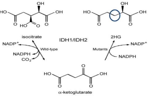
Figure 1. Comparing different pathways due to normal and mutant IDH1……….…………..… .4
Figure 2a. CT scan image showing the brain affected by GBM……….……...……...8
Figure 2b. Computed tomography: acquisition with one single slice (left) and multislice CT with four active acquisition channels………..………...……….……....8
Figure 3. Illustrations showing an overview on magnetic resonance imaging………..…………..9
Figure 4. MRI images before and after (left) of GBM-affected brain……….……..……...…. 10
Figure 5. Chemical structures of different chemotherapeutics………...………....….. 13
Figure 6. An illustration of the BBB structure showing its different components……...…... 19
Figure 7. A simple diagram illustrating the different stages of drug-loaded nanoparticle development to improve their target specificity ………...….……. 24
Figure 8. The EPR effect in the tumor tissue ...……….………...….…….25
Figure 9. pH-sensitive chitosan conjugated nanocarrier interactions ……..….…..……...…… 27
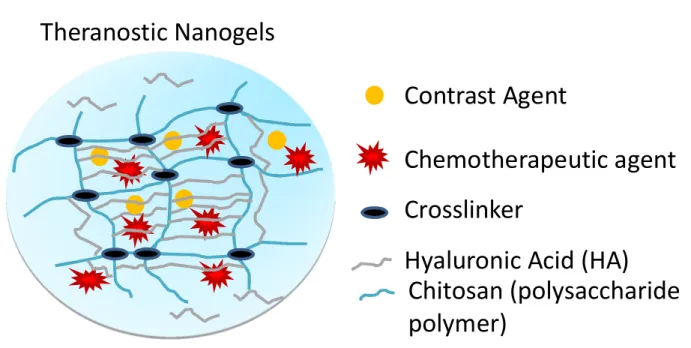
Figure 10. A hypothetical structure of the final drug-loaded chitosan/hyaluronic acid-based theranostic nanogels………...……….….……….…....…32
Figure 11. Illustrations for DLS technique.……….………...….……….….42
Figure 12. A typical DLS graph showing a single population of NPs……….43
Figure 13.Cuvette for Zeta potential (ZP) measurements containing two opposite electrodes…44 Figure 14. Zeta Potential of chitosan-based NPs measured by the ELS technique .……….45
Figure 15. The heat excess and DSC measurement technique for sample and reference ...….46
Figure 16. Thermogram of chitosan powder………47
Figure 17. A scheme showing FTIR Michelson technique ………...….48
Figure 18. Chitosan IR spectrum ………..……….. 49
List of abbreviations
2-HG 2-hydroxy glutarate ABC ATP-binding cassette ALPs alkyl-lysophospholipids ATP Adenosine tri-phosphate BBB Blood brain barrier
BCNU Carmustine
BCRP Breast cancer resistant proteins
BEV Bevacizumab
CAM Cell adhesion molecule
CBTRUS Central brain tumor registry of the United States
CCNU Lumostine
CH Chitosan
CIMP CpG island methylator phenotype
CK Cytokeratine
CpG DNA sequence where a cytosine base is followed by a guanine one
CT Computed tomography
Da Dalton
DC Dendritic cells
DDS Drug delivery system
DLE% Percentage of drug loading efficiency DLS Dynamic light scattering
DNA Deoxyribonucleic acid
DOX Doxorubicin
DSC Differential scanning calorimetry EE% Percentage of encapsulation efficiency EGFR Epidermal growth factor receptors ELS Electric light scattering
EPR Enhanced permeation retention
ET Edelfosine
FD Freeze-drying
FDA US Food and Drug Administration FTIR Fourier Transform Infrared
GBM Glioblastoma multiforme
Gd3+ Gadolinium
GFAP Glial fibrillary acidic protein GFP Green fluorescent protein GST Glutathione-S-transferase
HA Hyaluronic acid
IDH Isocitrate dehydrogenase
IgG Immunoglobulin G
IHC Immunohistochemistry
LC/MS-MS Liquid Chromatography Mass Spectrometry and Liquid Chromatography -Tandem Mass Spectrometry
mAbs Monoclonal antibodies MDM Murine double minute MDR Multi drug resistance
MGMT Methyl guanine methyl transferase
mo Month (unit used to measure mPFS & mOS) mOS Median overall survival
mPFS Median progression-free survival MPS Mononuclear phagocytes system MR Magnetic resonance
MRI Magnetic resonance imaging
MRPs Multi-drug resistance associated proteins mTOR Mechanistic target of rapamycin
mV Milli volt
MWCO Molecular weight cut-off
NG Nanogel
nm Nanometer
nM Nanomolar
NOS Not otherwise specified
NP Nanoparticle
PCL Poly (epsilon-caprolactone) PdI Polydispersity index
PdIf Final polydispersity index (i.e. after FD) PdIi Initial polydispersity index (i.e. prior to FD) PEG Polyethylene glycol
PEO-PCL Poly(ethylene oxide)-modified poly(epsilon-caprolactone) p-gp P-glycoprotein
PLGA poly(lactic-co-glycolic acid) PNGs Plasmonic nanogels
PY% Percentage of production yield
ref Reference
RF Radiofrequency
Sf Final size (i.e. after FD) Si Initial size (i.e. prior to FD) t1/2 Half-life time
TAAs Tumor associated antigens Tg’ Glass transition temperature
TMZ Temozolomide
TMZ-NP Temozolomide-loaded nanoparticles TPP Tripolyphosphate
TSAs Tumor specific antigens TTFs Tumor treating fields
UK United Kingdom
US/USA United States of America
USPIO Ultra-small super paramagnetic iron oxide
WHO World Health Organization
Contributions of coauthors
This thesis is based on two chapters. Part one of chapter two is an article containing the major part of our project. The contribution of each author to the article is described as below:
Chapter two (part one): Recoverability of freeze-dried doxorubicin-loaded, chitosan-based nanogels: a physicochemical study
Hoda Besheir1, Hugo Alarie1, Martin Jutras2, V. Gaëlle Roullin1, 2*
The NG synthesis, physicochemical characterization as well as their freeze-drying is a team work of Hoda Besheir and Hugo Alarie. The DSC, FT-IR experiments and release study were accomplished by Hoda Besheir. LC-MS/MS method development in the DOX quantification to obtain encapsulation efficiency as well as determining remaining DOX concentrations in the release study was done by Martin Jutras. The work follow up and review was done by the director of research prof. V. Gaëlle Roullin.
Acknowledgements
First of all I would like to thank my supervisor, Prof. V. Gaëlle Roullin, for the patient guidance, encouragement and advice she has provided throughout my time as her student.
I would also like to thank the members of my advisory committee, Prof. Jeanne Leblond-Chain and Prof. Grégoire Leclair, for their generosity and for taking the time to evaluate my work. Special thanks to my coworkers and coauthors:
- Martin Jutras, B.Sc., HPLC-MS/MS specialist, platform of biopharmacy, for his great help in developing methods to analyze our samples.
- Hugo Alarie, 3rd year biopharmacy student, for the contributions he made to my project particularly his work in the freeze-drying process and release kinetics experiments
- Dounia Boumahni, B.Sc. in biopharmacy, for helping us in the NGs synthesis and their freeze-drying.
Great thanks to my colleagues in the laboratory of pharmaceutical nanotechnology, Valéry Aoun (Post-Doc.), Soudeh Tehrani (Ph.D. candidate) and Florian Bernard (Ph.D. candidate) as well as all members of department of formulation and analysis for their guidance and for answering my inquiries whenever I asked for their help.
Thanks to Mary Salib, student in D.E.S.S drug development, B.Sc. biopharmaceutical sciences for editing the French summary.
Chapter one
1. Glioblastoma (GBM)
1.1 Background
Glioma is a general term used to describe brain tumors which affect glial cells, the glue-like supportive tissue of the brain.(1) A subtype of gliomas is the astrocytic tumors or so-called astrocytomas. Astrocytomas are named after the astrocytes, the star-shaped cells they grow from.(2) According to their degree of aggressiveness, astrocytomas are classified, following an ascending order into 4 grades from I to IV, grade I being the least aggressive while grade IV is the most aggressive one.(3) This classification also reflects the degree of growth and tendency of the tumor to recur.(3) For example, grades I and II are low-grade astrocytomas that grow relatively slowly and are considered benign tumors manageable by surgical excision. Both grades III and IV are malignant types and require more aggressive treatment strategies.(3, 4) Grade IV glioma, also called glioblastoma (GBM), is a fast-growing tumor with very low prognosis, hence its high degree of malignancy and aggressiveness. It also has a very short median survival rate (14.9 months).(5) Main characteristics of each glioma type are outlined in table 1.
Table 1: Different grades of glioma according to classification by world health organization (WHO).Adapted from reference (3)
WHO grade Histologic Characteristics
Grade I Includes lesions with low proliferative potential and a frequently discrete nature; surgical resection is the main treatment.
Grade II Includes lesions that are generally infiltrating and low in mitotic activity but recur. Some tumor types tend to progress to higher grades of malignancy. Grade III Includes lesions with histologic evidence of malignancy, generally in the
form of mitotic activity, clearly expressed infiltrative capabilities, and anaplasia.
Grade IV Includes lesions that are mitotically active with vascular proliferation, necrosis-prone, and generally associated with a rapid preoperative and postoperative evolution of disease.
Unfortunately GBM is not only the most aggressive and malignant tumor among the four types of astrocytomas but also the most common one, affecting about 3 cases per 100,000 person-years.(6) Another cause that contributes to the aggressiveness of GBM is the infiltrating features it bears,(6) mainly because of the star shape of the affected astrocytes. Affecting such types of cells with high-grade, fast-growing tumors generates a characteristic invasive nature of the tumor; therefore the surgical procedure is not enough. Sometimes it is even challenging to achieve the complete tumor resection due to the tumor existence near parts of the brain responsible for essential body functions. 1.2 Primary versus Secondary glioblastomas
All grades II, III and IV astrocytomas are also known as diffuse gliomas. GBM (grade IV glioma) alone accounts for 45-50% of all primary intrinsic brain tumors.(7) Brief definitions for both are provided below to understand the main differences:
Primary GBM: Represents the vast majority of GBMs and is defined as the one which arises de novo in the brain without clinical or histological evidence of a lesser malignant precursor lesion.(8)
Secondary GBMs: are brain tumors which develop due to evolution of pre-existing lower-grade gliomas.(8)
1.3 Understanding the poor prognosis of GBM from a pathological point of view From a histological point of view, both primary and secondary GBM are very indistinguishable but fortunately they differ in their genetic and epigenetic profiles. In 1940, Scherer, a young German neuropathologist scientist was, for the first time, able to enlighten the differences between both types.(9, 10) According to him, it is very important to distinguish primary from secondary glioblastomas as the latter ones are the cause of most of glioblastomas with long clinical duration.(9) The main differences between primary and secondary astrocytomas lie in their biological behaviors. In fact the answer was found after the discovery of IDH gene mutations. IDH
gene, with two subtypes IDH1 & IDH2, encodes for the enzyme isocitrate dehydrogenase, which catalyzes the conversion of isocitrate to alpha-ketoglutarate (Figure 1).
Mutations of both subtypes occur in glioblastomas.(10, 11) However, IDH2 mutations occur less frequently than IDH1.(10) The wild type IDH, non-mutant type, catalyzes the conversion of isocitrate to alpha-ketoglutarate. IDH mutant types follow an alternative procedure and catalyze the conversion of alpha-ketoglutarate to 2-hydroxy glutarate (2-HG) (Figure 1). 2-HG is an oncometabolite which accumulates at high levels, inhibiting alpha-ketoglutarate-dependent enzymes. It inhibits histone demethylase leading to the synthesis of a hypermethylated DNA resulting in a tumor called G-CIMP or what is known as Glioma CpG Island.(12) Actually what makes IDH1 mutant a reliable tool of differentiation between primary and secondary GBM is its abundance in the later with a percentage of >80% vs.˂5% in primary GBM.(13-16)
Fig. 1. Comparing different pathways due to normal and mutant IDH1
The normal IDH1 enzyme converts isocitrate into alpha-ketoglutarate. The mutant IDH1 enzyme converts alpha-ketoglutarate into 2-hydroxyglutarate (2-HG). “Originally published by {Mellai M, Caldera V, Annovazzi L, Schiffer D. The Distribution and Significance of IDH Mutations in Gliomas 2013 2013-02-27}. Available from: Ref. (17)
1.4 Epidemiology
According to different reports, GBM has a higher incidence in Caucasian than in African or Asian populations.(18) Males are at higher risk of developing GBM than in case of females. Statistics in Canada showed in 2012 that the estimated distribution rates of new cases per 100,000 population tested for brain cancer was 1.6 in males and 1.4 in females.(19) Although these data are relatively low, especially when compared to the same statistics for prostate and breast cancers, 27.2 and 25.6 respectively,(19) the total estimated numbers to be diagnosed and die from primary brain tumors alone, in the same year, were 2,800 and 1,850 cases respectively which were still high numbers and should not be ignored.(20)
Incidence of grade IV glioma, i.e. GBM, is fourfold higher in countries like Canada and US than in regions with lower incidence such as India and Philippines.(21) According to a report issued in 2012 by the Alberta Health Services, results showed that GBM alone accounted for 40 percent of all malignant brain tumors. In USA, among all tumors, GBM is the most commonly reported one after meningioma, 15.4% and 36.1% respectively.
Table 2 gives an idea about GBM relative high incidence, age group, gender-difference occurrence as well as its general total rate of incidence comparing to other types of astrocytomas.
Table 2 Median age at diagnosis and age-adjusted average annual (1998–2002) incidence rates of adult gliomas stratified by sex. Reported in CBTRUS 2005–2006 Statistical Report: Primary Brain Tumors in the United States, 1998–2002.
These results are obtained from studies performed in the US Adult glioma by major
histologic subtype No of Cases
Median age at
diagnosis (years) Total rate
a Male ratea
Female ratea
Glioblastoma 12,943 64 3.05 3.86 2.39
aRates are per 100,000 population, age-adjusted to the 2000 US (19 age-groups) standard, and based on data from the
following registries: Arizona, Colorado, Connecticut, Delaware, Idaho, Maine, Massachusetts, Minnesota, Montana, New Mexico, New York, North Carolina, Rhode Island, Texas, Utah and Virginia. Abbreviation: NOS, not otherwise specified.
1.5 Symptoms
Although malignant gliomas, including GBM, are the most common primary brain tumors affecting adults, only few studies give information on the early symptoms. The silent development of GBM can partially explain why GBM is still difficult to notice at the beginning. For high-grade gliomas, symptoms like focal neurological deficits, epilepsy and cognitive dysfunction are prominent and can be observed at any stage of the disease.(22) Generally symptoms of glioma vary and they greatly depend on the patient’s age as well as the tumor histopathological class.(23) 1.6 Diagnosis
There are always several techniques and laboratory tests needed to be performed before providing certain information about the type of glioma affecting the brain. Some are histological laboratory tests which are performed mainly to give initial pathological reports. These tests are based on Immunohistochemistry (IHC) which uses small biopsies specifically extracted for diagnosis and which, despite being expensive and relatively time-consuming, are considered one of the most reliable tests used for accurate differentiation between the metastatic malignant carcinoma (secondary GBM) and primary GBMs. Imaging techniques of diagnosis such as computed tomography (CT-scan) and magnetic resonance imaging (MRI) are usually performed directly on the patient’s body to specifically localize the regions affected by the tumor in the brain.(24)
1.6.1 Pathological reports produced to initially differentiate 1ry from 2ry GBMs,
Immunohistochemistry (IHC) tests
This kind of laboratory tests is performed using a brain biopsy tested by a panel of immunostains before determining the accurate diagnosis depending on the obtained results.(25) According to a publication in 1999 by David OH et. al. (25) whose study was performed on biopsies from 45 patients, among who 23 had GBM. Table 3 shows the results of immunoreactivity obtained by different stains when applied to biopsies from both primary and secondary GBM.(25)
Table 3 Positive results are shown in percentage after applying each of the six immunostains to biopsies from both types of GBMs. Adapted from ref (25)
Immunostain % Observed immunoreactivity in 1ry GBM biopsies % Observed immunoreactivity in 2ry GBM biopsies
Fibrillary acid protein (GFAP) 100% 13.6%
Ber-EP4* 0% 50% Antikeratin monoclonal antibodies AE1/3 95.7% 100% Antibodies to CAM 5.2 4.3% 100% Cytokeratin 7 (CK7) 4.3% 77.3% Cytokeratin 20 (CK20) 4.3% 40.9%
*Ber-EP4 is a monoclonal antibody that reliably labels epithelial tissues.(26)
Table 3 results suggest that, to distinguish poorly-differentiated secondary from primary GBMs, a combination of immunostains including GFAP and cytokeratins CAM 5.2 should be the best method to use. Their diagnostic capability is mainly due to fully stain, by 100%, one type of GBM with almost negligible effect on the other type. However, because a significant number of GBMs stains are labeled with antikeratin monoclonal antibodies (AE1/3), such markers should not be used to differentiate between the two types of glioblastomas due to their lack of specificity.(25)
1.6.2 Diagnosis using imaging techniques used to localize the tumor in the brain
1.6.2.1 Computed Tomography (CT-scan)
CT-scan was introduced to the field of medical examination in the late of 1980s.(27) In this diagnostic imaging procedure, X-rays are used to give an image of the biological material (figure 2-a). Briefly, the principle depends on reconstructing cross-section images of the body after measuring how much an X-ray beam is attenuated as it crosses the tissue under examination. Data acquisition is preformed either by uni- or multi-slice detectors (figure 2-b).(27) Despite having advantages such as being non-invasive and quick, CT scan sensitivity in detecting cancer tissues was found to be significantly less than other imaging techniques such as MRI.(28) In addition to
this, CT scan bears the risk of developing cancer in the future due to being an increasing source of radiation.(29)
Fig. 2. (a) A CT scan image showing the brain affected by GBM.Case courtesy of A. Prof Frank Gaillard, "http://radiopaedia.org/">Radiopaedia.org. From the case "http://radiopaedia.org/cases/4756">rID: 4756 (b) Computed tomography: acquisition with one single slice (left) and multislice CT with four active acquisition channels. Reproduced from.(30) by G. Bongartz, S. J. Golding, A. G. Jurik, M. Leonardi, E. van Persijn van Meerten, R. Rodríguez, K. Schneider, A. Calzado, J. Geleijns, K. A. Jessen, W. Panzer, P. C. Shrimpton, G. Tosi European Guidelines for Multislice Computed Tomography Funded by the European Commission Contract number FIGM-CT2000-20078-CT-TIP March 2004
N. B. (we chose to explain the second technique, magnetic resonance imaging (MRI), in more details because the diagnostic features of our nanoparticle formula will mainly depend on this principle).
1.6.2.2 Magnetic Resonance Imaging (MRI)
MRI is a technique that takes advantage of the magnetic properties found in atomic nuclei. A typical example of these nuclei is the protons found in hydrogen atoms of water molecules (H2O) which are abundant in all tissues of the human body.(31, 32) Those protons act as tiny magnets because of the positive charge they carry in addition to their fast spinning (figure 3-a). Since these
rather cancel out. Nevertheless, in the presence of a strong constant external magnetic field (B0), the spinning nuclei of hydrogen atoms undergo precession, i.e. a change in the orientation of their rotation axis. Of course some of them still cancel out but most of them will tend to align with the applied magnetic field in direction Z and this results in a net magnetization (Figure 3-b).(31) The net magnetization is the source of magnetic resonance (MR) that is used to produce the images. To guarantee the efficient transfer of energy, protons are allowed to constantly oscillate by applying radio waves (B1 field) perpendicular to the previously applied magnetic field. The applied radiofrequency energy (RF pulse), which comes in the form of rapidly changing magnetic and electric fields generated by electrons, causes the protons to oscillate back and forth (Figure 3-c). The time these protons take to reach the equilibrium state, after RF is stopped, is interpreted in terms of signals T1& T2 signals.(31)
Fig. 3. Illustrations showing an overview on magnetic resonance imaging
(a) Random spinning of hydrogen protons: their net magnetization is zero because they are tiny magnets that do not sum but they rather cancel out. (b) Applying an external magnetic field (B0) to the randomly spinning protons will cause most of them to precess and change
the orientation of their axes. Protons will still spin about their axes but aligned to the direction of B0, direction Z. The system now shows net magnetization. (c) Radiofrequency
energy (B1) is applied perpendicular to the external magnetic field (B0) causing the protons
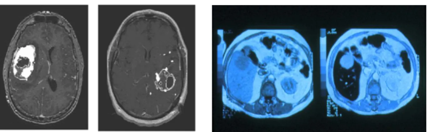
MRI technique is now accompanied by the use of contrast agents. Best examples of these are chelates using ions such as Gadolinium (Gd3+) and USPIO (Ultra Small super Paramagnetic Iron Oxide). The first ones shorten the T1 relaxation time and cause the part affected by the tumor to
appear brighter than its surroundings. Gd3+ chelates are therefore called positive contrast agent (figure 4-a). On the contrary, USPIO shorten the T2 relaxation time and produce an image in which
the tumor-affected part looks darker than the rest of the brain. Hence they are known as negative contrast agents (figure 4-b). In vivo distribution and pharmacokinetic properties of both agents vary widely depending on their chemical composition, molecular structure and overall size.(33)
Fig. 4. MRI images before (right) and after (left) injection of a contrast agent, in a GBM-affected brain. (a) MRI image using a Gd3+chelate as a contrast agent. Affected part is shown brighter
than its surroundings. (b) MRI image using USPIO as a contrast agent. Affected part is shown darker than its surroundings.(34)
(a) This image was originally published in JNM. Kristin R. Swanson, Gargi Chakraborty, Christina H. Wang, Russell Rockne, Hana L.P. Harpold, Mark Muzi, Tom C.H. Adamsen, Kenneth A. Krohn and Alexander M. Spence.Complementary but Distinct Roles for MRI and 18F-Fluoromisonidazole PET in the Assessment of Human Glioblastomas. J Nucl Med. 2009; 50:36-44. © by the Society of Nuclear Medicine and Molecular Imaging, Inc.
(b)This image was originally published in Neurospin Saclay website
2. Current treatments:
2.1 Currently available treatment strategies for GBMs
The treatment of patients with high-grade gliomas, either primary or secondary glioblastomas, has always represented a challenge to both neurosurgeons and drug researchers. Each line and option
of treatment has its own degree of success, restrictions and sometimes consequences. Each one of the currently available treatment options is discussed below in details.
2.1.1 Surgical intervention
Surgery is a first-line procedure to follow in the treatment strategy of GBM, whenever possible. However, there is still a controversy regarding the extent of the tumor that needs to be removed and whether it participates to an increase in the median survival rate of the patient or not.(35) The recommendations by many neurosurgeons to perform the tumor resection as extensively as possible, were further supported by studying the critical effect of the percentage of tumor removal on the increase in median survival rate.(35) Summarizing the study results, in which they compared a group of patients who underwent ˂ 98% removal of the diagnosed tumor to a second group who underwent ≥ 98% removal of their tumor, the results showed that the second group of patients demonstrated a significant increase in median survival rates, around 19 months compared to 10.9 months in people with lower percentage of tumor resection.(35)
Surgery as an only option for the high-grade glioma is never enough, since, as we previously mentioned the characteristic infiltrating feature of GBM and its potential presence next to essential brain parts restrict the 100% tumor elimination by surgical intervention.
2.1.2 Post-operative treatment
Although for many years surgery has been considered the initial treatment that ensured maximal safety and the highest degree of tumor elimination in GBM, clinical research was pursued to find postoperative treatment options that could help in increasing the median survival rate. For this reason therapists chose to resort to radiotherapy which increased the survival from 4-5 months to 9-12 months.(36) There were also results to support the role of using corticosteroids in controlling symptoms (mainly postoperative edema) although there was no information about their oncolytic
effects.(37) Starting from the early 1980s many scientists became more encouraged to study, discuss and publish the advantages of concomitant additive chemotherapeutics that could be used in combination with radiotherapy.(35) Nitrosoureas (Carmustine BCNU, Lumostine CCNU and others) remained included as first-line adjuvant therapy after surgery and they were combined with radiotherapy either alone or in a combination of Lumostine and procarbazine.(38) In 1999 published results proved the necessity to use Temozolomide (TMZ) as a first-line agent because of displaying better toxicity profiles than nitrosoureas.(39, 40) Since then, those were used as a second-line option.(38) The poor prognosis of GBM, jointly with the tumor recurrence inevitably after a median survival rate of 32 to 36 weeks,(41) motivated the researchers to look for more effective drug molecules. In the next part, we will discuss some of the promising chemotherapeutic agents which demonstrated efficacy against GBM either in vivo or in vitro.
2.1.3 In vivo and in vitro-effective drug entities in treating GBM
2.1.3.1 Nitrosoureas
They are also DNA alkylating agents with a high degree of lipophilicity, allowing them to cross the BBB and exert their cytotoxic effects on brain tumors.(38) Before 1999 nitosoureas (e.g. Carmustine (BCNU), Lumostine (CCNU) or Nimustine (figures 5-c, 5-d and 5-e respectively) were always included in first-line treatments of GBM.(39, 40)
That was before studies started to reveal the higher efficacy of TMZ especially for patients with recurrent GBM who had not received more than one course of nitrosourea treatment.(38) Another study also recommended TMZ over BCNU especially for patients with recurrent GBM due to better toxicity profiles of the former.(41) All this contributed to changing nitrosoureas from first-line treatments to second-first-line options. Acting by the same mechanism as BCNU, TMZ causes the emergence of the same resistance mechanisms during treatment with BCNU, leading to cross
resistance interaction between both agents.(41) However, they are still used as secondary-line treatment when TMZ has failed.(38)
Nitrosoureas monotherapy phase II and phase III clinical trials were performed to investigate the side effect profiles of drugs from this group.(42-44) Hematologic and long-lasting hepatic and pulmonary toxicity were found to be the predominant side effects when using BCNU or CCNU in recurrent GBM patients who had received TMZ before.(42-44) For nimustine the side effects reported were hematologic but no pulmonary fibrosis was observed.(45) The best findings were obtained when Fotemustine (figure 5-f), another nitrosourea agent which was administered continuously after TMZ administration as first line treatment. There were the characteristic grades 3 & 4 hematologic toxicities but the report rates were much less than with other agents.(46) Despite having cytotoxic effects on tumor cells, their toxicity side effects made them undesirable agents and greatly limit their use in cancer treatment.
Fig. 5. Chemical structures of different chemotherapeutics (a) Temozolomide, TMZ. (b) Doxorubicin HCl (c) Carmustine, BCNU (d) Lumostine, CCNU. (e) Nimustine (f) Fotemustine.
2.1.3.2 Temozolomide (TMZ) (Figure 5-a)
TMZ is a highly lipophilic DNA alkylating agent prodrug which exhibits antitumor activity.(47) Due to its favourable efficacy and safety profiles, it became the drug of choice for GBM starting from 2005.(48) The mode of action of this drug is to deliver a methyl group to purine bases of DNA in the tumor cells leading to a defected hypermethylated DNA followed by eventual apoptosis of the affected cell.(47) A critical problem of TMZ therapy is the inherent, as well as acquired, resistance by tumor cells to this drug.(49) Resistance is mainly caused by therapy neutralization by MGMT (Methyl guanine methyl transferase) which is a DNA repair protein generated by GBM tumor cells. Several studies investigated TMZ safety and efficacy.(49) First, it was investigated in TMZ-naïve patients who were pre-treated either by nitrosoureas or by a combination of surgery and radiation. In a second group TMZ rechallenge was evaluated in TMZ-pretreated patients.(38) Table 4 is an attempt to summarize all results obtained in TMZ-naïve patients
Table 4: Results observed from different clinical studies aimed to investigate efficacy and safety of TMZ. Table was adapted from ref. (38)
TMZ-treatment Naïve Patients Treatment cycle No. Of
studies Results Side effects
75 mg/m2/d for 21 d q28d 3 mPFS (mo)* = 3.8
mOS (mo)** = 9.3 Grade 3&4 hematologic toxicity occurred in a limited proportion of patients as thrombocytopenia, leukopenia, and neutropenia. Usually the toxicity resolved with one dose level reduction. 75 mg/m2/d for 42 d q70d 3 mPFS (mo) = 2.3 mOS (mo) = 7.7 150 mg/m2 1-week-on/1-week-off schedule 1 mPFS (mo)= NA mOS (mo)= 7 150-200 mg/m2, 5-day
cycles of 28 day cycle
5 mPFS (mo) = 2.1 mOS (mo) = 5.4 *mPFS (mo) = median progression free survival in months. **mOS (mo) = median overall survival in months.
When considering 6 studies performed on TMZ-pretreated patients,(50-55) a variety of similar dosing strategies as in table 3 were evaluated. PFS ranged from 23 to 53% while median OS ranged between 5.1 – 13 months. Toxicity following TMZ use as monotherapy in TMZ re-challenge studies was due to grade 3 and 4 hematologic adverse events but no data for cumulative toxicity was reported. Stupp et. al. demonstrated in their study that the addition of a chemotherapeutic agent such as Temozolomide significantly increased the median survival rates among patients recently diagnosed with GBM. The observed median survival increase was a factor of 2.5 months, from 10% with radiotherapy alone to 27% when the combination therapy was used.(56) Therefore, a combination of radiotherapy and temozolomide has been recommended, for higher efficacy, over radiation monotherapy as postoperative surgical treatment of GBM.(56)
2.1.3.3 Doxorubicin (figure 5-b)
Doxorubicin (DOX) is a well-known anticancer molecule (already on the market). It is an anthracycline molecule synthesized for the first time in the late 60s from mutation-induced strains of Streptomyces Peucetius.(57) It is widely used for the treatment of various peripheral cancers such as leukemia, lymphoma, bladder, stomach, breast and ovarian cancers.(58) It also still exhibits severe side effects which limit its use. For example, in addition to bone marrow suppression, mouth ulcers and alopecia, delayed cardiotoxicity remains a main limitation against the use of DOX.(59) Also the rapid clearance of DOX due to the hydrophilic structure (figure 5-b), reported average t1/2 of 5 to 10 minutes,(60) from the body greatly reduces its chances to reach the site of action.(61) Another added challenge to be considered when treating GBM is the crossing of the blood brain barrier (BBB).(62) Free DOX cannot even cross a disrupted BBB. All those problems contributed to the urgency of developing delivery system which is able to safely, effectively and selectively carry DOX to GBM-affected cells. The aimed formulation needed to be one able to hide DOX inside it, so that healthy body organs are protected from its serious side
effects, and at the same time possesses high affinity to cross BBB. A DOX-loaded formula sought to target GBM has also to be sensitive enough to specifically open in the brain tumor cells which are, relatively, highly acidic than interstitial space and surrounding healthy cells.(63)
Resorting to new pharmaceutical technologies such as pharmaceutical nanotechnology allowed resolving, to a great extent, the dilemma of using DOX. Doxil® is an excellent current example of a nanoliposomal system coated with polyethylene glycol (PEG). This coating helped mainly to protect the formula against opsonisation and rapid elimination from the body.(58) Doxil® showed a remarkable success in treating peripheral tumors for more than 20 years.(58) The targeted delivery properties of Doxil® mainly depended on the enhanced permeation retention (EPR) effect. EPR effect, in case of brain tumors, is noticed to be relatively weaker than in other types.(58, 64) Therefore while Doxil® is suitable for the treatment of peripheral tumors, a DOX formula that is more specific and sensitive to brain tumors has not been yet well-developed. Many clinical trials were performed using combination treatment of both temozlomide and PEGylated liposomal DOX.(65) Moreover many publications reporting the encapsulation of doxorubicin into chitosan-based nanoparticles showed that it can help overcoming DOX post-administration problems.(66) Progress of these trials will be discussed in a later section in an introductory context to our hypothesis.
2.1.3.4 Antiangiogenic agents
The most common example of this group is Bevacizumab (BEV), which is a human recombinant monoclonal antibody to vascular endothelial growth factor mediating tumor neoangiogenesis. It is suggested that antiangiogenic agents reorganize the blood vessels back to normal.(67) These agents displayed more efficacies in progressive disease than in newly diagnosed patients. In 2009
BEV was approved by the US Food and Drug Administration (FDA) for recurrent GBM based on uncontrolled phase II trials after showing 6-month improvement of progression-free survival.(67)
2.1.3.5 Novel chemotherapeutics
Generally, they can be categorized into two main approaches. The first one, which is known as targeted molecular therapy, alters the tumor growth pathways. For example some agents alter receptor tyrosine kinase pathway(68, 69) while others alter p53 pathway which is a tumor suppressor gene mutated in up to 85% of GBM.(67, 70) Another approach of novel chemotherapeutics is immunotherapy which mainly affects the tumor microenvironment through targeting tumor specific antigens (TSAs) and tumor associated antigens (TAAs).(71, 72) Immune response of the patient is not strong enough to destroy these antigens.(73)
2.1.3.6 Other alternative strategies (physical methods)
Based on a phase III trial, FDA approved NovoTTF-100A system (Novocure Ltd., Haifa, Israel) for recurrent GBM.(74) The concept of therapy is to deliver tumor treating fields (TTFs) in the form of low-amplitude, alternating electrical fields that disrupt cell division and tumor development. Fields are transferred to patient through electrodes fixed to the head for 20 -24 hrs daily. The use of TTF is now being tested in the treatment of newly diagnosed GBM. Preliminary results showed higher mOS and PFS data when using TTF plus TMZ compared to using TMZ alone in patients after receiving their initial radiotherapy.(75) There are still concerns regarding the price, the need to wear the device for a long time and also having to carry the source of energy in a backpack.(67)
2.2 Multidrug resistance (MDR)
MDR occurs mainly as a cross-resistance of a wide range of agents after antineoplastic treatment.(76) Drug resistance of tumor cells is conditioned by multiple factors, e.g. glutathione-S-transferase (GST), MGMT, p-glycoprotein (p-gp) and multi-drug resistance associated proteins
(MRPs).(76) All of them protect tumor cells from cytotoxic agents by different mechanisms. For example the role the BBB plays in brain homeostasis is regulated by actions of active-efflux transporters of the ATP-binding cassette (ABC) family including (p-gp), MRPs and breast cancer resistant proteins (BCRP).(77) All these proteins are overexpressed by high-grade gliomas.(77)
A candidate drug designed to cross the BBB has to possess both lipid solubility and affinity to the transporters on the BBB.(78) Many of the aforementioned substrates are substrates for these transporters e.g. MRPs & BCRP. Therefore, they compete with the drug to these pathways causing drug accumulation outside the brain.(78) A lot of anticancer drugs face MDR when they try to reach tumor tissues. Examples of these are doxorubicin, daunorubicin, docetaxel, epirubicin, etoposide, idarubicin, methotrexate, mitoxantrone, paclitaxel, teniposide, vinblastine, vincristine, etc(78)
Recently many molecules showed promising efficacy results when tested in-vitro on tumor cells. However, crossing physiological barriers such as the BBB always stood against their delivery in effective concentrations. Before explaining how to improve drug delivery systems to overcome this challenge, we will firstly give a detailed explanation of the BBB structure.
2.3 Understanding the blood-brain barrier (BBB)
Normally the blood capillary wall thickness in human body is about one cell thick with three exceptions.(79) Those exceptions are the placental barrier, blood-testicular barrier and the BBB. The BBB is a complex physiological structure, challenging many drug therapies and usually standing as a major obstacle against their crossing to the intended sites of action. Endothelial tissue of the capillary bed all over the human body shows fenestrations.(79) Those fenestrations are absent in the entire Circle of Willis,(79) a circulatory anastomosis (i.e. connection of two blood
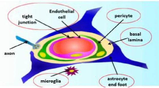
junctions between the adjacent endothelial cells.(79) Those junctions restrict the paracellular pathways. Thus, the endothelial continuous walls of the microcellular vessels act as a highly lipophilic barrier.(79) In addition to endothelial cells, the BBB is composed of a basement membrane, astrocytes end-feet ensheathing the vessels and pericytes which are embedded in the basement membrane.(79) They are thought to play an important role in angiogenesis. Microglia are glial cells found in the brain and spinal cord and they act as resident macrophages (figure 6).(79)
Fig. 6: An illustration of the BBB structure showing its different components such as the tightened adjacent endothelial cells, astrocyte end-foot and the resident
macrophages(microglia).(81)Reproduced with permission.
The whole protective structure of the BBB aims at protecting the brain from harmful substances that can be found in the periphery. There are efflux transport systems included in the BBB to regulate the entrance of some essential substances such as glucose.(79) Therefore, drug uptake into the brain only relies on a certain degree of lipophilicity, small size and affinity to transporters located on the BBB and blood-cerebrospinal fluid interfaces.(81)
3. Novel treatment strategies
Because the current therapeutic strategies are still away from safely and effectively eliminating GBM tumors, researchers are always moving forward to discover newly modified drug delivery systems and new drug entities. A very promising area of research is pharmaceutical nanotechnology. A historical background, principles and nanosystems which are currently under investigation are discussed below in details.
3.1 Pharmaceutical nanotechnology
Throughout the last two decades pharmaceutical nanoparticles started to be considered, in research scale, one of the evolutionary mechanisms of drug delivery. In fact the considered promising effects of this technique can be attributed to the advantageous features they have over other drug delivery systems (DDS). Among those unique features is a very small size (˂ 1000 nm) while, at the same time, the significant large surface-to-mass ratio of a NP compared to other entities. Another advantage is their quantum properties which facilitate their adsorption and the effective carrying of other drug molecules.(82)
A subtype of solid NPs is nanogels which are hydrogels synthesized in a nanoscale. Hydrogels were best described as hydrophilic three-dimensional polymer networks able to take up large amounts of water or physiological fluid, while maintaining their internal network structure.(83) Research has been focused on adapting NGs for in vivo applications, trying to make a benefit from the biocompatibility/biodegradability of certain polymers.(60) When such polymers are used in the synthesis of self-assembled NGs, an added advantage of safety, in addition to the previously mentioned ones, is warranted. The modifiable surface of NGs allows the incorporation of ligands to it and the subsequent target delivery of the encapsulated drug, thus reducing harm to healthy cells while maximizing efficacy of the dosage form.(84) Another important key feature is the
nanosize as well as the chemical properties of the polymers used in NG synthesis.(84) This type of NGs, known as stimuli-responsive, is being researched extensively as it is considered to exhibit promising selectivity in drug delivery. Examples of this NG type will be explained later in details.
3.1.1 Polymers and materials used in NP synthesis
Nanoparticles can be prepared from a variety of materials such as lipids, proteins, polysaccharides and synthetic polymers. Selection of the material to be used in the synthesis depends widely on the required size, release profile aimed for the drug, final site to target, drug physicochemical characteristics and also on the degree of biodegradability, biocompatibility and toxicity of the selected polymers.(85)
Another important factor which seems to play a great role in selecting polymers is the mechanism by which NPs are intended to be prepared. Among different approaches used in the preparation of NPs, nanoprecipitation and ionic gelation are two major ones.
In nanoprecipitation, or so called solvent displacement, NPs are prepared by dissolving both polymer and drug in a water-miscible solvent. The prepared solution is then added, drop by drop, to a nonsolvent to the polymer which is usually water. This addition will allow the diffusion of the organic phase into the aqueous one and vice versa. The result will be supersaturation and precipitation of the polymeric NPs with drug entrapped inside them.(86) Poly (d,l-lactide-co-glycolide) (PLGA) and polycaprolactone (PCL) are typical examples of polymers used in the nanoprecipitation method.
Another major approach for synthesizing NPs, particularly NGs, is ionic gelation. The mechanism of this method depends mainly on ionic bond formation. The bond is formed between the protonated amino acid groups in polymers like chitosan and the negatively-charged phosphate groups of a crosslinker such as tripolyphosphate. For this reaction to occur, pH adjustment of the
medium is critical because protons are required to be present at the amino groups of the selected polymer.(87, 88)
Chitosan, a very popular polymer in the NG synthesis, is a natural biodegradable polysaccharide derived from chitin.(89) It contains units from both glucosamine and N-acetyl-D-glucosamine (NAG). Chitosan is characterized by a ratio of glucosamine to NAG higher than one. Chitosan has wide contributions in food and pharmaceutical industries as an excipient that is able to help in tablet compression and disintegration. In addition to its role as an excipient, chitosan has attracted attention as a main component in NGs formulation thanks to its unique physicochemical properties. The acidic pKa of chitosan,
̴
6.3, allowed it to gain a proton in a medium of pH ˂5.5, thus turning chitosan into a positively-charged molecule that can easily form an ionic bond with negative phosphate groups of TPP.(89) Other polymers such as alginic acid can undergo the same mechanism (but with reverse negative charges) and get crosslinked with CaCO3 to form calcium alginate. However, the resulting chitosan gels appear to be more stable compared to those synthesized by calcium alginate especially in phosphate buffers. Therefore chitosan gels are expected to have longer life span.(90) Another potential component in NG synthesis is hyaluronic acid (HA) which is a polysaccharide composed of NAG and D-glucuronic acid. HA is an endogenous substance that exhibits excellent biocompatibility and biodegrabaility properties.(91) The structure of HA also includes carboxylic groups available for a reaction with protonated amino groups of chitosan, when both molecules are combined together.(92) In this way, incorporating HA into NG structure provide extra support to the matrix. In addition to this, CD4, a HA receptor abundant in tumor cells, facilitates the intracellular uptake of HA-based NGs into their target cells.(93)3.1.2 Different generations of nanoparicles
Nanoparticles can be classifiedinto four generations so far, according to their degree of ‘intelligence’, i.e. ability to act safely and effectively.
3.1.2.1 First generation
Those are conventional nanoparticles with a surface remaining unmodified. They are naïve drug carriers that are easily recognized by blood components (opsonins) such as IgG, complement C3 components (figure 7-a). These opsonins adsorb onto the nanoparticle surface, mostly a hydrophobic surface which favours opsonin adherence. As a consequence nanoparticles are massively cleared from the bloodstream, together with their carried drug, by macrophages of the mononuclear phagocytes system (MPS) such as liver, spleen, lungs and bone marrow.(94)
3.1.2.2 Second generation
They were developed from the first generation to produce long-circulating drug delivery systems. This was mainly achieved by surface modification of the conventional nanoparticles so they were not recognized by the circulating opsonins (figure 7-b). The modification is made either by a) surface-coating of the nanoparticles using specific adsorption of hydrophilic polymers/surfactants or b) grafting hydrophilic segments to the surface, example of these hydrophilic segments are polyethylene glycols (PEG), poloxamers, poloxamines and polysorbates.(94)
A good example of this is the PEGylation of liposomal DDS which were proved to avoid the in vivo uptake by MPS. Some of these ‘stealth’ systems are currently marketed e.g. Doxil® (doxorubicin-loaded PEGylated liposomes) and Abraxane® (albumin-bound paclitaxel nanoparticles) both used for the treatment of metastatic breast cancer.(95)
Nanoparticles from the first and second generations reach their site of action by passive targeting,(95) a pathway which benefits from angiogenesis abnormalities of tumor tissues to enhance the drug permeability. For better understanding, we will explain the tumor tissue lining
structure. Tumor tissue walls are characterized by angiogenesis abnormalities in which new blood vessels are generated to supply nourishment to the over-grown tumor cells.(96) This causes the tumor vascular lining to lose the smooth muscle layer and to have highly defective endothelial cells with wider fenestrations. Also permeability enhancing factors such as nitric oxide, prostaglandins, bradykinins, vascular permeability factor are overexpressed and they cause extravasation of the tumor tissue walls allowing the passage of macromolecules, a phenomenon known as enhanced permeability and retention effect (EPR) (Figure 8).(64)
Fig. 7: A simple diagram illustrating the different stages of drug -loaded nanoparticle development to improve their target specificity. (a) first generation, conventional and non-modified surface nanoparticles easily recognized by opsonins , (b) second generation non- modified-surface nanoparticles successfully avoided blood-circulating opsonins, (c) third generation nanoparticles in which ligands are grafted to their surfaces making them able to target specific cell receptors, (d) fourth generation nanoparticles, they are highly intelligent and sensitive
to triggers in the tumor environment.
Fig. 8: The EPR effect in the tumor tissue.
A comparison between the intact endothelial smooth muscle structure lining the normal tissues and the loose one lining the tumor tissues which results in EPR effect and allowing accumulation of the drug inside. Reproduced from www.wikipedia.org
The abnormal structure also allows for drug molecules to favour the accumulation in tumor tissues exerting their therapeutic effects.
Increasing the drug circulation period in the body by avoiding opsonisation and depending on the efficiency of the EPR effect provides a more controlled release of the drug. However, there is still a random drug delivery of the drug. For example, tumor vascularisation does not only vary in its degree of porosity but also EPR phenomenon is not restricted to tumor tissues and extends to normal tissues although with much lower degree.(95) In fact when it comes to brain tumor, the noticed EPR effect is relatively weak. For instance, the macromolecules and nanoparticles need to be less than 100 nm to significantly extravasate from the tumor vasculature. These particles also
Normal tissue
Erythrocytes
Small
molecules
Macro-molecules
Endothelial
smooth muscles
Lymphatic
vessel
Tumor tissue
need to be higher than 20 nm to be able to remain in the tumor environment and not inefficiently get back to the circulation or undergo rapid renal filtration.(97)
Therefore, closely looking at the degree of intelligence of first and second generations of nanoparticles, one can easily conclude that the specific targeting of the site of action by nanocarriers is not yet achieved. As a result developing nanocarriers continued for the purpose of achieving specific targeting.
3.1.2.3 Third generation (Active targeting)
In this generation nanoparticle surface was subjected to further modification. Ligands were grafted to the outer surface of the nanoparticles to enable active targeting of the tumor cells (figure 7-c). The whole concept depends on an efficient interaction, active targeting, between the grafted ligand and the receptor at the tumor cell membrane.(95) Some tumor cells are known for overexpressing some specific antigens or receptors on their surfaces.(95) Example of moieties that can be grafted to target specific cell receptors are proteins (mainly antibodies and their fragments), peptides, nucleic acids (aptamers), small molecules, or others (vitamins or carbohydrates).(95)
Although they seem to be relatively highly specific in targeting the tumor cell, the third generation nanoparticles are limited by many factors. These factors include the extent of the selective expression of the target cell receptor relative to non-target cells, the receptor availability on the target cell surface, the rate of internalization vs. shedding of that surface receptor following ligand binding, etc.(98) Moreover the expression of a promising tumor-targeting receptor may not be occurring homogenously throughout the whole tumor. Therefore, the need to produce nanoparticles with higher degree of intelligence necessitated the quest for identifying conditions that are highly specific to tumor tissues alone and not the healthy ones.
3.1.2.4 Fourth generation (dynamic or self-triggered nanoparticles)
Fourth generation nanoparticles are highly ‘intelligent’ (i.e. ‘smart’) nanosystems demonstrating target-specificity characteristics. Moreover, in these systems, the drug release effect at the target site is mostly triggered by factors that could be either internal or external (figure 7-d). Examples of internal factors are enzymes or low pH of the tumor environment, while external factors could be light, temperature, magnetic field, and ultrasound.(98)
Example of self-triggered nanoparticles is pH-sensitive nanocarriers which are able to stabilize the drugs inside their nanoparticles at the physiological pH while releasing it in the tumor tissues, in which a higher degree of acidity is normally caused by glycolysis-upregulated hypoxia. Solid tumors have an acidic pH of 5 – 6.8. Chitosan, a polysaccharide polymer used to synthesize pH-sensitive nanogels, has a pKa of 6.3. Since the interstitial space of solid tumors has a pH in a range of 6.8 – 7.2, chitosan-based nanogels should be able to specifically release their encapsulated drug in the microenvironment of the tumor tissue (figure 9).(96)
Fig. 9: pH-sensitive chitosan conjugated nanocarrier interactions: (a) aggregated form of nanocarrier in blood or natural tissues. (b) Swollen form in the acidic sites. Repulsion between protonated amino group on the chitosan skeleton and protons leads to release the anti-tumor drugs. Reproduced with permission from (96)
USPIO, magnetized iron oxide synthesized as nanoparticles and used as diagnostic agents, represent another good example of an ideal fourth generation. These NPs consist of a coated magnetized iron oxide core, the coated surface usually being PEG. Their average hydrodynamic diameter measures around 10 nm.(99) The hydrophilic nature of the coating protects USPIO against opsonisation and makes them long-circulating particles. In addition to this, the magnetic properties of the core allowed its specific delivery in response to the application of an extra magnetic field. Thus, after iv infusion of USPIO-containing micro-bubbles in healthy mice, the accumulation of diagnostic USPIO were visualized and quantified using MRI. By the same technique the co-delivery of anticancer drug can be rendered specific.(100)
3.1.3 Promising role of pharmaceutical nanotechnology in brain cancer treatment
A safe and effective drug delivery system (DDS) needs to be able to control the fate of the drug. Many academic researches were started to investigate the safety and efficacy of pharmaceutical nanotechnology as DDSs. There are several challenges that stand against the drug delivery in therapeutic concentrations to the specific site of actions. These challenges are faced by the drug throughout its journey starting from administration until its elimination from the body. Among these encountered problems are poor solubility, crossing the BBB, increased resistance to the drug and nonspecific targeting resulting in the drug accumulation in healthy tissues causing serious side effects.(101)
3.1.4 Examples of overcoming formulation challenges by pharmaceutical nanotechnology
The following are some, but not all, general examples about how pharmaceutical nanotechnology could overcome the potential problems encountered during the drug journey to its site of action.
3.1.4.1 Improving bioavailability of poorly soluble drug
hydrophilic or hydophobic. Around 40% molecules recognized to be active substances are limitedly used because of their poor water solubility and consequent difficulty to be formulated.(102) A good example of this is the hydrophobic anticancer camptothecin which was successfully delivered to cancer cells and induced their death after its loading into mesoporous silica nanoparticles.(103) Thanks to their small size, NPs can be administered intravenously for targeted delivery. Also targeting moieties can be conjugated to their surfaces for further increase of therapeutic efficiency of the drug.(104) Testing chitosan-based NGs loaded with temozolomide showed that drug release was specifically enhanced in response to pH changes.(105) Further in vitro cytotoxicity experiments proved their efficacy compared to the unloaded NGs.(106) Chitosan NGs also gave an advantage to DOX by turning the latter in to an orally bioavailable molecule. Such experiments offered a proof of chitosan absorption-enhancing properties.(107)
3.1.4.2 Increasing the half-life time of drug in the body
Delivery of hydrophilic drugs to brain tumors remains a great challenge in the field of pharmaceutical formulation. Many hydrophilic molecules, e.g. methotrexate (MTX) and DOX exhibited in vitro cytotoxic effects to isolated brain tumors. However, they still have limited or no in vivo application because of their short half-life time. Chitosan polymer and its derivatives as main components of NGs offer the advantage of having modifiable surfaces. A recent in vivo trial compared results after iv administration of free MTX solution, nanodipersions of unmodified MTX-loaded chitosan NGs (UMNGs) as well as polysorbate-80-modified-MTX-loaded chitosan NGs (MNGs). Both UMNGs and MNGs produced 2.33- and 8.52-fold area under the plasma concentration–time curve within the test time period (AUC 0–t) compared with the free MTX. This observation was suggested to be due to some drug preserving mechanism(s), e.g. physical masking, exerted by the nanoparticles on the loaded MTX. This physical masking protected the
drug molecules against being rapidly eliminated by natural elimination mechanisms of the body.(108)
Another example is a doxorubicin-loaded dextrin-based nanogels which showed higher sustained release compared to doxorubicin-poly lactide nanoparticles over 72 h.(109)
3.1.4.3 Decreasing the resistance of tumor cells to anti-cancer drugs
MDR remains a major challenge against the chemotherapeutic efficacy in cancer cells. As previously mentioned, many factors are involved in MDR. These include decreased drug uptake, enhanced DNA repair, overexpression of drug transporters such as P-gp, MRP1, MRP2, and (BCRP). Polymeric nanoparticles have been investigated as vectors to overcome drug resistance by diverting ABC-transporter mediated drug efflux mechanisms.(110) A famous example of this is free DOX which is resisted by MCF-7 cell lines in breast cancer cells. After loading DOX into chitosan-coated magnetic NPs, the drug was investigated by fluorescence microscopy in comparison to free DOX. Unlike the free drug which did not sufficiently accumulate in DOX-resistant MCF-7, the loaded one showed higher accumulation rates similar to those found in sensitive cells.(111) A similar approach was the targeting specificity of chlorotoxin-conjugated nanoparticles.(112) This formula significantly reduced IC50 compared to TMZ-NP and free TMZ which means a decrease in GBM resistance to TMZ therapy.(112)
Although pharmaceutical nanotechnology is relatively a new science, it still has a lot of secrets to reveal especially in challenging conditions such as GBM. In our work, we tried to benefit from the previously mentioned advantages trying to prove the below hypothesis by setting and applying the objectives mentioned after.