HAL Id: hal-01830595
https://hal.archives-ouvertes.fr/hal-01830595
Submitted on 11 Jul 2018
HAL is a multi-disciplinary open access
archive for the deposit and dissemination of
sci-entific research documents, whether they are
pub-lished or not. The documents may come from
teaching and research institutions in France or
abroad, or from public or private research centers.
L’archive ouverte pluridisciplinaire HAL, est
destinée au dépôt et à la diffusion de documents
scientifiques de niveau recherche, publiés ou non,
émanant des établissements d’enseignement et de
recherche français ou étrangers, des laboratoires
publics ou privés.
Arhgef1 in Humans
Maria Luigia Carbone, Jérémy Brégeon, Nabila Devos, Gilliane Chadeuf, Anne
Blanchard, Michel Azizi, Pierre Pacaud, Xavier Jeunemaître, Gervaise Loirand
To cite this version:
Maria Luigia Carbone, Jérémy Brégeon, Nabila Devos, Gilliane Chadeuf, Anne Blanchard, et al..
Angiotensin II Activates the RhoA Exchange Factor Arhgef1 in Humans. Hypertension, 2015, Equipe
2, 65 (6), pp.1273–1278. �10.1161/HYPERTENSIONAHA.114.05065�. �hal-01830595�
1273
T
he RhoA-Rho kinase signaling has been recognizedas a major regulator of vascular tone and arterial blood pressure.1 Overactivation of this pathway has been
sug-gested to participate to the pathogenesis of hypertension in several experimental models2–5 and in humans.6 RhoA acts
as a molecular switch that cycles between an inactive GDP-bound form and an active GTP-GDP-bound form.7 The transition
from the inactive to the active state requires a Rho guanine exchange factor (GEF) that promotes the release of GDP in exchange for GTP.8,9 In the active GTP-bound form, through
the activation of its effector Rho kinase, the phosphoryla-tion of the myosin phosphatase target (MYPT) subunit and the consequent inhibition of the myosin light chain phospha-tase activity, RhoA is responsible for the Ca2+ sensitization of
the contractile proteins that underlies the tonic component of vascular smooth muscle contraction.10 Functional and
phar-macological analyses using Rho kinase inhibitors have fur-ther suggested that RhoA-dependent pathways are involved in the increased vascular resistance associated with hyper-tension.5,11 Although these observations suggest a possible
pathophysiological role for RhoA-Rho kinase in the develop-ment of hypertension, the molecular mechanism(s) and the GEF(s) responsible for the overactivation of RhoA remain largely unknown. Several mouse models of RhoA GEF dele-tion have thus been developed during the few past years and used to address this question. Recent reports described that the RhoA GEF Arhgef12 (Larg) plays a key role in salt-induced high blood pressure, whereas Arhgef1 mediates the activation RhoA by type 1 angiotensin II (Ang II) receptor (AT1R) acti-vation and is essential for Ang II–dependent hypertension.12,13
Whether these GEFs are relevant to hypertension in humans remains to be proven.
RhoA-Rho kinase activity assessed in peripheral blood mononuclear cells (PBMCs) is increased in hypertensive patients and can be reduced by antihypertensive treatments.14
This method is proposed for assessing RhoA-Rho kinase activity in patients with hypertension and other cardiovas-cular diseases.15 In this study, we extended this concept to
the analysis of GEF activity in human. We developed an in vitro approach to evaluate RhoA GEF activity and to identify
Abstract—Although a causative role for RhoA-Rho kinase has been recognized in the development of human hypertension,
the molecular mechanism(s) and the RhoA guanine exchange factor(s) responsible for the overactivation of RhoA remain unknown. Arhgef1 was identified as a RhoA guanine exchange factor involved in angiotensin II (Ang II)– mediated regulation of vascular tone and hypertension in mice. The aim of this study was to determine whether Arhgef1 is activated and involved in the activation of RhoA-Rho kinase signaling by Ang II in humans. In vitro stimulation of human coronary artery smooth muscle cells and human peripheral blood mononuclear cells by Ang II (0.1 μmol/L) induced activation of Arhgef1 attested by its increased tyrosine phosphorylation. Silencing of Arhgef1 expression by siRNA inhibited Ang II–induced activation of RhoA-Rho kinase signaling. In normotensive subjects, activation of the renin–angiotensin system by a low-salt diet for 7 days increased RhoA-Rho kinase signaling and stimulated Arhgef1 activity in peripheral blood mononuclear cells. In conclusion, our results strongly suggest that Arhgef1 mediates Ang II–induced RhoA activation in humans. Moreover, they show that measurement of RhoA guanine exchange factor activity in peripheral blood mononuclear cells might be a useful method to evaluate RhoA guanine exchange factor activity in humans. (Hypertension. 2015;65:1273-1278. DOI: 10.1161/HYPERTENSIONAHA.114.05065.)
•
Online Data SupplementKey Words: angiotensins ■ arteries ■ hypertension ■ leukocytes
■ Rho GTP-binding proteins ■ Rho guanine nucleotide exchange factors ■ signal transduction
Received December 15, 2014; first decision December 18, 2014; revision accepted March 20, 2015.
From Inserm UMR 1087, CNRS UMR 6291 and University of Nantes, Nantes, France (M.L.C., J.B., G.C., P.P., G.L.); CHU Nantes, l’Institut du Thorax, Nantes, France (P.P., G.L.); Inserm, UMR 970, Paris Cardiovascular Research Center, Paris, France (N.D, X.J.); Université Paris Descartes, Sorbonne Paris Cité, Paris, France (A.B., M.A., X.J.); Assistance Publique, Hôpitaux de Paris, Hôpital Européen Georges Pompidou, Paris, France (A.B., M.A., X.J.); Inserm CIC 1418, Paris, France (A.B., M.A.); and Laboratorio di Genomica e Proteomica funzionale, Universta di Bari, Bari, Italy (M.L.C.).
*These authors contributed equally to this work.
The online-only Data Supplement is available with this article at http://hyper.ahajournals.org/lookup/suppl/doi: 10.1161/HYPERTENSIONAHA. 114.05065/-/DC1.
Correspondence to Gervaise Loirand, Institut du Thorax, IRS-UN, 8 quai Moncousu, BP70721, 44007 Nantes cedex 7, France. E-mail [email protected]
Angiotensin II Activates the RhoA Exchange
Factor Arhgef1 in Humans
Maria Luigia Carbone,* Jérémy Brégeon,* Nabila Devos, Gilliane Chadeuf, Anne Blanchard,
Michel Azizi, Pierre Pacaud, Xavier Jeunemaître, Gervaise Loirand
© 2015 American Heart Association, Inc.
Hypertension is available at http://hyper.ahajournals.org DOI: 10.1161/HYPERTENSIONAHA.114.05065
Renin–Angiotensin System
by guest on July 11, 2018 http://hyper.ahajournals.org/ Downloaded from by guest on July 11, 2018 http://hyper.ahajournals.org/ Downloaded from by guest on July 11, 2018 http://hyper.ahajournals.org/ Downloaded from by guest on July 11, 2018 http://hyper.ahajournals.org/ Downloaded from by guest on July 11, 2018 http://hyper.ahajournals.org/ Downloaded fromwhether Arhgef1 is activated by Ang II in humans. We show that Arhgef1 is activated by in vitro Ang II stimulation of human coronary artery smooth muscle cells (HCASMCs) and PBMC, and that Arhgef1 activity in PBMC is increased by in vivo stimulation of the renin–angiotensin system (RAS) by a low-salt diet.
Methods
In Vitro Studies: HCASMC Culture and siRNA
HCASMC (3 different lots; Promocell, Heidelberg, Germany) were cultured in smooth muscle cell (SMC) growth medium 2 (Promocell). The medium was changed every 2 or 3 days. When cells reached confluence, subculture was obtained by harvesting the cells with 0.2% ethylenediaminetetraacetic acid and 0.25% trypsin. Cells were used at passage 2 to 4 and were serum-starved for 24 hours before experiments. HCASMC were stimulated by Ang II (0.1 μmol/L) in the presence of PD123319 (1 µmol/L;
Sigma-Aldrich, Saint-Quentin Fallavier, France) added 1 hour before Ang II stimulation to avoid potential type 2 Ang II recep-tor-mediated effects.
The sense strands of siRNAs (Eurogentec, Seraing, Belgium) used for Arhgef1 silencing was: 5′-GCAGCUCUGAGAACGGCAA-dTdT-3′.16 A scrambled, nontargeting siRNA has been used as
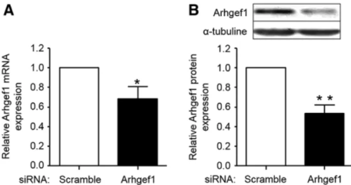
negative control (5′-UUCUCCGAACGUGUCACGU-dTdT-3′). The siRNA were introduced into HCASMCs with Jet PEI reagent (Polytransfection, Illkirch, France) according to the manufactur-er’s recommendations. Cells were incubated with or without 1.2 µg of specific or scrambled siRNA for 48 hours before functional assays were conducted. The efficiency of siRNA-mediated gene silencing assessed by real-time quantitative polymerase chain re-action or by Western blot at 48 hours post-transfection was ≈50% (Figure 1), with no significant effect on other RGS-Rho GEFs (not shown). This assay has been performed in triplicates, in 4 inde-pendent experiments.
Real-Time Quantitative Polymerase Chain Reaction
siRNA-mediated Arhgef1 silencing was validated by real-time quantitative polymerase chain reaction performed on the iCy-cler iQ Detection System (Bio-Rad S.A., Marnes la Coquette, France) as previously described17 with the following primers for
Arhgef1 (up: 5′-TCACCACCAGGGAGATCAGAC-3′, down: 5′-TTGGTTGCTTCTCCTATTCTTGTC-3′) and GAPDH (up: 5′-GACAGTCAGCCGCATCTTCTT-3′, down: 5′- AGTTAAAAGC AGCCCTGGTGA-3′).
Human Study: Subjects and Protocol
We studied 47 normotensive (office blood pressure<140/90 mm Hg after 5 minutes rest in the supine position) healthy male nonsmoking subjects (mean age, 23.7±0.5 years; mean body mass index, 22.9±0.3 kg/m2). Informed consent for participation in the study was obtained
from all subjects. The study protocol was performed in accordance with the Declaration of Helsinki guidelines and institutional guide-lines, and was approved by the Comité Consultatif de Protection des Personnes se prêtant à des Recherches Biomédicales (Paris-Cochin, France). Each subject has received a low-sodium/high-potassium diet for 7 days (sodium, ≤20 mmol/d and potassium, >120 mmol/d). Plasma hormones, blood pressure, and heart rate were measured before (day 0) and at the end of the 7-day low-salt/high-potassium diet (day 7).
Laboratory Analysis and Measurement of Blood Pressure
The methods used for measuring blood pressure, collecting blood samples and for quantifying plasma active renin, total renin, and aldo-sterone were as described previously.18
Isolation of PBMC
Whole blood was collected into EDTA tubes. PBMC were isolated from whole blood under sterile conditions by density gradient cen-trifugation using Unisep tubes (Eurobio, Courtaboeuf, France). PBMC pellets were either stored at −80°C until used or immediately resuspended in RPMI and stimulated with Ang II, then processed for Western blot analyses.
Western Blot Analysis
Cells (HCASMCs or PBMC) were lysed in NETF buffer (NaCl 100 mmol/L, EGTA 2 mmol/L, Tris HCL 50 mmol/L, NaF 50 mmol/L, 1% NP-40, protease inhibitor and Ser/Thr phosphatase inhibitors [Sigma, cocktail 1]). Cell lysates were subjected to SDS-PAGE, transferred to nitrocellulose membranes then incubated with spe-cific antibodies. Immunoprecipitation of phosphotyrosine protein was performed using the 4G10 antibody (Upstate Biotechnology; Millipore, Molsheim, France) Assessment of the phosphorylation level of the Rho kinase target MYPT subunit 1 (P-MYPT) with a rab-bit polyclonal antibody to phospho-MYPT (P-MYPT1; Santa Cruz Biotechnology, CliniSciences, Naterre, France). Antibody to Arhgef1 was purchased from Santa Cruz Biotechnology. Anti-MYPT-1 was purchased from Cell Signaling. ATR1 antibody was from Santa Cruz Biotechnology. Immunoreactive proteins were detected by enhanced chemiluminescence detection procedure (Amersham Pharmacia Biotech, GE Healthcare, Velizy-Villacoublay, France) and quantified using QuantityOne (Bio-Rad). Equal loading was checked by reprob-ing the membrane with β-actin or α-tubulin antibody (Santa Cruz Biotechnologies).
AT1R mRNA Expression
Total RNA was extracted from PBMC using TRIzol reagent (Life technologies) according to the manufacturer’s instructions. RNA (1 μg) was reverse transcripted with moloney murine leukemia virus reverse transcriptase (Invitrogen) and deoxyribonucleic random hexamer as primers (Invitrogen). Real-time gene expres-sion analysis of AT1 receptor was performed with predesigned TaqMan Gene Expression Assay (Hs00258938_m1; Life technolo-gies) using the StepOnePlus Real-Time polymerase chain reaction System (Applied Biosystems). AT1 receptor mRNA expression was normalized to HPRT1 mRNA expression (Hs02800695_1) used as housekeeping control gene. Cycle threshold and relative quantification by the 2−Ct method values were determined using the
StepOne Software v2.1.
Statistical Analysis
Results are presented as mean±SEM. All reported P values were 2-sided, and a P<0.05 was considered statistically significant. Figure 1. siRNA targeting Arhgef1 decreases Arhgef1 mRNA
(A) and protein expression (B) in human coronary artery smooth muscle cell. Analysis is performed 48 hours after siRNA transfection. Arhgef1 mRNA and protein expression are normalized to GAPDH and α-tubuline, respectively. Results are expressed relative to the control (scramble siRNA). The data presented are representative of 3 independent experiments. *P<0.05; **P<0.01.
by guest on July 11, 2018
http://hyper.ahajournals.org/
Carbone et al Ang II Activates Arhgef1 in Humans 1275
Nonparametric Mann–Whitney (for HCASMC data) and Wilcoxon signed-rank tests were used. Relations between variables were deter-mined by Spearman correlation coefficients analysis. The data were processed using the software package GraphPad Prism 5.
Results
Arhgef1 Is Required for Ang II–Mediated RhoA-Rho Kinase Activation in HCASMC
Stimulation of control HCASMCs by Ang II (0.1 µmol/L) for 5 and 60 minutes induced RhoA-Rho kinase activation (Figure 2). Knockdown of Arhgef1 by siRNA prevented Ang II–induced RhoA-Rho kinase activation at both 5 and 60 minutes (Figure 2). This suggests that, as in rodent SMC, the RhoA exchange factor Arhgef1 is essential for the activation of RhoA signaling induced by Ang II in human SMC.
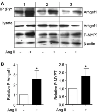
In Vitro Stimulation of Human PBMC by Ang II Induces Activation of Arhgef1
To validate whether PBMC can be used to monitor Arhgef1 activation by Ang II in humans, we first assessed whether Arhgef1 was activated by in vitro stimulation of PBMC by Ang II. Ang II activated Arhgef1 through Jak2-mediated Tyr738 phosphorylation.12 We, therefore, measured the activation of
Arhgef1 in PBMC by Western blot analysis of its phosphory-lation on Tyr residues. Stimuphosphory-lation of PBMC by Ang II (0.1 µmol/L) for 60 minutes increased both the phosphorylation of
Arhgef1 on Tyr, attesting its activation, and the phosphoryla-tion of MYPT indicating the activaphosphoryla-tion of RhoA-Rho kinase signaling (Figure 3).
In Vivo Activation of RAS by Low-Sodium/High-Potassium Diet Induces Activation of Arhgef1 in Human PBMC
After 7 days of the low-sodium/high-potassium diet, as expected, the plasma total and active renin and aldosterone concentrations were markedly and significantly increased compared with those measured at day 0, thereby demonstrat-ing the activation of the RAS (Table 1). Regression analysis confirmed that plasma aldosterone correlated with active renin level (Figure S1A in the online-only Data Supplement). This activation of RAS was associated with an increase in uri-nary Na+ excretion and a reduction of K+ excretion (Table 1).
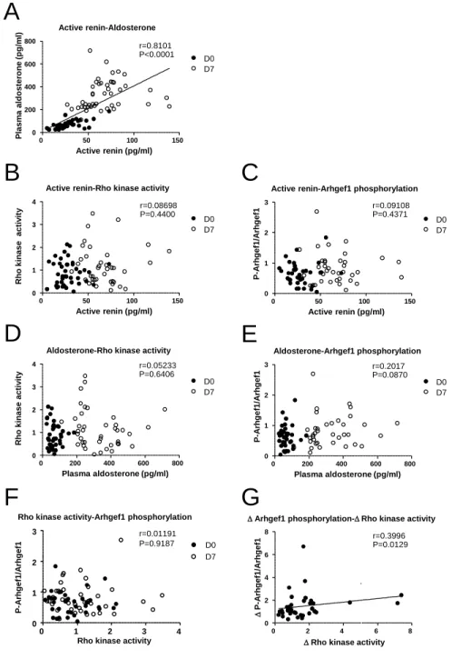
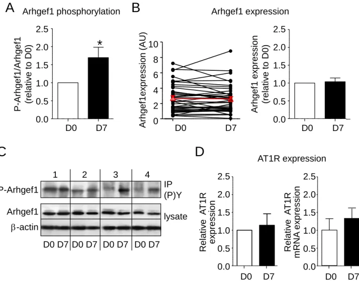
Systolic blood pressure slightly decreased at day 7 of the low-sodium/high-potassium diet (not shown). To analyze whether in vivo stimulation of the RAS activates Arhgef1, we next measured the activity of RhoA-Rho kinase signaling and the phosphorylation of Arhgef1 in PBMC collected at day 0 and at day 7 of the low-sodium/high-potassium diet. Activity of RhoA-Rho kinase was significantly increased after 7 days of low-sodium/high-potassium diet (Figure 4A). This is associ-ated with an increased phosphorylation of Arhgef1 attesting its activation (Figure 4B). The low-sodium/high-potassium diet for 7 days induced a 1.7-fold increase in Arhgef1 phos-phorylation from its initial level at day 0 (Figure S2A), with-out change neither in Arhgef1 expression (1.04±0.10 at day 7 normalized to day 0; Figure S2B and S2C) nor in AT1R pro-tein and mRNA expression (1.14±0.32 and 1.34±0.34 at day 7 normalized to day 0, respectively; Figure S2D). Leukocyte Rho kinase activity and Arhgef1 phosphorylation did not cor-relate with plasma active renin or aldosterone levels (Figure S1B–S1E). Leukocyte Rho kinase activity did not correlate with Arhgef1 phosphorylation level, although the stimula-tion of Rho kinase activity and the increase in Arhgef1 phos-phorylation induced by the low-sodium/high-potassium diet are weakly correlated (Figure S1F and S1G). These data thus provide evidence that activation of the RAS leads to increase the leukocytes activity of Arhgef1 and RhoA-Rho kinase signaling.
Discussion
Our study demonstrates that Ang II activates Arhgef1 in HCASMC and human PBMC in vitro and that in vivo physi-ological activation of the RAS by a low salt intake increases the activity of Arhgef1 in PBMC of normotensive subjects. The present data thus suggest a potential role of Arhgef1 in the regulation of blood pressure by Ang II in humans. Furthermore, our study provides evidence that measurement of Rho GEF activity in PBMC might be a useful method to evaluate Rho GEF activity in humans.
Recent works in both the rodent models of hypertension and transgenic mice have greatly increased our knowledge on the role of RhoA-Rho kinase signaling in blood pressure regulation and hypertension.1 In particular, the identification
of GEFs responsible for the increased RhoA activity, such as Arhgef12 for salt-dependent hypertension and Arhgef1 Figure 2. siRNA-mediated Arhgef1 silencing inhibits the
activation of RhoA-Rho kinase induced by angiotensin II (Ang II) stimulation (0.1 µmol/L) of human coronary artery smooth muscle cell for 5 and 60 minutes. RhoA-Rho kinase activity is assessed by the measurement of myosin phosphatase target (MYPT) phosphorylation by Western blot and quantified by densitometric analysis. Results are normalized to MYPT expression and expressed relative to the value obtained in the absence of Ang II in each experimental condition. Expression of Arhgef1 and α-tubuline was also shown. (*P<0.05, vs same condition in control [scramble] cells, n=4).
by guest on July 11, 2018
http://hyper.ahajournals.org/
for Ang II–dependent hypertension, supports the concept that RhoA GEFs involved in the overactivation of RhoA are probably different according to the pathological context.12,13
Accordingly, RhoA GEFs represent new target for antihyper-tensive therapies to selectively reverse the hyperactivity of RhoA occurring in different forms of hypertension, allowing adapted and targeted antihypertensive treatments. Translating these results from rodents to human hypertension is, there-fore, an important and promising challenge. In vitro experi-ments performed here confirm that, as previously described in rat and mouse SMCs, Arhgef1 is involved in Ang II–induced RhoA/Rho kinase activation in human arterial SMCs.
In humans, both genetic studies and pharmacological data indirectly support a potential role of Rho proteins in the gen-eration of high blood pressure. Indeed, high basal blood pres-sure and increased systemic vascular resistance was shown to be associated with the nonsynonymous Thr431Asn Rock2 variant of the Rho kinase gene.19 More recently, a lower risk
of high blood pressure was associated with a major haplotype block at the Rock2 locus in a recessive manner.20 About RhoA
GEFs, although no study has been performed in the context of hypertension, genetic analyses have defined RhoA GEFs as susceptibility genes for other cardiovascular risk factors. Arhgef11 and Arhgef12 have been identified as candidate
genes for type 2 diabetes mellitus.21,22 From a
pharmacologi-cal point of view, the first data suggesting a role of RhoA-Rho kinase in the pathogenesis of human hypertension were obtained by showing that the increase in forearm blood flow and the decrease in forearm vascular resistance produced by the Rho kinase inhibitor fasudil are significantly stronger in patients with essential hypertension than in the normotensive group of patients, suggesting that hypertension-associated vascular dysfunction depended, at least in part, on Rho kinase activation.6 Interestingly, Rho kinase activity in leukocytes
correlates with the forearm blood flow response to Rho kinase inhibitors indicating that leukocyte Rho kinase activity is also an index of vascular RhoA-Rho kinase activity.15 Yet systolic
blood pressure was not found to correlate with leukocyte Rho-associated kinase activity.23 Results from this recent
pro-spective study in a large population, however, demonstrated that leukocyte Rho kinase activity is a potential biomarker for predicting cardiovascular events.23 The molecular
mecha-nisms that regulate Rho kinase activity or those involved in the dysregulation of RhoA/Rho kinase signaling in human are not known. Only study in leukocytes from patients with Bartter and Gitelman syndromes has indirectly suggested a role of Arhgef1 and Rho kinase signaling pathway in the Ang II effect in humans.24,25
Our results confirm that Arhgef1 is expressed in human PBMC and activated RhoA/Rho kinase signaling in response to Ang II stimulation in vitro. They further show that Arhgef1 activity and RhoA/Rho kinase signaling are increased by the physiological activation of the RAS. However, the increase in PBMC Arhgef1 activity and Rho kinase activity observed when the RAS was stimulated are only weakly correlated, and neither Arhgef1 activity nor Rho kinase activity correlated with plasma active renin and aldosterone. Several reasons could potentially explain this apparent discrepancy between in vitro and in vivo results. In vivo activation of the RAS induces multiple pathways as well as positive and negative feedback loops.26 These events lead to the generation of
sec-ondary circulating mediators, such as reactive oxygen species, chemokines, cytokines, or vasoactive mediators that can also positively or negatively regulate exchange factors and RhoA/ Rho kinase signaling. In whole organism, the level of PBMC Arhgef1 and RhoA/Rho kinase activation could thus be not directly related to the level of RAS activity and AT1R activa-tion, but may result from complex interactions of numerous upstream signals. Furthermore, even standardized protocols were used and phosphatase inhibitors included in the lysis buf-fer, the level of Arhgef1 and MYPT phosphorylation in human PBMC depends on cellular protein phosphatase activity that
Table 1. Hormonal and Urine Parameters Before (Day 0) and After a 7-Day Low-Sodium/High-Potassium Diet (Day 7; n=47)
Parameter Day 0 Day 7 P Value
Plasma active renin, pg/mL 27.4±1.9 68.3±3.6 <0.0001 Plasma total renin, pg/mL 330.5±14.1 433.1±18.5 <0.0001 Plasma aldosterone, pg/mL 69.9±5.3 331.2±19.9. <0.0001 Natriuresis, mmol/24 h 169.1±12.6 22.9±1.2 <0.0001 Kaliuresis, mmol/24 h 75.1±4.4 140.2±4.1 <0.0001 Figure 3. Angiotensin II (Ang II; 0.1 µmol/L, 60 minutes)
induces activation of Arhgef1 and phosphorylation of myosin phosphatase target (MYPT) in human peripheral blood mononuclear cells. A, Activation of Arhgef1 is assessing by examining the Tyr phosphorylation of Arhgef1 by Western blotting with specific anti-Arhgef1 antibody of phosphotyrosine protein immunoprecipitates (IP (P)Y). Total amount of Arhgef1, MYPT phosphorylation, and β-actin expression are checked in total lysate. Typical results obtained in 3 normotensive subjects are shown. B, Quantification of Arhgef1 phosphorylation (normalized to total Arhgef1) and MYPT phosphorylation (normalized to β-actin) expressed relative to control in the absence of Ang II (*P<0.05).
by guest on July 11, 2018
http://hyper.ahajournals.org/
Carbone et al Ang II Activates Arhgef1 in Humans 1277
could be different among subjects and might inconsistently affect protein phosphorylation. Finally, analysis of Arhgef1 phosphorylation required a step of immunoprecipitation of phosphoproteins that adds to the complexity of the measure-ment and would be solved by the developmeasure-ment of phospho-specific Arhgef1 antibodies.
To summarize, the activation of Arhgef1 by Ang II in human vascular smooth muscle in vitro and the inhibition Ang II–induced RhoA-Rho kinase activation by siRNA-mediated Arhgef1 silencing strongly suggest that as in mice,12
Arhgef1 mediates the activation of RhoA downstream AT1R in humans. Although Arhgef1 is also expressed in human PBMC and activated by Ang II in vitro, its activation, and that of RhoA/Rho kinase signaling by the stimulation of RAS in normotensive subject, do not correlate with the level of RAS activity, suggesting that regulation of RhoA/Rho kinase sig-naling in whole organism is complex and probably involved multiple mechanisms.
Perspectives
To our knowledge, our present work is the first study address-ing GEF activity in humans. We show that analysis of GEF activity in PBMC is a minimally invasive method for assess-ing GEF activity in humans because PBMC are easy to obtained and preserved. Although we found that the GEF acti-vation induced in PBMC in vitro reproduced that observed in similar condition in human vascular SMCs, measurement of GEF activity in endothelial cells and vascular SMCs from
normotensive and hypertensive subjects would enable more specific confirmation of vascular GEFs activity. Nevertheless, we proposed that the use of PBMC can represent a relevant and useful functional assay of GEF activity in humans.
Acknowledgments
We thank Cindy Schleder (UMR_S1087, Nantes) for his contribu-tion to human coronary artery smooth muscle cell cultures and experiments.
Sources of Funding
This work was supported by grants from the French Agence Nationale de la Recherche (08-GENO-040-01 and ANR-11-BSV1-013-01) and the Fondation pour la Recherche Médicale (DEQ20090515416). M-L.C. was supported by grants from Laboratorio di Genomica e Proteomica funzionale, Universta di Bari, Bari, Italy.
Disclosures
None.
References
1. Loirand G, Pacaud P. The role of Rho protein signaling in hypertension.
Nat Rev Cardiol. 2010;7:637–647. doi: 10.1038/nrcardio.2010.136. 2. Chrissobolis S, Sobey CG. Evidence that Rho-kinase activity contributes
to cerebral vascular tone in vivo and is enhanced during chronic hyperten-sion: comparison with protein kinase C. Circ Res. 2001;88:774–779. 3. Mukai Y, Shimokawa H, Matoba T, Kandabashi T, Satoh S, Hiroki J,
Kaibuchi K, Takeshita A. Involvement of Rho-kinase in hypertensive vascular disease: a novel therapeutic target in hypertension. FASEB J. 2001;15:1062–1064.
4. Seko T, Ito M, Kureishi Y, Okamoto R, Moriki N, Onishi K, Isaka N, Hartshorne DJ, Nakano T. Activation of RhoA and inhibition of myo-sin phosphatase as important components in hypertension in vas-cular smooth muscle. Circ Res. 2003;92:411–418. doi: 10.1161/01. RES.0000059987.90200.44.
5. Uehata M, Ishizaki T, Satoh H, Ono T, Kawahara T, Morishita T, Tamakawa H, Yamagami K, Inui J, Maekawa M, Narumiya S. Calcium sensitization of smooth muscle mediated by a Rho-associated protein kinase in hypertension. Nature. 1997;389:990–994. doi: 10.1038/40187.
6. Masumoto A, Hirooka Y, Shimokawa H, Hironaga K, Setoguchi S, Takeshita A. Possible involvement of Rho-kinase in the pathogenesis of hypertension in humans. Hypertension. 2001;38:1307–1310.
7. Jaffe AB, Hall A. Rho GTPases: biochemistry and biology. Annu
Rev Cell Dev Biol. 2005;21:247–269. doi: 10.1146/annurev. cellbio.21.020604.150721
8. Bos JL, Rehmann H, Wittinghofer A. GEFs and GAPs: critical elements in the control of small G proteins. Cell. 2007;129:865–877. doi: 10.1016/j. cell.2007.05.018.
9. Rossman KL, Der CJ, Sondek J. GEF means go: turning on RHO GTPases with guanine nucleotide-exchange factors. Nat Rev Mol Cell
Biol. 2005;6:167–180. doi: 10.1038/nrm1587.
10. Somlyo AP, Somlyo AV. Ca2+ sensitivity of smooth muscle and nonmus-cle myosin II: modulated by G proteins, kinases, and myosin phosphatase.
Physiol Rev. 2003;83:1325–1358. doi: 10.1152/physrev.00023.2003. 11. Loirand G, Guérin P, Pacaud P. Rho kinases in cardiovascular
physiol-ogy and pathophysiolphysiol-ogy. Circ Res. 2006;98:322–334. doi: 10.1161/01. RES.0000201960.04223.3c.
12. Guilluy C, Brégeon J, Toumaniantz G, Rolli-Derkinderen M, Retailleau K, Loufrani L, Henrion D, Scalbert E, Bril A, Torres RM, Offermanns S, Pacaud P, Loirand G. The Rho exchange factor Arhgef1 mediates the effects of angiotensin II on vascular tone and blood pressure. Nat Med. 2010;16:183–190. doi: 10.1038/nm.2079.
13. Wirth A, Benyó Z, Lukasova M, Leutgeb B, Wettschureck N, Gorbey S, Orsy P, Horváth B, Maser-Gluth C, Greiner E, Lemmer B, Schütz G, Gutkind JS, Offermanns S. G12-G13-LARG-mediated signaling in vas-cular smooth muscle is required for salt-induced hypertension. Nat Med. 2008;14:64–68. doi: 10.1038/nm1666.
14. Hata T, Soga J, Hidaka T, Idei N, Fujii Y, Fujimura N, Mikami S, Maruhashi T, Kihara Y, Chayama K, Kato H, Noma K, Liao JK, Higashi
Figure 4. Activation of the renin–angiotensin system by low-sodium/high-potassium diet increases the activity of RhoA-Rho kinase (A) and Arhgef1 (B) in human peripheral blood mononuclear cells (PBMC). Graphs show individual changes in Rho kinase activity and Arhgef1 phosphorylation in PBMC collected at day 0 and day 7 of low-sodium/high-potassium diet. Rho kinase activity was assessed by the ratio of myosin phosphatase target (MYPT) phosphorylation to MYPT expression and Arhgef1 phosphorylation was measured as the ratio of Tyr phosphorylated Arhgef1 to total Arhgef1. Red symbols corresponded to mean. *P<0.05.
by guest on July 11, 2018
http://hyper.ahajournals.org/
Y; ROCK Study Group. Calcium channel blocker and Rho-associated kinase activity in patients with hypertension. J Hypertens. 2011;29:373– 379. doi: 10.1097/HJH.0b013e328340902d.
15. Hata T, Goto C, Soga J, Hidaka T, Fujii Y, Idei N, Fujimura N, Maruhashi T, Mikami S, Kihara Y, Chayama K, Noma K, Liao JK, Higashi Y. Measurement of Rho-associated kinase (ROCK) activity in humans: validity of leukocyte p-MBS/t-MBS in comparison with vascular response to fasudil. Atherosclerosis. 2011;214:117–121. doi: 10.1016/j.atherosclerosis.2010.10.005. 16. Rosenfeldt H, Castellone MD, Randazzo PA, Gutkind JS. Rac inhibits
thrombin-induced Rho activation: evidence of a Pak-dependent GTPase crosstalk. J Mol Signal. 2006;1:8. doi: 10.1186/1750-2187-1-8.
17. Cario-Toumaniantz C, Boularan C, Schurgers LJ, Heymann MF, Le Cunff M, Léger J, Loirand G, Pacaud P. Identification of differentially expressed genes in human varicose veins: involvement of matrix gla pro-tein in extracellular matrix remodeling. J Vasc Res. 2007;44:444–459. doi: 10.1159/000106189.
18. Azizi M, Boutouyrie P, Bissery A, Agharazii M, Verbeke F, Stern N, Bura-Rivière A, Laurent S, Alhenc-Gelas F, Jeunemaitre X. Arterial and renal consequences of partial genetic deficiency in tissue kallikrein activity in humans. J Clin Invest. 2005;115:780–787. doi: 10.1172/JCI23669. 19. Seasholtz TM, Wessel J, Rao F, Rana BK, Khandrika S, Kennedy BP,
Lillie EO, Ziegler MG, Smith DW, Schork NJ, Brown JH, O’Connor DT. Rho kinase polymorphism influences blood pressure and systemic vascular resistance in human twins: role of heredity. Hypertension. 2006;47:937–947. doi: 10.1161/01.HYP.0000217364.45622.f0. 20. Rankinen T, Church T, Rice T, Markward N, Blair SN, Bouchard C. A
major haplotype block at the rho-associated kinase 2 locus is associated
with a lower risk of hypertension in a recessive manner: the HYPGENE study. Hypertens Res. 2008;31:1651–1657. doi: 10.1291/hypres.31.1651. 21. Kovacs P, Stumvoll M, Bogardus C, Hanson RL, Baier LJ. A functional
Tyr1306Cys variant in LARG is associated with increased insulin action in vivo. Diabetes. 2006;55:1497–1503.
22. Ma L, Hanson RL, Que LN, Cali AM, Fu M, Mack JL, Infante AM, Kobes S, Bogardus C, Shuldiner AR, Baier LJ; International Type 2 Diabetes 1q Consortium. Variants in ARHGEF11, a candidate gene for the linkage to type 2 diabetes on chromosome 1q, are nominally associated with insulin resistance and type 2 diabetes in Pima Indians. Diabetes. 2007;56:1454– 1459. doi: 10.2337/db06-0640.
23. Kajikawa M, Noma K, Maruhashi T, Mikami S, Iwamoto Y, Iwamoto A, Matsumoto T, Hidaka T, Kihara Y, Chayama K, Nakashima A, Goto C, Liao JK, Higashi Y. Rho-associated kinase activity is a predictor of cardiovascular outcomes. Hypertension. 2014;63:856–864. doi: 10.1161/ HYPERTENSIONAHA.113.02296.
24. Pagnin E, Semplicini A, Sartori M, Pessina AC, Calo LA. Reduced mRNA and protein content of rho guanine nucleotide exchange factor (RhoGEF) in Bartter’s and Gitelman’s syndromes: relevance for the pathophysiology of hypertension. Am J Hypertens. 2005;18:1200–1205. doi: 10.1016/j. amjhyper.2005.03.747.
25. Calò LA, Pessina AC. RhoA/Rho-kinase pathway: much more than just a modulation of vascular tone. Evidence from studies in humans.
J Hypertens. 2007;25:259–264. doi: 10.1097/HJH.0b013e328010d4d2. 26. Crowley SD, Coffman TM. Recent advances involving the
renin-angio-tensin system. Exp Cell Res. 2012;318:1049–1056. doi: 10.1016/j. yexcr.2012.02.023.
What Is New?
•
This study is the first study demonstrating that angiotensin II activates RhoA exchange factor in humans.•
Physiological activation of the renin–angiotensin system by a low salt intake increases the activity of Arhgef1 and RhoA/Rho kinase signaling in peripheral blood mononuclear cells of normotensive subjects.•
These data provides evidence that measurement of Rho guanine ex-change factor activity in peripheral blood mononuclear cells might be a useful method to evaluate Rho guanine exchange factor activity in humans.What Is Relevant?
•
Deciphering the mechanisms regulating RhoA-Rho kinase activity in hu-mans may define new therapeutic targets against hypertension.Summary
This study shows that Arhgef1 mediates RhoA-Rho kinase activa-tion in human smooth muscle cells and that physiological activaactiva-tion of the renin–angiotensin system in normotensive subjects increas-es Arhgef1 and Rho kinase activity in circulating leukocytincreas-es.
Novelty and Significance
by guest on July 11, 2018
http://hyper.ahajournals.org/
Michel Azizi, Pierre Pacaud, Xavier Jeunemaître and Gervaise Loirand
Maria Luigia Carbone, Jérémy Brégeon, Nabila Devos, Gilliane Chadeuf, Anne Blanchard,
Angiotensin II Activates the RhoA Exchange Factor Arhgef1 in Humans
Print ISSN: 0194-911X. Online ISSN: 1524-4563
Copyright © 2015 American Heart Association, Inc. All rights reserved.
is published by the American Heart Association, 7272 Greenville Avenue, Dallas, TX 75231 Hypertension
doi: 10.1161/HYPERTENSIONAHA.114.05065
2015;65:1273-1278; originally published online April 13, 2015;
Hypertension.
http://hyper.ahajournals.org/content/65/6/1273
World Wide Web at:
The online version of this article, along with updated information and services, is located on the
http://hyper.ahajournals.org/content/suppl/2015/04/13/HYPERTENSIONAHA.114.05065.DC1
Data Supplement (unedited) at:
http://hyper.ahajournals.org//subscriptions/
is online at: Hypertension
Information about subscribing to Subscriptions:
http://www.lww.com/reprints
Information about reprints can be found online at: Reprints:
document. Permissions and Rights Question and Answer
this process is available in the
click Request Permissions in the middle column of the Web page under Services. Further information about Office. Once the online version of the published article for which permission is being requested is located,
can be obtained via RightsLink, a service of the Copyright Clearance Center, not the Editorial Hypertension
in
Requests for permissions to reproduce figures, tables, or portions of articles originally published Permissions:
by guest on July 11, 2018
http://hyper.ahajournals.org/
Ang II activates the RhoA exchange factor Arhgef1 in humans
Maria Luigia Carbone*, Jérémy Brégeon*, Nabila Devos, Gilliane Chadeuf, Anne
Blanchard, Michel Azizi, Pierre Pacaud, Xavier Jeunemaître, Gervaise Loirand
From Inserm UMR 1087, CNRS UMR 6291 and University of Nantes, F-44000
Nantes, France. (ML.C., J.B., G.C., P.P., G.L.); CHU Nantes, l'institut du thorax
(P.P., G.L.), F-44000 Nantes France; Inserm, UMR 970, Paris Cardiovascular
Research Center, F-75015 Paris, France (N.D, X.J.); Université Paris Descartes,
Sorbonne Paris Cité, Paris, F-75006, France (A.B., M.A., X.J.); Assistance
Publique, Hôpitaux de Paris, Hôpital Européen Georges
Pompidou, F-75015 Paris, France (A.B., M.A., X.J.); Inserm CIC 9201, F-75015
Paris, France (A.B., M.A.), Laboratorio di Genomica e Proteomica funzionale,
Universta di Bari, Bari, I-70126 Italia. (ML.C.).
* Equal contributors
Correspondance to Gervaise Loirand, Institut du Thorax, IRS-UN, 8 quai
Moncousu, BP70721, 44007 Nantes cedex 7, France Phone: 33 2 28 08 01 16 Fax:
33 2 28 08 01 30.
E-mail:
[email protected]
Figure S1. Scatter plots showing the relationships between various parameters measured
before (D0, empty circle) and after 7 days of low-sodium/high potassium diet (D7, black
circle). Parameters analyzed are indicated above each graph. Rock activity is determined by
the ratio MYPT/MYPT and Arhgef1 phosphorylation corresponds to the ratio
P-Arhgef1/Arhgef1.
∆ Arhgef1 phosphorylation and ∆ Rho kinase activity are estimated by
the ratio of Arhgef1 phosphorylation at D7 to Arhgef1 phosphorylation at D0 and the ratio
of Rho kinase activity at D7 to Rho kinase activity at D0, respectively.
Active renin-Aldosterone 0 50 100 150 0 200 400 600 800 D0 D7 r=0.8101 P<0.0001 Active renin (pg/ml) P lasm a al d o st er o n e ( p g /m l)
Active renin-Rho kinase activity
0 50 100 150 0 1 2 3 4 D7 D0 r=0.08698 P=0.4400 Active renin (pg/ml) R h o ki n ase act iv it y
Active renin-Arhgef1 phosphorylation
0 50 100 150 0 1 2 3 D7 D0 r=0.09108 P=0.4371 Active renin (pg/ml) P -A rh g ef 1/ A rh g ef 1
Aldosterone-Rho kinase activity
0 200 400 600 800 0 1 2 3 4 D7 D0 r=0.05233 P=0.6406 Plasma aldosterone (pg/ml) R h o ki n ase act iv it y r=0.3996 P=0.0129 ∆ Arhgef1 phosphorylation-∆ Rho kinase activity
0 2 4 6 8 0 2 4 6 8
∆ Rho kinase activity
∆ P -A rh g ef 1/ A rh g ef 1
A
G
B
D
C
Aldosterone-Arhgef1 phosphorylation 0 200 400 600 800 0 1 2 3 D7 D0 r=0.2017 P=0.0870 Plasma aldosterone (pg/ml) P -A rh g ef 1/ A rh g ef 1E
D0 D7 r=0.01191 P=0.9187Rho kinase activity-Arhgef1 phosphorylation
0 1 2 3 4 0
1 2 3
Rho kinase activity
P -A rh g ef 1/ A rh g ef 1