© Sarah Malvaut, 2019
The role of different subtypes of olfactory bulb
interneurons in olfactory behavior
Thèse
Sarah Malvaut
Doctorat en neurobiologie
Philosophiæ doctor (Ph. D.)
The role of different subtypes of olfactory
bulb interneurons in olfactory behavior
Thèse
Sarah MALVAUT
Sous la direction de :
RÉSUMÉ
Le bulbe olfactif (BO) représente dans le cerveau le premier relai dans le traitement des informations olfactives. Au niveau de cette structure, plusieurs types de neurones sont impliqués dans la modulation de l’information odorante, avant même que celle-ci ne soit envoyée vers des structures corticales supérieures. Parmi eux se trouvent les cellules granulaires (CGs), une population d’interneurones régulant de manière importante l’activité des cellules principales du BO. De manière intéressante, le BO est capable à l’âge adulte de produire et régénérer une partie de sa population interneuronale via le processus de neurogénèse adulte. Il est ainsi possible de faire la distinction entre les CGs générées au cours de la période postnatale (CGs postnatales) des CGs générées à l’âge adulte (CGs nouvellement générées). Le rôle que jouent ces CGs dans le traitement olfactif mais aussi dans les différents comportements olfactifs a pendant très longtemps donné lieu à des interprétations contradictoires. Le manque de cohérence au niveau des données peut s’expliquer par le fait que pendant longtemps, les CGs ont été considérées comme étant une population homogène de cellules. Néanmoins, des études ont montré que les CGs peuvent exprimer différents marqueurs neurochimiques. Notamment, nous nous sommes intéressés dans le cadre de notre étude à deux de ces marqueurs : la protéine kinase calcium calmoduline dépendante IIα (CaMKIIα) et la Calrétinine (CR). Une telle hétérogénéité au sein des cellules interneuronales du BO pourrait également refléter une hétérogénéité fonctionnelle, chaque sous-population de CGs pouvant contribuer de façon propre et unique au traitement des informations olfactives et donc au comportement olfactif.
Dans la première partie de ces travaux, nous avons étudié le rôle fonctionnel des cellules exprimant la CaMKIIα et l’avons comparé à la population générale de CGs. De manière intéressante, nous montrons que, bien que ces deux populations de cellules soient en tous points semblables au niveau morphologique, les cellules CaMKIIα reçoivent un niveau d’inhibition moindre par rapport à leurs homologies négatives, les rendant plus susceptibles d’être activées suite à des tâches
comportementales spécifiques. De plus, l’inhibition spécifique des cellules CaMKIIα-positive entraine une perturbation des performances de discrimination fine.
Dans la seconde partie de ces travaux, nous nous sommes intéressés cette fois-ci à la sous-population de CGs exprimant la CR, en tenant compte également de la période développementale de ces cellules (i.e CGs post-natales ou nouvellement générées). Nous montrons que les cellules nouvellement générées exprimant ou non la CR, ainsi que les cellules CR-positives postnatales diffèrent quant à leurs propriétés électrophysiologiques. De plus, tout comme les cellules exprimant la CaMKIIα, les cellules exprimant la CR présentent un niveau d’activation plus important à la suite de certaines tâches comportementales et sont également nécessaires à la bonne réalisation de tâches de discrimination olfactive.
ABSTRACT
The olfactory bulb (OB) is considered as the first relay in the brain during olfactory processing. Several types of neurons are involved at the level of this structure in the refinement of the olfactory information before it is sent to higher cortical structures. Among the cell types involved is the population of granule cells (GC), a population of interneurons largely regulating the activity of OB principal cells. Interestingly, the OB retain during adulthood the ability to produce and renew part of its interneuronal pool through a process called adult neurogenesis. Therefore, it is possible to distinguish in the adult OB between GCs born during the early postnatal period (early-born GCs) to the one that were generated during adulthood (adult-born GCs). Several studies aimed at determining the precise role played by GC in olfactory processing and olfactory behavior, giving rise quite often to conflicting results. This absence of coherence in the data could come from the fact that for long, the population of GCs was considered as a homogeneous cell population. However, GCs were shown to express diverse neurochemical markers. In this study we investigated more particularly into two of those markers, showed to be expressed by GCs: the Ca2+/calmodulin-dependent protein kinase IIα (CaMKIIα) and Calretinin (CR). Hence,
such a heterogeneity in the phenotype of OB interneurons could also underlie a functional heterogeneity of the different GC subpopulation, each one contributing in a unique way to olfactory processing and thus olfactory behavior.
In the first part of this work, we investigated the functional role of CaMKIIα-expressing cells and compared it to the general population of GCs. Interestingly we revealed that CaMKIIα-positive GCs are more prone to activation following specific behavioral tasks, likely due to a decreased level of inhibition as compared to their negative counterparts. Moreover, the specific inhibition of this GC subpopulation let to alteration of animals’ fine discrimination abilities.
In the second part of our work, when focusing this time on the subpopulation of CR-expressing GCs, taking this time also into account the developmental period at which they were generated (i.e early- versus born cells), we showed that adult-born CR-expressing and non-expressing GCs, but also early-adult-born CR-expressing
GCs display different electrophysiological characteristics. Moreover, as for CaMKIIα-positive GCs, CR-CaMKIIα-positive GCs present a higher level of activation following specific olfactory tasks and are also important for a proper ability to perform olfactory discrimination tasks.
TABLE
OF
CONTENTS
RÉSUMÉ ... II ABSTRACT ... IV TABLE OF CONTENTS... VI LIST OF FIGURES ... X LIST OF TABLES ... XII LIST OF ABBREVIATIONS ... XIII REMERCIEMENTS ... XVI AVANT-PROPOS ... XVIII
INTRODUCTION ... 1
STRUCTURE AND ORGANIZATION OF THE MAMMALIAN OLFACTORY BULB ... 1
REGENERATION OF THE OLFACTORY BULB DURING ADULTHOOD: THE PROCESS OF ADULT NEUROGENESIS ... 5
A. Adult neurogenesis: an overview ... 5
a. From stem cells to neuronal progenitors ... 7
b. Neuroblasts migration in the adult brain ... 9
c. Becoming a functional neuron in a pre-existing network ... 9
B. Adult neurogenesis generates a specific subset of olfactory bulb interneurons ... 12
a. Properties of adult-born neurons... 12
b. Responses of adult-born cells to sensory stimulations ... 15
FUNCTIONS OF OLFACTORY BULB INTERNEURONS IN OLFACTORY FUNCTIONING AND BEHAVIOR ... 17
A. Role of OB interneurons in socially relevant behaviors ... 17
B.Role of OB interneurons in spontaneous odor-behavior ... 19
C.Role of OB interneurons in associative odor-behavior ... 21
THE HETEROGENEITY OF OLFACTORY BULB INTERNEURONS: DIFFERENT CELLS FOR DIFFERENT FUNCTIONS ... 23
A.The heterogeneity of periglomerular cells ... 24
B.The heterogeneity of granule cells ... 26
C.Another level of heterogeneity: the differential wiring of olfactory bulb interneurons 29 a. Methods for mapping brain connections in vivo ... 29
b. Towards a differential connectivity of OB interneurons ... 31
CHAPTER I : CAMKIIΑ EXPRESSION DEFINES TWO FUNCTIONALLY DISTINCT POPULATIONS OF GRANULE CELLS INVOLVED IN DIFFERENT TYPES OF ODOR
BEHAVIOR. ... 35 RÉSUMÉ ... 36 ABSTRACT ... 37 HIGHLIGHTS ... 38 ETOC BLURB ... 38 INTRODUCTION ... 39 RESULTS ... 40
A. Functionally heterogeneous populations of GCs in the adult OB ... 40
B. GCs can be divided into two functionally different subtypes based on the expression of CaMKIIα ... 41
C. The population of CaMKIIα+ GCs is sensitive to olfactory experience ... 43
D. Structuro-functional properties of CaMKIIα+ and CaMKIIα− GCs... 45
E. Perceptual learning activates CaMKIIα− GCs ... 46
F. CaMKIIα+ GCs are essential for spontaneous and go/no-go odor ... 48
DISCUSSION ... 50
STAR METHODS ... 54
A. Key resource table ... 54
B. Contact for reagent and resource sharing ... 56
C. Experimental model and subject details ... 56
METHOD DETAILS ... 57
A.Stereotaxic injections ... 57
B.Cranial window surgery and in vivo two-photon calcium imaging ... 58
C.Immunohistochemistry ... 59
D.Morphological analysis... 60
E.Electrophysiological recordings ... 60
F. Novel odor stimulation ... 61
G.Sensory deprivation ... 61
H.Behavioral procedures ... 62
a. Odor discrimination ... 62
b. Perceptual learning ... 62
c. Go/no-go olfactory discrimination learning ... 62
d. Long-term associative memory ... 63
e. Pharmacogenetic inhibition of CaMKIIα+ GCs ... 64
REFERENCES ... 67
FIGURES ... 72
ACKNOWLEDGMENTS ... 82
SUPPLEMENTARY MOVIE LEGENDS ... 82
CHAPTER II : THE ROLE OF CALRETININ-EXPRESSING GRANULE CELLS IN OLFACTORY BULB FUNCTIONS AND ODOR BEHAVIOR ... 83
RÉSUMÉ ... 84
ABSTRACT ... 85
INTRODUCTION ... 86
RESULTS ... 87
A. CR+ and CR− GCs display similar morphological characteristics ... 87
B. CR+ and CR− GCs receive similar excitatory but different inhibitory inputs ... 90
C. Spontaneous odor discrimination increases the activation of CR+ GCs ... 92
D. The go/no-go olfactory learning task induces the activation of CR+ GCs ... 94
E. The pharmacogenetic inhibition of CR+ GCs diminishes the fine olfactory discrimination ability of mice ... 95
DISCUSSION ... 97
MATERIAL AND METHODS ... 100
A. Animals ... 100
B. Stereotaxic injections ... 101
C. Immunohistochemistry ... 102
D.Patch-clamp recordings ... 103
E.Morphological analysis... 105
F. Analysis of gephyrin immunolabeling ... 105
G.Behavioral procedures ... 106
a. Odor discrimination ... 106
b. Go/no-go olfactory discrimination learning ... 106
c. Pharmacogenetic inactivation of CR-expressing GCs ... 108
H.Statistical analysis ... 109
REFERENCES ... 109
FIGURES ... 114
TABLES ... 121
ACKNOWLEDGEMENTS ... 125
DISCUSSION AND FUTURE DIRECTIONS ... 126
A. The birthdate as a factor of GCs heterogeneity ... 127
B. The chemical makeup as a factor of GCs functional heterogeneity ... 130
TOWARDS A DIFFERENTIAL WIRING OF GCS SUBTYPES ... 135
A. Identification of olfactory bulb projecting regions (OPR) of interest ... 135
B. The role of basal forebrain inputs in olfactory processing ... 137
GENERAL CONCLUSION ... 139
REFERENCES ... 140
ANNEX: THE ROLE OF ADULT-BORN NEURONS IN THE CONSTANTLY CHANGING OLFACTORY BULB NETWORK ... 157
ABSTRACT ... 158
INTRODUCTION ... 159
ADULT OB NEUROGENESIS, AN UNUSUAL FORM OF STRUCTURAL AND FUNCTIONAL PLASTICITY ... 160
A. Plasticity in the OB network by the addition of new neurons ... 161
B. Adult-born neurons, a population of cells remarkably different from their pre-existing counterparts ... 161
C. Plasticity through changing morpho-functional properties of new neurons ... 162
IMPLICATION OF ADULT-BORN NEURONS IN OLFACTORY BEHAVIOUR ... 163
ADULT-BORN NEURONS: DISTINCT FUNCTIONS FOR DIFFERENT SUB-POPULATIONS OF CELLS? ... 166
CONCLUSION ... 169
LIST
OF
FIGURES
Figure 1.1: Schematic representation of the OB. ... 2
Figure 1.2: Reciprocal dendrodendritic synapse between mitral/tufted cells and granule cells ... 3
Figure 1.3: Modulation of granule cells activity influences odor discrimination performances ... 5
Figure 1.4: Adult neurogenesis in the olfactory bulb ... 8
Figure 1.5: Synaptic development on adult- and early-born GCs. ... 11
Figure 1.6: Adult-born neurons are more sensitive to novel odors ... 13
Figure 1.7: Plasticity of adult-born neuron spines is driven by sensory activity ... 15
Figure 1.8: Adult-born neurons are sensitive to the level of olfactory activity ... 16
Figure 1.9: The two forms of odor-reward conditioning tasks ... 22
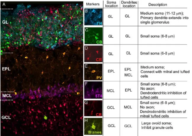
Figure 1.10: Olfactory bulb interneurons diversity ... 25
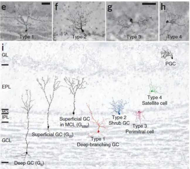
Figure 1.11: The 4 types of adult-born GCs, based on morphological and GCL location characterization ... 28
Figure 1.12: Example of strategies for brain connections mapping ... 30
Figure 1.13: GCs receive inputs from different brain regions based on Allen Brain website. ... 32
Figure 2.1: In vivo Ca2+ imaging of GC activity under baseline and odor stimulation conditions. ... 72
Figure 2.2: Expression of CaMKIIα in the adult olfactory bulb ... 73
Figure 2.3: Sensory stimulation induces higher Ca2+ activity in CaMKIIα+ GCs. ... 74
Figure 2.4: CaMKIIα+ cells are more responsive to sensory activity ... 75
Figure 2.5: Morphological and functional characteristics of CaMKIIα+ and CaMKIIα−GCs ... 76
Figure 2.6: CaMKIIα+ GCs are not required for perceptual learning ... 78
Figure 2.7: CaMKIIα+ GCs are required for go/no-go odor discrimination learning and spontaneous odor discrimination ... 79
Figure S2.1: GCs subtypes activation during long-term associative memory. ... 81
Figure 3.1: Morphological characterization of early-born and adult-born CR+ GCs and adultborn CR− GCs ... 114
Figure 3.2: The excitatory postsynaptic inputs of CR+ and CR− GCs are indistinguishable ... 116
Figure 3.3: CR+ GCs receive weaker inhibitory inputs than CR− GCs and have fewer gephyrin+ puncta on their primary dendrites ... 117
Figure 3.4: Spontaneous odor discrimination induces the activation of CR+ GCs. 118 Figure 3.5: Odor discrimination learning activates CR+ GCs ... 119 Figure 3.6: The pharmacogenetic inhibition of CR+ GCs reduces the olfactory discriminatory ability of mice ... 120 Figure 4.1: Expression of some GABAA receptors subunits and gephyrin in the OB
and by CaMKIIα+ GCs ... 131 Figure 4.2: Transsynaptic identification of OB projecting regions ... 136 Figure 4.2: Basal forebrain activity is linked to sniffing ... 138
LIST
OF
TABLES
Table 2.1: Key resource table ... 54 Table 3.1: Morphological properties of CR+ and CR− GCs ... 121
Table 3.2: Intrinsic electrophysiological properties of early- and adult-born CR+ GCs and adult-born CR− GCs. ... 122 Table 3.3: EPSC amplitudes and frequencies of early-born and adult-born CR+ GCs and adult-born CR− GCs ... 123 Table 3.4: IPSC amplitudes and frequencies of early- and adult-born CR+ GCs and adult-born CR− GCs, and number of gephyrin immunopuncta on adult-born CR+ and CR− GCs ... 124
LIST
OF
ABBREVIATIONS
AAV adeno-associated virusACSF artificial cerebrospinal fluid
AMPA 2-amino-3-(3-hydroxy-5-methyl-isoxazol-4-yl) propanoic acid
AON anterior olfactory nucleus
AP action potential
AraC -D-arabinofuranoside
BDNF brain-derived neurotrophic factor
BF basal forebrain
BMI bicuculline methochlorine
BrdU bromo-desoxy-uridine
CAG cytomegalovirus early enhancer/chicken β actin CaMKIIα Ca2+/calmodulin-dependent protein kinase IIα CaMKIV Ca2+/calmodulin-dependent protein kinase IV
CB calbindin
CBA chicken -actin
cFos cellular oncogene response 1
ChR2 channel-rhodopsin
CNO clozapine-N-oxide
CR calretinin
DAB diaminobenzidine
dpi day post-injection
DREADDs Designer receptors exclusively activated by designer drugs
DV dorso-ventral
EF1α elongation factor 1α
EGFR epidermal growth factor receptor
EPL external plexiform layer
EPSC excitatory post-synaptic current
GABA γ-aminobutyric acid GC granule cell
GCL granule cell layer
GFAP glial fibrillary acidic protein
GFP green fluorescent protein
GL glomerular layer
HDB horizontal limb of diagonal band of Broca
IEG immediate early gene
IPL internal plexiform layer
IPSC inhibitory post-synaptic current
Kyn kynurenic acid
LTP long-term potentiation
M/T cells mitral/tufted cells
MC mitral cell
MCPO magnocellular preoptic nucleus
mGluR2 metabotropic glutamate receptor 2
mIPSC miniature inhibitory post-synaptic current
ML medio-lateral
NCAM neural cell adhesion molecule
NeuN neuronal nuclei
NpHR halorhodospin
NSC neural stem cell
OB olfactory bulb
OE olfactory epithelium
ONL olfactory nerve layer
OPR olfactory bulb projecting region
OSN olfactory sensory neuron
OT olfactory tubercle
PC piriform cortex
PCNA proliferating cell nuclear antigen
PFA paraformaldehyde
PG periglomerular cell
PGC periglomerular cell
PV parvalbumin
RFP red fluorescent protein
RMS rostral migratory stream
ROI region of interest
SAC short-axon cell
SD standard deviation
sEPSC spontaneous excitatory post-synaptic current
SHF spine head filopodia
sIPSC spontaneous inhibitory post-synaptic current
SVZ subventricular zone
Syn synapsin
TC tufted cell
TH tyrosine-hydroxylase
TTX tetrodotoxin
TVA avian tumor virus receptor A
Val66Met variant brain-derived neurotrophic factor
wpi week post-injection
REMERCIEMENTS
Je tiens tout d’abord à remercier mon directeur de thèse, le Dr. Armen Saghatelyan pour m’avoir offert la chance il y a maintenant presque 6 ans de commencer un doctorat dans son laboratoire. Il a toujours su se montrer disponible pour me guider, discuter et répondre à mes parfois nombreuses interrogations tout au long de ces années. Venir étudier au Québec est probablement ce que je considère comme l’un des plus grands défis de ma vie et cette expérience dans son laboratoire m’aura permis d’enrichir à la fois mon panel de connaissances et d’expertises, tout en développant un intérêt et une curiosité pour les approches d’optique et d’imagerie.
Je tiens également à remercier tous les membres de mon jury, le Dr. Paul de Koninck, le Dr. Christophe Proulx ainsi que le Dr. Diane Lagace, pour avoir accepté de réviser ma thèse.
Je remercie tous mes collègues du laboratoire du Dr. Saghatelyan, actuels ou anciens, Marina, Cédric, Archana, Delphine, Arthur, Tiziano, Alessandra, Aymeric, Vlad, Majid, Qian, Marcos, Caroline, et Rodrigo.
Les prochains remerciements vont à mes amis, tant outre-atlantique : Amélie, Rémy et Sophie, qu’à ma famille du Québec : Cédric et Thierry, Archana, Axelle et Yoann, Léa, Louis, Mélina et Mike, Aymeric, Karen et Armina, Alessandra, Romina, Delphine et Flavien. Être loin de sa famille et de son pays peut parfois, même souvent, être difficile, et je n’oublierai jamais tous nos repas, brunchs du dimanche, soirées ou tout simplement nos moments partagés ensemble ainsi que votre présence et soutien lors d’évènements importants de ma vie. Je ne doute pas qu’il y en aura beaucoup d’autres et pour tout cela, un énorme merci !
Je remercie maintenant tout naturellement ma famille, et notamment mon père, ma mère, ainsi que mes deux (petites mais grandes) sœurs Fanny et Raphaëlle. Partir loin de vous pour faire ma thèse a probablement été la décision la plus dure à prendre, et même si celle-ci n’a pas forcément fait l’unanimité au départ, vous avez toujours
été présents pour moi dans tous les moments, les bons comme les moins bons, ces 6 dernières années et pour cela je vous en remercie tellement. Comme l’a dit une fois une poète de la famille « la distance n’enlève en rien tout l’amour que j’ai pour vous ». Enfin, je remercie mon mari Julien. Tu es celui qui m’accompagne au quotidien, a su me supporter et me soutenir dans les dernières étapes de mon doctorat. Tu sais toujours me redonner le sourire par ton humour (certes un peu spécial…) et me remonter le moral lorsque je doute ou stresse, tout simplement.
AVANT-PROPOS
La première partie des résultats présentés dans ce manuscrit de thèse, et qui représente le projet principal sur lequel j’ai travaillé au cours de mon doctorat, a fait l’objet d’une publication dans le journal Current Biology en Octobre 2017. Dans le cadre de cet article, j’ai réalisé l’intégralité des expériences d’imagerie calcique in
vivo, ainsi qu’une grande majorité des expériences comportementales ayant permis
de caractériser par immunohistochimie l’activation des sous-types cellulaires d’intérêt dans l’étude. Plusieurs personnes ont également collaboré à la réalisation de ce travail. Delphine Hardy a participé à l’analyse de la morphologie et de l’activation des cellules à la suite des paradigmes comportementaux. Linda David a réalisé les enregistrements électrophysiologiques. L’équipe du Dr. Alain Trembleau, composée des Drs. Simona Gribaudo, Laura Daroles, Alain Trembleau et Isabelle Caillé, a également contribué à l’analyse morphologique des populations neuronales d’intérêt ainsi qu’à l’analyse de l’activation de ces populations site à différents comportements olfactifs. Les Drs. Martin Holzenberger et Zayna Chaker nous ont permis d’obtenir des cerveaux d’animaux transgéniques CaMKIIa-cre-tdTomato utilisés lors de notre analyse immunohistochimique. Les expériences d’imagerie deux-photons in vivo ont été effectuées sur le système du Dr. Daniel Côté et l’analyse de ces imageries calciques a été permise grâce à l’élaboration de codes Matlab par le Dr. Simon Labrecque, le Dr. Paul de Koninck et Marie-Anne Lebel-Cormier. Ce projet a été supervisé par le Dr. Armen Saghatelyan, avec qui j’ai écrit l’article, en tenant compte des commentaires et remarques émis par chacun des co-auteurs.
Dans le cadre de l’étude présentée en seconde partie de cette thèse, la majorité des résultats a été obtenue par ma collègue Delphine Hardy qui a réalisé les analyses électrophysiologiques, morphologiques et immunohistochimiques. Nous avons toutes deux également réalisé des tâches de comportements olfactifs (conditionnement opérant pour sa part et discrimination olfactive pour ma part) ayant permis de caractériser l’activation cellulaire à la suite de ces différentes tâches. Le Dr. Vincent Breton-Provencher a contribué à la conception des expériences d’électrophysiologie
aux premières étapes de développement du projet. Enfin le Dr. Armen Saghatelyan a supervisé le projet.
INTRODUCTION
The olfactory environment by which we are surrounded and live is in constant flux. The sense of smell plays a vital role for some species, allowing them to find food, detect potential threats, and interact with conspecifics. Animals encounter new odors every day. To detect these new odors, they need to discriminate them from previously experienced ones. Among the structures involved, the olfactory bulb (OB) is pivotal since it represents the first relay of olfactory inputs coming from the olfactory epithelium (OE). It has the ability to decode the olfactory information before sending it, without any thalamic relay, to other brain regions such as the olfactory cortex, amygdala, olfactory tubercle, hippocampus, and entorhinal cortex (Davis, 2004; Ghosh et al., 2011; Miyamichi et al., 2011; Sosulski et al., 2011). Interestingly, the OB, even during adulthood, retains the ability to regenerate some of its neuronal population by constantly integrating new cells through a process called adult neurogenesis (Altman, 1969; Alvarez-Buylla and Garcia-Verdugo, 2002; Lagace et al., 2007; Lepousez et al., 2015; Lois and Alvarez-Buylla, 1994). This regenerative process poses a “stability-plasticity” dilemma in that the normal functioning of the OB network must be maintained despite its constant structuro-functional reorganization.
In this chapter, I will first briefly describe the architecture and cellular organization of the OB. I will then present and discuss the current state of knowledge concerning the processes by which the OB can partly regenerate its cellular pool and also adapt to an environment undergoing constant change. In addition, I will present the roles that such phenomena may play in the bulbar network and odor behavior. Lastly, I will discuss how the olfactory bulb functioning is shaped and complexified by the existence of subgroups of cells that play distinct roles in the bulbar network functioning but also sensory processing.
Structure and organization of the mammalian olfactory bulb
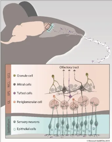
The structure of the OB has been well characterized, and several cellular layers are easily identifiable, depending on their location in the OB (Figure 1.1). The outermost layer, the olfactory nerve layer (ONL), conveys the axons of olfactorysensory neurons (OSNs) that project to a very well-organized structure in the glomerular layer (GL), the glomerulus. Interestingly, axons from OSNs scattered throughout the OE that express the same olfactory receptor gather together in the same glomeruli, creating an odorant map in the OB (Mombaerts et al., 1996; Mori et al., 2006). Inside the glomeruli, OSN axons form synapses with the primary dendrites of OB principal cells, that is, mitral and tufted cells (MCs and TCs). Short-axon cells (SACs) and periglomerular cells (PGs), the first type of OB resident interneurons, also reside in the GL and play important roles in information processing. The external plexiform layer (EPL) lies below the GL and is home to the dendrites of MCs, the apical dendrites of granule cells (GCs), and the soma and lateral dendrites of TCs and parvalbumin-expressing neurons (Miyamichi et al., 2013).
Figure 1.1: Schematic representation of the OB.
Volatile odorant molecules, once they enter the nasal cavity, bind to olfactory receptors on the surface of OSNs in the OE. Odorant information is then transduced into an electrical signal and is transmitted to the glomerular layer of the olfactory bulb via OSN axons. Each OSN expresses one of the thousands of olfactory receptors and all OSNs expressing the same olfactory receptor project to the same pair of glomeruli. Inside the glomeruli, OSN axons form synapses with MCs and TCs, the output neurons of the OB. PGs are the first population of resident interneurons and are also found inside the GL. The cell bodies of TCs reside in the EPL, which is located deeper in the OB. The inner part of the EPL is lined with the MCs layer, which encloses the cell bodies of MCs. The GCL is the largest layer of the OB and is composed of GCs (source: Massouh bioMEDia).
The inner part of the EPL is delimited by the mitral cell layer (MCL), which is formed by the cell bodies of MCs. The granule cell layer (GCL), the largest and deepest layer of the OB, is composed of GC cell bodies. GCs are axon-less resident
interneurons in the OB. They send long, ramified secondary dendrites toward the EPL to form synapses with the lateral dendrites of principal cells. Lastly, the OB portion of the rostral migratory stream (RMS-OB) is the innermost layer of the OB and constantly replenishes the OB with immature neurons (Figure 1.1).
In the OB network, GC and PG interneurons play a major role in modulating the output of olfactory information sent by principal cells to higher brain regions. Interestingly, GCs constitute the most numerous cell population, and it is estimated that individual MCs can receive inputs from approximately 100 individual GCs and that each GC may contact hundreds of MCs (Shepherd, 1972). This important inhibitory drive on bulbar principal neurons allows the tight spatio-temporal modulation of information processing (Arevian et al., 2008; Gschwend et al., 2015; Urban, 2002; Yokoi et al., 1995). To modulate principal cell activity, PGs and GCs form a unique type of synapse: the reciprocal dendro-dendritic synapse (Isaacson and Strowbridge, 1998; Jahr and Nicoll, 1982) (Figure 1.2). Following their activation, MCs and TCs release glutamate onto GC or PG spines, which in turn leads to the release, within the same synapse, of the inhibitory neurotransmitter γ-aminobutyric acid (GABA) back to principal cell dendrites (Rojas-Libano and Kay, 2008; Whitman and Greer, 2007).
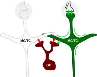
Figure 1.2: Reciprocal dendrodendritic synapse between mitral/tufted cells and granule cells
Action potentials from OSNs arrive at glomeruli (gray circles) and excite (short green arrows) MC\TC. This depolarization propagates from the postsynaptic membrane through the entire cell (long green arrows), resulting in excitation of GCs through glutamate liberation at the dendro-dendritic synapse. The excitation of granule cells causes them to liberate GABA (red arrows), eliciting recurrent and lateral inhibition of M/T cells. Note that due to the geometry of the arrangement, the same dynamical interactions can be elicited by antidromic activation of M/T cells
Individual MCs/TCs respond to only a few odorants by changing their firing rates, supporting the idea of sparse encoding of odor information in the OB (Bathellier et al., 2008; Davison and Katz, 2007; Fantana et al., 2008; Nagayama et al., 2004). Moreover, the inhibition of principal cells by GCs at dendrodendritic reciprocal synapses generates fast oscillations in the gamma range (20-80 Hz) (Fukunaga et al., 2014), resulting in the synchronization of MC activity (Arevian et al., 2008; Lagier et al., 2004; Lagier et al., 2007; Lepousez and Lledo, 2013). The proper functioning of the recurrent inhibition of MCs by GCs and the generation of gamma rhythms is also of particular relevance in the context of odor discrimination (Abraham et al., 2010; Lepousez and Lledo, 2013). In fact, modulating the imbalance between the excitation/inhibition of these synapses using picrotoxin, a GABA(A)-receptor antagonist, is sufficient to decrease the frequency of gamma oscillations and concomitantly alter the performance of animals in odor discrimination tasks (Lepousez and Lledo, 2013). This effect has been corroborated by inhibiting GC activity using a pharmacogenetic approach (use of designer receptors exclusively activated by designer drugs or DREADDs), which decreases pattern separation and slows down the learning of an odor discrimination task involving complex odor mixtures (Gschwend et al., 2015) (Figure 1.3). On the other hand, optogenetically activating GCs using channel-rhodopsin (ChR2) resulted in increased pattern separation (Gschwend et al., 2015) (Figure 1.3). These results thus highlight the importance of GCs and their inhibitory drive on principal cells regarding the decorrelation of overlapping MC/TC ensembles, an ability that underlies the capacity of animals to properly discriminate related odorants. The inhibition provided by PGs is also important for information processing and coding in the OB. PGs coordinate theta activity in the OB and regulate baseline and odor-evoked inhibition (Fukunaga et al., 2014). These GC- and PG-associated inhibitory networks thus control OB activities at different timescales, with PGs being involved in regulating the theta rhythm and GCs controlling gamma oscillations (Fukunaga et al., 2014).
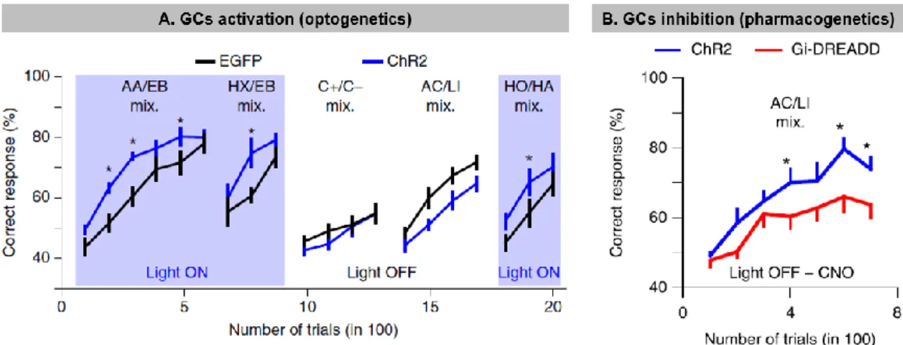
Figure 1.3: Modulation of granule cells activity influences odor discrimination performances
A. Optogenetic stimulation of GCL improves odor discrimination learning. Discrimination
performances for different groups of mice, which received injection of an adeno-associated virus (AAV) expressing either ChR2 or EGFP) in the GCL. Performances of ChR2-expressing mice (blue lines, n = 7) were specifically improved by photo-stimulation in comparison with EGFP-expressing mice (black lines, n = 7; Light ON: repeated-measures ANOVA, F = 8.3, P = 0.015, post hoc Fisher test at least *P < 0.034; Light OFF: repeated-measures ANOVA, F = 3.7, P = 0.08). Blue boxes indicate light ON episodes. Data are presented as mean ± sem. The odor pairs used were (all gas mixes): amyl acetate/ethyl butyrate (AA/EB) 60/40 versus AA/EB 40/60, ethyl butyrate/3-hexanone (EB/HX) 60/40 versus EB/HX 40/60, (+)-Carvone/(-)Carvone (C+/C–) 60/40 versus C+/C– 40/60, acetophenone/limonene (AC/LI) 60/40 versus AC/LI 40/60, and heptanol/hexanal (HO/HA) 60/40 versus HO/HA 40/60. B.Pharmacogenetic inhibition of GCL neurons deteriorates odor discrimination learning. CNO injection (2 mg per kg; all groups injected) decreased learning performances in Gi-DREADD–expressing mice in comparison with ChR2-expressing mice (n = 9 mice in each group; repeated-measures ANOVA F = 6.32 P = 0.023, post hoc LSD test at least *P < 0.045). Data are presented as mean ± sem. Odor pair used: AC/LI 60/40 versus AC/LI 40/60 (gas mixes). Adapted
from Gschwend et al., 2015.
Regeneration of the olfactory bulb during adulthood: the
process of adult neurogenesis
A. Adult neurogenesis: an overview
The OB is in direct contact with the outside environment through its contacts with OSNs. As already mentioned, the OE is responsible for the transduction of olfactory information to the OB via the axons of OSNs. Interestingly, the OE has a marked ability to regenerate since OSNs are in direct contact with the external environment. They thus have a short lifetime and are constantly being replenished (Calof et al., 1998; Graziadei and Graziadei, 1979; Murray and Calof, 1999). The axons of these newly generated OSNs form new connections with MCs and TCs in
the glomeruli of the OB (Mombaerts, 2001) and induce morpho-functional rearrangements of the GL circuitry.
The constant regeneration of OSNs and axonal remodeling in the glomeruli implies that an adaptive process that adjusts the bulbar network should also occur intrinsically in the OB. One current assumption is that the turnover of OSNs in the OE may underlie another regenerative process in the OB. Adult neurogenesis, which will be more extensively discussed below, provides the OB daily with thousands of newly generated cells by constantly renewing approximately 10-15% of GCs and 30% of PGs each (Ninkovic et al., 2007). Until now, no clear evidence has emerged of a direct link between these two neurogenic processes in the OE and the OB. However, it appears that both phenomena are sensitive to the level of sensory activity. For instance, olfactory enrichment results in increased OE neurogenesis and survival of adult-born OB interneurons (Cavallin et al., 2010; Rochefort et al., 2002; Rochefort and Lledo, 2005; Watt et al., 2004; Woo et al., 2006). On the other hand, sensory deprivation via a mechanical blockade of olfactory input entry or a lesion of the sensory epithelium results in a reduction in the thickness of the OE, a loss of olfactory sensory neurons and basal cells in the occluded side (Cavallin et al., 2010; Farbman et al., 1988), and a reduction in OB volume (Cummings et al., 1997) and the number of adult-born interneurons (Corotto et al., 1994; Petreanu and Alvarez-Buylla, 2002; Saghatelyan et al., 2005; Yamaguchi and Mori, 2005). It remains, however, to be determined whether the constant arrival of new sensory axons drives synaptic remodeling between interneurons and principal cells in the OB network and whether neurogenesis in the OE drives neurogenesis in the OB. OB neurogenesis involves a succession of processes, beginning with stem cell division and ending with the morpho-functional integration of adult-born neurons into the OB neuronal circuitry. Below, I briefly describe the generation and migration of neuronal precursors, with an emphasis on the developmental processes leading to the maturation and integration of adult-born neurons into the OB.
a. From stem cells to neuronal progenitors
The adult neural stem cells (NSCs) that produce neuronal precursors for the OB are located at the border of the lateral ventricle in the subventricular zone (SVZ) (Figure
1.4, Insight 1). These stem cells are derived from embryonic radial glial cells
(Fuentealba et al., 2015; Furutachi et al., 2015; Redmond et al., 2019) and express astrocytic markers such as glial fibrillary acidic protein (GFAP) and brain-lipid binding proteins (Codega et al., 2014; Fuentealba et al., 2015). NSCs exist in either a quiescent or proliferative state (Codega et al., 2014). Once activated, GFAP+ B1
cells express the epidermal growth factor receptor (EGFR) and give rise to rapidly dividing progenitors (type C cells) (Codega et al., 2014; Fuentealba et al., 2015; Obernier and Alvarez-Buylla, 2019). These cells in turn divide to generate migrating neuroblasts (Figure 1.4, Insight 1). Cells that express both GFAP and EGFR (or proliferative markers such as proliferating cell nuclear antigen (PCNA) and Ki67) are activated B1 stem cells (Codega et al., 2014; Pastrana et al., 2009). On the other hand, GFAP+ cells that do not express EGFR and proliferative markers may be either
quiescent stem cells or SVZ niche astrocytes. These two populations can be further distinguished by the expression of prominin, which is present on the primary cilia of NSCs and ependymal cells, but not niche astrocytes (Beckervordersandforth et al., 2010; Codega et al., 2014; Mirzadeh et al., 2008). It is currently thought that some of embryonic radial glia enters a long quiescent state until their re-activation in adulthood (Fuentealba et al., 2015; Furutachi et al., 2015). Adult NSCs are located in a specialized microenvironment and encounter multiple inter-cellular interactions and myriads of molecular factors derived from blood vessels, cerebrospinal fluid, niche astrocytes, neuroblasts, and other cells in the SVZ (Gengatharan et al., 2016; Obernier and Alvarez-Buylla, 2019). Adult NSCs have to decode and integrate all these microenvironmental factors to either remain in a quiescent state or become activated to generate neurons and glia. Once activated, adult NSCs undergo self-renewing or differentiating divisions that either lead to the expansion and maintenance of the NSC pool or to the production of rapidly dividing transient amplifying C-type cells (Obernier et al., 2018). C-type cells in turn generate neuroblasts (Alvarez-Buylla and Garcia-Verdugo, 2002; Doetsch et al., 1999; Luskin,
1993) that migrate long distances toward the OB (Gengatharan et al., 2016; Saghatelyan, 2009).
Figure 1.4: Adult neurogenesis in the olfactory bulb
A scheme of the adult mouse forebrain showing the different steps of adult neurogenesis, from the subventricular zone (SVZ) to the olfactory bulb (OB). Insight 1: Cellular organization of the SVZ: Type B cells are neural stem cells (NSCs). Once activated, these NSCs either self-renew or give rise to transient amplifying C-type cells, which in turn generate neuroblasts. Insight 2: Migration in the rostral migratory stream (RMS). Once neuroblasts enter the RMS, they migrate tangentially in chains along blood vessels and inside astrocytic tunnels. Insight 3: Differentiation and maturation of adult-born neurons in the OB. In the case of GCs, immature neurons go through five developmental stages. Neuroblasts first finish migrating tangentially in the RMS-OB (class 1 cells) and then migrate radially in the OB to reach their final location in the GCL where they become class 2 cells. In the GCL, immature GCs (class 3 cells) start extending their primary dendrites toward the EPL. Their dendritic arborization will later complexify and form spineless secondary dendrites in the EPL (class 4 cells). Lastly, type 5 GCs form spines and fully integrate the OB network, forming synapses with principal cells. Source: Massouh bioMEDia.
b. Neuroblasts migration in the adult brain
To reach the OB, neuroblasts follow the RMS, a long and intricate migratory pathway (Figure 1.4, Insight 2). The neuroblasts first migrate tangentially along the RMS and, once in the OB, turn to migrate radially and individually out of the RMS into the GCL and GL. In the adult RMS, neuroblasts travel in chains unsheathed by astrocytic processes (Kaneko et al., 2010; Lois and Alvarez-Buylla, 1994), and their migration is under the control of various signaling molecules, including those derived from astrocytes (Gengatharan et al., 2016). Cellular interactions between neuroblasts and blood vessels are also required for faithful migration toward the OB. Neuronal precursors use blood vessels that topographically outline the RMS for their migration (Snapyan et al., 2009; Whitman et al., 2009). Furthermore, endothelial cells release trophic factors that foster neuronal migration (Snapyan et al., 2009), indicating that blood vessels provide not only a physical scaffold but also molecular cues required for neuroblast migration. In addition to factors derived from astrocytes and endothelial cells, factors released by neuroblasts themselves affect their own migration. For example, the autocrine action of GABA on neuronal migration through GABAA receptors on neuroblasts has been documented (Bolteus and Bordey, 2004;
Platel et al., 2008; Snapyan et al., 2009). Some 30,000 to 40,000 neuroblasts arrive in the OB every day (Petreanu and Alvarez-Buylla, 2002). This massive and unique migration of neuronal precursors in the adult brain is under the tight control of several molecular signals (Gengatharan et al., 2016; Kaneko et al., 2017).
c. Becoming a functional neuron in a pre-existing network
The vast majority of neuroblasts (approximately 90%) in the OB become GCs that mostly populate the deep layer of the GCL (Imayoshi et al., 2008), whereas the remaining immature neurons differentiate into PGs (Winner et al., 2002). Approximately four weeks are required from the moment immature neuronal precursors are generated in the SVZ to the full morpho-functional maturation of adult-born neurons in the OB (Carleton et al., 2003; Petreanu and Alvarez-Buylla, 2002). As they mature through the development periods, adult-born neurons undergo increasingly complex changes in their morphology, functions, and
channel/receptor repertoires. Five developmental classes (Figure 1.4, Insight 3) have been described for GCs (Carleton et al., 2003; Petreanu and Alvarez-Buylla, 2002). Class 1 cells correspond to neuroblasts that migrate tangentially in the RMS-OB. Class 2 cells correspond to neuroblasts that migrate radially in the OB toward their final destination in the GCL. Once installed in the GCL, immature class 3 GCs start to extend their primary dendrite toward the EPL, with no contacts with MCs. At the end of the second week, adult-born GCs undergo the final maturational steps. They first extend and complexify their dendritic arborization to form spineless secondary dendrites in the EPL (class 4 cells) and ultimately, after a process of spinogenesis, fully integrate the bulbar circuitry and form synapses with MCs as class 5 GCs (Carleton et al., 2003; Petreanu and Alvarez-Buylla, 2002).
From a functional point of view, adult-born neuroblasts express both AMPA and GABA receptors and start to receive spontaneous synaptic inputs at early stages of their maturation, that is, shortly after completion of the migration phase (Carleton et al., 2003). They first receive excitatory glutamatergic and inhibitory GABAergic inputs on their soma and proximal part of their apical dendrites (Kelsch et al., 2008; Panzanelli et al., 2009; Whitman and Greer, 2007) (Figure 1.5). Driven partly by these synaptic inputs, adult-born neurons mature and extend their dendrites from the GCL to the EPL, which is followed by the formation of axo-dendritic inputs impinging on their basal dendrites and dendro-dendritic inputs on their secondary dendrites. The end of GC synaptic maturation is marked by the establishment of the major output synapses that GCs establish in the OB, that is, the dendro-dendritic inhibitory synapses with MCs/TCs (Kelsch et al., 2008) (Figure 1.5). This sequential acquisition of input and output synapses differentiates adult-born cells from early-born ones, with early-early-born cells acquiring outputs and inputs simultaneously (Kelsch et al., 2008) (Figure 1.5). Another particularity of adult-born GC maturation is delayed action potential (AP) firing (Carleton et al., 2003) which may allow adult-born GCs to silently integrate into the bulbar network, without disturbing it by “uncontrolled” neurotransmitter release (Lledo et al., 2004).
Figure 1.5: Synaptic development on adult- and early-born GCs.
A. Schematic of the dendritic domains of a GC, showing the basal (blue), proximal (green) and apical
(red) domains of the dendritic tree. B. Diagrams illustrating the developmental pattern of input (left diagram) and output (right diagram) synapses during maturation of adult-born GCs. C. Diagrams illustrating the developmental pattern of input (left diagram) and output (right diagram) synapses during maturation of early-born GCs. Adapted from Kelsch et al., 2008.
Unlike GCs, adult-born PGs fire APs, have fully developed voltage-gated sodium and potassium conductances at early stages of their maturation (Belluzzi et al., 2003), and reach their final location in the OB faster than the GCs despite the fact that they have to move over longer distances (Hack et al., 2005). Interestingly,
in vivo two-photon imaging combined with sensory stimulation has shown that while
adult-born PGs are still migrating in the GL, approximately half have functional responses to odor stimulation that are undistinguishable from their early-born counterparts (Kovalchuk et al., 2015). The structuro-functional maturation and integration of PGs thus occurs very rapidly and concurrently in the GL.
B. Adult neurogenesis generates a specific subset of olfactory bulb interneurons
The odor environment is in constant flux and the OB has to adapt and reorganize its network to deal with these changes. Adult-born cells are constantly supplied to the bulbar network, which could facilitate the adaptive response of the bulbar network to changing environmental conditions. In the following paragraphs, I discuss the unique properties intrinsic to adult-born interneurons as well as the specific roles played by this particular population of cells in OB functioning and behavior.
a. Properties of adult-born neurons
Early-born and adult-born interneurons undergo different survival and targeting dynamics. It has been reported that early-born interneurons have longer lifetimes than their adult-born counterparts (Lemasson et al., 2005; Petreanu and Alvarez-Buylla, 2002). These two populations of GCs also colonize different parts of the GCL. GCs born during the first post-natal weeks are found mostly in the superficial GCL (Lemasson et al., 2005). On the contrary, a genetic ablation of adult-born GCs results in a loss of cells predominantly in the deep GCL, while the superficial GCL is spared (Imayoshi et al., 2008). Since no morphological differences between deep and superficial GCs have been observed, and since deep GCs may form synapses preferentially with MCs whereas superficial GCs tend to form synapses with TCs (Greer, 1987; Shepherd et al., 2007), it is conceivable that early-born GCs mainly regulate the synaptic activity of TCs whereas adult-born GCs regulate the activities of both MCs and TCs. Indeed, it has been shown that the ablation of neurogenesis using an antimitotic drug results in a 45% decrease in the inhibition received by MCs after 28 days (Breton-Provencher et al., 2009). The same decrease has also been observed following sensory deprivation (Saghatelyan et al., 2005). This marked effect of adult-born cells on the activity of principal cells is corroborated by the fact that adult-born cells display higher excitability than pre-existing neurons in the OB. Indeed, both fully mature adult-born GCs and PGs display larger sodium currents with steeper conductance-voltage relationships and more negative activation
voltages than early-born interneurons. This may result in greater excitability to incoming sensory inputs and thus greater recruitment for odor processing.
Adult-born neurons appear to play a major role in mediating the ability of the OB network to rapidly adapt to changes in the external olfactory environment. Indeed, an analysis of immediate early genes has revealed that adult-born neurons have stronger responses to novel odors than early-born interneurons, at least during the second week after their birth, a period known as their critical period (Belnoue et al., 2011; Magavi et al., 2005) (Figure 1.6). This critical period also coincides with the time window during which a process of long-term potentiation of excitatory inputs on the proximal dendrites of adult-born cells can be induced (Nissant et al., 2009). Similarly, in vivo two-photon targeted recordings of adult-born PGs at different developmental stages has revealed that their responsiveness increases after the start of their synaptic integration, even when they are still migrating in the GL (Livneh et al., 2014).
Figure 1.6: Adult-born neurons are more sensitive to novel odors
A. Mice receiving only clean, HEPA-filtered air have very few c-fos-positive (white) GCs. The specks
in the ventral glomerular layer are autofluorescent fixation artifacts and are easily distinguished from genuine cellular labeling at higher magnification. B. Novel odor exposure dramatically increases the
number of c-fos-positive GCs, MCs, and PGs. C-F. Confocal imaging reveals that Bromo-desoxy-uridine (BrdU, green) adult-born cells in the GCL differentiate into mature NeuN-expressing neurons (blue) and express c-fos (red) in response to olfactory stimulation. The arrows indicate activated adult-born neurons. The arrowhead indicates an inactive adult-adult-born neuron. Scale bar, 10μm. G. Adult-born neurons are most responsive soon after they synaptically integrate; as they mature, they become less responsive. Pre-existing granule neurons maintain a stable response. Adapted from Magavi et
al., 2005.
OB interneurons remain plastic well beyond their final maturational stages. In fact, spine turnover, i.e., the addition of new spines and the elimination of pre-existing ones, remains at the level of the secondary dendrites of mature GCs for extended periods of time, even up to one year following the generation of the cells in the SVZ (Sailor et al., 2016). It has been estimated that approximately 20% of the spines on GCs, either pre-existing or adult-generated, are being continuously renewed (Sailor et al., 2016). However, although this constant synaptic turnover could be one of the mechanisms underlying OB adaptability and plasticity in response to environmental changes, synaptogenesis is a slow process and cannot cope with rapid changes in the odor environment. The bulbar network thus requires a fast adaptation process to rapidly respond and adjust to the changing olfactory environment. In line with this, a new phenomenon of structural plasticity in the OB has recently been described and involves the relocation of mature GC spines in an activity-dependent manner (Figure 1.7). Two-photon in vitro and in vivo experiments have revealed that adult-born GCs, in response to a stimulus, are able to extend a protrusion or spine head filopodia (SHF) from their spines and toward an activated principal cell (Breton-Provencher et al., 2016). SHFs are found almost exclusively on the spines of the secondary dendrites of adult-born neurons and are guided by MC odor-evoked activity. The protrusion of SHFs is followed by the relocation of the spine in the direction of the activated MC, an event that depends on the secretion of brain-derived neurotrophic factor (BDNF) by the principal cell (Breton-Provencher et al., 2016). Such rapid structural modifications, which have also been observed in other brain regions during the critical period (Majewska and Sur, 2003; Weinhard et al., 2018), may allow for rapid adaptive responses in the bulbar network to changing environmental conditions. Indeed, a modeling study has corroborated this notion by showing that as few as 3% of the spines relocating to activated MC dendrites may
allow the synchronization of MCs in response to a sensory stimulation (Breton-Provencher et al., 2016).
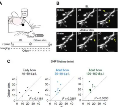
Figure 1.7: Plasticity of adult-born neuron spines is driven by sensory activity
A-B. Experimental procedure and time-lapse imaging during sensory stimulation in vivo. Scale bar,
5μm. C. Quantification of effects of sensory stimulation on the SHF lifetime in vivo for early-born GCs at 45–60 days post-injection (dpi). and adult-born GCs at 30–50 and 120–150 d.p.i. (n=7, 8 and 7 cells from 6, 6 and 4 mice, respectively). Adapted from Breton-Provencher et al., 2016.
b. Responses of adult-born cells to sensory stimulations
It is well now established that environmental factors, specifically sensory experiences, have a substantial impact on the fate of adult-born OB neurons (Figure
1.8). Exposure for an extended period of time to an enriched olfactory environment
results in a noticeable increase in the survival and maturation of adult-born neurons (Moreno et al., 2009; Rochefort et al., 2002; Rochefort and Lledo, 2005). In vivo two-photon imaging of mature adult-born PGs and GCs has revealed that their synapses stabilize following odor enrichment (Livneh and Mizrahi, 2012) and that depriving animals of olfactory inputs negatively impacts the survival rate of immature adult-born OB interneurons, resulting in an overall decrease in the size of the deprived OB. This effect occurs more specifically at the moment of their synaptic integration
into the pre-existing bulbar network, i.e., the critical period of adult-born cells (Corotto et al., 1994; Petreanu and Alvarez-Buylla, 2002; Saghatelyan et al., 2005; Yamaguchi and Mori, 2005). These data are also corroborated by a recent in vivo imaging study suggesting that while most adult-born GCs survive in the OB following their arrival, sensory deprivation reduces the survival rate and maintenance of adult-born neurons (Platel et al., 2019).
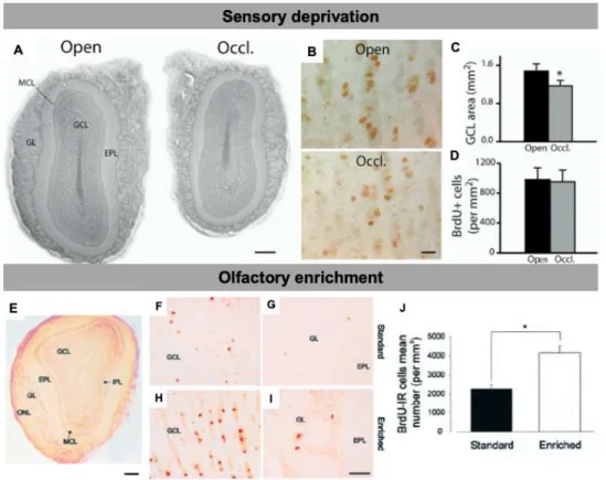
Figure 1.8: Adult-born neurons are sensitive to the level of olfactory activity
A. Photomicrographs of coronal sections displaying the different layers of the MOB. Note the size
difference between the control (Open) and the occluded (Occl.) bulb sections of the same animal taken at the same rostrocaudal position. B. BrdU-positive cells in the granule cell layer of the control (Open) and odor-deprived (Occl.) bulbs. C. Decrease in the GCL area following odor deprivation. D. Density of BrdU-labeled cells in the control and odor-deprived bulbs. BrdU counting was performed 21 days following BrdU injections and 33 days following unilateral nostril occlusion. *p < 0.05 with a Student’s t test. E. Photomicrograph of a coronal section of the main olfactory bulb displaying the different layers. F-I. BrdU-IR cells in the GCL (F,H) and in the GL (G,I ) of standard (F,G) and enriched (H,I) mice. H, Mean number of BrdU-IR cells per millimeters cubed. *p<0.05 with a Student’s t test. Scale bars: E, 300 µm; F-I, 20 µm. IPL, internal plexiform layer. Adapted from Saghatelyan et al.,
Sensory deprivation also has an effect on the maturation of adult-born cells, resulting in a decrease in the spine density of their secondary dendrites and a decrease in overall dendritic length (Kelsch et al., 2008; Saghatelyan et al., 2005). Interestingly, despite such drastic morphological changes, adult-born GCs retain their functional role in the OB network by increasing their excitability, indicating that adult-born interneurons possess intrinsic adaptive plasticity mechanisms that respond to changing environmental conditions (Saghatelyan et al., 2005).
All the unique features provided by adult-born neurons to the OB network underscore their importance as well as their specific signature and contribution to olfactory processing. In fact, they are one of the ways the OB can adapt and respond to new incoming odorant inputs. Moreover, if one considers the populations of adult-born and early-adult-born interneurons as physiologically independent cell populations, it is conceivable that adult-born neurons also contribute in unique way to different olfactory behaviors.
Functions of olfactory bulb interneurons in olfactory
functioning and behavior
The process of adult neurogenesis remains a fascinating topic given that the brain was once considered as being unable to replenish its neuronal pool. Nevertheless, the reasons for and the role played by such a process in the OB remain to be fully elucidated. When it comes to odor behavior, it is possible to distinguish between odor-based behaviors that have a social relevance for the animals and behaviors involving neutral odorants in ambient air and that are divided into spontaneous and reward-associated behaviors. Below, I discuss the role of OB interneurons in these different types of olfactory behaviors.
A. Role of OB interneurons in socially relevant behaviors
Olfaction intervenes at the different steps of an animal’s development, shaping the interactions or reactions that an individual has with its conspecifics and with predators. The involvement of bulbar adult neurogenesis in ethologically relevant odor behavior differs depending on the sex of the animal and the type of behavior.
In fact, the ablation of neurogenesis in mice using either an irradiation or a genetic strategy counteracts several innate behaviors, including male-male aggression, sexual behavior toward females, and recognition of predators (Feierstein et al., 2010; Sakamoto et al., 2011). Neurogenesis also increases in males that interact with their offspring during the postnatal period. This increase is required for paternal recognition once the offspring reaches adulthood (Mak and Weiss, 2010).
The link between adult neurogenesis in the SVZ and social behavior has been more extensively studied in females, more specifically in the context of pregnancy and maternal behavior. Pregnancy per se has been reported to have different effects on adult neurogenesis, including a prolactin-mediated increase in SVZ proliferation during gestation in rodents (Larsen and Grattan, 2010; Shingo et al., 2003; Wang et al., 2013) and a downregulation of neurogenesis in parturient ewes compared to non-pregnant ewes (Brus et al., 2014). Another study reported a more specific effect on the dendritic arborization of adult-born cells (Belnoue et al., 2016), indicating that these cells may contribute to the OB network and olfactory processing. In line with this, in vivo recordings of neurons generated during the pregnancy of lactating mice revealed enhanced synaptic integration reflected by the stabilization of their spines compared to naïve subjects (Kopel et al., 2012). Moreover, pregnancy appears to increase the specificity of adult-born cells toward certain types of odorants (Vinograd et al., 2017). Motherhood also increases the inhibitory drive of interneurons onto MCs, which respond more strongly to behaviorally relevant odors and more sparsely to neutral odors (Vinograd et al., 2017).
Several studies have also tried to link adult neurogenesis in the OB to the establishment of maternal behavior. In mother ewes, interactions with lambs result in an increased activation of adult-born neurons in the GCL, an effect that is specific to lamb-related odorants (Corona et al., 2017). It has also been shown that the ablation of neurogenesis using a genetic approach (Sakamoto et al., 2011) or the reduction of the pregnancy-induced increase in neurogenesis using bromocriptine, a prolactin secretion suppressor, affects maternal interactions with their offspring (Larsen and Grattan, 2010). In contrast, focal irradiation of the SVZ spares the
establishment of normal maternal behavior in treated females and does not affect maternal discrimination of their own pups from strange pups (Feierstein et al., 2010). Maternal recognition is, however, altered in gestationally stressed mothers in which the beneficial effect of motherhood on the dendritic arborization of adult-born GCs is counteracted (Belnoue et al., 2016). Hence, all the results presented above highlight the importance of OB adult neurogenesis in socially relevant behaviors.
B. Role of OB interneurons in spontaneous odor-behavior
Spontaneous odor behaviors are based on the motivation of the animals to perform the task. The time spent exploring is recorded, which gives an indication as to whether the animal performed the task correctly or not. The involvement of adult-born neurons in spontaneous odor behavior tasks such as the odor detection threshold, habituation/dishabituation odor discrimination, short-term odor memory, and perceptual learning has been investigated. The odor detection threshold corresponds to the minimal concentration at which an animal is able to detect the presence of an odorant and is reflected by an increase in exploration time. Habituation/dishabituation odor discrimination is based on the successive exposure of the animal to the same odorant (habituation phase), which is reflected by a decrease in the exploration time of the odorant in successive sessions. During the second phase of the test, a novel odor (dishabituation odor), which is different from the habituation odor, is introduced, leading to an increase in exploration time and thus discrimination between the two odorants. Different versions of this task exist, depending on the molecular similarity of the habituation and dishabituation odors. The use of chemically similar odorants increases the difficulty of the task. In the short-term olfactory memory task, the animals are exposed twice to the same odorant separated by various time intervals ranging from several minutes to several hours. Once again, a decrease in exploration time during the second presentation of the odor attests to the animal’s memory of the odor. For the perceptual learning task, the animals are first submitted to a habituation/dishabituation task using very similar odorants that animals are naturally unable to discriminate. Following a 10-day period
of olfactory enrichment with these two odorants, the animals are resubmitted to the discrimination task and are now able to complete it.
Many studies have addressed the role(s) of adult-born neurons in olfactory behavior but have given conflicting results. The approaches used to modulate the levels of adult-born cells arriving in the OB, the behavioral tests used, and heterogeneous adult-born populations, with each subtype playing a distinct role in an odor behavioral task, may explain the discrepancies (see below). Olfactory enrichment, a process known to enhance the survival of adult-born cells in the OB (Moreno et al., 2009; Rochefort et al., 2002; Rochefort and Lledo, 2005) also has a positive effect on the discrimination ability of rodents that were unable to discriminate between two enantiomers prior to enrichment (Mandairon et al., 2006c; Mandairon et al., 2006d; Moreno et al., 2012; Moreno et al., 2009). This effect on discrimination ability can be counteracted when an anti-mitotic drug is infused during olfactory enrichment (Moreno et al., 2009). Discrimination is also affected in transgenic animals in which the migration of neuroblasts is affected by a deficiency in neural cell adhesion molecule (NCAM) or variant brain-derived neurotrophic factor (Val66Met) (Bath et al., 2008; Gheusi et al., 2000). It should be noted, however, that constitutive gene knock-out and knock-in may affect the development and function of the olfactory system and may lead indirectly to changes in odor behavior. Similarly, olfactory enrichment may also affect sensory neuron turnover and plasticity in the OB (Jones et al., 2008; Watt et al., 2004). When the generation of adult-born neurons is ablated using -D-arabinofuranoside (AraC), an anti-mitotic drug, no changes in spontaneous odor discrimination are observed (Breton-Provencher et al., 2009). These results show that different approaches for modulating neurogenesis can have different impacts on behavioral outcomes. Furthermore, the use of similar or dissimilar odorants in the discrimination task may also impact the behavioral results since adult-born neurons may be involved in more complex odor discrimination tasks.
Olfactory enrichment is also beneficial for short-term odor memory. Enriched animals displaying increased survival of adult-born neurons are able to recall previously encountered odorants for much longer periods than non-enriched mice
(Rochefort et al., 2002). In line with this, the ablation of adult neurogenesis by AraC infusion impairs short-term odor memory (Breton-Provencher et al., 2009) while the genetic ablation of 2-containing nicotinic acetylcholine receptors, which increases the survival of adult-born cells, improves short-term odor memory (Mechawar et al., 2004).
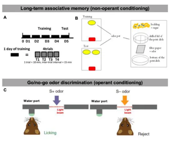
C. Role of OB interneurons in associative odor-behavior
The role of adult-born neurons in odor-reward conditioning tasks has also been extensively studied. Among those, it is possible to distinguish between non-operant and operant conditioning tasks (Figure 1.9). In long-term associative odor memory tasks, a form of non-operant conditioning tasks, partially food-deprived animals are trained for 4 days to discriminate between two odorants presented independently and hidden under clean bedding (Mandairon et al., 2011) (Figure 1.9) . A day of training is composed of a session of 4 randomized trials during which one of the two odors is associated with a sugar reward and presented for 2 trials. During the 2 remaining trials, the second odorant is presented alone, without any reward. During the test day, which can be 24 h or several days after the training day, both odorants are presented simultaneously but without a sugar reward. The time spent digging above each odorant is recorded, and if the animals are able to discriminate an odor previously associated with a sugar reward, they dig for a much longer time.
The pharmacological (AraC) or genetic ablation of adult neurogenesis has no effect on long-term associative memory tasks (Breton-Provencher et al., 2009; Imayoshi et al., 2008). Similarly, the suppression of programmed cell death in BAX knock-out mice has no effect on the taste aversion paradigm (Kim et al., 2007). However, it has been also reported that the modulation of neurogenesis has an impact on long-term associative memory (Lazarini et al., 2009; Sultan et al., 2010). It should be noted, however, that Sultan et al. (2010) studied the role of adult-born neurons in long-term memory retention and not in memory acquisition (Sultan et al., 2010). Similar results have been obtained in odor-cued fear conditioning following focal SVZ irradiation (Valley et al., 2009). These findings were further corroborated by another study that took advantage of the tag-and-ablate genetic strategy. Following a treatment with tamoxifen, this model makes it possible to “tag” a large