The role of microglia in the olfactory bulb
Mémoire
Helia Tenza Ferrer
Maîtrise en neurobiologie
Maître ès sciences (M. Sc.)
Québec, Canada
Resumé
Les microglies sont petites cellules gliales du système nerveux centrale (SNC) considérées comment les cellules immunitaires pour la ressemblance avec les macrophages et la capacité de phagocyter. Bien quelles sont distribués partout le SNC, la densité, la morphologie et l’état d’activation de la microglie diffère selon la région du cerveau. Il a été signalé récemment que les microglies sont fortement activées dans le bulbe olfactif (OB) des souris saines. C’est cet état constant d’activation qui nous suggère que les microglies peuvent jouer un rôle crucial dans l'OB. Dans cette étude, nous avons cherché à expliquer le rôle de la microglie en l’OB.
Nous avons évalué les changements survenus dans la densité et le niveau d’activation de la microglie avec: l’abolition de la neurogenèse, la deprivation sensorielle et la présentation des molécules d’odeur. Fait intéressant, en utilisant des souris transgéniques portant le gène de la luciférase sous le contrôle transcriptionnel du promoteur de TLR2, nous avons observé que la présentation d’odeur a induit une activation de la microglie.
Nos résultats suggèrent que les microglies dans l'OB peuvent être impliqués dans le traitement sensoriel et jouer donc un rôle important dans le comportement animal.
Abstract
Microglia are small glial cells present in the central nervous system (CNS) that are considered as resident immune cells because of their resemblance with macrophages and capacity to phagocyte. Although they are distributed throughout all the CNS, the density, morphology and state of activation of microglia vary according to brain region. It has been recently reported that microglia are highly activated in the olfactory bulb (OB) of healthy mice. It remains however unclear why these cells are present in the activated alert state in the OB and what is their role in the bulbar network stability and/or function. In this study we aimed to examine the role of microglia in the OB.
We evaluated changes in the density and level of activation of microglia following: ablation of adult neurogenesis, sensory deprivation as well as presentation of odor molecules. Interestingly, using a transgenic mouse bearing the luciferase gene under the transcriptional control of a murine TLR2 promoter, we observed that odor presentation induces a dramatic increase in the microglia activation.
Our findings suggest that microglia in the OB may be involved in the sensory processing and play thus an important role in animal behaviour.
Table of contents
RESUMÉ ... III ABSTRACT ... V TABLE OF CONTENTS ... VII LIST OF FIGURES ... IX ABBREVIATIONS ... XI ACKNOWLEDGEMENTS ... XIII
1. INTRODUCTION... 1
1.1 MICROGLIA CELLS ... 4
1.1.1 Classification of microglia cells ... 4
1.1.1.1 Amoeboid microglia ... 5
1.1.1.2 Ramified microglia ... 7
1.1.1.3 Activated microglia... 8
1.1.2 Role of microglia ... 8
1.1.2.1 Toll like receptors: the case of TLR2 ... 9
1.1.2.2 The role of microglia in the healthy brain ... 10
1.1.2.3 The role of microglia under pathological conditions ... 12
1.2 OLFACTION AND OLFACTORY BULB ... 14
1.2.1 The olfactory bulb ... 15
1.2.1.1 Structure of OB... 15
1.2.1.2 Olfactory bulb as a first relay in the odor processing ... 18
1.3 ADULT NEUROGENESIS ... 19
1.3.1 OB neurogenesis ... 19
1.3.1.1 Neurogenesis in the olfactory epithelium ... 20
1.3.1.2 Neurogenesis in the olfactory bulb ... 20
1.4 OLFACTION AND NEUROGENESIS ... 23
1.5 THE ROLE OF MICROGLIA IN THE OB ... 24
2. GENERAL OBJECTIVE AND HYPOTHESIS ... 27
2.1 HYPOTHESIS ... 29
3. MATERIALS AND METHODS ... 31
3.1 ANIMALS ... 33
3.2 OSMOTIC MINIPUMPS INSTALLATION ... 33
3.3 SENSORY DEPRIVATION ... 34 3.4 BRAIN COLLECTION ... 35 3.5 BRDU ADMINISTRATION ... 35 3.6 MINOCYCLINE ADMINISTRATION ... 35 3.7 IMMUNOHISTOCHEMISTRY ... 36 3.7.1 Diaminobenzidine staining ... 36 3.7.2 Immunofluorescence labelling ... 36 3.8 IMAGE ACQUISITION ... 37
3.9 COUNTING IBA1 AND CD68 LABELED MICROGLIA CELLS IN OB ... 37
3.10 WESTERN BLOT ... 37
3.11 ODOR APPLICATION ... 38
3.11.1 Odors used ... 39
3.11.1.1 Carvone ( C10H14O ) ... 39
3.11.1.2 Terpinen-4-ol ( C10H18O ) ... 39
3.12 BIOLUMINESCENCE IMAGING IN VIVO ... 40
3.13 STATISTICAL ANALYSIS ... 41
4. RESULTS... 43
5. DISCUSSION ... 61
List of figures
FIGURE 1: REPRESENTATION OF DIFFERENT MORPHOLOGIES OF MICROGLIA DEPENDING ON
THEIR STATE OF ACTIVATION.. ... 6
FIGURE 2: THE OB ORGANIZATION. ... 17
FIGURE 3: ADULT NEUROGENESIS IN THE OB.. ... 22
FIGURE 4: CHEMICAL STRUCTURE OF (-)CARVONE. ... 39
FIGURE 5: CHEMICAL STRUCTURE OF (+)TERPINEN-4-OL.. ... 40
FIGURE 6: EXPERIMENTAL PROCEDURES OF THE INTRAVENTRICULAR TREATMENT WITH THE ANTIMITOTIC DRUG ARAC.. ... 46
FIGURE 7: DENSITY OF MICROGLIA IN OB AFTER INTRACEREBRAL TREATMENT WITH ARAC.. ... 47
FIGURE 8: ACTIVATION OF MICROGLIA IN OB AFTER INTRAVENTRICULAR INFUSION OF ARAC… 49 FIGURE 9: WESTERN BLOT ANALYSES OF MICROGLIA ACTIVATION IN OB.. ... 50
FIGURE 10: MAC1-SAPORIN AFFECTS MICROGLIA IN OB.. ... 52
FIGURE 11: EFFECT OF MINOCYCLINE ON MICROGLIA IN OB.. ... 53
FIGURE 12: UNILATERAL SENSORY DEPRIVATION INCREASES THE DENSITY OF MICROGLIA IN THE OB.. ... 55
FIGURE 13: INCREASE IN THE MICROGLIA DENSITY IN THE OB FOLLOWING SENSORY DEPRIVATION IS NOT ASSOCIATED TO CHANGES IN THE CELL PROLIFERATION.. ... 57
FIGURE 14: SENSORY DEPRIVATION INCREASES MICROGLIA DENSITY.. ... 58
Abbreviations
CNS: Central nervous system EPL: External plexiform layer GC: Granule cellGCL: Granule cell layer GL: Glomerular layer
IPL: Internal plexiform layer MCL: Mitral cell layer OB: Olfactory bulb OE: Olfactory epithelium
OSNs: Olfactory sensory neurons
PAMPs: Pathogen-associated molecular patterns PG: Periglomerular
RMS: Rostral migratory stream
RMSob: Rostral migratory stream in olfactory bulb SGZ: Subgranular zone
SVZ: Subventricular zone TLRs: Toll-like receptors
Acknowledgements
I thank Dr. Armen Saghatelyan for the opportunity of learning in his laboratory. For believing in me at the beginning, this helps a lot when you are in a foreign country.
I thank Dr. Jasna Kriz and her lab, for their collaboration in this project with their resources. And Dr. Jasna Kriz again, and Dr. Yves De Koninck for accepting being part of my committee, it’s an honor that so known and busy scientists as you, accept to take part in this.
Thanks to Katalin Toth, director of the program, for trying to help me in tough moments with her best suggestions.
Thanks to all my colleagues and friends in the lab and the research center. To meet you was one of the best gifts I will bring with me wherever I go. You taught me almost every thing I know of working in a lab. And of course, you gave to me the best funny and beautiful moments I had in Québec. These two years would not be the same without you.
To my friends in Barcelona: There are many years that I am far away but every time that I have the opportunity of seeing you I can feel that the time have not passed. This is possible only because of your warm company. “Guapes”, thanks for your pictures and conversations and support, you make me feel more like home.
A special thank to my family:
“Mare, ets una dona molt forta, has passat per moments complicats però sempre te n’has ensortit. I malgrat tot, sempre has estat al meu costat, gràcies. No deixis passar un sol dia sense un somriure i una voluntat per a que tot sigui encara millor”.
“Pare, sempre intentant ser positiu, no deixis que el temps t’ho prengui (jo ho intento copiar, malgrat no sempre sigui fàcil). Aprofita també els bons moments per a petits que siguin”.
“A la meva germana Alba. Una germana que s’ha preocupat sempre per a mi com una mare (tot i que no ho he sabut valorar moltes vegades). Sé que no t’agrada que estigui lluny de tu, que em necessities al teu costat, però has de saber que estigui on estigui sempre estem juntes”.
“I als que ja no esteu aquí: àvia i Salus. No és fàcil escriure-us però alhora no podia deixar dincloure-us en aquest treball després de com n’estàveu dorgullosos de mi. Tan de bo us ho pogués agrair en persona”.
Many people might not believe in destiny, but each day that passes I am surer that “the destiny” brought me Alex. Since the beginning, you are teaching me in my personal and professional life. Thanks for being by my side and for believing in me even when I really wanted to renounce to everything. I could not imagine a better partner in this life. I love you.
The central nervous system (CNS) of all vertebrates comprises several cellular elements that can be clearly distinguished on the basis of both histological and physiological features. Neurons have traditionally been considered as the most important cellular elements of the CNS because of their crucial role in the transmission of nerve impulses. During the last decades, however, a substantial amount of work revealed that glial cells also play an important role in the CNS development and function by regulating neuronal activity, plasticity and survival.
Among the different types of glial cells described in the CNS, macroglia and microglia are found. The macroglia includes astrocytes and oligodendrocytes, which are the most numerous and well characterized glial population.
In addition to macroglia cells, the microglia constitute an important part of the CNS glial population. Since their discovery, these cells have always been an enigmatic glial lineage whose existence and roles in the CNS have been debated. Nowadays, these small glial cells have become the subject of intense study regarding their critical importance in terms of brain’s immune system, neurological disease and normal CNS function (Ziv et al., 2006; Wake et al., 2009; Tremblay et al., 2010). Since microglia were thought to be quiescent or inactive cells in the non-pathological brain, studies on microglia were largely focused on the injured and diseased CNS (Fan et al., 2007; Wake et al., 2009). Although these studies have provided the general basis of the microglia morphology, behaviour and functions, crucial aspects of microglia biology during healthy conditions have remained a neglected research endeavour.
The discovery that microglia are not quiescent in healthy conditions prompted the studies on the function of these cells in the uninjured CNS. However, the role of microglia in the healthy olfactory bulb (OB) remains unknown.
1.1 Microglia cells
Early in the XX century, Pio del Rio Hortega designed the name "microglia cell" (Castellano and Gonzalez, 1996). After a series of studies, Hortega showed that the so-called third element of Cajal (the first and second elements were neurons and astroglial cells, respectively) was also constituted by two different strains: cells with a few extensions that he named oligodendrocytes (Del Rio-Hortega, 1921), and cells with particular characteristics called microglia or microgliocits (Del Rio-Hortega, 1924). The origin of microglia in the CNS has been debated during many years, but today there is a general consensus that, unlike the other brain cells, microglia are derived from mesodermal/mesenchymal progenitors that migrated from the periphery. Specifically, microglia progenitors derive from yolk sacs during early development. Once in the brain, these progenitor cells give rise to microglia, which will proliferate and differentiate to adult microglia (Alliot et al., 1999; Chan, et al., 2006; Ginhoux et al., 2010).
Microglia are considered as the resident immune cells of the CNS for their resemblance with macrophages and their capacity to phagocyte. They are surveillan cells that scrutinize the environment for pathogens and harmful molecules. In response to the brain insult, microglia undergo morphological changes that enable them to engulf microorganisms, immunogenic products and cell debris. Microglia can be distinguished using three parameters: morphology, function and their developmental states. These aspects will be discussed below.
1.1.1 Classification of microglia cells
Microglia cells vary in morphology depending on the microenvironment. Thus, their morphology in the adult brain in normal conditions is different than their morphology during the CNS development or after an injury.
Although microglia display a remarkable range of morphology, several parameters can be used to distinguish resting (in the absence of brain injury) and activated
(immunologically stimulated) populations. For example, the different microglia states can be classified according to the number and type of processes. Non-activated microglia present many fine processes (ramified microglia) while the most activated microglia display thick and retracted branches (Figure 1). It is generally thought that some intermediate activated states of microglia take place between resting and activated microglia (Jonas et al., 2012). Since the ramified and activated morphologies are not an all or none form, the frontier between these two morphologies is unclear, making difficult the functional classification of the intermediate states based on morphology.
1.1.1.1 Amoeboid microglia
Amoeboid microglia are similar to the peripheral macrophages, regarding the morphology. They have a big cellular body that projects only few or no processes. Like the macrophages, amoeboid microglia display the abilities of migration (Rieske et al., 1989) and adhesion to surfaces (Frei et al., 1988). Amoeboid microglia are ultrastructurally different from other types of microglia, not only by the morphology, but also by the presence of vacuoles and dense lysosomes in their cytoplasm (Del Río-Hortega, 1932; Dalmau et al., 1997). This type of microglia is abundant in the brain where they perform a fundamental role during the early stages of development. For example, studies have shown that microglia are involved in the elimination of the cell debris (Cuadros et al., 1993), synaptogenesis, gliogenesis and release of neurotrophic factors (Nakajima and Kohsaka, 1993). The high number of amoeboid microglia observed during development is thought to be due to the self-proliferative capacity of these cells (Castellano and González, 1996).
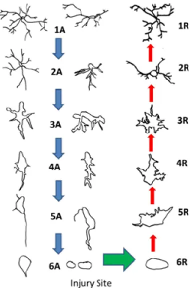
Figure 1: Representation of different morphologies of microglia depending on their state of activation.
Microglia’s morphology from the most “resting” (1A and 1R) to the most activated (6A and 6R). 1A to 6A depict different stages of microglia activation. From 6R to 1R depict the different stages that microglia pass when the injury is healed. In this case, microglia turn from the active to the resting states. Adapted from Jonas et al., 2012.
1.1.1.2 Ramified microglia
With the development of the CNS, amoeboid microglia is replaced by ramified microglia. The differentiation of amoeboid to ramified microglia is a process that occurs rapidly during the first two weeks of postnatal life (Dalmau et al., 1992; Wu et al., 1993). In this period the amoeboid microglia cells, located mainly in the white matter areas, migrate to invade areas of gray matter. At the same time, the cells begin to grow some extensions that are short and thick at the beginning, but longer and thinner later (primitive ramified microglia). During this differentiation process, the amoeboid microglia lose some distinctive features, such as their morphology and expression of certain enzymes (Ling and Wong, 1993). These ramified microglia also display changes in the antigenic profile in comparison to the amoeboid form (Ling et al., 1990; Ling et al., 1991). Some studies indicate that not all amoeboid cells differentiate into ramified microglia cells (Imamoto and Leblond, 1978; Wu et al., 1992). About two-thirds of the amoeboid cell population disappears (Imamoto and Leblond, 1978), leaving unclear whether they degenerate or they migrate out from the nervous tissue (Reid et al., 1992; Gehrmann et al., 1995).
A small soma and large processes that are highly arborized characterize the ramified microglia (Dalmau et al., 1997). Unlike amoeboid microglia, the ramified ones have neither vacuoles nor phagosomes. Ramified microglia are distributed heterogeneously in all brain regions (Lawson et al., 1990). Although they were thought to be “inactive” cells, often called quiescent or resting microglia, different functions of these cells have been discovered over the past years. For example, microglia interact directly with both presynaptic and postsynaptic parts of chemical synapses, under non-pathological conditions and controls the neuronal activity (Li et al., 2013; Wake et al., 2009). Additionally, they actively scan the brain microenvironment with their motile processes in a time scale of minutes to detect a possible injury or pathogen (Nimmerjahn et al., 2005). The constant surveying role of resting microglia is believed to be important in the maintenance of brain homeostasis (Nimmerjahn et al., 2005).
1.1.1.3 Activated microglia
Following brain injury, such as infection, trauma or neurodegenerative diseases, the microglia undergo rapid and profound changes in their morphology and gene expression that lead to the “activated” state of microglia.
The term of activated microglia should be understood as a transient state of alert that can either go back or lead to a superior degree of reactivity. Activated microglia also called reactive microglia shows signs of reactivity including morphological changes, expression of new antigens and changes in metabolic synthesis and secretion of certain substances. The activation of microglia is not an all or none process: these cells can acquire distinct functional states (Schwartz et al., 2006; Hanisch et al., 2007), show different morphological shapes (Jonas et al., 2012) and express different molecular signals while active. The microglia when active can acquire a phagocytic state, an advanced microglia reactivity culminating in the transformation into a macrophage cell. For this, ramified microglia retract their processes and turn into the amoeboid form. The active microglia not only can be similar to macrophages in the morphology, but they can also express the same molecular cues (Kreutzberg, 1996). Under this state, microglia are called “the phagocytes of the CNS”. Activated and phagocytic microglia play a crucial role in maintaining homeostasis in adult brain after pathologic conditions (Beyer et al., 2000).
I will discuss in more details the functions of microglia under pathological and healthy conditions in the chapter 1.2 “Role of microglia”.
1.1.2 Role of microglia
Since microglia are the resident immune cells of CNS, their main biological role is to scan the environment for signs of pathogens or tissue damage. Damaged or apoptotic cells release chemoattractant molecules so-called “find-me” signals like cytokines (ILs andTNF-α) (Smith, 2012), chemokines (fractalkine/CX3CL1) (Truman et al., 2008) and extracellular nucleotides (ATP, UDP) (Elliott et al., 2009). These substances recruit
microglia to the injured site, which discriminate the “eat-me” from the “don’t-eat-me” signals.
Microglia can express different type of receptors that allow them to fulfill their functions. Among these receptors are: neurotransmitter receptors, cytokine and chemokine receptors and pattern-recognition receptors. Among the neurotransmitter receptors, the activation of the AMPA receptors leads to a rapid remodelling of microglia cytoskeleton (re and depolarization of actin) and these changes play a role in the regulation of the motility and phagocytosis of activated microglia (Christensen et al., 2006). Microglia can express cytokine receptors such as the fractalkine receptor (CX3CR1), which promotes phagocytosis of apoptotic cells (Noda et al., 2011). This phagocytosis takes place once the cell membrane of microglia contacts the molecule to phagocyte through the receptor-ligand interaction. Microglia, as the principal resident immune cells of the CNS, express pattern-recognition receptors such as toll-like receptors (TLRs) (Olson and Miller, 2004).
1.1.2.1 Toll like receptors: the case of TLR2
Toll-like receptors (TLRs) are a class of proteins that initiate innate immune responses via recognition of molecules derived from pathogens, also known as pathogen-associated molecular patterns (PAMPs) (Akira et al., 2006). These receptors are important in various CNS diseases like infection, neurodegeneration and autoimmunity (Kettenman et al., 2011). At least 13 mouse TLRs have been described. These proteins act as surface-expressing TLRs (TLR1, 2, 4, 5, 6 and 10), which recognize mainly microbial membrane components or intracellular expressing TLRs (TLR3, 7/8 and 9) that recognize microbial nucleic acids (Akira et al., 2006; Kawai and Akira, 2010).
The TLRs (except TLR3) share a common signalling pathway called MyD88-dependent (Takeda and Akira, 2004). The MyD88-MyD88-dependent responses initiate when the receptors are activated by their ligand resulting in recruitment of the adaptor protein MyD88. MyD88, then, binds to the IL-1 receptor-associated kinases such as IRAK-4, which phosphorylate and activate the TNF receptor-associated factor6 (TRAF6). Activated TRAF6 binds to and induces IKK-β phosphorylation by TAK1. Finally, activation of the IKK complex leads to the phosphorylation of MAP kinases (JNK, p38 MAPK) and NF-κB
(Takeda and Akira, 2004; Kawai and Akira, 2010). This pathway induces adaptive responses including the synthesis and secretion of various inflammatory cytokines and chemokines.
The activation of TLR signalling in microglia induces morphological changes that turn these cells active having immune activity. In response to infection, activated TLR2 mediates the production of a number of important pro-inflammatory cytokines and chemokines including TNF-α, IL-1β, and CXCL1 (Medzhitov, 2001). Importantly, TLR2 has a putative role in microglia activation. For example, TLR2 seems to be necessary for the activation of microglia in the spinal cord (after peripheral nerve injury), hippocampus (during excitotoxic cell death) and whole brain (under ischemia) (Lalancette-Hebert et al., 2009; Hayward and Lee, 2014). TLR2 is usually found forming heterodimers with TLR1 or TLR6, which results in different ligand specificity. The heterodimer TLR1-TLR2 recognizes triacylated lipopeptides from Gram-negative bacteria while the heterodimer TLR2-TLR6 recognizes diacylated lipopeptides from Gram-positives (Kawai and Akira, 2010). This difference in recognition is due to distinct conformational changes in the protein structure. TLR2-TLR1 complex forms three lipid chains (two in TLR2 and one in TLR1) that bind to the triacylated lipopeptide of bacteria. For the dimer TLR2-TLR6, only two lipid chains interact with the PAMP because TLR6 lacks a hydrophobic channel. TLR2 also acts with other co-receptors to recognize PAMPs. For example, heterodimer TLR2-TLR6 is reported to interact with CD36 (Takeda and Akira, 2004; Kawai and Akira, 2010).
1.1.2.2 The role of microglia in the healthy brain
Because microglia are mostly studied under pathologic conditions, the comprehension of the microglia functions in healthy brain is still limited.
Microglia in non-pathologic conditions are being studied for their relation with formation and maintenance of synapses. During the development, the microglia play a role in the synaptic pruning – process where a large number of immature synapses are permanently eliminated while a subset of synapses is maintained and strengthened (Hua and Smith, 2004). Microglia can interact directly with synapses and control the neuronal activity (Li et al., 2013 and Wake et al., 2009). They contact pre- and postsynaptic
terminals and their processes display enlarged extremities or bulbous endings. The frequency of these contacts depends on the synaptic activity (Wake et al., 2009; Tremblay et al., 2010).
Additionally, microglia cells actively scan the brain microenvironment with their motile processes (Nimmerjahn et al., 2005). The constant surveying role of resting microglia is believed to be important in the maintenance of brain homeostasis since it allows detection of a possible injury or pathogen. Using in vivo experiments, Nimmerjahn and colleagues showed that under healthy conditions microglia act as a constant sensor of homeostasis of the brain. The authors found that the microglia continuously extend and retract their processes in the brain parenchyma, which leads to changes in their cellular morphology on a time scale of minutes (Nimmerjahn et al., 2005). The high degree of motility, which depends on actin polymerization (Nimmerjahn et al., 2005), facilitates efficient immune surveillance of the brain.
In healthy conditions, phagocytosis of apoptotic debris is performed by microglia (Neumann et al., 2009). This phagocytosis of apoptotic debris is beneficial for the brain because it reduces the secretion of pro-inflammatory cytokines, chemoattractants, and the migration of T lymphocytes (Magnus et al., 2001). It has been shown that, under healthy conditions, microglia can engulf different tissue components (Nimmerjahn et al., 2005; Tremblay et al., 2012). Indeed, Tremblay and colleagues observed phagocytic inclusions that resembled terminals, spines, and vesicles, in the cell bodies and processes of microglia under healthy conditions These inclusions increased correspondingly with the aging (Tremblay et al., 2012).
In early development, microglia activity contributes to neurogenesis (Antony et al., 2011), by removing the excess of debris generated by the proliferation of neuronal progenitors (Perry et al., 1985) and controlling the synapse formation by synaptic pruning (Paolicelli et al., 2011). In contrast, in adulthood, they have an interesting two-face role (Ekdahl et al., 2009). In the hippocampus, where the role of microglia in neurogenesis is more studied, the activation of microglia can be beneficial (Ziv et al., 2006) or detrimental (Ekdahl et al., 2003). In one hand, the microglia release pro-inflammatory mediators that contribute for clearing infections and repairing tissue. However, if left uncontrolled, the over pro-inflammatory response can perpetrate the neural insult.
As mentioned above, microglia also maintain homeostasis by engulfing cellular debris, lipids, and apoptotic cells in the non-inflamed state. This role represents a major regulatory influence on the process of adult neurogenesis (Sierra et al., 2010; Neumann et al., 2009) since majority of apoptotic cells are cleared out through phagocytosis by microglia. This phagocytic process can be done by ramified microglia. Unchallenged microglia phagocyte the apoptotic newborn cells through terminal or branches forming “ball-and-chain” structures (Sierra et al., 2010).
Numerous indications suggest that microglia cells regulate the synaptic activity in the normal brain by controlling the extracellular concentration of neurotransmitters and neuromodulators (Castellano, 1987). For example, it has been demonstrated that microglia cells have the necessary enzymes to promote degradation of adenosine and inosine phosphoesters to adenosine and inosine, respectively (Castellano, 1987). Two of these enzymes, the ATPase and nucleoside diphosphatase (NDPasa), were found in the cell membrane of ramified microglia (Castellano, 1987).
1.1.2.3 The role of microglia under pathological conditions
Microglia become activated in response to brain injuries or immunological stimuli (Kreutzberg, 1996). These cells undergo dramatic morphologic alterations upon activation, changing from resting microglia into activated amoeboid microglia (Kreutzberg, 1996). Then, complement receptors and major histocompatibility complex molecules are upregulated in the surface of activated microglia (Graeber et al., 1988).
It is generally thought that under CNS injury, the amoeboid microglia can become from resident microglia or monocyte-derived macrophages that infiltrate from peripheral systems (Simard et al., 2006). These cells are ontogenetically distinct from two myeloid populations: one originated from erythromyeloid precursors in the embryonic yolk sac (resident microglia) and another derived from hematopoietic stem cells in the bone marrow (Alliot et al., 1999; Chan, et al., 2006; Ginhoux et al., 2010; Schulz et al., 2012). Interestingly, monocyte-derived macrophages infiltrate into CNS only after resident microglia have been activated. This occurs due to production of chemoattractive molecules driven by resident activated microglia (Okamura et al., 2012). Once there,
monocytes-derived macrophages accumulate at the site of injury and influence on both tissue damage and repair. Distinctions between these two states of activated microglia within CNS lesion sites are difficult to perceive because they display similar amoeboid shape and apparently express the same markers (Streit et al., 1988; London et al., 2013; Bsibsi et al., 2014). Thus, many authors prefer to name these cells as macrophage/microglia.
Macrophages/microglia undergo two main phenotypes in response to environment-derived signals. The “M1” phenotype is the classical activation, in which the microglia display an IL-12high, IL-23high and IL-10low profile (Mantovani et al., 2013). IFN-γ and LPS induce this phenotype. Evidences show that M1 phenotype is associated with cytotoxic and anti-tumoral properties due to the secretion of interleukin-1β (IL-1β) and tumour necrosis factor-α (TNF-α), and therefore, it is considered as pro-inflammatory (Mantovani et al., 2004). CD16 and CD32 markers can identify this class of macrophages/microglia (David and Kroner, 2011). On the other hand, microglia with the alternative M2 phenotype express an IL-12low, IL-23low and IL-10high profile (David and Kroner, 2011). Interleukins (IL-4, IL-10), IL-1R agonists, immune complexes and TLRs have been described to induce this phenotype. M2 pattern is considered as anti-inflammatory due to the secretion of anti-inflammatory cytokines such as IL-10, resulting in immunoregulatory functions and tissue remodelling properties (Mantovani et al., 2004). Immunoreactivity to CD206 (mannose receptor) and arginase 1 (Arg1) markers can identify this class of macrophages/microglia cells (David and Kroner 2011).
In situations such as cerebral ischemia, contact between microglia processes and synapses are prolonged and usually followed by the disappearance of synapses (Wake et al., 2009). The activation of microglia can last several months and it is not restricted only to the injured site of the brain (Lalancette-Hebert et al., 2009).
The presence of reactive microglia has been described in many neurodegenerative diseases such as Alzheimer's, Parkinson's and Huntington. There is some controversy about whether the presence of activated microglia in these disorders is beneficial or detrimental (Bard et al., 2000; Fan et al., 2007). While a balanced and short-term inflammatory response is unlikely to promote secondary cell damage, the chronic inflammation otherwise can cause detrimental tissue effects, which overcomes the primary benefits. In Alzheimer's disease, for example, microglia’s phagocytosis is beneficial whereas the inflammation
factors released in this process are considered detrimental (Fan et al., 2007). Bard and colleagues have shown that the clearance of Aβ plaques by microglia increases the survival/cognitive deficits of mice model of Alzheimer disease (AD) and prevents the senile plaque expansion in the brain (Bard et al., 2000). On the other hand, other studies found that the suppression of the inflammatory response by microglia attenuated the symptoms of a mouse model of Alzheimer's disease (Fan et al., 2007).
Thus, microglia have different morphologies and functions depending on their environment and are considered as the resident immune cells of the CNS. These cells have become an interesting subject for scientists because these are versatile cells involved in many different processes in the brain.
1.2 Olfaction and Olfactory bulb
The olfactory sensory system is one of the most ancient senses responsible for detecting and processing chemical sensory information from the environment. These include general odorants, pheromones, and in some species, carbon dioxide (Ma, 2007). We constantly capture molecules in our environment that provide us important information about food, pleasure or danger. What humans generally think of as a single odor, like the odour of a flower, is usually a complex mixture of dozens of different odor molecules.
First of all, olfactory sensory neurons (OSNs) in the olfactory epithelium (OE) detect odor molecules through receptors located on the cilia of the OSNs. The binding of the odorant to its receptor leads to the generation of electrical signal that travel through the axon of the OSNs. The axons of OSN enter into the OB and make contact with the bulbar principal neurons. Then, the information is processed locally and transmitted from the OB to the piriform cortex. In the following sub-chapters, I will discuss the organization of the olfactory system in more detail.
1.2.1 The olfactory bulb
The OB is the structure of the brain responsible for the odor information processing (Fletcher et al., 2009), and it is one of the two regions in the brain that maintain ongoing neurogenesis during adult life (Lois and Alvarez-Buylla, 1994). The OB is supported and protected by the cribriform plate of the ethmoid bone, which in mammals separates it from the OE, and which is perforated by olfactory nerve axons. The bulb is divided into two distinct structures: the main and accessory OBs. This chapter will concentrate on the main OB, its principal function and its special characteristic: adult neurogenesis.
1.2.1.1 Structure of OB
The OB is a multilayered structure consisting of both gray matter and white matter arranged in six distinct concentric layers. From superficial to deep, these layers are: olfactory nerve, glomerular layer, external plexiform layer, mitral cell layer, internal plexiform layer, granule cell layer and rostral migratory stream of the OB (Roy and Roy, 2012). The mammalian olfactory nerve is composed from the axons of the primary OSNs, the cell bodies of which are located in the OE. The axons of these cells cross the cribriform plate, enter into a specialized structure in the OB called glomeruli and form the outermost layer of the OB: the olfactory nerve layer (ONL). The set of glomeruli forms the
glomerular layer (GL), where each glomerulus is devoid of cell somas and consists of
incoming OSN axons, the dendrites of the principal cells of OB - mitral and tufted cells-, local interneurons, and the processes of astrocytes. Inside the glomeruli, OSNs make synapses with the dendrites of mitral cells transmitting the odor information from the periphery to the OB.
The external plexiform layer (EPL) represents the next layer of the OB and is composed from the lateral dendrites of principal output neurons (mitral and tufted cells) and distal dendrites of granule cells (GCs). In this layer the major type of OB synapse, the dendro-dendritic synapses, are present between the dendrites of principal cells and GCs. EPL also contains the cell bodies of tufted cells and diverse population of interneurons making synapses with output neurons or other interneurons (Fletcher et al., 2009).
Just after the EPL, there is the mitral cell layer (MCL) where the soma of mitral cells is present. The mitral cells transfer information to the higher brain areas, such as piriform cortex, entorhinal cortex and amygdala.
Between the mitral cell and the granule cell layers there is a narrow layer relatively free of cell bodies, the internal plexiform layer (IPL), which mainly consists of granule cell dendrites passing through on their way to the EPL.
The innermost layer of the OB is the granule cell layer (GCL) that contains the cell bodies of the interneurons that give the name to this layer. These are axonless neurons that have basal and apical dendrites. The apical dendrite is composed of an unbranched proximal segment (also called primary dendrite) and highly branched distal segments (secondary dendrites) with spines. The spines of GCs are constituents of dendro-dendritic synapses that these interneurons form with the lateral dendrites of mitral/tufted cells. In dendro-dendritic reciprocal synapses, the glutamate is released from the lateral dendrites of the mitral/tufted cells onto the spine of a GC, which in turn induces the release of GABA back onto the principal cell dendrites (Price and Powell, 1970). The dendro-dendritic synapses are output synapses of GCs in the OB and are responsible for the recurrent and lateral inhibition of principal neurons (Isaacson and Strowbridge, 1998) (Figure 2).
The rostral migratory stream of the olfactory bulb (RMSob) is the region of the rostral migratory stream (RMS) inside of the OB. The RMS serves as a migratory route for neuronal precursors and consists mostly from neuroblasts, astrocytes and endothelial cells.
Figure 2: The OB organization. Distribution of the different layers and cells of the OB, from the most
external (the bottom of the figure) to the most internal (the top of the figure) (From Doty, 2012).
1.2.1.2 Olfactory bulb as a first relay in the odor processing
The mammalian olfactory system detects and discriminates a large variety of odor molecules. This is partially achieved by more than 1000 odorant receptors expressed on the cilial membrane surface of the OSNs in the OE (Buck and Axel, 1991). The odor molecules are volatiles and enter to the nose where they are dissolved in the mucus. Then, the odor molecules bind and activate the odorant receptors on OSNs.
The odorant receptors are members of a large family of seven transmembrane G-protein-coupled receptors (Godfrey et al., 2004). Studies of imaging and electrophysiology in flies and mice revealed that the odorant molecules can bind multiple receptors, activating different OSNs in the OE (Rubin, 1999). Interestingly, each OSN presents only one kind of receptor and all neurons expressing the same receptor project their axons into one or two glomeruli (typically one lateral and one medial glomerulus) (Gao, et al., 2000; Feinstein and Mombaerts, 2004). Such precise topographical targeting of OSNs axons leads to the precise spatially-restricted representation of olfactory signals in the OB and so-called spatial coding (Ma et al., 2012). Once the odor molecule binds to the receptor, it activates a G-protein situated in the cytoplasmatic side, and at the same time, this activates adenylyl cyclase, an enzyme embedded in the plasma membrane of the cilia. This enzyme catalyzes the conversion of ATP to cAMP – a second messenger – in the cytosol, that opens a sodium channel letting Na+ entry into the cell. This influx of Na+ depolarizes OSN and leads to the generation of action potential (Menini, 1999).
All this electrical information travels through the axons of the sensory neurons that reach the OB where they make excitatory synapse with dendrites of mitral and tufted cells inside the glomeruli. It is important to note that a given glomerulus receives input from OSNs expressing a given odorant receptor (Vassar et al., 1994). This molecular specificity established in the epithelium is maintained because each mitral cell receives afferent input in only two glomeruli, where all the incoming axons express the same odorant receptor.
Two classes of interneurons modulate mitral cells activity and thus information processing on the OB: the periglomerular (PGs) cells and the granule cells (GCs). The inhibition is provided via the dendrodendritic synapse formed by the dendritic spine of the interneuron and the dendrite of the mitral cells. This inhibition is involved in the temporal
coding of the odors, characterized by the synchronization of the activity between mitral cells in response to an odor (Schoppa and Westbrook, 2001). The constant change of GCs and, therefore, of the remodelling of this dendrodendritic synapses give an important plasticity in OB depending in the olfactory experience.
1.3 Adult neurogenesis
The neurogenesis, a process of generating new neurons from neural precursors, occurs not only during embryonic and perinatal stages, but also in adulthood in few regions of the mammalian brain.
Some decades ago, Altman showed the first evidence of neurogenesis with the presence of newborn GCs in the postnatal rat hippocampus (Altman and Das, 1965). Now it is known that neurogenesis is present during the entire lifespan of animals in almost all mammals examined (Eriksson et al., 1998).
The adult neurogenesis is present in two areas of the brain under normal conditions: the subgranular zone (SGZ) on the dentate gyrus of the hippocampus, and the SVZ where new neurons are generated and then migrate along the RMS into the OB (Alvarez-Buylla, 2002). Neurogenesis in other adult CNS regions is generally believed to be absent or very limited under normal physiological conditions but could be induced after injury (Gould, 2007). This section will only focus in OB neurogenesis.
1.3.1 OB neurogenesis
The neuronal circuitry of the OB is continuously remodelled throughout the life of animals (Lledo et al., 2006). Neurogenesis occurs not only in the SVZ but also in the OE. I will discuss these two neurogenic sites separately in the sub-chapters that follow.
1.3.1.1 Neurogenesis in the olfactory epithelium
As mentioned above, synaptic reorganization also occurs at primary synapses, in the GL between the axons of OSNs and dendrites of principal cells. In the 1940, Y. Nagahara reported that the neuronal replacement in the OE was evidenced by mitotic activity in the basal cells of the mouse OE (Nagahara, 1940). By the late 1970s, it became much more evident that neurons were continuously replaced in the adult OE in both normal physiological and pathological conditions (Farbman, 1992). The OE contains supporting cells, the ORNs and basal or progenitor cells, each of which occupies a defined place in the epithelium. The cell bodies of sensory neurons are located in the OE and the axons of these cells form synapses in the OB (Farbman, 1992).
The dendrites of ORNs, which contain the olfactory receptors, are in direct contact with the outside world. This location makes them very susceptible to damage caused by substances in the environment and by mechanical injury. This vulnerability is commonly used as an argument to explain the need for continuous neuronal replacement in the OE.
Neuronal replacement in the OE begins with the proliferation of stem cells. The basal cells divide asymmetrically every 30-50 days producing another stem cell and a neural precursor that rapidly divide several times, resulting in several immature neurons that migrate away from the basal membrane of the epithelium while they differentiate (Mackay-Sim and Kittel, 1991). Sensory neurons, which have typical life span of 30-60 days, are continuously renewed in the OE from a population of stem cells at the base of the epithelium (Crews and Hunter, 1994).
1.3.1.2 Neurogenesis in the olfactory bulb
While I will focus on the SVZ as the main proliferative zone of the OB newborn cells, it is important to note that the OB neurogenesis is not restricted to the wall of the lateral ventricles facing the striatum. The RMS also contains proliferative cells (Merkel et al., 2007). In adult SVZ, stem cells are astrocyte-like cells (also known as B-type cells) that divide slowly and give rise to transit-amplifying cells (C-type cells), which then divide
rapidly and give rise to neuroblasts (A-type cells) (Lois and Alvarez-Buylla, 1994), which express the migratory markers doublecortin (DCX) and PSA–NCAM. Neuroblasts migrate along the RMS, a well-delineated pathway, into the OB where they differentiate into GC or PG cells (Alvarez-Buylla and Garcia-Verdugo, 2002; Lledo et al., 2006).
Neuroblasts migrate tangentially in chains through the RMS in close association with astrocytes (Lois and Alvarez-Buylla, 1994) along blood vessels that not only serve as a physical scaffold for migrating neuroblasts but also provide essential molecular cues (Snapyan et al., 2009). When neuroblasts reach the OB, they detach from the chains and migrate radially to the GCL (David et al., 2013). The majority of these newborn cells (approximately 95%) differentiate into GC cells (Lledo and Saghatelyan, 2005), while the remaining population differentiate into PG cells (Figure 3). These new interneurons can replace dead cells in the OB, maintaining the integrity of the neural network (Imayoshi et al., 2008). Interestingly, within 15 to 45 days after their generation, approximately 50% of the newly generated cells die (Petreanu and Alvarez-Buylla, 2002). Then, the number of new cells remains constant for several months. The decrease in the number of new cells is probably caused by programmed cell death (Petreanu and Alvarez-Buylla, 2002). It has been hypothesized that the constant elimination of cells in the OB abolishes the transmission cycle of pathogens originated from the OSNs (Loseva et al., 2009). In addition, the regeneration process can even increase the plasticity of the olfactory network and enhance odor memory and discrimination (Ortega-Perez et al., 2007). Neurogenesis in the OB thus contributes to the capacity of the brain to change and adapt to the environment.
Figure 3: Adult neurogenesis in the OB. (Left) Newborn cells generated in the SVZ migrate tangentially
along the RMS toward the OB. Once they reach the OB, the new cells migrate radially and integrate into the OB. (Right) Magnification of the blue box on the left. Organization of the different layers and cells in the OB. Adapted from Breton-Provencher V., Saghatelyan A., 2012.
1.4 Olfaction and neurogenesis
Olfaction represents a particular interesting system for investigating the physiological impact of newborn neurons. Neurogenesis continues throughout adult life in the olfactory neurons within the OB and the OE (Farbman, 1994), suggesting that newborn cells may contribute to olfaction. The newborn neurons that will become GC will form inhibitory GABAergic synapses with the mitral cells. It is thought that this inhibitory network is important for the proper functioning of the olfactory system because it is involved in the temporal coding of the odors characterized by synchronizing the activity of mitral cells in response to an odor (Schoppa and Westbrook, 2001). Moreover, some new cells can also become periglomerular cells in the GL layer. These cells regulate intraglomerular activity by contributing to the capacity of the glomerulus to detect specific odors (Schoppa and Urban, 2003).
A number of investigations have revealed that manipulating adult OB neurogenesis does not affect odor detection per se. For example, the odor detection, odor discrimination and associative olfactory learning are not affected by impairing neurogenesis in adulthood (Lazarini et al., 2009; Imayoshi et al., 2008; Breton-Provencher et al., 2009), in contrast to perceptual learning and short-term olfactory memory (Breton-Provencher et al., 2009).
There were conflicting findings on the role of the OB neurogenesis in the olfactory long-term memory (Imayoshi et al., 2008; Lazarini et al., 2009; Breton-Provencher et al., 2009; Sultan et al., 2010). Studies using operant conditioning tasks showed that neurogenesis was necessary for long-term olfactory memory (Lazarini et al., 2009). However, other studies found no changes in long-term associative olfactory memory when non-operant association tasks were carried out (Imayoshi et al., 2008; Breton-Provencher et al., 2009). These differences in the findings were proposed to be due to the different behavioural approaches used among the studies (operant vs non-operant conditioning tasks) (Breton-Provencher and Saghatelyan, 2012). Indeed, Mandairon et al., (2011) have recently demonstrated different activation of cerebral structures known to be involved in olfactory memory and they found higher number of activated cells in the orbital cortex in the operant
than non-operant conditioning tasks. These data suggests that the olfactory information is differently processed depending on the form of learning paradigm (Mandairon et al., 2011).
Sensory activity appears to be important for the integration and maturation of newborn cells. Sensory deprivation by unilateral nostril occlusion markedly affects OB neurogenesis by reducing the survival of newborn GCs (Yamaguchi and Mori 2005), dendritic arborisation and spine density (Saghatelyan et al., 2005). On the other hand, odor enrichment enhances the survival of newborn interneurons and improves odor memory (Rochefort, et al., 2002; Rochefort and Lledo, 2005; Mandairon et al., 2006; Sultan et al., 2010).
1.5 The role of microglia in the OB
While the microglia have been studied in pathological (Wake et al., 2009; Lalancette-Hebert et al., 2009) and healthy conditions in other brain regions (Sierra et al., 2010; Neumann et al., 2009), little is known about their role in the adult OB, adult neurogenesis and sensory processing.
The relationship between the different states of microglia and neurogenesis is still unclear, although some studies have begun to shed light on this issue. It is well established that approximately 50% of newborn neurons die once they arrive in the OB (Petreanu and Alvarez-Buylla, 2002) and that continuous structural remodelling in the GL occurs between OSN axons and the dendrites of principal neurons (Crews and Hunter, 1994). The high cell death rate in the OB implies that microglia likely play a role in removing cell debris from the microenvironment and maintaining neuronal homeostasis. This possibility is supported by observations showing that the OB contains a much higher density of microglia than other brain regions (Lawson et al., 1990; Yang, et al., 2012).
Lalancette-Hébert et al. (2009) used a transgenic mouse model bearing a dual reporter system (luciferase and green fluorescent protein) under the transcriptional control of TLR2 promoter, to show that the microglia in the OB are activated in normal conditions (Lalancette-Hébert, et al. 2009). Given the high density of activated microglia in the OB,
this finding raises an interesting question about the role of these cells in OB neuronal remodelling and odor processing.
Sensory deafferentation of the mice OB results in the transient activation of the microglia and a reduction of the adult OB neurogenesis (Lazarini et al., 2012). However, when the mice were treated with an anti-inflammatory drug to prevent microglia activation, olfactory deafferentation did not reduce adult neurogenesis, suggesting that activated microglia cells per se, and not the lack of sensory experience, is related to the survival of adult-born neurons. The study by Lazarini and colleagues (Lazarini et al., 2012) is the only one to date that has addressed the role of microglia in the OB. It should be noted, however that the authors used a quite drastic model of sensory deafferentation that resulted in a massive ablation of the sensory epithelium.
It is still unclear whether microglia play a role in the un-injured OB and, if so, which particular process they regulate. To address this issue, we designed series of experiments to investigate the role of microglia in the healthy OB.
General objective
The aim of this study is to determine the role of microglia in the healthy OB.
2.1 Hypothesis
As it is mentioned in the introduction, little is known about the role of microglia in OB adult neurogenesis. Since their phagocytic role in the hippocampus neurogenesis is being debated (Sierra et al., 2010), it is possible that these cells play a similar role in OB. Phagocytic removal of tissue debris done by microglia has an important role in creating a pro-regenerative environment in the CNS (Neumann et al., 2009). In the adult OB neurogenesis, more newborn cells than needed arrive from the SVZ to the OB. The majority of these newborn cells undergo apoptosis, and microglia may play an important role in removing the cell debris, as it has been observed during hippocampal neurogenesis (Sierra et al., 2010). Therefore, we hypothesize that the large number of activated microglia observed in the OB of healthy mice is due to the important role of these cells in phagocyting the interneurons undergoing continuous turnover in this brain area. If activated microglia are involved in removing debris of dying cells born in the SVZ, the number and/or their state of activation should be decreased when neurogenesis is impaired. In other words, the decreased number of neuroblasts arriving into the OB should reduce the total number of dying cells and thus, recruitment and activation of microglia. Thus, our first hypothesis is continuous arrival and elimination of new neurons determines the number
and activation status of microglia in the adult OB.
Anatomically, the OB is located very close to the outside environment. The OE (which contains sensory neurons, basal cells and supporting cells), the lamina propria, and the cribriform plate (Purves et al., 2005) are the structures that protect the OB from outside. Since 1930, it has been postulated that neurotropic viruses (Sabin et al., 1937) and other molecules such as lipopolysaccharides (a component of gram-negative bacteria cell wall)
can pass from the nose to the brain along the olfactory pathway. In contrast to other brain structures, these viruses and molecules do not cross the blood-brain barrier to get inside of the OB. Instead, they find another kind of barriers composed of immunological defenses in the nasal mucosa and the tight junctions between olfactory neurons and supporting cells, among others. Based on these evidences, our second hypothesis is that high density and
increased activation of microglia in healthy OB are due to a constant “alerted” situation to prevent the pathogen entry into the CNS. We hypothesized that if microglia are
involved in the OB immunoprotection, the number and/or their state of activation should be decreased by closing the route of arrival of pathogens. We thus performed nostril closure to test this hypothesis.
The microglia make specific and direct contact to synapses, in vivo, under non-pathological conditions (Wake et al., 2009) and this interaction is modulated by the visual experience (Tremblay et al., 2010). While it remains completely unknown how microglia react on odor stimuli, it should be mentioned that the OB is the only brain region where the microglia are activated in non-injured mice (Lalancette-Hébert et al., 2009). Using a transgenic mouse model bearing the luciferase and green fluorescent – reporter proteins under the transcriptional control of the TLR2 promoter, Lalancette-Hébertet al. have revealed high level of microglia activation in the healthy OB (Lalancette-Hébert et al., 2009). Therefore, our third hypothesis is that the microglia are involved in the processing
3.1 Animals
Experiments were performed on adult C57/Bl6 male mice (two- to three-month-old) housed in a humidity-controlled facility maintained on a 12-h light/dark cycle (lights on at 7:00 a.m.). All animals were kept with food and water available ad libitum. The mice were purchased from Charles River Laboratories and the Animal Care Committee of the Université Laval approved the experiments. Mice used for bioluminescence experiments were kindly provided by Dr. Kriz and were previously described by Lalancette-Hébert and co-authors (Lalancette-Hébert et al., 2009). These transgenic mice carried out a dual reporter system for luciferase and green fluorescent protein (GFP) under the transcriptional control of the murine TLR2 (toll-like receptor 2) promoter.
3.2 Osmotic minipumps installation
The osmotic minipumps (Alzet®) were assembled with Brain infusion Kit 1 (Alzet®) under sterile conditions. The canula of the brain infusion kit was cut to 2.45 mm or to 0.3 mm in order to target the lateral ventricle and the dorsal part of the olfactory bulb, respectively, located at 2.45 and 0.3 mm from the surface of the scalp. The osmotic minipumps were filled with anti-mitotic drug AraC (2%), Mac1-saporin, NaCl (0.9%), as a control for AraC infusion, and IgG-saporin (0,02µg/µl) as a control for Mac1-saporin infusion. The infusion of AraC into the lateral ventricle effectively blocks adult olfactory bulb neurogenesis as demonstrated previously in our lab (Breton-Provencher et al., 2009). Mac1-saporin consists in the saporin toxin conjugated with the antibody Mac1 that recognizes an antigen present specifically in the microglia. The infusion of Mac1-saporin in the olfactory bulb allows kill microglia locally, only in this part of the brain. The pumps were submerged in NaCl (0.9%) at 37ºC during 4h before installation.
Mice were deeply anesthetised with a mixed solution of ketamine-xylazin (10 mg/ml) and placed in the stereotaxic injection setup. To prevent corneal drying, mouse eyes were covered with a lubricating gel.
Once properly mounted, the animal’s scalp was opened just enough to expose the major skull landmarks and midscapular area. A subcutaneous pocket in the midscapular area was prepared to place the osmotic pump. After the craniotomy, the cannula attached to the pump was implanted into the lateral ventricle of the left hemisphere for AraC or NaCl infusions using the following coordinates, relative to the bregma: anterior–posterior 0.40 mm; medial–lateral 0.90mm. For infusion of Mac1-saporin and IgG-saporin into the olfactory bulb the following coordinates relative to the bregma were used: anterior– posterior 4.75 mm; medial–lateral 0.40. AraC and NaCl were infused into the lateral ventricle during 14 days, whereas Mac1-saporin and IgG-saporin were infused during 7 days. After the pump installation and suture, all mice were housed individually. All the experiments were approved by Université Laval’s animal care and use committee guidelines.
3.3 Sensory deprivation
Animals were anaesthetized and placed in a sterile surgical mat. The naris occlusion was performed according to Cummings and co-authors (Cummings et al., 1997) to block the sensory input into the olfactory bulb. Polyethylene nose plugs were constructed out of polyethylene (PE) tubing (Becton Dickinson, Parsippany, NJ) and silk surgical suture thread (Figure 12). An eight-knot was done in the thread that was placed inside the tube. Then, the tube was inserted into the left nostril of the anesthetised mouse, which obstructed the nasal aperture orifice during 14 days. When the plugs were inserted properly, only 2 mm of hair or floss extended from the external naris. Thus, although animals were unable to grasp the plugs, they could be removed with forceps at any time.
3.4 Brain collection
The animals were anaesthetized by an i.p injection of penthobarbital (24 mg/ml) and transcardially perfused with 0.9% saline followed by 4% paraformaldehyde (PFA) dissolved in PB (phosphate buffered) and distilled water. Brain was then extracted and post fixed overnight in 4% PFA. After equilibration with PBS (phosphate buffered saline), brains were covered with agar 4% or gelatin-ovoalbumin polymerized with glutaraldehyde (for the occlusions experiment) and the OB was sliced into coronal sections using a tissue vibratome (Leica 1000VT).
3.5 BrdU administration
To assess cell proliferation, BrdU (bromodeoxyuridine, 50 mg/kg) diluted in saline 0.9% and NaOH (0.4 N) was injected i.p (100 μl) in the mice 2 h before perfusion. BrdU is a replication marker and integrates into the DNA of dividing cells during DNA synthesis. This allows identification of dividing cells during the period of BrdU exposure.
3.6 Minocycline administration
To deactivate microglia cells, minocycline (75mg/kg), diluted in saline 0.9%, was injected i.p (100 μl) in mice, twice a day, during 7 days. Minocycline is an antibiotic with anti-inflammatory properties, which has been reported to inhibit microglia activation (Yrjänheikki et al., 1998).
3.7 Immunohistochemistry
3.7.1 Diaminobenzidine staining
3,3-diaminobenzidine (DAB) staining was performed in PFA fixed slices of OB, treated with 3% H2O2 for 30 minutes. After PBS washing, OB slices were incubated with
0.2 % Triton-X for 2 hours followed by anti-Iba1 (1:400; rabbit polyclonal; Wako) primary antibody overnight at 4ºC. The next day, slices were washed again with PBS and subsequently immersed for 3 h with biotinylated anti-rabbit secondary antibody (biotinylated horseradish peroxidase; 1:500), which is oxidized in presence of DAB, producing an easily observable brown color. Finally, sections were dehydrated and coverslipped in DPX.
3.7.2 Immunofluorescence labelling
For immunofluorescence experiments, the tissue slices were incubated overnight at 4ºC with the following primary antibodies: anti-Iba1 (1:400; rabbit polyclonal; Wako,), anti-DCX (1:1000; mouse; Santa Cruz Biothech), anti-TH (1:1000; mouse; Immunostar), anti-BrdU (1:200; mouse; AbD serotec), or anti-CD68 (1:1000; rat; AbD serotec). After PBS washing, the slices were incubated with the corresponding fluorescent secondary antibody at a 1:1000 dilution for 3 h at room temperature. Finally, sections were mounted and coverslipped using Dako mounting medium.
3.8 Image acquisition
The immunofluorescence images were captured with a FV1000 confocal microscope (Olympus, Center Valley, PA) equipped with lasers Ar 488, HeNe 543 and HeNe2 633. Images were acquired in 1 µm z-step and analyzed with the software FluoView 6.0 (Olympus).
The diaminobenzidine images were captured using a standard microscope (Olympus) equipped with a motorized stage BX51 (Prior; MediaCybernetics, Bethesa, MD). This microscope allowed acquisition of the entire slices of OB with 20x objective. To assess the density of microglia cells in OB, the number of microglia cells was related to the surface of every OB layer, using the software Image-Pro Plus 6.0 software (MediaCybernetics).
3.9 Counting Iba1 and CD68 labeled microglia cells in OB
Numerical density of Iba1- (Iba1+) and CD68-positive cells (CD68+) were evaluated in 50 μm-thick coronal sections of mice OB to estimate the number and state of activation of microglia cells, respectively. Images were captured from all layers within the OB. Counting was carried out using 20x magnifying lens on a total of 12 OB slices per mouse (1 out of every 3 slices) for Iba1 counting, and 3 slices per mouse (1 of every 12 slices) for CD68 counting.
3.10 Western blot
OB extracted from AraC or NaCl treated mice as well as from the sensory deprived animals (occluded and control OB) were homogenized with 100 µl of lysis buffer containing a protease inhibitor cocktail (Calbiochem, San Diego, CA). Then they were
sonicated four times during 5 seconds, and centrifuged 20 minutes at 13000g and 4 ºC. The protein concentration was estimated according to the Bradford method. Similar amount of protein extracts (around 40 to 70 µg) diluted in NuPAGE ® LDS Sample Buffer, NuPAGE ® Reducing Agent (1µl) and water (to obtain 10µl), were denatured at 70 ºC during 10 minutes. Then, the samples were run in the NuPAGE® gel at a constant voltage (200 V) for 1 hour to separate proteins by molecular weight. After the electrophoresis, the proteins were transferred to a PVDF membrane (Hybond-LFP; GE Healthcare Life Science) at 17 V overnight at 4 ºC in agitation with magnetic stirrer. The following day, when the transfer was done, the membrane was incubated with blocking buffer (7% low fat milk in 500 mM TrisBase, 150 mM NaCl and 1% Triton X-100) during 1 hour at room temperature. Blots were incubated overnight at 4 ºC with each corresponding antibodies: CD68 (1:500; Santa Cruz), Iba1 (1:5,000; Wako), TH (1:1,000; Immunostar), DCX (1:1,000; US Biological), Actin (1:10,000; Sigma). In the next day, membranes were incubated 1 hour at room temperature with HRP-labeled secondary antibodies (IgG-HRP 1:1,000). Chemiluminescence detection was developed using Amersham ECL Prime Western Blotting Detection Reagent (GE healthcare Life Science) and Amersham Hyperfilm. The optical density of each blot was analysed using Image-Pro Plus (Media Cybernetics).
3.11 Odor application
The undiluted odors (-)Carvone, and (+)Terpinen-4-ol (experimental group); or distilled water (control group) were individually presented in cotton swab inside of the clean cage for each individually housed animal. The cotton swab was placed inside a sterile closed petri dish with holes in the cover and for the control, the same protocol of cotton inside a petri dish was performed but without odor. The experiments were performed during the dark cycle of the animals. During all the time, the animals had access to water and food ad libitum.
3.11.1 Odors used
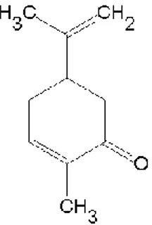
3.11.1.1 Carvone ( C10H14O )
Carvone is a member of a class of compounds called terpenes that are produced by plants, often to attract beneficial insects. This chemical has a pleasant smelling and it is often used as a flavouring agent in liqueurs and added to soaps and perfumes to improve their aromas. Carvone forms two mirror image or enantiomers: R-(–)Carvone that smells like spearmint and S-(+)-Carvone that smells like caraway (Figure 4). The two Carvone odors elicit different patterns of neuronal activity in the olfactory bulb in mice, however, with partially overlapping activated areas (Clarin et al., 2010).
Figure 4: Chemical structure of (-)Carvone. Chemical structure of
(5R)-5-Isopropenyl-2-methyl-2-cyclohexen-1-one (Carvone).
3.11.1.2 Terpinen-4-ol ( C10H18O )
Terpinen-4-ol is one of the 100 compounds of the tea tree oil (Figure 5). A study suggests that tea tree oil may potentially control inflammatory responses to foreign antigens in the skin (Hart et al., 2000).