HAL Id: tel-01806951
https://pastel.archives-ouvertes.fr/tel-01806951
Submitted on 4 Jun 2018HAL is a multi-disciplinary open access archive for the deposit and dissemination of sci-entific research documents, whether they are pub-lished or not. The documents may come from teaching and research institutions in France or abroad, or from public or private research centers.
L’archive ouverte pluridisciplinaire HAL, est destinée au dépôt et à la diffusion de documents scientifiques de niveau recherche, publiés ou non, émanant des établissements d’enseignement et de recherche français ou étrangers, des laboratoires publics ou privés.
by time-resolved circular dichroism
Marco Schmid
To cite this version:
Marco Schmid. Conformational dynamics of G-quadruplex DNA probed by time-resolved circu-lar dichroism. Optics [physics.optics]. Université Paris Saclay (COmUE), 2017. English. �NNT : 2017SACLX107�. �tel-01806951�
Conformational dynamics of
G-quadruplex DNA probed by
time-resolved circular dichroism
Thèse de doctorat de l'Université Paris-Saclay préparée à Ecole Polytechnique
École doctorale : Interfaces (ED 573)
Spécialité de doctorat: Physique
Thèse présentée et soutenue à l’Ecole Polytechnique, le 13 décembre 2017
Marco Schmid
Composition du Jury :Valérie Gabelica
IECB, Université de Bordeaux Rapporteur
Martin Volk
Department of Chemistry, University of Liverpool Rapporteur Stefan Haacke
IPCPM, Université de Strasbourg Président du jury
Frank Wien
DISCO, Synchrotron Soleil Examinateur
François Hache
LOB, Ecole Polytechnique Directeur de thèse
Pascale Changenet-Barret
LOB, Ecole Polytechnique Co-Directrice de thèse
NNT : 2 0 1 7 S A CL X 1 0 7
sondern das Erwerben, nicht das Dasein, sondern das Hinkommen, was den gr¨oßten Genuß gew¨ahrt.
A dissertation is in general of a long and stony with many obstacles. Therefore, I am grateful for having two great supervisors who gave me a lot of support and advice. On that note, I want to thank sincerely to Pascale Changenet-Barret and Fran¸cois Hache for those three rewarding years. I would like to also give special thanks to my jury: my two reviewers Val´erie Gabelica and Martin Volk as well as our collaborator and member of the jury Frank Wien and the jury’s president Stefan Haacke. Their reports and questions have helped us to deepen the understanding of our research.
In order to mount our experiments we had to add many modifications, so with-out Xavier Solinas and Jean-Marc Sintes our eventual success would not have been possible. Equally, I want to thank Christelle Fran¸cais and Laure Lachapelle who, as our secretaries, did all the indispensable administrative work during my stay.
I also had fruitful discussions in my first year with Vincent Kemlin who proposed to measure temperature changes via a change of the refractive index of water. Addi-tionally, as an pH indicator to monitor temperature changes, Hubert Becker proposed phenol red, which turned out to be applicable at 293 nm. This facilitated alignment and temperature control significantly. He further helped me with Mayla to perform a gel electrophoresis and introduced me kindly to it.
Three years of working on a interesting scientific project is quite joyful. However, without human relationships is is not completely satisfying. Therefore, I have been quite lucky to perform my thesis in the LOB. Besides the helpfulness of permanent researchers as well as students, there have been several get-togethers, evenings and daily visits. Added to this, we had many discussions at lunch about politics and science.
In this sense, I want to thank my collegues Lipsa, Olga, Ravi, Jos´ephine, Maila, Thuy, Xiujun, Pascal, Max and all the other students. Additionally I also want to give thanks to all the other members of the LOB for this rewarding and mind-opening three years’ journey.
my thankfulness. Also I want to thank my brother and sister and the rest of the family.
Les quadruplexes de guanines (G4) sont des structures secondaires de l’ADN qui consistent en l’assemblage de t´etrades de guanines reli´ees par des liaisons hydrog`ene Hoogsteen. La formation des G4 n´ecessite g´en´eralement la pr´esence de cations m´etalliques (typiquement N a+ ou K+) qui stabilisent la structure. Les G4 sont des
structures tr`es polymorphes dont la topologie d´epend non seulement de la s´equence d’ADN, mais aussi de la nature des cations m´etalliques et de leur environnement. Ils peuvent ˆetre form´es `a partir d’un, deux, trois ou quatre brins d’ADN. Les G4 form´es `a partir d’un seul brin d’ADN (i.e. monomol´eculaires) sont les structures les plus pertinentes biologiquement. Ces derni`eres d´ecennies, l’int´erˆet pour ces structures monomol´eculaires a fortement augment´e avec l’identification dans divers g´enomes d’un grand nombre de s´equences riches en guanine capables d’adopter une confor-mation G4 in vitro, dans des conditions biologiques. Longtemps contest´ees, il existe aujourd’hui un ensemble de preuves exp´erimentales qui attestent de l’existence des G4 in vivo et de leurs implications dans d’importants processus cellulaires, comme la maintenance des t´elom`eres, la r´eplication, la transcription ou la translation des g`enes. Toutefois, malgr´e le nombre croissant des ´etudes des G4, tr`es peu d’entre elles ont abord´e les aspects dynamiques de leur formation. La plupart de ces ´etudes qui se sont focalis´ees sur des s´equences t´elom`eriques humaines tr`es polymorphes ont conduit `a des images contradictoires de leurs m´ecanismes de repliement et d´epliement.
Dans le cadre de ma th`ese, nous avons choisi d’´etudier la dynamique de repliement et de d´epliement de plusieurs s´equences G4 mod`eles, constitu´ees d’une vingtaine de base, au moyen d’exp´eriences pompe-sonde de saut de temp´erature ( T-jump )
coupl´ees `a une d´etection de dichro¨ısme circulaire (CD) r´esolu en temps. Le CD, qui est la diff´erence d’absorption d’une onde polaris´ee circulairement gauche et d’une onde polaris´ee circulairement droite, est particuli`erement sensible `a l’arrangement des guanines dans les G4. Il offre la possibilit´e de sonder la dynamique des changements structuraux de s´equences d’ADN non-modifi´ees, sur une tr`es large fenˆetre temporelle. Afin d’initier le d´epliement de l’ADN, nous avons utilis´e des exp´eriences de T-jump
Toutefois les exp´eriences de T-jump conventionnelles, o`u le changement de
temp´erature de l’´echantillon est g´en´eralement induit par l’absorption par le solvant d’un faisceau de lumi`ere infra-rouge puls´e nanoseconde (1,4 µm pour H2O), ne
per-mettent pas de mesurer les dynamiques de repliement/d´epliement des G4 au-del`a de quelques millisecondes. Afin d’´elargir notre fenˆetre d’observation jusqu’aux secondes pour capturer l’int´egralit´e de ces dynamiques, nous avons remplac´e notre source laser puls´ee IR nanoseconde par une diode laser IR continue (3 W @ 1,48 µm) et un ob-turateur rapide pour contrˆoler le temps de chauffage des ´echantillons. Pour mesurer la faible variation des signaux CD (i.e. ∆DO 10−4) induite par l’´el´evation de
temp´erature des ´echantillons d’ADN, nous avons transpos´e le principe de mesure d’un spectropolarim`etre statique sur notre montage pompe-sonde. Pour cela, la po-larisation de la sonde g´en´er´ee par un oscillateur Titane saphir femtoseconde accord-able est alternativement polaris´ee circulairement gauche et droite `a la fr´equence de 50 kHz `a l’aide d’un modulateur photo-´elastique (PEM) coupl´e `a un amplificateur lock-in. Grˆace `a la modulation rapide de la polarisation de la sonde, nous avons pu ainsi r´eduire consid´erablement le bruit de nos mesures (∆DO = 10−5). Cependant
quelques cycles du PEM sont n´ecessaires pour extraire un signal CD de l’amplificateur lock-in, limitant ainsi la r´esolution temporelle de notre d´etection `a quelques millisec-ondes.
Grˆace `a ce nouveau montage exp´erimental nous avons mesur´e les dynamiques de d´enaturation et de renaturation de s´equences G4 pr´esentant deux topologies dis-tinctes, parall`ele et antiparall`ele. Du fait de leur grand polymorphisme, nous nous avons choisi des s´equences bien caract´eris´ees, telles que les s´equences t´elom`eriques Tel21 (5’-GGG(TTAGGG)3-3’ ) et Tel22 (5’-AGGG(TTAGGG)3-3’ ), la s´equence de
l’aptam`ere de la thrombine TBA G4 (5’-GGTTGGTGTGGTTGG-3’ ) et la s´equence promotrice oncog`ene c-MYC (5’-TGAGGGTGGGTAGGGTGGGTAA-3’ ).
Pr´ealablement aux ´etudes r´esolues en temps, en collaboration avec l’´equipe DISCO au synchrotron SOLEIL, nous avons mesur´e les spectres CD statiques (SRCD) de cha-cune de ces s´equences G4, `a diff´erentes temp´eratures. Dans la r´egion spectrale en dessous de 260 nm, les signaux SRCD qui d´ependent fortement de la s´equence d’ADN refl`etent majoritairement la contribution des interactions intra-brins des bases. En revanche, aux longueurs d’onde plus grandes (λ > 260 nm), les signaux SRCD qui
Les ´etudes de CD r´esolues en temps des s´equences G4 ont r´ev´el´e, dans tous les cas, des dynamiques de d´enaturation et de renaturation de l’ADN biexponentielles, avec des constantes de temps de quelques centaines de millisecondes et quelques secondes qui d´ependent fortement de l’amplitude du saut de temp´erature. Nous avons ´egalement observ´e des cin´etiques de d´enaturation et de renaturation de l’ADN tr`es diff´erentes qui sugg`erent l’existence de voies bien distinctes de repliement et de d´epliement. Par ailleurs, l’´etude des effets de la concentration des cations sur la cin´etique de d´enaturation et de renaturation de Tel21 a mis en ´evidence leur rˆole important `a la fois dans les propri´et´es thermodynamiques et dynamiques des G4. L’ensemble de ces tendances a pu ˆetre reproduit `a l’aide d’un mod`ele empirique de diffusion de la population le long d’une surface de potentiel en forme d’entonnoir comparable `a celui propos´e pour la formation de structures simples double-brin de l’ADN. Toutefois les cin´etiques de formation des G4 mesur´ees sont 2 `a 3 ordres de grandeur plus lentes, une diff´erence qui pourrait s’expliquer par les fortes interactions intra-brin des guanines qui doivent ˆetre surmont´ees pour que l’ADN se replie.
Enfin, nous avons men´e une ´etude compar´ee des dynamiques de d´enaturation et renaturation de la s´equence TBA G4 avec une s´equence tronqu´ee TBA G3 (5’-GGTTGGTGTGG-3’ ) connue pour former une structure triplex (G3). Cette ´etude a r´ev´el´e des vitesses de repliement et d´epliement du G3 4 fois plus lentes que celles du G4. Ces toutes premi`eres ´etudes dynamique d’une structure G3 semblent donc exclure l’hypoth`ese g´en´eralement avanc´ee dans la litt´erature de la formation d’un interm´ediaire triplex de courte dur´ee de vie au cours du repliement des G4 antipar-rall`eles.
1 Introduction 1
2 An overview of G-quadruplex structures 5
2.1 What defines a G-quadruplex . . . 6
2.2 Polymorphism of G-Quadruplexes . . . 7
2.2.1 Effect of Loop length . . . 9
2.2.2 Effect of Cations . . . 9
2.2.2.1 Specific Binding . . . 9
2.2.2.2 Non-specific binding and ionic strength . . . 10
2.2.2.3 G4 stability according to cation type . . . 10
2.2.3 Effect of Cosolutes and Molecular Crowding . . . 12
2.3 Biological Pertinence and Applications of G4 . . . 12
2.3.1 G4 Ligands . . . 13
2.3.2 G-Quadruplexes as nanomachines and -devices . . . 15
2.4 State of Art of the Investigations on G4 Folding . . . 16
2.4.1 Structure characterization of G4 . . . 16
2.4.1.1 UV & CD melting experiments . . . 16
2.4.1.2 Gel Electrophoresis . . . 17
2.4.1.3 X-ray Crystallography . . . 17
2.4.1.4 NMR . . . 18
2.4.1.5 CD spectroscopy . . . 18
2.4.2 Studies of G4 folding/unfolding pathways . . . 19
2.4.2.1 Rapid mixing experiments . . . 19
2.4.2.2 Single molecule FRET studies . . . 21
2.4.2.3 Single-molecule force spectroscopy . . . 23
2.4.2.4 Other experimental approaches . . . 25
2.4.2.5 Theoretical Approaches . . . 26
2.4.3 Conclusion . . . 27 i
3 Techniques and methods 29
3.1 Preface . . . 31
3.2 Circular Dichroism . . . 31
3.2.1 Definitions . . . 31
3.2.1.1 Difference in absorption coefficients . . . 31
3.2.1.2 Ellipticity . . . 32
3.2.2 Artifacts . . . 33
3.2.3 Theoretical Description . . . 35
3.2.4 Dipole-Dipole Coupling . . . 37
3.2.5 Origin of CD in G Quadruplexes . . . 38
3.3 Temperature jump (T-jump) . . . 41
3.3.1 Direct heating vs. indirect heating . . . 43
3.3.2 Nanosecond T-jump . . . 43
3.3.3 Millisecond T-jump . . . 44
3.3.4 Probing Temperature Changes in situ . . . 46
3.3.4.1 Bromothymol Blue . . . 46
3.3.4.2 Phenol Red . . . 47
3.3.4.3 Refractive Index of Water . . . 49
3.4 CD Spectrometer with Temperature Automation . . . 50
3.4.1 Setup, Principle and Implementation . . . 51
3.4.2 Signal processing . . . 52
3.4.3 CD spectrometer for T-jump measurements . . . 54
3.5 Combination of T-jump with TRCD using a Pockel’s cell . . . 58
3.5.0.1 Heating path . . . 58
3.5.0.2 Probe modulation via Pockels cell . . . 58
3.6 A new TRCD setup: one-pulse TRCD . . . 59
3.6.1 One-pulse experiment . . . 59
3.6.2 Two-pulse experiment . . . 61
3.6.3 Outlook . . . 63
3.7 Combination of T-jump with TRCD by using PEM . . . 64
3.7.0.1 Possible Artefact . . . 65
4 Steady-state CD spectra 67 4.0.1 Preface . . . 67
4.1 Sample preparation . . . 67
4.2 Studied G4 forming sequences . . . 68
4.2.1 Human telomeric sequences . . . 69
4.2.3 Modified oncogene promoter sequence c-MYC . . . 69
4.3 Static SRCD Spectra of G4 . . . 70
4.3.1 Melting curve - fit procedure . . . 70
4.4 Antiparallel human telomeric G4 . . . 72
4.4.1 Tel21 with Sodium . . . 72
4.4.2 Tel22 with Sodium . . . 78
4.4.3 22CTA with Potassium . . . 80
4.5 hybrid-type human telomeric G4 . . . 82
4.5.1 Tel22 with Potassium . . . 82
4.5.2 2GKU with Potassium . . . 84
4.6 Thrombine binding aptamer . . . 86
4.6.1 TBA G4 with Potassium . . . 86
4.6.2 TBA G3 . . . 88
4.7 c-MYC . . . 90
4.8 Conclusion . . . 92
5 Folding dynamics of Tel21 & Tel22 with Sodium 93 5.1 Preface . . . 94
5.2 Dynamics on the millisecond time-scale . . . 94
5.2.1 Results of Tel21 with Sodium . . . 94
5.2.1.1 Multiexponential fitting of results . . . 99
5.2.1.2 Data Treatment of biexponential fitting . . . 101
5.2.2 First model: 1D diffusional folding model . . . 103
5.2.3 Extension of our model: rugged free-energy folding landscapes 108 5.2.3.1 Data treatment with kinst . . . 108
5.2.4 Comparison with Tel22 with Sodium . . . 112
5.2.5 Conclusion . . . 115
5.3 Effect of Sodium concentrations on dynamics . . . 119
5.3.1 Tel21 in 25 mM sodium solution . . . 119
5.3.2 Tel21 in 900 mM sodium solution . . . 121
5.4 Folding dynamics of Tel21 on microsecond time-scale . . . 123
5.4.0.1 Fitting function . . . 124
5.4.1 Conclusion . . . 125
6 Dynamics of TBA G4 and G3 & c-MYC 127 6.1 Preface . . . 128
6.2 TBA Quadruplex with Potassium . . . 128
6.4 TBA G4 and G3 on microsecond time-scale . . . 137
6.5 Conclusion . . . 138
6.6 c-MYC with Sodium . . . 140
6.6.1 Nanosecond Dynamics . . . 143
6.6.1.1 Conclusion . . . 144
6.6.2 Absorption Dynamics of c-MYC at 265 nm . . . 145
G4 structure Abbreviation Sequence obtained b y NMR cation typ with cation: h uman telom eric sequences T el21 5’-GGG TT A GGG TT A GGG T T A GGG -3’ mixture N a + T el21 5’-GGG TT A GGG TT A GGG T T A GGG -3’ mixture K + T el22 5’-A GGG TT A GGG TT A GGG TT A GGG -3’ an tiparallel bask et-t yp e [1] N a + T el22 5’-A GGG TT A GGG TT A GGG TT A GGG -3’ mixture K + 2GKU 5’-TT GGG TT A GGG TT A GGG TT A GGG A-3’ h ybrid-t yp e form 1 [2] K + 22CT A 5’-A GGG CT A GGG CT A GGG CT A GGG -3’ an tiparallel chair-t yp e [3] K + T el23 5’-T A GGG TT A GGG TT A GGG TT A GGG -3’ h ybrid-t yp e form 1 [4] K + T el24 5’-TT A GGG TT A GGG TT A GGG TT A GGG -3’ mixture N a + T el26 5’-TT GGG TT A GGG TT A GGG TT A GGG TTT-3 ’ mixture K + 26TT A 5’-TT A GGG TT A GGG TT A GGG TT A GGG TT-3’ h ybrid-t yp e form 2 [5] K + T el27 5’-TT A GGG TT A GGG TT A GGG TT A GGG TT A-3’ mixture N a + other structures TBA G4 5’-GG TT GG TGT GG TT GG -3’ an tiparallel chair-t yp e [6] K + TBA G3 5’-GG TT GG TGT GG -3’ triplex structure [7] K + c-MYC 5’-TGA GGG T GGG T A GGG T GGG T A A-3’ all-parallel [8] K + c-MYC2 5’-TGA GGG T GGG GA GGG T GGG T AA-3’ all-parallel [9] K + hTER T 5’-G GGG GCT GGG CCGGGGA CCCGGGA GGGGTCA CG GGG C GGG G-3’ ? K + T able 1: Abbreviation of G4-forming sequences used in the presen t w ork. Guanine s that participate in the G-quartet are colored blue. Underlined abb rev ations are structures, whose CD sp ectra w ere studied in the presen t w ork. Bold are those whose dynamics w ere also studied.
Introduction
Nature is beautiful. This is reflected in its simplicity as well as in its complexity. Based on a simple concept of merely four nucleobases, Nature is capable to construct an enormous diversity of living beings. Even by using only guanylic acid, Gellert et al. found a non-canonical secondary DNA structure that forms in situ containing only Guanines in its center: the Guanine quadruplex (G4) [10] (see Fig. 1.1). Cation coordination in the center tremendously increase G4 stability [11].
Figure 1.1: Left: G4 quartet. Right: Structure of Tel22 with N a+.
Since the determination of the DNA double helix [12], it has been commonsense that only the double helix forms in vivo. However, advances in recent years have proven that DNA can adopt a variety of structures. Among them G-quadruplexes (G4) are non-canonical structures that result from the hydrophobic stacking of guanine-tetrads stabilized by cations (typically N a+ and K+). Recently, the interest in G4
has grown considerably, due to the identification in various eukaryotic and prokary-otic genomes, of many guanine-rich regions capable of forming such structures in solution, in vitro. There is now a body of experimental evidence for the occurrence of G4 in vivo and their participation in several biological functions related to their
folding mechanism, such as chromosomal integrity, gene replication, transcription and translation [13, 14].
Notably, the interest in G4 has been amplified by the understanding of telomere maintenance and the discovery of telomerase [15]. In humans, the telomeric sequence is rich in guanine and thus has the potential to form G4: (GGGT T An) [16, 17].
Telomerase is an enzyme that maintains the length of telomeres [18]. In approxi-mately 90% of all malignant tumors, telomerase has been detected and inhibits tumor cell to undergo senescence due to telomere shortening [19]. Consequently, telomerase and human telomeres have become good potential target structures for novel drugs in cancer treatment [20, 21]. The telomeric overhang has to be bound to proteins so that telomerase can interact [22]. Therefore, by adding ligands that favor G4 formation in this overhang, telomerase cannot interact and telomere maintenance is impeded.
Besides the design of ligands targeting G4, most of the experimental studies car-ried out so far on G4, have been focused on the characterization of their 3D structure and stability highlighting their large conformational diversity. Although the litera-ture devoted to G4 has exploded considerably, experimental studies of their folding remain scarce and are mainly focused on the human telomere sequence. Elucidating G4 folding mechanisms is however a crucial issue for understanding their biological functions. Despite recent experimental and theoretical efforts, there is not yet a clear picture these mechanisms [20–22]. One of the reasons may be due to fact that most of the studies of G4 folding have focused on the highly polymorphic human telom-eric sequence, due to its biological pertinence [23, 24]. On the other hand, most of the studies of G4 folding have yielded a limited structural information with time resolution generally limited to milliseconds or seconds.
Laser-induced temperature jump is an established method to study unfolding dynamics of proteins [25, 26]. However, it only allows to monitor dynamics up to several ms in situ [27].
Laser-induced temperature jump (T-jump) is an established method to study the folding dynamics of proteins and to a lesser extend DNA hairpin [26, 28]. Combined with various optical detections, it provides the opportunity to probe conformational changes of biomolecules on shorter time scale than single-molecule spectroscopy or rapid mixing techniques often used to study G4 folding. The principle of T-jump is to heat up the solvent with an infrared laser pulse and to monitor the resulting DNA denaturation as a function of time. However most of experimental studies performed on DNA have used fluorescence or FRET detection that requires the use of covalently attached fluorescent labels that may affect the thermodynamic and the mechanical properties of G4. In contrast, circular dichroism can provide information on the
secondary structure of biomolecules [29].
Recently, our laboratory has implemented a T-jump set-up combined with time-resolved CD detection [28], for measuring the folding dynamics of biomolecules within the time window of ca. 10 ns and 1 ms. In the frame of my PhD work, we have extended this method to probe slower dynamics on the time window of a few ten milliseconds up to the seconds. With this new setup, both denaturation and consec-utive renaturation of DNA can be measured. In combination with our conventional T-jump, it becomes therefore possible to measure the thermal-induced changes of DNA secondary structure over a time-scale of more than 8 orders of magnitude.
This manuscript is organized as follows:
• Chapter 2 addresses the state of art of the knowledge on G4 focusing on the dynamical studies of their folding.
• Chapter 3 is dedicated to the description of the principles and the implemen-tation of the used experimental techniques.
• Chapter 4 describes the static synchrotron radiation CD spectra of various G4-forming sequences displaying distinct topologies (antiparallel, hybrid and parallel) measured in collaboration with DISCO/SOLEIL.
• Chapter 5 discusses the results of our time-resolved studies of human telomeric G4-forming sequences (Tel21 and Tel22) in N a+ phosphate buffer.
• Chapter 6 presents the results of the shortest forming G4 sequence: the throm-bin throm-binding aptamer G4 (TBA G4) [6] as well as on its related triplex structure (TBA G3) [7]. Finally, the dynamics of a modified G4-forming sequence of the oncogene promoter sequence, c-MYC, have been investigated and are presented here.
An overview of G-quadruplex
structures
Such as we are made of, such we be. William Shakespeare
Contents
2.1 What defines a G-quadruplex . . . 6 2.2 Polymorphism of G-Quadruplexes . . . 7 2.2.1 Effect of Loop length . . . 9 2.2.2 Effect of Cations . . . 9 2.2.3 Effect of Cosolutes and Molecular Crowding . . . 12 2.3 Biological Pertinence and Applications of G4 . . . 12 2.3.1 G4 Ligands . . . 13 2.3.2 G-Quadruplexes as nanomachines and -devices . . . 15 2.4 State of Art of the Investigations on G4 Folding . . . 16 2.4.1 Structure characterization of G4 . . . 16 2.4.2 Studies of G4 folding/unfolding pathways . . . 19 2.4.3 Conclusion . . . 27
2.1
What defines a G-quadruplex
The double helical structure of B-DNA was determined in 1953 [12]. Various alter-native structures have been discovered since. One of these structures is the so-called guanine quadruplex (G4). It seems to be the most biological relevant structure after the B-form due to its high stability at neutral pH [13, 30–32]. G4 consist of a DNA sequence that is abounding in guanine bases. In a G4 core, a planar tetrad made of 4 guanine bases is found as well as cations such as K+ and N a+ 1. It is called
a G-quadruplex when there are at least two tetrads stacked on each other [35]. In these tetrads the guanines are connected to each other by Hoogsteen-hydrogen bonds (see Fig. 1.1). Due to the negative dipolar charge of the oxygen atoms pointing to the tetrad center, a cation is necessary to compensate these negative charges and stabilize the G4 structure.
Formation of G4 was first discovered in 1962 [10] in a concentrated solution of guanosine monophosphate (GMP) that formed a gel over night. In that case, the G4 structure is made of guanine monomers. G4 can also form from association of different DNA strands. Tetramolecular, trimolecular as well as bimolecular and monomolecular G4 are possible [36]. Typical intramolecular G4 forming sequences are as follows: G≥3NxG≥3NxG≥3NxG≥3 with x between 1 and 7 and N being any base.
Longer connections destabilize the G4 structure [37]. My work, however, focuses only on intramolecular G4, e.g. monomolecular G4. In that case, guanines that form the quartets are connected directly by a negatively charged phosphate backbone in a way that adjacent bases do not participate on the same quartet, but are stacked on each other [38]. In human telomeric sequences (5’-(GGGTTA)n-3’ ) three quartets form
a G4. The stacked quartets are slightly twisted [39] and are spatially separated by ca. 3.25 ˚A [40].
In the example of the human telomeric G4 sequence Tel22 with N a+ [1] three
quartets are formed (see Fig. 1.1 right). The four GGG run are connected by three loops. The length of the loops are known to influence the relative orientation of the GGG runs, which can be all parallel (all in same direction), antiparallel (all in opposite direction) or hybrid (one in opposite direction). Besides the primary DNA sequence, the environment can also influence the relative direction of the GGG runs, such as the nature of cations, the presence of cosolvents or crowding agents.
1Exeptions exist in which guanines are replaced by cytosines [33] or some guanines are missing
2.2
Polymorphism of G-Quadruplexes
G4 can form a vast variety of structures. For a better understanding, we discuss here the features that allow to discriminate different G4 structures. The human telomeric G4 have been the most studied ones up to date [41]. These sequences exhibit the largest structural polymorphism [42]. All different human telomeric G4 structures found up to now are depicted in Fig. 2.1.
Figure 2.1: All characterized telomeric G4 topologies. (a) Antiparallel basket-type form [1]. (b) antiparallel chair-type structure [3]. (c) all-parallel G4, propeller-type form [43]. (d) (3+1) [4, 44]. (e) (3+1) form 2 (hybrid2) [4]. (f) antiparallel G4, sequence variant d[(TTAGGG)3TTA(BrG)GGTTA] [45]. (g) basket-type with only
two quartets observed [33]. anti guanines are colored grey and syn guanines are colored white. M, N and W indicate medium, narrow and wide grooves, respectively. The strand direction goes from the 5’ to the 3’ end.
The first crucial factor that narrows significantly the number of possible G4 con-formations is the primary DNA sequence. For example, a sequence that contains only one GGG run forms tetrameric G4 [36]. The second important feature that enhances G4 polymorphism is the syn and anti orientations of the glycosidic bond angles of guanosines. Fig. 2.2 shows the two orientation angles. As indicated in Fig. 2.2, guanosines with syn and anti orientation angle are colored in white and in grey, re-spectively. These colors will consequently be maintained throughout the manuscript. In Fig. 2.2 bottom left, a rotation sense is depicted. Looking from the top of the
G4 (indicated by the 5’ end) to the G4 one can see that the twist of the quartets is anti-clockwise (e.g. right-handed). This is valid for all G4 with only one exception (see Phan et al. [46]). As previously mentioned, the relative strand orientations of the GGG runs in the G4 can be either antiparallel (Fig. 2.1(a,b,f,g)) [1], all-parallel (c) or hybrid (d,e) [47].
Figure 2.2: Top: Guanosine monophosphate with an anti and syn glycosidic angle. Bottom left: G4 quartet with anitclockwise rotation (-) from the donor NH to the acceptor C=O hydrogen bonds. Bottom right. Illustration of different groove sizes. syn guanines appear white and anti guanines in grey. M, N and W indicate medium, narrow and wide grooves, respectively.
A third feature of G4 structures is the loop orientation, which can be diagonal, lateral (edgewise) and external (propeller) [36]. All quadruplex structures have four grooves, whose dimensions depend on the overall topology and the nature of the loops. G4 grooves are classified according to their size: Narrow (N), medium (M) and wide (W).
2.2.1
Effect of Loop length
The number of nucleotides between G runs is known to determine partly the folding topology of G4. A sequence G3T1G3T1G3T1G3with solely one nucleoside between the
G runs forms the parallel propeller-type G4, most likely independent of flanking loops [48]. Bugaut and Balasubramanian studied a number of sequences containing only G and T with loops containing only T. By varying the loop lengths, they observed some trends: parallel G4 are the most stable structure, when there is only one nucleoside in their loops [49]. In general, an increase of the loop length decreases the G4 stability up to 5 nucleosides. Longer loops do not significantly destabilize the G4 structure compared to 5 thymines [49]. However, this cannot be used as a general statement for all sequences. The Mergny group performed a more general essay, in which they used a sequence G3LaG3LbG3LcG3 [50]. As loop bases La,b,c they chose T or TTA
loops. Salt conditions were either 100 mM K+ or N a+. An increase of loop length
showed for both cations a decrease of the melting temperature Tm. It is, however,
more pronounced and more linear for K+ than for N a+.
Additionally, some sequences with loop lengths of one to three nucleotides can even form G4 in the presence of the complementary DNA strand, which reflects their stability [51–53]. A G4 formed by the c-MYC promoter sequence in 100 mM KCl remains stable even in the presence of its complementary DNA strand up to at least 70◦C [54]. It has two loops with one nucleotide and one loop with two nucleotides
and forms a parallel structure. It has been shown in some cases that for two linking loops containing only one nucleoside, the length of the third one does not influence the G4 topology in potassium. In that case only parallel quadruplexes are formed [49, 50, 55]. With sodium there can be more diversity [50]. Quadruplexes with loop lengths of three nucleosides do not fold only in one specific topology. They rather form a mixture of parallel and antiparallel conformations, depending on their sequence, the nature of the cations in solution and the presesence of cosolvent [56]. However, parallel quadruplexes are energetically more favorable, even for loop lengths of three nucleosides [56].
2.2.2
Effect of Cations
2.2.2.1 Specific Binding
There are several factors that contribute to G4 stabilization: stacking interactions [57] between the nucleosides, hydrogen bonding [53, 58], solvation and cation binding [59–61]. Without cations, G4 do practically not form. Cations located inside the G4 core play a key role in the G4 stability and are classified as specific binding cation.
Those cations can be located inside the G-tetrad plane or between two G-tetrads.
A cation located in the plane of a G-quartet has 4 oxygen binding sides, one from each Guanine. When it is located between two planes, there are even 8 oxygen atoms available for coordination. Due to the nature of the cavity, no anions enter. Hence, they have a weak influence on G4. Gu et al. have stated that according to their ab inito calculations the coordination of the cation contributes more to the G4 stability than the Hoogsteen hydrogen bonds between the guanines [62].
In literature it is often stated that N a+ is located inside the plane of G-tetrads
because it is small enough. In contrast, K+ and N H+
4 for example are too large and
are thus located between the G-tetrads of the G4 core [63]. In fact, there are other parameters that contribute to the exact location of cations in the G4 core [61]. For instance, the presence of several cations inside the G4 core induces ionic repulsion (example with N a+ see [64]). Furthermore, the location of cations inside the G4 core
also depends on the presence of the cations in the phosphate backbone or on top of the terminal G-quartet.
2.2.2.2 Non-specific binding and ionic strength
Once all the cavities are filled with cations, no more cations go inside the G4 core. However, the negatively charged phosphor backbone allows more ions to be coordi-nated in a non-specific manner outside the G4 core. Gray et al. proposed that Tel22 can bind up to five K+in a non-specific manner [65]. A study on c-MYC has focused
on the effect of K+ concentration on G4 stability. It has been shown that addition
of cations once the G4 is filled, leads solely to non-specific binding. The increase of the concentration of cations results in a stabilization of the quadruplex structure, reflected by an increase in the melting point Tm[66].
2.2.2.3 G4 stability according to cation type
Physiological relevance is especially assigned to K+, due to its high intracellular
concentration [67], but also to N a+. The melting temperature (T
m) for G4 is in
general higher with K+ than with N a+ [68, 69]. Since specific binding inside the G4
core necessitates a complete dehydration of cations, this effect is due to their sizes and their energies of hydration. K+is rather coordinated to 8 oxygen atoms, whereas N a+
coordinates to 4 oxygen atoms. Hence, these different coordinations and G4 oxygen-ion distances are partly responsible for the difference in the free energy between N a+
and K+ [69]. Hydration energies for monovalent ions depend roughly on their ionic
energy distributions of coordination and hydration determines the selectivity of G4 to a specific cation [68, 70–72]. Gray et al. have shown that by adding K+to an already
formed human telomeric G4 (Tel22) with N a+, K+ replaces N a+ and changes the
G4 structure [73].
G4 do not only show different stabilities in the presence of different cations, but they also form different structures [74]. Therefore, the melting temperatures of G4 with K+ and N a+ cannot be compared directly. In the case of Tel22, two hybrid
structures form in the presence of K+, whereas merely one antiparallel (basket-type)
structure forms in the presence of N a+ [73]. Interestingly, NMR measurements have
shown that cations in the G4 core can exchange with the bulk [75]. 23N a NMR
measurements have shown that it takes 180 µs at 20◦C for a sodium cation to be
exchanged by another one in a dimeric G4 made of the sequence d(G4T4G4). This
suggests that no quadruplex opening or conformational change is needed, since bi-molecular G4 folding dynamics are known to be slower [76].
In addition to N a+ and K+ there are several other ions whose presences induce
quadruplex formation: Rb+, Cs+ [60, 77],N H+
4 [78, 79] and T l+ [80]. The work of
Wong et al. revealed the following order for G4 stability: K+ > N H+
4 > Rb+ >
N a+ > Cs+ > Li+, where K+ yields the most stable G4 and Li+ the weakest one
[81]. Subsequent studies revealed a slightly different order: K+ > N H+
4 > N a+ >
Rb+> Li+
≥ Cs+ [59–61], as illustrated in Fig. 2.3. As one can see, between K+and
N a+decreasing the cation size exhibits the same tendency as decreasing stabilization
effect. Li+ and Cs+ are too small and too big, respectively, to fit in the G4 cavity
[36].
Figure 2.3: Monovalent ions with decreasing stability effect on G4 from left to right. K+ being the most stabilizing one, Cs+ is the least stabilizing one according to [61].
Relative ion radii are reflected by size of spheres.
Not only with monovalent cations G4 can form, but also with divalent cations as Sr2+ [60, 82]. As well as for monovalent cations, a similar trend in quadruplex
stability is observed: Sr2+>> Ba2+ > Ca2+ > M g2+. However, it seems that there
is a more complex effect of bivalent cations on G4 stability. In the case of Tel24 Sr2+
interesting effects, such as destabilizing effects of quadruplex structures by bivalent cations have been reported [36, 66, 83]. Since in the present work only monovalent cations have been investigated, these effects are not discussed here.
2.2.3
Effect of Cosolutes and Molecular Crowding
Human telomeric quadruplexes can adopt a variety of different structures depending on their sequence. In K+ solution four structures have been reported so far [84] (see
Fig. 2.1(b,d,e,g)). A change from solution to solid state, e.g. G4 crystals, the very same sequence Tel22 with the same amount of cations changes from two coexisting hybrid to a sole all-parallel structure (Fig. 2.1). Since in crystals the relative dis-tance between two G4 is reduced, one can think that this structural change is due to molecular crowding. Heddi and Phan have found that human telomeric G4 with K+ adopts the all-parallel topology under molecular crowding conditions [84]. This
is an important result, since crowding effects are expected to reflect in vivo-like envi-ronment [85]. Crowding conditions result in a reduction of G4 hydration [85]. Under such conditions the G4 structure with the minimum hydration energy should prevail. According to the work of Heddi and Phan this is the propeller-type quadruplex [84]. A commonly used cosolute is polyethylene glycol (PEG) [36, 86]. It is considered to not interact with the quadruplex. However, a study of the group of Chaires suggests that PEG rather binds to the grooves of propeller-type quadruplexes and does not act as a crowding agent [87]. Other used cosolutes are ethanol, Ficoll 400, acetonitrile or DMSO [84]. It was shown that for all these agents the same parallel quadruplex structure formed out of different other structures [84], which is in accordance with the suggestions of Heddi and Phan.
2.3
Biological Pertinence and Applications of G4
It is extremely difficult to detect G4 in vivo, in part because reagents to detect G4 have been laking [32]. Nevertheless, advances in recent years have shown accumu-lated evidence that G4 also form in vivo [13, 30–32, 88]. Some remaining questions are: where would they appear and what would be their exact role. An algorithm developed by Balasubramanian’s group has scanned the human genome in search of potential quadruplex forming sequences. In their work of 2005 they have considered only sequences that can form 3 or more tetrads [37]. The following sequence motif was used: G≥3NxG≥3NxG≥3NxG≥3. With those conditions, a number of over 350,000
potential G4 forming sequences have been identified. These motifs are found at very specific regions of the genome, such as telomeres or promoter regions.
Human telomeres consist of thousands of DNA repeats (TTAGGG) and have a single-stranded 3’-tail. Short telomeric repeats in vivo form quadruplexes (see e.g. [89]). Furthermore, telomeres play an important role in cancer cells and aging. Conse-quently, they are considered as potential targets for the development of anti-cancer therapy [90].
There are some speculations about the possible role of G4 within the genome. G4 in telomeres might be there to protect the genome. G4 could also help telomeres to form DNA t-loops more easily [30]. Furthermore, it is possible that G4 form during the replication process when duplex DNA is unwound. The fact that a couple of helicases exist to unwind quadruplexes supports this suggestion [91].
Interestingly, many promoters of oncogenes also contain G4 forming motifs: c-KIT, wt c-MYC, k-RAS, B-RAF, BCL-220 and RET [90].
2.3.1
G4 Ligands
Due to the potential biological relevance of G4 there is a big field of research dedicated to the design of G4 ligands. This approach to target specific DNA regions by either stabilizing G4 or inducing their formation provides a strategy for elaboration of new disease treatments. Special interest has been dedicated to the inhibition of telomerase and promoter regions of some oncogenes to pave the way for effective anti-cancer targets [90]. Another target can be potential RNA G4 motifs of ribosomal viruses as HIV [92].
In promoters, ligand binding is expected to lead to an inhibition and to down-regulate the expression of the targeted gene [93]. Telomerase and telomere were the first targets for small ligand molecules [94]. However, up to now no ligands have been reported that are 100% specific to one particular G4 structure [90]. Under physio-logical conditions, it seems furthermore almost impossible to specifically target only one specific G4. Single G4 may be not the best candidates. Some studies show that higher order G4 structures have very particular surfaces to provide a higher ligand specificity [95].
Currently, more than 1000 G4-binding ligands have been reported. They are classified in two main groups so far: i) hetero-aromatic polycyclic ring systems, and ii) macrocycles with linked hetero-aromatic compounds [101]. One specific binding side of a G4 is its planar surface G-quartet. This is still the most explored one, but side grooves can also serve to increase ligand specificity. Theoretical studies have suggested that groove dimensions and loop sizes are crucial factors for the design of selective ligands [102].
Figure 2.4: Examples of G4 ligands. Quarfloxin, the first G4-binding molecule that underwent clinical trials [96]. Tetra-substituted naphthalene diimide, containing two different side-chaines to increase G4-binding affinity [97]. Five-ring quinolino-benzo-[5, 6]-dihydroisoquindolium, having a high specifity to discriminate between c-MYC and telomeric G4 [98]. Telomestatin, cyclic G4 ligand with a high selectivity and affinity for G4 [99, 100]
.
first one, quarfloxin is the only compound used for clinical trials so far [96]. It consists of a heteroaromatic planar core with two distinct side chains. The tetra-substituted naphthaline diimide illustrates another type of highly potent G4 binding agents [97]. The third example is a five-ring quinolino-benzo-[5,6]-dihydroisoquindolium. Its core consists of a positively charged heteroaromatic structure and in its sole side chain it exhibits a charged pyridine ring to increase water solubility [90]. This compound preferentially binds to wt c-MYC (parallel G4) vs. telomeric G4 (antiparallel G4) [98]. The last example is telomestatin, a macrocycle without a polycyclic core. It displays a high G4 selectivity and affinity over duplex DNA [99, 100]. For example, it stabilizes specifically anti-parallel human telomeric G4 [103]. But it has a very low aqueous solubility. In addition, its demanding isolation procedures and current synthetic approaches are reasons why it has not been developed further as a therapeutic agent [36].
2.3.2
G-Quadruplexes as nanomachines and -devices
Beside the interest of G4 in therapeutics, their polymorphism and self-assembly prop-erties can serve to create nanodevices. A few examples of G4 applications are pre-sented below.
For instance, functionalization of G4 has been performed by the group of Fet-tinger (e.g. [104]) who created lipophilic G4 by using modified lipophilic guanosine monomeres. The phosphate group has been replaced by a nonpolar silicon residue and the hydroxy groups have been protected with an acetalic nonpolar residue. Hence, these nanostructures are stable in nonpolar solvents. Still needing a cation to form, they can extract salts from water into nonpolar solvents [40]. While anions have little influence on the formation of G4 made of oligonucleotides, the group of Davis showed that they influence the properties of lipophilic G4 in organic solvents [40]. By increasing the basicity of anions, the rearrangement of G4 octamers can also be halted [105]. Hence, anions could be used to control regioselectivity of G4 assemblies. Additionally, lipophilic guanosines and folate analogues form dynamic crystals, that can be controlled by changing parameters as ions, solvents or temperature [40]. Fo-late derivatives of guanines, for example, form a smectic phase. Introducing cations changes the assembly to a columnar discotic phase similar to abreast columns of stacked guanine quartets [106]. Based on NMR studies that show that K+ are only
bound for milliseconds to G4 [107], oligomers or polymers of stacked guanine quadru-plexes can function as ion channels. In this respect, lypophilic G4 could be used as transmembrane ion channels [40].
On the other hand, G4 built out of monomers can be used as self-assembly ionophores [40]. Ion-selectivity can be achieved by influencing the non-covalent bonds. This is a big advantage in terms of synthesis, since established ionophores such as crown ethers and calixarenes demand a considerable synthetic effort to have ion-selectiviy [108, 109].
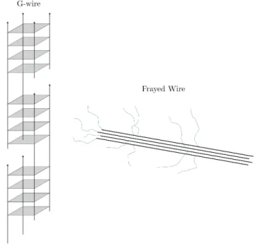
Nanometer assemblies of G-rich DNA have been discovered by the groups of Hen-derson and Sheardy [110]. Marsh and HenHen-derson established the term ”G-wires” to describe these parallel-stranded superstructures (see Fig. 2.5). This term is further justified, since G4 is capable of transporting electric current [111]. In this respect, G4 assemblies are interesting candidates, since guanine has the lowest potential of all ncucleic bases. ”G-wires” can also be used as a cation channel for N a+, K+ and
N H4+ with the best conduction for N a+ [112].
According to Mergny’s group, switching between double-stranded duplex and quadruplex can be considered as a nanomolecular machine [113]. Conformational changes of a G-rich 21-mer DNA have been studied. An alternating switching
be-tween duplex and G4 was achieved by adding C-rich strands to unfold quadruplexes and G-rich strands to fold G4 again. In a more recent work a combination of i-motif (cytosine analogue to G4) and G4 was synthesized in one strand [114]. Formation of the two structures can be controlled independently by changing the pH and the concentration of cations in solution. At acid pH without cations, solely the i-motif forms, while basic pH with cations mostly G4 is present. Many more applications do already exist. The potential of further guanine-based nanodevices is paramount and depends on the new G4 structures that will be discovered in the future.
Figure 2.5: Scheme of a G-wire formed out of d(G4T2G4) and a frayed wire formed
out of d(G15A15) according to [40].
2.4
State of Art of the Investigations on G4
Fold-ing
2.4.1
Structure characterization of G4
2.4.1.1 UV & CD melting experiments
The simplest way to verify G4 formation is to monitor the spectral changes induced on the absorption or the circular dichroism spectra of DNA as a function of the temperature. Upon G4 formation, a hyperchromism is generally observed around 295 nm concomitant with a hypochromism around 260 nm in the absorption spec-trum of DNA. The melting temperature (Tm) corresponds to the temperature at
which half of the population is folded and half is unfolded (Keq = 1). By taking the
first derivative of the melting curve, one can simply determine Tm. Further analysis
assuming a folding model allows the determination of thermodynamic parameters of the reaction: the Gibbs free-enthalpy (∆G0), the enthalpy (∆H0) and the standard
entropy (∆S0) [115, 116].
In addition to absorption and CD, formation of G4 can be also investigated by fluorescence either by FRET or by substitution of certain bases in the G4 loops by a fluorescent base such as 2-aminopurine (2-AP) [117]. While absorption and CD yield global information of the secondary DNA structure, FRET provides insight into the distance between the 5’ and 3’ endings of the G4 strand. This allows to distinguish between the relative positions of the first and last GGG run [23]. On the other hand, fluorescence of 2-AP monitors the stacking in the different G4 loops in the G4 [23]. As a matter of fact, fluorescence melting provides a better sensitivity than UV absorption and CD melting.
2.4.1.2 Gel Electrophoresis
Polyacrylamide gel electrophoresis (PAGE) is a method to separate molecules accord-ing to their weight [118]. Therein, molecules in a porous polymer gel move due to an applied electric field. Their mobility depends on their molecular weight, i.e. number of nucleotides. By adding a denaturating agent, one can verify the sequence’s length and its purity. Additionally, in a nondenaturating environment, the DNA secondary structure remains intact. Therefore, it allows to distinguish between unimolecular G4 and higher order G4 [119].
2.4.1.3 X-ray Crystallography
X-ray crystallography provides information about the molecular structure of G4 crys-tals [43]. In 2002 the first crystal structure of the human telomeric sequence Tel22 in the presence of K+has been determined [43]. Tel22 adopts an all-parallel topology in
crystals. With X-ray crystallography, positions of most of all atoms of G4 can be de-termined e.g. the absolute configuration. Furthermore, parameters such as distance between two G4 quartets, the tilt angles and the cation positions and distances can be determined [43]. Despite the fact that X-ray crystallography is a powerful tool to determine the absolute configuration of G4, X-ray radiations can easily destroy DNA [120]. Additionally, structure determination via X-ray scattering needs monocrystals, whose growth is a long and demanding process (see e.g. [43]).
2.4.1.4 NMR
In contrast to crystallography, nuclear magnetic resonance spectroscopy (NMR) en-ables structure determination in solution, which is closer to in vivo conditions [1]. Cross peaks of the nuclear Overhauser effect (NOE) reveal information about the spatial distance between H protons. From that structural information is derived. The general procedure to determine the full structure is as follows [1, 3, 4, 7, 33, 45]: Spatial proximity of atoms yields a first idea of G4 structure. Consecutively, it is used as an input to perform structure calculations. NMR analysis reveals important parameters such as tilt, loop orientation, syn, anti orientation of the glycosidic bond angles of guanines and yields information about spatial distances [1, 3, 4, 7, 33, 45].
2.4.1.5 CD spectroscopy
Crystallography and NMR spectroscopy are used for a precise structure analysis. However, these experiments are time-consuming and require a large amount of DNA. Circular dichroism (CD) spectroscopy provides a fast method to check G4 structures and requires a small amount of DNA. CD spectroscopy, which is very sensitive to the guanine arrangement in the G4 core, allows to distinguish between parallel, anti-parallel and hybrid topologies, as shown in Fig. 2.6. The origin of the CD signals in G4 will be discussed in Sec. 3.2.5.
Figure 2.6: Left: CD spectrum of an antiparallel basket-type G4 (Tel22 with N a+),
all-parallel G4 (c-MYC with N a+) and hybrid1 G4 (2GKU with K+). Right: TBA
quadruplex (G4 with K+) and triplex (G3 with K+), respectively.
In a parallel G4 (c-MYC), in which all glycosidic bond angles of guanines are in anti, an intense peak is observed at 265 nm [36]. In antiparallel G4 (Tel22), in which the glycosidic bond angles of guanines exhibit both syn and anti orientation, one positive peak at 295 nm and a negative one at 265 nm are observed [36]. Hybrid G4
structures (e.g. 2GKU) are mixtures between antiparallel and parallel topologies and display two positive peaks centered around 270 nm and 290 nm [121].
2.4.2
Studies of G4 folding/unfolding pathways
2.4.2.1 Rapid mixing experiments
The folding/unfolding processes of biomolecules are known to be complex mecha-nisms involving multidimensional energy landscapes reflecting all possible conforma-tions. From the experimental point of view, it is quite difficult to determine these energy landscapes, since the perturbation that is used to trigger folding or unfold-ing processes may also influence its shape [22]. One way to study foldunfold-ing is to use rapid-mixing techniques. In the absence of cations G4 structure hardly form [21]. Therefore, G4 formation (folding) can be induced by a rapid adjunction of cations in an aqueous solution of unfolded DNA [2, 23, 73, 122–124]. Another method, less used, consists to add a solution of the complementary DNA strand to a solution containing the folded G4, in order to induce a destabilization of the G4 structure for measuring the unfolding dynamics [125, 126]. The group of Chaires has monitored cation-induced folding kinetics of several human telomeric sequences by stopped-flow combined with absorption, CD detection, FRET and 2-AP fluorescence [23, 73, 122].
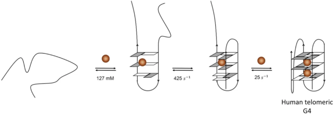
Figure 2.7: Proposed folding pathway for Tel22 in K+ [23, 127]. A random coil
undergoes transition to a hairpin structure, followed by the formation of antiparallel chair-type G4. A consecutive triplex formation allows the folding to the corresponding hybrid structure. K+ is indicated by brown spheres.
re-vealed multiphasic CD changes spanning the time scale of hundred milliseconds to a few hours [23]. Under these conditions, Tel22 is known to form two structures, hybrid1 and hybrid2. In order to explain the observed multiphasic CD changes, Gray et al. have proposed a four-step sequential folding mechanism (U *) I1 *) I2 *) I3 *) F , see Fig. 2.7) leading to the formation of the equilibrium of the two hybrid conforma-tions [23]. From the singular value decomposition (SVD) analysis of the time-resolved CD spectra, they propose the formation of an antiparallel topology (I2) in ca. 100 ms from a short-lived hairpin structure (not observed here), followed by the formation of a triplex (I3) in ca. 4000 s which rapidly form the final hybrid conformation in ca. 800 s, as shown in Fig. 2.7.
In contrast to the group of Chaires, very recent studies of Tel22 by stopped-flow combined with FRET detection have revealed faster folding kinetics, upon addition of 100 mM K+ [124]. The observed biphasic fluorescence decays (200 ms and 1.5 s)
have been suggested to arise from the existence of multiple folding pathways rather than a sequential folding process. Interestingly, biphasic folding kinetics have also been reported previously by the group of Balabrasumanian for the human telomeric sequence Tel21 upon the addition of 90 mM K+ [123]. In that case, a sequential
folding model involving the formation of one triplex intermediate preceded by a fast pre-equilibration step with the binding of a single K+ has been proposed as the most
plausible model (see Fig. 2.8). A similar model has been also proposed for RNA G4 and a non-telomeric G4 [123].
Figure 2.8: Proposed folding pathway for Tel22 in K+ according to the group of
Balasubramanian [123]. A random coil undergoes transition to a hairpin structure, followed by the formation of a triplex formation allowing the folding to the corre-sponding hybrid structure. K+ is indicated by brown spheres.
Besides rapid-mixing technique, one recent study using an ultrafast microfluidic mixer has highlighted the existence of events on the microsecond time scale in the
folding process of the G4 sequence Tel24 [128]. Such fast events would be consistent with the idea of the formation of a hairpin structure as the first step of the G4 folding process.
On the other hand, the group of Schwalbe has performed slow-mixing experiments combined with NMR on the 2GKU sequence with K+. This high-resolution method
allowed them to discriminate the absolute structures of DNA involved in the folding mechanism. They have found that it takes several hours at 25◦C for the G4 to reach its
final hybrid1 conformation. Several folding pathways have been identified involving the formation of several misfolded conformations, such as the hybrid2 conformation and a partially unfolded conformation. These observations are consistent with a multipathway model of folding.
2.4.2.2 Single molecule FRET studies
Besides rapid-mixing techniques, single-molecule approaches have been used to probe the folding mechanism of G4. In contrast to ensemble experiments, single-molecule FRET microscopy allows to probe metastable and heterogeneous states of one molecule at a time.
Single molecule measurements are typically implemented as depicted in Fig. 2.9. A single DNA strand containing a FRET marker (Dye1) and a G4 forming sequence is attached to a glass surface. A complementary strand with a second FRET marker (dye2) is then added in order to form a double helix as shown in Fig. 2.9. Formation of the G4 structure is induced by addition of cations. G4 formation results in an increase of the FRET efficiency.
The earliest studies of Balasubramanian carried out on Tel21 in the presence of 100 mM N a+ or K+ have shown the existence of two folded ensembles, they
attributed to two distinct G4 structures in both cases: an antiparallel and a parallel topologies, respectively [129]. In contrast, the subsequent study of Lee et al. [130] of Tel21 with various concentrations of K+ has highlighted the presence of an unfolded
conformation in addition to the two folded conformations. Each folded topologies have been found to exhibit long (minutes) and short (seconds) lifetimes. To explain these results, they proposed a complex kinetics scheme involving two parallel folding pathways.
The studies of Okamoto et al. of Tel22 with 100 mM K+ have revealed the
presence of two major conformations, attributed to an unfolded one and a folded one, respectively. By modifying certain guanines via bromide substitution, they have proposed the existence of a triplex topology as a possible intermediate in the folding mechanism of Tel22 [132]. Subsequent studies of Tel22 and two other human telomeric
Figure 2.9: Schematic representation of a single molecule FRET experiment on G4. A DNA strand containing the G4 forming sequence at its end is attached to a linker. To assure that there is no interaction of the G4 forming sequence with the surface a complementary strand is added in a way that the G4 sequence is attached to a DNA double helix. One dye (Dye1) is connected to the 5’ end of the G4 sequence whereas Dye2 is attached to the complementary strand. By adding cations G4 topologies form and the distance between the two dyes decreases which increases FRET efficiency.
sequences, Tel23 and 2GKU with 100 mM N a+ or K+have clearly shown the impact
of the protocol of G4 preparation on the observed conformations [133]. These studies of Long and Stones may explain the different distributions of conformations that have been observed in different experiments performed on similar sequences. The overall results of Long and Stones have been found to be consistent with a multi-pathway folding model.
Very recently, the group of Birkedal has published two studies of Tel22 in the presence of 100 mM N a+, Li+ and K+ [131]. Despite a similar protocol of
prepara-tion than Long and Stone (i.e. a slow annealing process), they have identified four different conformations (one unfolded and three folded) of Tel22 in the presence of N a+. Their populations were found to be strongly dependent on the concentration of
cations. With Li+, a sole conformation has been observed, attributed to an unfolded
structure, as expected. With K+, the existence of several conformations of Tel22 has
been observed. Their thermodynamic and kinetic properties have been attributed to a multi-pathway folding model involving several marginally stable conformations (possible misfolded states) as folding intermediates (see Fig. 2.10).
Figure 2.10: Figure taken from [131]. Different possible intermediates and folded structure (horizontal lines) and their stabilities and folding rates from unfolded species (dotted line).
2.4.2.3 Single-molecule force spectroscopy
Additional single molecule studies have utilized optical and magnetic tweezers to study the folding mechanisms of G4 [134–137]. Fig. 2.11 depicts the principle of an optical tweezers experiment. Typically, a DNA strand contains beads on both ends that are trapped via lasers. The G4 forming sequence is placed in the center of the DNA strand. After G4 formation, one can elongate the distance between the two laser foci to apply a stretching force on the quadruplex. This results in G4 unfolding which can be measured as a function of extension for a given applied force [134].
Sugiyama and coll. have performed several studies on different human telomeric G4 sequences [134, 138]. Notably, they have compared the mechanical properties of Tel27, with that of the truncated human telomeric sequence, T T A(GGGT T A)3
in the presence of 100 mM N a+. They have observed a thermodynamically and
mechanically stable species for the truncated sequence that they attributed to a triplex scaffold. Interestingly, the study of Tel27-mer has revealed the existence of two distinct conformations, one having an extension comparable to that of the putative triplex species. In subsequent works, Sugiyama and coll. have shown the formation of misfolded conformations as the number of TTAGGG repeats increases (4 vs 8). However, the number of misfolded conformations has been found to decrease if the length of the loops is increased.
Figure 2.11: Scheme of an optical tweezers experiment according to [134]. Blue spheres reflect beads that are trapped in laser foci (red profile). DNA is attached to the beads depicted as black double helices. In the center of the DNA double strand a quadruplex forming sequence is employed. This is shown by a type-1 forming G4.
telomeric sequence Tel27 with 100 mM K+ [135]. By substituting one thymine of
the G4 loops by one uridine, they have built 6 different DNA constructs in which ds-DNA is attached at different locations of the G4 sequence. They found that the G4 unfolding trajectories of these 6 DNA constructs might follow a funneled energy landscape. In another study [139], Yu et al. have identified several conformations in the mechanical unfolding/refolding of a longer G4 sequence, the 41-mer h-TERT in the presence of 100 mM K+. Kinetics and thermodynamics properties of these
conformations have been attributed to a multi-pathway folding mechanism involving both sequential and cooperative pathways. No evidence of the existence of a triplex intermediate has been found in these experiments.
The use of magnetic tweezers is another possibility to study mechanically induced folding/unfolding of G4. Its principle is similar to that of optical tweezers. However, a slightly different experimental setup is needed. One end of the ds-DNA is attached to a surface, whereas the other end is linked to a magnetic bead [136, 137]. This magnetic bead is trapped by a magnetic field. Finely tuned extension of DNA is realized by slightly moving the magnets that trap the bead [136]. Li et al. performed magnetic tweezer studies on a repeat of the human telomeric sequence Tel21. To reduce polymorphism they have chosen to work with 100 mM N a+solution [136]. By
finely adjusting the force exerted on DNA, they have observed several conformations in the mechanical unfolding/refolding of Tel21, they have attributed it to a sequential mechanism involving the formation of a triplex conformation.
Another sequence, Tel26, has been studied with magnetic tweezers by You et al. in the presence of 100 mM K+. In these conditions, Tel26 may form a mixture of
hybrid1 and hybrid2 conformations in solution. Measurements by force spectroscopy have suggested a sequential folding mechanism involving the formation of a short-lived intermediate with a lifetime of 5 seconds and a subsequent long-lived intermediate with a lifetime of tens of seconds (comparable to [137]).
2.4.2.4 Other experimental approaches
A thorough work has been published by the group of Gabelica [21]. They have used electrospray mass spectrometry in order to characterize the folding pathways of various G4 sequences, Tel21, Tel21-T, Tel22, 22CTA, Tel23, 2GKU and c-MYC2. Titration experiments showed that for low concentration of cations, the short se-quences (Tel21, Tel22, Tel21-T and 22CTA) preferentially form an antiparallel G4 structure with 2 G-quartets and one K+. For the longer sequences (> 22 bases),
K+-binding cooperativity has been found to increase. Kinetics of K+ binding of the
3 human telomeric sequences, Tel23, 2GKU and 26TTA, in addition to the sequence c-MYC2 have been found to be consistent with a multiple branching pathways as il-lustrated in Fig. 2.12. The antiparallel 2-G-quartet structure observed for low cation concentration has been suggested to be one misfolded intermediate of G4 folding (an off-pathway structure). Despite the fact that there is no direct evidence, the forma-tion of a hairpin and a G-triplex structure often invoked as G4 intermediates of the folding/unfolding mechanism of G4 are not excluded [21].
Figure 2.12: Scheme of possible folding pathways taken from [21].
com-bined with time-resolved absorption detection has been performed on Tel2, by the group of Garc´ıa. T-jump experiments measure conformational changes induced by a fast temperature rise, as will be explained in detail in the next chapter. Typical T-jump experiments explore the time-range of some nanoseconds up to 1 ms [27]. First T-jump measurments on Tel22 in presence of 150 mM N a+ or K+ have revealed
kinetics in the time scale of a few ten microseconds. Despite the fact that these measurements have been done at low temperature (i.e. 22.5◦C), far from the melting
temperature, they have observed unexpected significant changes in the absorption of DNA after a T-jump of 2.5◦C that has been attributed to the transition from the G4
structure to a triplex one.
2.4.2.5 Theoretical Approaches
Many experimental approaches require the modification of the G4 forming sequence in order to monitor the conformational changes during folding/unfolding (FRET, 2-AP, optical/magnetic tweezers). These modifications are suspected to alter significantly the folding landscapes of G4. In fact, most of the experimental studies have revealed complex G4 folding mechanisms that involve several intermediates. While the nature of these species remains elusive, it comes out that their formation and their kinetics do not only depend on the primary sequence of DNA but also on the environment (cations, proximity of duplex DNA, ionic strength, ...). In this context, it is difficult to compare experimental results obtained in different conditions. In addition, most of the experimental approaches used up to now have a limited time-resolution (in general millisecond or seconds) and/or lead to a limited structural information. In order to get an atomistic picture of G4 folding, advanced molecular dynamics (MD) simulations have been recently developed, notably by the group of ˇSponer [140–142]. Like experimental approaches, MD also have to face several drawbacks correlated, for instance, to the time-scale of the simulations and the sampling methods. A review explaining the basic principles and limitation of MD methods has been published in 2017 [20]. In 2007, Mashimo and Sugiyama have proposed a folding mechanism involving the formation of a G-triplex intermediate from a G-hairpin conformation for Tel22 in the presence of K+ [127]. With a combination of ab initio calculations
and MD simulations, they have shown that the formation of a G-hairpin and then a G-triplex structure are energetically favorable intermediates in the formation of hybrid1 and hybrid2 topologies of Tel22 with K+. GG base pairs have been found to
have comparable energetic stability than AT base pairs but are less stable than GC base pairs. Their calculations have also shown that G-quartets are more stable than GG base pairs. The folding process has been found to be facilitated by K+ binding
at each step of the process.
The group of ˇSponer has also performed a series of (MD) simulations on G4 folding and unfolding dynamics. Their works have suggested that the formation of antiparallel G-hairpin conformations may take place at all stages of the folding process of G4 [142]. The existence of various antiparallel structures has been taken as an indication of a multi-pathway nature of G4 folding. In contrast to antiparallel hairpins parallel ones have been found to be very unstable and may not form. It is noteworthy that a stable G-hairpin structure resulting in a complex topology that includes a chain reversal arrangment of the backbone in the continuous G-tract has been recently observed for the 5’-GTGTGGGTGTG-3’ sequence [143]. Other MD simulations of the ˇSponer group have shown that antiparallel G-triplex conformations may have a sufficient lifetime (i.e. a few tens of microseconds) to be a G4 folding intermediate. At this point, it is noteworthy that an antiparallel G-triplex conformation (TBA G3) has also been predicted from metadynamics simulations of the shortest G4 forming sequence, TBA G4, by Limongelli et al. [7]. Such a G-triplex structure has been then observed in solution by NMR spectroscopy from the 3’-end truncated TBA sequence [144].
In addition to putative G-triplex intermediates, recent simulations of ˇSponer and coll. [140, 145] as well as experimental approaches [2, 21] have pointed to the impor-tance of misfolded structures in G4 folding, such as those with shifted strands with respect to the final G4 structure and a reduced number of tetrads. These structures, that have to unfold to form the final G4 structure, are expected to have a much longer lifetime and to be more stable than G-triplex structures. The existence of such misfolded structures indicates extremely rugged G4 folding energy landscapes. The natural kinetic partitioning of G4 energy landscapes has been attributed to the propensity of G-rich sequences to populate a large number of syn-anti patterns in their G-tracts [20].
2.4.3
Conclusion
Several experiments and simulations exploring the folding pathways of G4 have been performed. Most of these studies have focused on human telomeric G4 sequences. However, due to their high polymorphism resulting in part from their extreme sensi-tivity to the environment (solvent, cations, ...), it is difficult to compare the results of different experiments and simulations carried out under different conditions.
All studies of G4 folding have revealed complex mechanisms, but there is not yet a unified picture of these mechanisms. While ensemble experiments are generally in favor of a sequential folding mechanism, single molecule experiments and simulations
![Figure 2.4: Examples of G4 ligands. Quarfloxin, the first G4-binding molecule that underwent clinical trials [96]](https://thumb-eu.123doks.com/thumbv2/123doknet/2921795.76512/31.918.158.754.117.479/figure-examples-ligands-quarfloxin-binding-molecule-underwent-clinical.webp)