For Peer Review
Partially segregated neural networks for spatial and contextual memory in virtual navigation.
Journal: Hippocampus Manuscript ID: HIPO-07-130.R1 Wiley - Manuscript type: Research Article
For Peer Review
Partially segregated neural networks for spatial and contextual memory in virtual navigation
Géraldine Rauchs(1), Pierre Orban(1,2), Evelyne Balteau(1), Christina Schmidt(1), Christian Degueldre(1), André Luxen(1), Pierre Maquet(1) and Philippe Peigneux(1, 3 ‡).
(1)
Cyclotron Research Center, University of Liège, Belgium. (2)
Functional Neuroimaging Unit, University of Montréal, Canada (3)
Neuropsychology and Functional Neuroimaging Research Unit, Université Libre de Bruxelles, Belgium
Running title: Spatial and contextual memory in navigation.
Keywords: fMRI; Hippocampus; Parahippocampal cortex; Ecologically valid
environment;
Number of text pages: 35
Number of figures: 5 (including one in Supplemental Data) Number of tables: 9 (including one in Supplemental Data)
‡
To whom correspondence should be addressed: Prof. Philippe Peigneux
Université Libre de Bruxelles (ULB)
UR2NF - Neuropsychology and Functional Neuroimaging Research Unit Av. F.D. Roosevelt 50, CP191 B-1050 Bruxelles BELGIUM Phone: +32 2 650 26 39 3 4 5 6 7 8 9 10 11 12 13 14 15 16 17 18 19 20 21 22 23 24 25 26 27 28 29 30 31 32 33 34 35 36 37 38 39 40 41 42 43 44 45 46 47 48 49 50 51 52 53 54 55 56 57 58 59 60
For Peer Review
Fax: +32 2 650 22 09 Email: [email protected] 3 4 5 6 7 8 9 10 11 12 13 14 15 16 17 18 19 20 21 22 23 24 25 26 27 28 29 30 31 32 33 34 35 36 37 38 39 40 41 42 43 44 45 46 47 48 49 50 51 52 53 54 55 56 57 58 59 60For Peer Review
Abstract
Finding our way in a previously learned, ecologically valid environment concurrently involves spatial and contextual cognitive operations. The former process accesses a cognitive map representing the spatial interactions between all paths in the environment. The latter accesses stored associations between landmark objects and their milieu. Here, we aimed at dissociating their neural basis in the context of memory-based virtual navigation. To do so, subjects freely explored a virtual town during one hour then were scanned using fMRI while retrieving their way between two locations, under four navigation conditions designed to probe separately or jointly the spatial and contextual memory components. Besides prominent commonalities found in a large hippocampo-neocortical network classically involved in topographical navigation, results yield evidence for a partial dissociation between the brain areas supporting spatial and contextual components of memory-based navigation. Performance-related analyses indicate that hippocampal activity mostly supports the spatial component, whereas parahippocampal activity primarily supports the contextual component. Additionally, the recruitment of contextual memory during navigation was associated with higher frontal, posterior parietal and lateral temporal activity. These results provide evidence for a partial segregation of the neural substrates of two crucial memory components in human navigation, whose combined involvement eventually leads to efficient navigation behavior within a learned environment. 3 4 5 6 7 8 9 10 11 12 13 14 15 16 17 18 19 20 21 22 23 24 25 26 27 28 29 30 31 32 33 34 35 36 37 38 39 40 41 42 43 44 45 46 47 48 49 50 51 52 53 54 55 56 57 58 59 60
For Peer Review
Introduction
Route retrieval and way finding in a previously learned environment are critical prerequisites to successfully carry out most of our daily activities. These cognitive abilities involve the creation of a cognitive map of the environment, where are coded its landmarks, paths and their spatial relationships (Maguire et al., 1998a; Berthoz, 2001; Pazzaglia and De Beni, 2001). Neuroimaging studies have revealed that navigation in a virtual environment involves an extended neural network, mostly including hippocampal and parahippocampal areas, frontal, posterior parietal and occipital cortices as well as the caudate nucleus (Aguirre et al., 1996; Ekstrom et al., 2003; Maguire et al., 1998b; Hartley et al., 2003; Voermans et al., 2004; Peigneux et al., 2004; Orban et al., 2006). Within these areas, spatial memory-based navigation prominently relies upon activity in the hippocampal formation (e.g. Burgess et al., 2002; Maguire et al., 1998b), also crucially involved in episodic memory. In contrast, procedural memory-based navigation (i.e., moving along a well-known pathway in a kind of automatic fashion) is rather contingent upon activity in the striatal complex (e.g. Packard and Knowlton, 2002; Hartley et al., 2003; Iaria et al., 2003; Orban et al., 2006). Additional experiments have indicated a role for the parahippocampal gyrus in the storage of object location as a part of the neural mechanisms underlying successful navigation (Janzen and van Turennout, 2004) and retrieval of objects’ spatial context (Burgess et al., 2001). Recently, promising attempts have been made to track the neural correlates of spontaneous mentalizing and behaviors during virtual navigation (Spiers and Maguire, 2006a, 2006b).
In the present study, we have focused on another possible dissociation between cognitive processes engaged in route retrieval and way-finding, considering that these actions are supported by manifold memory processes in which two prominent 3 4 5 6 7 8 9 10 11 12 13 14 15 16 17 18 19 20 21 22 23 24 25 26 27 28 29 30 31 32 33 34 35 36 37 38 39 40 41 42 43 44 45 46 47 48 49 50 51 52 53 54 55 56 57 58 59 60
For Peer Review
cognitive components may be identified. The first one, spatial representation memory, involves creation of and/or access to a cognitive map of the environment, where are specified the spatial relationships between the streets independently of the salient features of the environment. For instance, when attempting to reach the school from the church, one can keep in mind an “abstract” map-like representation indicating the appropriate direction to follow at each crossroad, independently of specific environmental cues along the way. Besides this “streets configuration” component, a second, complementary process can be used. It refers to a contextual representation memory (or “landmarks memory”) where are stored specific associations between salient landmark objects and their milieu. For instance, one may remember that from church to school, there is a right turn just after the library and then a left turn in front of the red telephone box close to the grocery store. In most cases though, both memory constituents are engaged simultaneously during route retrieval or way finding in a previously learned environment, in that navigation involves the creation of a cognitive map coding both the relationships between paths and between landmarks in episodic memory (Maguire et al., 1998a; Berthoz, 2001; Pazzaglia and De Beni, 2001).
Using functional magnetic resonance imaging (fMRI), we investigated the neural bases of spatial (map-like streets configuration) and contextual (milieu-related landmarks) components of navigation memory. To do so, 16 volunteers were scanned under four complementary memory conditions during route retrieval. Before testing, they freely explored during one hour a complex 3D virtual town, composed of three different surroundings in the same city, with distinctive wall and ground features, and landmark objects (Peigneux et al., 2004, 2006; Orban et al., 2006). In the Natural testing condition, they were positioned in the same environment as during the training 3 4 5 6 7 8 9 10 11 12 13 14 15 16 17 18 19 20 21 22 23 24 25 26 27 28 29 30 31 32 33 34 35 36 37 38 39 40 41 42 43 44 45 46 47 48 49 50 51 52 53 54 55 56 57 58 59 60
For Peer Review
period and asked to reach target locations from various starting points. This classical testing condition (e.g., Maguire et al., 1998b; Peigneux et al., 2004, 2006; Orban et al., 2006) actually does not dissociate the spatial and contextual components of memory-based navigation since circulating in such an enriched environment may rely on either or both, or even rely on a stimulus-response associations strategy mediated by the striatum (Iaria et al., 2003; Voermans et al., 2004; Bohbot et al., 2004; Orban et al., 2006). Thus, to engage subjects using more specifically the spatial memory component of navigation, they had, in the Impoverished testing condition, to reach target locations in this same environment after removal of all landmark objects and after that the walls and ground were made uniform. In this spatial condition indeed, subjects must rely on their abstract knowledge of the spatial relationships of the streets to find a way towards the target. Likewise in the Alternate condition, the scene was the same as in the Natural task but the optimal path between the starting location and the designated target was blocked by an impassable barrier. In this case, previously identified landmark objects and other contextual features become less relevant for navigation: subjects should rather rely on a more spatial, less contextualized representation of the navigation space to build an alternate route. Thus, this condition further allowed investigating the spatial component by promoting alternative route-finding strategies. And finally, to specifically assess the contextual component of memory in navigation, subjects were asked in the Recognition condition to follow the same pathways while paying particular attention to the potential changes made to the town’s scenery. To minimize the requirements for spatial information, they had to follow color dots positioned on the ground all along the pathway. Additionally, a forced-choice recognition task was proposed after each walk to reveal a posteriori the presence or absence of subjects’ awareness of 3 4 5 6 7 8 9 10 11 12 13 14 15 16 17 18 19 20 21 22 23 24 25 26 27 28 29 30 31 32 33 34 35 36 37 38 39 40 41 42 43 44 45 46 47 48 49 50 51 52 53 54 55 56 57 58 59 60
For Peer Review
contextual changes. Besides targeted differences, it should be mentioned that all these conditions involve episodic memory, although additional contribution of procedural memory cannot be excluded.
The analysis of brain imaging data aimed at unraveling the specificity of the spatial and contextual memory components of memory-based navigation, by conducting between-tasks comparisons. Additionally, correlation analyses between functional imaging data and behavioral performance aimed at evidencing the brain structures whose activity differentiates efficient from poor navigation at the within-subject level, and good from bad navigators, at the between-within-subject level, across the various experimental conditions. We hypothesized that although all tasks would elicit grossly similar activity in the neural network classically engaged in navigation (Aguirre et al., 1996; Maguire et al., 1998b; Burgess, 2002; Hartley et al., 2003; Peigneux et al., 2004), those conditions relying more on the spatial memory component should induce higher activity in the hippocampal/parahippocampal region as well as posterior cortical areas. In contrast, tasks more based on the contextual memory component should rather rely upon frontal (Ranganath and Knight, 2003) and lateral temporal (Ojemann et al., 2002) cortices activity, as well as the parahippocampal gyrus known to be involved in visual recognition memory (Meunier et al., 1993; Rauchs et al., 2006).
Materials and Methods
Subjects
Sixteen healthy right-handed volunteers (8 females, 8 males; mean age of the group: 22.1 years; range: 18-30 years) gave their informed, written consent to participate in this experiment approved by the Ethics Committee of the University of Liège. They 3 4 5 6 7 8 9 10 11 12 13 14 15 16 17 18 19 20 21 22 23 24 25 26 27 28 29 30 31 32 33 34 35 36 37 38 39 40 41 42 43 44 45 46 47 48 49 50 51 52 53 54 55 56 57 58 59 60
For Peer Review
were free of neurological or psychiatric disease and had a normal structural MRI brain scan on visual inspection.
Navigation task
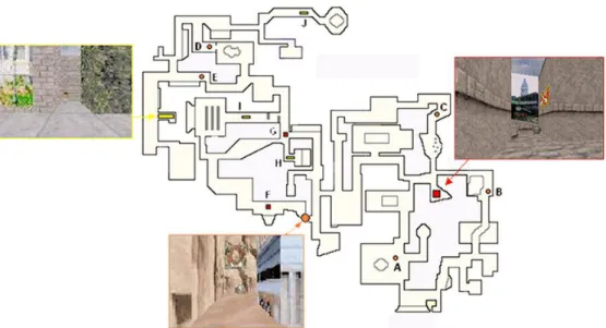
Subjects were trained in a virtual environment developed and validated in our laboratory (Peigneux et al., 2004, 2006; Orban et al., 2006), adapted from a commercially available computer game (Duke Nukem 3D, 3D Realms Entertainment, Apogee Software Ltd., Garland TX) using the editor provided (Build, Ken Silverman, Realms Entertainment). The environment was a complex town composed of three districts (Far West, Urban and English) that were made distinct from each other by different visual backgrounds and objects along the streets. Each of these districts contained a target location identified by a rotating medallion (Fig. 1). The virtual town also contained 10 starting points that were each 35 virtual units apart (optimal path) from their associated target location. Subjects navigated at a constant speed within the environment at the ground level using a four-direction keypad with their right hand. During training, the virtual environment was presented on a desktop 800-MHz Pentium-III PC (screen size, 17’). For testing in the scanner, a mirror allowed the participants to see the display of the virtual town projected on a screen.
Learning phase: Participants were trained outside of the scanner during four 15-minute exploration periods. They were explicitly instructed to learn the layout of streets, districts and target locations by moving freely within the environment. During the entire training session, pictures of the three target locations and their associated names were continuously available to the subject.
3 4 5 6 7 8 9 10 11 12 13 14 15 16 17 18 19 20 21 22 23 24 25 26 27 28 29 30 31 32 33 34 35 36 37 38 39 40 41 42 43 44 45 46 47 48 49 50 51 52 53 54 55 56 57 58 59 60
For Peer Review
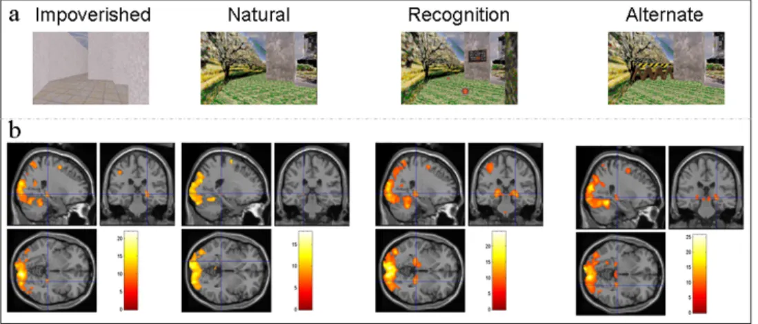
Test conditions: At the end of the training session, subjects were scanned using fMRI while performing four different tasks that aimed at assessing spatial and contextual components involved in memory-based navigation (Fig. 2a).
In the Natural, Impoverished and Alternate conditions, subjects had to retrieve, as fast as possible and in no more than 35 s, the route between two locations in the learned environment. In the Impoverished condition, the environment was made plainly uniform by removing all wall/ground features and objects. In the Natural and Alternate conditions, the environment was identical as during the exploration. In the Alternate condition however, optimal pathways between starting and target points were blocked by an impassable barrier to promote alternative route-finding strategies and to prevent from using a routine navigation behavior. Thus, the Impoverished condition allows to assess the spatial memory component and the Natural condition allows the investigation of both memory processes (spatial and contextual). The Alternate condition was designed to assess spatial memory since, in this task, previously identified landmark objects and contextual features become less relevant for navigation. Indeed, subjects should rather rely on a more spatial, less contextualized representation of the navigation space to build an alternate route. For each of these three tasks, the fMRI scanning session consisted of 10 blocks of tests, each lasting for 35 s, that alternated with 10 blocks of rest, during which a black screen was displayed for a duration randomly lasting from 10 to 17 s. During rest periods, subjects were instructed not to think to anything in particular and to relax as the study is long and demanding. Within the last two seconds of the rest period preceding each test, the target location for the test was indicated orally through MR compatible headphones. After test time elapsed, a quantitative measure of route retrieval performance was determined as the distance remaining between the subject’s 3 4 5 6 7 8 9 10 11 12 13 14 15 16 17 18 19 20 21 22 23 24 25 26 27 28 29 30 31 32 33 34 35 36 37 38 39 40 41 42 43 44 45 46 47 48 49 50 51 52 53 54 55 56 57 58 59 60
For Peer Review
actual location and the target to reach, proportional to the length of the optimal path between the starting point and the target. Although various indicators of performance can be computed (e.g. success, walked distance, effective navigation speed, crossroads, dead end errors, …), the distance remaining to target was selected as the main behavioral measure of navigation, in reference to previous studies in our group (Peigneux et al., 2004, 2006) in which the remaining distance (or conversely the distance towards destination) was shown the most sensitive measure of navigation ability. At variance with the Impoverished and Natural conditions in which all targets were 35 virtual units apart from the starting point, the use of barriers in the Alternate condition makes that the average optimal distance was 52 virtual units (SD = 5.8, range 39-58). Therefore, to render performance in the Alternate condition comparable with those in the two other conditions, performance was measured as the distance remaining between the subject’s actual location and the position located at 35 virtual units from the starting point on the shortest path towards the target. For the Impoverished, Natural and Alternate conditions, the same 10 tests were administered in a counterbalanced order, both at the between- and within-subjects levels.
In the Recognition condition, subjects had to pay attention to the environmental features of the town during 35s while following colored dots on the ground that signaled the path to follow between the starting and target points. They were instructed to determine whether and where environmental changes were made as compared to the town explored during the learning phase. All changes were easily detectable and did not necessitate stopping along the walk. At the end of each 35-s walk, they were presented with a four-choice panel composed of three pictures taken along the route just previously followed, and a white square (Supplementary Fig. 1). Using a keypad with their left hand, they had to indicate in no more than 10 s the 3 4 5 6 7 8 9 10 11 12 13 14 15 16 17 18 19 20 21 22 23 24 25 26 27 28 29 30 31 32 33 34 35 36 37 38 39 40 41 42 43 44 45 46 47 48 49 50 51 52 53 54 55 56 57 58 59 60
For Peer Review
modified image, or to select the white square if they thought that no modification was made. For the Recognition condition, 22 tests were administered in a pseudo-randomized order (4 possible lists), and alternated with short blocks of rest during which a black screen was displayed for 10-17 seconds. Behavioral performance was measured as the number of correct recognitions. This measure was used to associate a performance index with brain imaging data obtained during the walk between the starting and target points, here deemed as a contextual recognition task as subjects were actively engaged in the detection of potential modifications in the learned environment.
For all subjects, the four tasks were proposed in the following fixed order: Impoverished, Recognition, Natural then Alternate. Although administrating the various tasks in a randomized order would have ruled out the possibility of decreased medial temporal lobe activation as the pathways become familiar (Nyberg, 2005), it was much more important for the purpose of the present study to avoid as much as possible interference between the four tasks. Thus, to minimize re-learning of the contextual details of the environment or of the spatial layout of the routes, the Impoverished and Recognition conditions were administered before the Natural navigation condition. Additionally, although only optimal paths were blocked in the Alternate condition, some subjects never discovered these during the initial exploration period and actually learned an alternate route in which they perseverated. Therefore, we analyzed our subjects’ data in the Alternate condition based on the pathways (either optimal or not) followed by them in the immediately preceding Natural condition. Two strategies were differentiated in this condition: true alternate way finding and routine strategy (see Brain Imaging and Results sections for details). 3 4 5 6 7 8 9 10 11 12 13 14 15 16 17 18 19 20 21 22 23 24 25 26 27 28 29 30 31 32 33 34 35 36 37 38 39 40 41 42 43 44 45 46 47 48 49 50 51 52 53 54 55 56 57 58 59 60
For Peer Review
fMRI data acquisition.
Brain imaging data were obtained using a 3 Tesla head-only MRI system (Allegra, Siemens, Erlangen, Germany) equipped with an actively shielded gradient coil system (max gradient amplitude 40 mT/m). For each testing session, the functional multi-slice T2*-weighted images were obtained using a blood oxygen level dependent (BOLD) sensitive single-shot echo planar (EPI) sequence (TR = 2130 ms; TE = 40 ms; flip angle = 90°; FoV = 220x220 mm²; matrix size = 64x64x32) covering the whole brain (128 mm high). Each functional volume consisted of 32 slices, with a thickness of 3 mm (inter-slice gap = 1 mm) and a voxel size of 3.4x3.4x3 mm3. The four initial scans of each session were discarded to control for magnetic saturation effects.
A high-resolution structural MRI scan was also acquired for each subject using a standard three-dimensional T1-weighted sequence (TR = 1960 ms; TE = 4.43 ms; flip angle = 8°; 176 slices; FOV = 230x173 mm²; matrix size = 256x192x176; voxel size = 0.9x0.9x0.9 mm³). The mean and individual MR images were used for a precise identification of loci of activation.
fMRI data analysis.
Functional volumes were pre-processed and analysed using the Statistical Parametric Mapping software SPM2 (Wellcome Department of Cognitive Neurology, London, UK, http://www.fil.ion.ucl.ac.uk/spm/spm2) implemented in MATLAB (Mathworks Inc., Sherbom, MA). For each subject, spatial preprocessing included realignment and adjustment for movement related effects, co-registration of functional and anatomical data, spatial normalization into standard stereotactic MNI space, and spatial smoothing using a Gaussian kernel of 6 mm full width at half maximum (FWHM). 3 4 5 6 7 8 9 10 11 12 13 14 15 16 17 18 19 20 21 22 23 24 25 26 27 28 29 30 31 32 33 34 35 36 37 38 39 40 41 42 43 44 45 46 47 48 49 50 51 52 53 54 55 56 57 58 59 60
For Peer Review
Data were analyzed using a mixed-effects model, aimed at showing stereotypical effect in the population from which the subjects are drawn (Penny and Holmes, 2003). This procedure was implemented in two processing steps accounting respectively for fixed and random effects. For each subject, changes in BOLD responses were estimated in a first-level intra-individual analysis using a general linear model at each voxel. For each experimental condition, the regressors of interest were built using boxcar functions corresponding to each block of navigation convolved with the canonical hemodynamic response function. In the Impoverished, Natural and Alternate conditions, the 10 pre-test 2-s periods during which subjects were orally indicated the name of the target to reach were modeled explicitly. In the Recognition condition, the time during which subjects were presented the four-choice response panel and made their response was also modeled explicitly.
Additionally, the strategy used by the subjects in the Alternate condition was modeled in the design matrix as follows. For each test in this condition, if a subject had followed the optimal path in the Natural condition, its brain activity was analyzed separately for the time spent in the detour portion of the walk, outside of its usual pathway. Otherwise if the subject had followed a non-optimal path during the Natural condition for a given test, and did not encounter the barrier in the Alternate condition for the same test, its brain activity during this period of time was not taken as a detour-related activity. It was rather analyzed as a routine-related navigation activity if the pathway was the one previously followed by the subject or explicitly modeled as a “lost” condition when the subject appeared to be completely disoriented in the environment as compared to his/her behaviour in the Natural condition (i.e., going round in circles, going in a totally wrong direction, entering already known dead 3 4 5 6 7 8 9 10 11 12 13 14 15 16 17 18 19 20 21 22 23 24 25 26 27 28 29 30 31 32 33 34 35 36 37 38 39 40 41 42 43 44 45 46 47 48 49 50 51 52 53 54 55 56 57 58 59 60
For Peer Review
ends…). Only the results of comparisons between detour and routine strategies are of interest here.
Furthermore, in order to test whether modifications of neuronal activity in navigation–related areas were linked to behavioral performance (navigation accuracy or recognition performance), performance regressors were added to the model. This allowed computing at the within-subject level the correlation between navigation (and recognition) performance and the navigation-related regional BOLD response. Hence, within-subjects’ correlation analyses looked for cerebral structures in which BOLD response during tests of place finding (or during the recognition task) correlated with subject’s trial-to-trial variations in performance.
In all individual analyses, movement parameters derived from realignment of the functional volumes (translations in x, y and z directions and rotations around x, y and z axes) were included as covariates of no interest in the design matrix. High-pass filtering was implemented in the matrix design using a cut-off period of 128 seconds to remove low frequency drifts from the time series. Serial correlations in fMRI signal were estimated with a restricted maximum likelihood (ReML) algorithm, using an intrinsic autoregressive model during parameter estimation. The effects of interest (i.e., main effects of condition, direct comparisons between the four tasks and intra-individual modulations by performance) were then tested by linear contrasts, generating statistical parametric maps [SPM(T)]. Since no inference was made at this fixed effects level of analysis, individual summary statistic images were thresholded at p<0.95 (uncorrected).
The individual summary statistics images resulting from these contrasts were then further spatially smoothed (6 mm FWHM Gaussian kernel) and entered in a second-level analysis, corresponding to a random effects (RFX) model in which 3 4 5 6 7 8 9 10 11 12 13 14 15 16 17 18 19 20 21 22 23 24 25 26 27 28 29 30 31 32 33 34 35 36 37 38 39 40 41 42 43 44 45 46 47 48 49 50 51 52 53 54 55 56 57 58 59 60
For Peer Review
subjects are considered random variables. A global null analysis (Friston et al., 2005) computed at the RFX level aimed at highlighting the brain areas commonly engaged during navigation in the four experimental conditions. Restricted maximum likelihood estimates of variance components were used to allow possible departure from the sphericity assumptions in conjunction analyses (Friston et al., 2002). It should be noted that a significant conjunction does not mean all contrasts were individually significant (i.e., conjunction of significance), but rather means that the contrasts were consistently and jointly significant (Friston et al., 2005). Additionally, we investigated the neural activity that differentiated good navigators from bad ones in our population. To do so, correlations were computed at the RFX level between each subject’s individual contrast image of the main effect of navigation and its average behavioral performance for this condition.
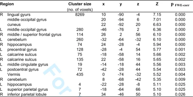
The resulting set of voxel values for each contrast constituted a map of the t statistic [SPM(T)], thresholded at p<0.001 (uncorrected for multiple comparisons). Statistical inferences were then obtained after corrections at the voxel level using Gaussian random field theory (Worsley et al., 1996), either pcorr<0.05 (FWE) corrected for multiple comparisons in the whole brain volume, or psvc(10mm) <0.05, corrected in a small spherical volume (radius 10 mm) around a priori locations of activation in structures of interest, taken from the literature (Table 1, Supplemental Data). Uncorrected values at p < .001 are reported descriptively only.
Finally, posterior probability maps enabled conditional or Bayesian inferences about regionally specific effects, allowing us to ensure that a lack of significant statistical effect in a given contrast was not merely due to a failure to detect this effect using classical inferences (Friston and Penny, 2003).
3 4 5 6 7 8 9 10 11 12 13 14 15 16 17 18 19 20 21 22 23 24 25 26 27 28 29 30 31 32 33 34 35 36 37 38 39 40 41 42 43 44 45 46 47 48 49 50 51 52 53 54 55 56 57 58 59 60
For Peer Review
For all activated voxels, anatomical localization was determined based both on stereotactic coordinates using the co-planar stereotactic atlas of the human brain (Talairach and Tournoux, 1988) after coordinates conversion using M. Brett’s set of linear transformations (http://www.mrc-cbu.cam.ac.uk/Imaging/mnispace.html) and an automatic algorithm labelling (AAL toolbox; Tzourio-Mazoyer et al., 2002). Confirmation of precise anatomical localization was made based on individuals’ and mean structural MR images.
Results
Behavioral performance
In the Impoverished, Natural and Alternate conditions, subjects were required to reach within 35 s a given target from a designated starting point. A quantitative estimate of navigational performance was given by the distance remaining between the subject’s actual location at the end of testing and the target, relative to the total length of the shortest possible route. Thus, a value comprised between 0 and 1 indicates that the subject moved towards the target on the optimal path and the smaller the value the closest the subject was from the target. In contrast, a value >1 indicates a displacement in the direction opposite to the target.
As expected given the absence of contextual cues during route retrieval, performance in the Impoverished condition was weaker than in the Natural condition (Table 1a, p<0.001). In order to render performance comparable in the Alternate condition with the index obtained in the other tasks, we measured not only the absolute performance (mean distance remaining to the target relative to the total length of the path = 0.40; SD = 0.16) but also a corrected index relative to an imaginary point located 35 units apart from the starting point on the new optimal path. 3 4 5 6 7 8 9 10 11 12 13 14 15 16 17 18 19 20 21 22 23 24 25 26 27 28 29 30 31 32 33 34 35 36 37 38 39 40 41 42 43 44 45 46 47 48 49 50 51 52 53 54 55 56 57 58 59 60
For Peer Review
This latter measure allows a direct comparison of the subjects’ efficiency to find their way between the Alternate and the other conditions. Note that both measures were correlated at the within-subject level (r = 0.78, p<0.001). Results indicate that corrected performance in the Alternate condition was better than in the Impoverished (p<0.001) but did not differ from the Natural (p>0.6) condition (see Table 1a). This result indicates that the Impoverished condition was harder than the two other conditions. In contrast, forcing subjects to find an alternative route to a given target does not reduce their navigation efficiency as performance, when expressed relative to the same path length, did not differ in the Alternate and Natural conditions.
Additionally, we found that performance in the Natural condition was correlated with performance in the Recognition memory task (i.e. number of correct recognitions; p<0.01) and in the Impoverished condition (p<0.01; see Table 1b). Performance in the Impoverished condition was also correlated with performance in the Alternate condition (p<0.05; see Table 1b). These results indicate that both spatial and contextual memory components contribute to accurate navigation in the Natural condition. It also suggests that performance in the Alternate condition, where subjects had to devise an unfamiliar path to reach the target, relies more on the capacity to elaborate an accurate cognitive map of the town, likewise in the Impoverished condition.
Brain imaging data
Navigation-related activations
A conjunction analysis across the Natural, Impoverished, Alternate and Recognition conditions revealed an increase in navigation-related blood oxygen level dependent (BOLD) responses in an extended hippocampo-neocortical network, including the 3 4 5 6 7 8 9 10 11 12 13 14 15 16 17 18 19 20 21 22 23 24 25 26 27 28 29 30 31 32 33 34 35 36 37 38 39 40 41 42 43 44 45 46 47 48 49 50 51 52 53 54 55 56 57 58 59 60
For Peer Review
hippocampus bilaterally as well as occipital, parietal and frontal areas (pcorr<0.05; Table 2; see Fig. 2b for navigation effects in separate conditions).
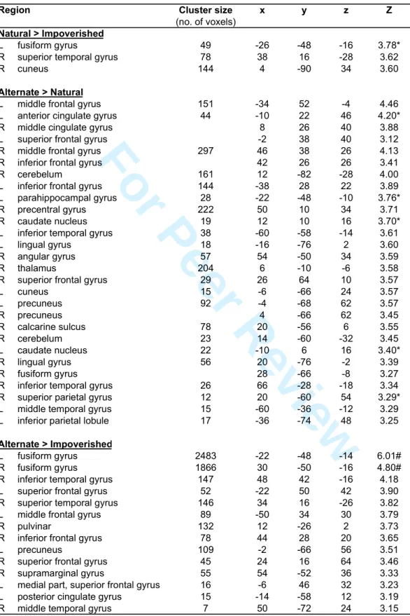
Besides these commonalities, differences in navigation-related brain activity were found between the four conditions. There was higher activity in the Natural than in the Impoverished condition in the left fusiform gyrus (psvc<0.05), right superior temporal gyrus and cuneus (p<0.001; Table 3). The reverse comparison was non significant. As compared to the Natural condition, there was higher activity in the left parahippocampal gyrus (psvc<0.05), the bilateral frontal areas (anterior cingulate and superior, inferior and middle frontal gyri) and in the caudate nuclei (psvc<0.05) in the Alternate condition. The reverse comparison was non significant. As compared to the Impoverished condition, there was higher activity mainly in the bilateral fusiform gyri (pcorr< 0.05; Table 3) in the Alternate condition, but also in the lateral temporal and frontal areas (p<0.001, uncorrected). The reverse comparison did not yield any significant activation. Finally, navigation in the Recognition condition was associated with higher activity than in the three other conditions in frontal areas, lateral temporal cortex (superior and middle temporal gyri), precuneus, retrosplenial and posterior parietal cortices (p<0.001, uncorrected, see Table 4).
In the Alternate condition, the regional cerebral activity was analyzed separately as a function of the subjects’ behavior during navigation. The underlying hypothesis was that finding an alternative to the pathway previously learned as optimal by the subject should engage more actively the hippocampus and parahippocampal area than navigating along a well-known route. We also speculated that navigating along the subject’s usual path could possibly rely on a response-based (habit) navigation strategy associated with caudate activity (Iaria et al., 2003; Orban et al., 2006). Therefore, the regional cerebral activity associated with navigation in the 3 4 5 6 7 8 9 10 11 12 13 14 15 16 17 18 19 20 21 22 23 24 25 26 27 28 29 30 31 32 33 34 35 36 37 38 39 40 41 42 43 44 45 46 47 48 49 50 51 52 53 54 55 56 57 58 59 60
For Peer Review
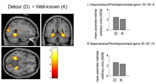
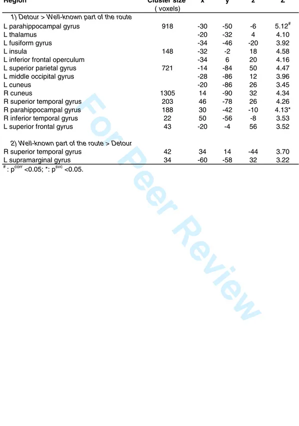
known portion of the route (i.e. corresponding to the portion of the route taken by the subject in the immediately preceding Natural condition) was contrasted within each trial with the activity associated with the portion of the route that corresponded to a detour. This analysis revealed higher activity in the detour than in the well-known part of the route bilaterally in the left ([-30 –50 -6], Z score = 4.38, pcorr<0.05) and right ([30 –42 -10] Z score = 4.13, psvc< .05; Fig. 3) parahippocampal gyrus, as well as in fusiform and frontal, parietal and occipital areas (p<0.001 uncorrected; Table 5). In contrast, the analysis failed to evidence differential activation in the caudate nucleus between routine and detour behaviors. Posterior probability maps (Friston and Penny, 2003) indicated a low probability to disclose a caudate activation in the detour part of the road (all P < 0.27, range 0.05-0.27).
Performance-related activations
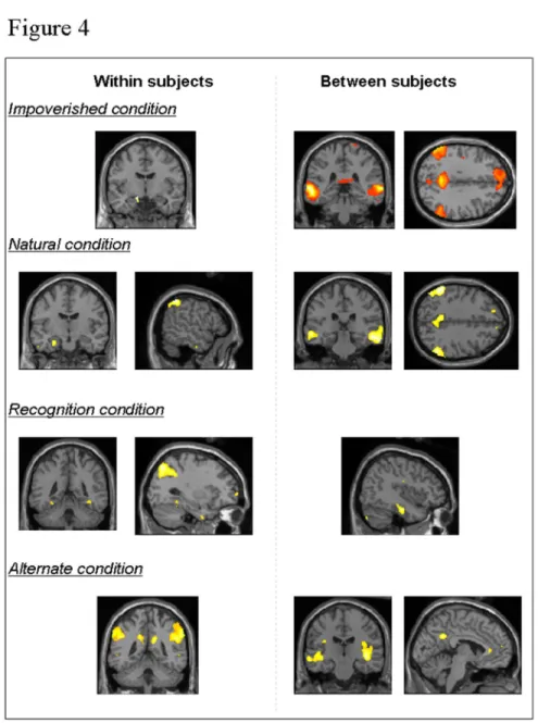
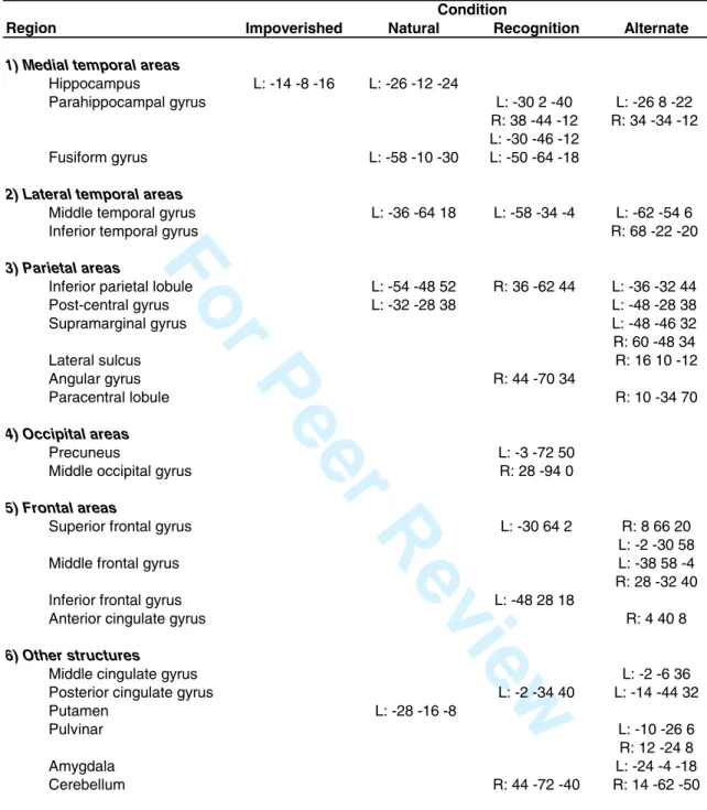
Next, to evidence the brain areas where activity varies from trial to trial according to navigation efficiency within an individual, we looked at the correlation between BOLD response during navigation and the performance measure at each trial within each condition (i.e., performance was the distance remaining to target in the Natural, Impoverished and Alternate conditions; the correct recognition score in the Recognition condition). This analysis highlighted correlation profiles between performance and activity in medial temporal areas (Fig. 4): left hippocampal activity was correlated with navigation performance in the Impoverished and Natural conditions, whereas bilateral parahippocampal activity was associated with performance in the Recognition and Alternate conditions. Additionally, performance was correlated with left parietal activity in the Natural condition, with right or bilateral parietal activity in the Recognition and Alternate conditions respectively, and 3 4 5 6 7 8 9 10 11 12 13 14 15 16 17 18 19 20 21 22 23 24 25 26 27 28 29 30 31 32 33 34 35 36 37 38 39 40 41 42 43 44 45 46 47 48 49 50 51 52 53 54 55 56 57 58 59 60
For Peer Review
with predominant left frontal activity in the Recognition condition (Table 6). Finally, an interaction analysis revealed that activity in the left hippocampus, parahippocampal gyri, parietal lobe (mainly on the right side), frontal areas, precuneus and caudate nuclei correlated more with navigation performance in the Alternate than in the Natural condition (Table 7).
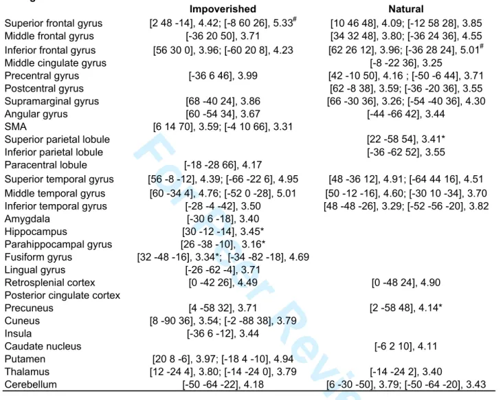
In a second step, we determined the brain areas where activity varies between subjects according to their global navigation efficiency, i.e. the brain regions that differentiate good from poor navigators. At the between-subjects level, we evidenced correlations between subjects’ average performance score and their navigation-related BOLD response within each condition. Positive correlations indicate brain areas that activated more in good than poor navigators. Results are summarized in Table 8 and illustrated in Fig. 4. Positive correlations with hippocampal activity were found only in the Impoverished condition (psvc<0.05). At variance, performance was positively correlated with activity in the right lateral temporal cortex in all conditions, and with activity in the precuneus, inferior parietal lobule and frontal areas in the Impoverished, Natural and Alternate conditions.
Discussion
The present study aimed at unravelling the neural substrates of spatial and contextual components of spatial memory-based navigation during route retrieval in an ecologically valid environment. Besides prominent commonalities found in a large hippocampo-neocortical network known to be involved in topographical learning in humans (Aguirre et al., 1996; Maguire et al., 1998b; Burgess et al., 2002; Hartley et al., 2003; Voermans et al., 2004; Peigneux et al., 2004, 2006; Orban et al., 2006; Spiers and Maguire, 2006a), our results yield evidence for a partial dissociation 3 4 5 6 7 8 9 10 11 12 13 14 15 16 17 18 19 20 21 22 23 24 25 26 27 28 29 30 31 32 33 34 35 36 37 38 39 40 41 42 43 44 45 46 47 48 49 50 51 52 53 54 55 56 57 58 59 60
For Peer Review
between the brain areas that sustain these primary memory components, eventually leading to efficient navigation within a newly learned environment.
Between-tasks comparisons revealed subtle differences in navigation-related brain activity that may relate to various cognitive processes. We interpret the higher fusiform, superior temporal and cuneus activity in the Natural than in the Impoverished condition (also found for the fusiform gyrus in the comparison between Alternate and Impoverished conditions) as being induced by the profuseness of visual details (objects, colors, textures, …) in the former case, in line with Maguire et al. (1998a) who found increased activation in fusiform and parahippocampal regions when the navigated environment was visually enriched. Better performance in the Natural than in the Impoverished condition is also in accordance with rodent studies having demonstrated that exposure to an enriched environment enhances memory performance in spatial tasks (Rosenzweig and Bennett, 1996; Frick et al., 2003). In the Alternate condition, in which subjects had to find a new way to reach the target, higher activity was observed in the left parahippocampal gyrus and frontal areas than in the Natural condition. Since the degree of enrichment was similar between those conditions, we propose that higher parahippocampal activity in the Alternate condition reflects an increased dependency upon map-like representations of the environment to find an alternative route near the target. At variance, higher prefrontal involvement may be explained by increased requirements for strategy switching in the presence of obstacles, a reading consistent with prior studies having used tasks with similar cognitive demands (Maguire et al., 1998b; Rosenbaum et al., 2004; Spiers and Maguire, 2006a). Frontal activations are also in line with a role for these areas in planning (Shallice, 1982) and decision making (Fellows, 2004 for review) in the 3 4 5 6 7 8 9 10 11 12 13 14 15 16 17 18 19 20 21 22 23 24 25 26 27 28 29 30 31 32 33 34 35 36 37 38 39 40 41 42 43 44 45 46 47 48 49 50 51 52 53 54 55 56 57 58 59 60
For Peer Review
Alternate condition, and increased working memory demands when updating topographical information (Gron et al., 2000).
In the Recognition task, cerebral activity was consistently higher than in the three other conditions in a large set of brain areas encompassing the precuneus, retrosplenial and posterior parietal cortices, and frontal and lateral temporal cortices. Frontal areas are well known to be involved in contextual and source memory tasks (Ranganath and Knight, 2003). Their involvement in the Recognition condition could therefore reflect the effortful process associated with retrieving the environmental information acquired during the exploration period, in order to ascertain possible differences with the actual scene when following the track. Also, fMRI studies have highlighted a role for the precuneus and posterior parietal cortex in source memory tasks (Lundstrom et al., 2003, 2005; Cavanna and Trimble, 2006), and activations in these regions, extending to posterior cingulate and retrosplenial cortices as well as to the inferior parietal lobule, have been consistently linked to episodic retrieval processes (Wagner et al., 2005). Finally, higher activity in the lateral temporal cortex in the Recognition condition is in line with the results of intracerebral recording in superior, middle and temporal regions in patients performing a series of memory tasks (Ojemann et al., 2002). The authors observed significant changes of neural activity during a recognition task mainly in the superior temporal gyrus and the superior part of the middle temporal gyrus, confirming the role of lateral temporal cortex in memory.
It is worth noticing that activation of the hippocampus per se was not different between the four navigation conditions. Beyond its well-known role in human navigation, numerous studies indicate that the hippocampus is also involved in relational memory (Davachi, 2006, for review; Tendolkar et al., 2007). Such a double 3 4 5 6 7 8 9 10 11 12 13 14 15 16 17 18 19 20 21 22 23 24 25 26 27 28 29 30 31 32 33 34 35 36 37 38 39 40 41 42 43 44 45 46 47 48 49 50 51 52 53 54 55 56 57 58 59 60
For Peer Review
role could explain why hippocampal activity is observed in all memory tasks, even if they were designed to tap selectively spatial or associative (contextual) memory processes. A lack of differential involvement of the hippocampus across tasks could also be due to the fixed order in which we administered the tasks. Indeed, such an order constraint, that limits interference between conditions, could have masked hippocampal activation since neural activity in this region is known to decrease as stimuli become more familiar (Nyberg, 2005). Nevertheless, even if the same pathways were presented several times, objects were presented in an unexpected context in the Recognition condition. Detection of contextual novelty also implies the hippocampus (Nyberg, 2005) and may have compensated, at least in part, for the repetition of pathways. Furthermore, in the Alternate condition, the fact that we added barriers along the road created another form of novelty, and may have compensated for the fact that this condition was the last administered. Though, a comparable hippocampal involvement both in spatial and contextual memory is consistent with the report of independent codes for spatial and episodic memory within this same region (Leutgeb et al., 2005). Independent encoding patterns may enable a simultaneous representation of spatial and episodic information, giving additional ground for these two cognitive components to be generally both involved in navigation. However, when brain activity in the Alternate condition was analyzed separately as a function of the subjects’ behavior, we found that the hippocampus/parahippocampal area and the fusiform gyrus were more activated in the detour than in well-known part of the route. This result can not be attributed solely to a novelty effect as the alternate routes to reach the target are, for the most part, known by the subjects. This confirms that two different navigation strategies were embedded in the Alternate condition, that must be segregated to find out the neural correlates of 3 4 5 6 7 8 9 10 11 12 13 14 15 16 17 18 19 20 21 22 23 24 25 26 27 28 29 30 31 32 33 34 35 36 37 38 39 40 41 42 43 44 45 46 47 48 49 50 51 52 53 54 55 56 57 58 59 60
For Peer Review
the spatial component. Indeed, it is mostly in the detour portion of the test trial that subjects actually need to rely more heavily on a mental map of the environment to find an alternative route to reach the target, and therefore activate more regions known to be involved in spatial memory. Notwithstanding, an alternative interpretation could be that differences in brain activity between these two conditions relate to behavioral or attentional processing differences, as subjects might have attended more to particular aspects of the environment during the detour than in the routine segments. These results also confirm those reported by Rosenbaum et al. (2004) but depart from those of Spiers and Maguire (2006a), who disclosed an involvement of the retrosplenial cortex in planning new routes, i.e. when topographical representations need to be updated, whereas we did not. However, this apparent discrepancy could be resolved considering that the retrosplenial cortex is always activated throughout the navigation process, as acknowledged by these authors themselves. Therefore, a direct comparison between navigation conditions would be unable to evidence a retrosplenial activity-related behavior. Finally, when comparing brain activity associated with routine and detour behavior within the Alternate condition, no differential activation of the caudate nucleus was evidenced (although caudate activity was globally higher than in the Natural condition). Prior studies indicate that navigation-related caudate activity is present when the environment is well-learned (Packard and Knowlton, 2002; Hartley et al., 2003) and/or consolidated for the long-term after sleep (Orban et al., 2006). Therefore, a lack of differential caudate activation between routine and detour strategies suggests an incomplete consolidation of the town’s knowledge at the time of testing.
Although the hippocampus is involved in all navigation conditions, hints for a dedicated association of the hippocampus with the spatial memory component come 3 4 5 6 7 8 9 10 11 12 13 14 15 16 17 18 19 20 21 22 23 24 25 26 27 28 29 30 31 32 33 34 35 36 37 38 39 40 41 42 43 44 45 46 47 48 49 50 51 52 53 54 55 56 57 58 59 60
For Peer Review
from correlation analyses in which we seek the brain areas associated with variations in behavioral performance. At the within-subject level, trial-by-trial variations in place-finding efficiency was correlated with hippocampal activity in the Impoverished and Natural conditions, in line with prior studies indicating that the hippocampus is involved in accurate navigation (e.g., Maguire et al., 1998b; Hartley et al., 2003; Peigneux et al., 2004). Similarly at the between-subjects level, we found greater hippocampal activation in accurate than in poor navigators, like Maguire et al. (1998b) and Orban et al. (2006), but only in the Impoverished condition. This latter correlation, exclusively disclosed in the most difficult task, suggests that only those subjects who were truly able to create a hippocampus-related spatial representation of the town during the exploration period have been able to rely successfully on this map-like representation at the time of testing in a non-enriched, un-contextualised environment. As a whole, these findings concur with the idea that hippocampus activity sustains accurate way-finding in man (Hartley et al., 2003). Contrary to our expectations however, no correlation was found with hippocampal activity in the Alternate condition, also designed to probe the spatial memory component. A potential explanation for this lack of effect could be due to the fact that this condition came last in the protocol, which may have contributed to the reduction in individual performance variability (SD = .13) as compared to the other conditions (SD > .2), decreasing the power of correlation.
At variance in the Recognition condition, correlations between navigation-related cerebral activity and trial-by-trial variations in correct recognition scores were found in the parahippocampal area, noticeably in the perirhinal cortex (Brodmann areas 35 and 36) known to be a key region for visual recognition memory (Meunier et al., 1993; Rauchs et al., 2006). The parahippocampal cortex, and notably the 3 4 5 6 7 8 9 10 11 12 13 14 15 16 17 18 19 20 21 22 23 24 25 26 27 28 29 30 31 32 33 34 35 36 37 38 39 40 41 42 43 44 45 46 47 48 49 50 51 52 53 54 55 56 57 58 59 60
For Peer Review
parahippocampal place area, is also known to selectively respond to and identify individual visual scenes (Epstein and Higgins, 2007) and encode the geometry of the local environment (Epstein and Kanwisher, 1998). Still, this role cannot solely explain the correlations found in the contextual condition, in which subjects had to recognize landmarks in their specific context. In contrast, our results are consistent with the idea that the parahippocampal cortex is also involved in binding processes, mediating contextual associations between spatial and non-spatial stimuli (Aminoff et al., 2007). Such a function has already been aforementioned for the hippocampus. Nevertheless, it appears that a clear-cut dissociation between the contribution of the hippocampus and parahippocampal cortex to memory is not so obvious (see for example Gold et al., 2006). However, Bohbot et al. (1998) found that some patients with hippocampal lesions were still able to retain information over half an hour in a human adaptation of the Morris water maze task, whereas other patients with parahippocampal areas damage were not, suggesting implication of the parahippocampal cortex itself in spatial memory. Aminoff et al. (2007) additionally provided evidence for a functional dissociation within the parahippocampal cortex, with its anterior part involved in associations of non-spatial elements, and its posterior part (overlapping with the parahippocampal place area) mediating associations of spatial stimuli. In our study, correlations were observed in the perirhinal cortex (i.e., the anterior part of the parahippocampal gyrus) in a condition in which subjects had to associate landmarks and their specific context. In the framework of the dissociation proposed by Aminoff et al. (2007), this suggests that performance in the contextual condition was indeed achieved with minimal requirements for the spatial memory component.
Beside segregated correlations between hippocampal (respectively parahippocampal) activity and spatial (respectively contextual) memory, we found 3 4 5 6 7 8 9 10 11 12 13 14 15 16 17 18 19 20 21 22 23 24 25 26 27 28 29 30 31 32 33 34 35 36 37 38 39 40 41 42 43 44 45 46 47 48 49 50 51 52 53 54 55 56 57 58 59 60
For Peer Review
that trial-by-trial, within-individual variations in navigation efficiency also correlate with cerebral activity in a set of neocortical regions essentially encompassing the inferior parietal lobe (IPL) and the left frontal cortex. Activity in the left IPL was correlated with performance in the Natural, Recognition and Alternate conditions, confirming the role of this brain area in episodic retrieval and retrieval success (Wagner et al., 2005). Since IPL activity was linked to behavioral indexes both in the Alternate, Recognition and Natural conditions, it suggests that this area integrates spatial and contextual information to enable accurate navigation. Conversely, performance in the Recognition task was correlated with left frontal activity, which confirms the crucial role of the left frontal cortex in retrieval success (Cabeza and Nyberg, 2000; Konishi et al., 2000), and the involvement of frontal areas in recognition or source memory tasks, as evidenced using fMRI (Fan et al., 2003). Finally, activity in bilateral parietal cortices was correlated with navigation performance in the Alternate condition. Recruitment of the right hemisphere and more particularly of the right infero-posterior parietal cortex, known to be involved in spatial processes (e.g., Woelbers et al., 2004), is compatible with increased reliance upon a spatial map when forced to find an alternative route to reach the target after a barrier has prevented using the optimal path. Note however that correlations between performance and activity in the parahippocampal gyrus were found both in the Recognition and Alternate conditions. These data suggest that besides a crucial involvement of the spatial component in the Alternate condition, concurrent processing and remembering of contextual information is not to be excluded.
Finally, correlation analyses conducted at the between-subject level aimed at evidencing the neural structures whose activity differentiates good from poor navigators, based on their average measure of performance in each condition. Our 3 4 5 6 7 8 9 10 11 12 13 14 15 16 17 18 19 20 21 22 23 24 25 26 27 28 29 30 31 32 33 34 35 36 37 38 39 40 41 42 43 44 45 46 47 48 49 50 51 52 53 54 55 56 57 58 59 60
For Peer Review
study essentially confirms the results obtained by Hartley et al. (2003) who made a comparison between navigation-related activity in novel versus well-learned routes. Here, we have additionally extended these observations to other navigation-related behaviors or strategies. As previously reported (Hartley et al., 2003), there was a significant association between lateral temporal activity and performance in all experimental conditions. Furthermore, in all experimental conditions except the Recognition one, navigation performance was correlated with BOLD responses in a network including the frontal areas bilaterally, the precuneus and the parietal areas notably including the inferior parietal lobule. An association between performance levels and activity in frontal areas may reflect the successful involvement of executive functions in planning routes (Maguire et al., 1998b; Hartley et al., 2003), whereas posterior parietal activations are consistent with a role for this area in visuospatial attention (Grefkes and Fink, 2005) and in mental imagery (Cavanna and Trimbles, 2006). One may speculate that the most accurate navigators are those individuals who, besides higher hippocampal involvement, more efficiently allocate their visuospatial attention and executive functioning resources to the task.
To sum up, the present study provides evidence for a partial segregation of the neural bases of two primary memory processes usually embedded during active navigation in humans. Although these memory components primarily rely on the integrity of a large hippocampo-neocortical network during navigation, behavior-based analyses suggest that activity in the hippocampus mostly sustains spatial memory, whereas parahippocampal activity preponderantly supports contextual memory. Their combined action eventually leads to successful route retrieval in an ecologically valid, virtual environment. Further investigations should assess whether dissociation between the spatial and contextual memory components may be observed 3 4 5 6 7 8 9 10 11 12 13 14 15 16 17 18 19 20 21 22 23 24 25 26 27 28 29 30 31 32 33 34 35 36 37 38 39 40 41 42 43 44 45 46 47 48 49 50 51 52 53 54 55 56 57 58 59 60
For Peer Review
in patients with circumscribed hippocampal lesions, and how compensatory cognitive mechanisms may be promoted to help them coping with their memory impairment in everyday navigation. 3 4 5 6 7 8 9 10 11 12 13 14 15 16 17 18 19 20 21 22 23 24 25 26 27 28 29 30 31 32 33 34 35 36 37 38 39 40 41 42 43 44 45 46 47 48 49 50 51 52 53 54 55 56 57 58 59 60
For Peer Review
References
Aguirre GK, Detre JA, Alsop DC, D'Esposito M. 1996. The parahippocampus subserves topographical learning in man. Cereb Cortex 6:823-829.
Aminoff E, Gronau N, Bar M. 2007. The parahippocampal cortex mediates spatial and nonspatial associations. Cereb Cortex 17:1493-1503.
Berthoz A. 2001. [Neural basis of spatial orientation and memory of routes: topokinetic memory or topokinesthesic memory]. Rev Neurol (Paris) 157:779-789.
Bohbot V, Kalina M, Stepankova K, Spackova N, Petrides M, Nadel L. 1998. Spatial memory deficits in patients with lesions to the right hippocampus and to the right parahippocampal cortex. Neuropsychologia 36:1217-1238. Bohbot VD, Iaria G, Petrides M. 2004. Hippocampal function and spatial memory:
evidence from functional neuroimaging in healthy participants and performance of patients with medial temporal lobe resections. Neuropsychology 18:418-425.
Burgess N, Maguire EA, Spiers HJ, O'Keefe J. 2001. A temporoparietal and prefrontal network for retrieving the spatial context of lifelike events. Neuroimage 14:439-453.
Burgess N, Maguire EA, O'Keefe J. 2002. The human hippocampus and spatial and episodic memory. Neuron 35:625-641.
Cabeza R, Nyberg L. 2000. Imaging cognition II: An empirical review of 275 PET and fMRI studies. J Cogn Neurosci 12:1-47.
Cavanna AE, Trimble MR. 2006. The precuneus: a review of its functional anatomy and behavioural correlates. Brain 129:564-583.
Davachi L. 2006. Item, context and relational episodic encoding in humans. Curr Opin Neurobiol 16:693-700.
Ekstrom AD, Kahana MJ, Caplan JB, Fields TA, Isham EA, Newman EL, Fried I. 2003. Cellular networks underlying human spatial navigation. Nature 425:184-188.
Epstein R, Kanwisher N. 1998. A cortical representation of the local visual environment. Nature 392:598-601.
Epstein RA, Higgins JS. 2007. Differential parahippocampal and retrosplenial involvement in three types of visual scene recognition. Cereb Cortex 17:1680-1693.
Fan J, Snodgrass JG, Bilder RM. 2003. Functional magnetic resonance imaging of source versus item memory. NeuroReport 14:2275-2281.
3 4 5 6 7 8 9 10 11 12 13 14 15 16 17 18 19 20 21 22 23 24 25 26 27 28 29 30 31 32 33 34 35 36 37 38 39 40 41 42 43 44 45 46 47 48 49 50 51 52 53 54 55 56 57 58 59 60
For Peer Review
Fellows LK. 2004. The cognitive neuroscience of human decision making: a review and conceptual framework. Behav Cogn Neurosci Rev 3:159-172.
Frick KM, Stearns NA, Pan JY, Berger-Sweeney J. 2003. Effects of environmental enrichment on spatial memory and neurochemistry in middle-aged mice. Learn Mem 10:187-198.
Friston KJ, Glaser DE, Henson RN, Kiebel S, Phillips C, Ashburner J. 2002. Classical and Bayesian inference in neuroimaging: applications. Neuroimage 16:484–512
Friston KJ, Penny W. 2003. Posterior probability maps and SPMs. Neuroimage 19:1240-1249.
Friston KJ, Penny W, Glaser DE. 2005. Conjunction revisited. Neuroimage 25:661-667.
Gold J.J., Smith C.N., Bayley P.J., Shrager Y., Brewer J.B., Stark C.E., Hopkins R.O., Squire L.R. 2006. Item memory, source memory, and the medial temporal lobe: concordant findings from fMRI and memory-impaired patients, Proc. Natl. Acad. Sci. U. S. A. 103:9351–9356.
Grefkes C, Fink GR. 2005. The functional organization of the intraparietal sulcus in humans and monkeys. J Anat 207:3-17.
Gron G, Wunderlich AP, Spitzer M, Tomczak R, Riepe MW. 2000. Brain activation during human navigation: gender-different neural networks as substrate of performance. Nat Neurosci 3:404-408.
Hartley T, Maguire EA, Spiers HJ, Burgess N. 2003. The well-worn route and the path less travelled: distinct neural bases of route following and wayfinding in humans. Neuron 37:877-888.
Iaria G, Petrides M, Dagher A, Pike B, Bohbot VD. 2003. Cognitive strategies dependent on the hippocampus and caudate nucleus in human navigation: variability and change with practice. J Neurosci 23:5945-5952.
Janzen G, van Turennout M. 2004. Selective neural representation of objects relevant for navigation. Nat Neurosci 7:673-677.
Konishi S, Wheeler ME, Donaldson DI, Buckner RL. 2000. Neural correlates of episodic retrieval success. Neuroimage 12:276-286.
Leutgeb S, Leutgeb JK, Barnes CA, Moser EI, McNaughton BL, Moser MB. 2005. Independent codes for spatial and episodic memory in hippocampal neuronal ensembles. Science 309:619-623.
Lundstrom BN, Petersson KM, Andersson J, Johansson M, Fransson P, Ingvar M. 2003. Isolating the retrieval of imagined pictures during episodic memory: activation of the left precuneus and the left prefrontal cortex. Neuroimage 20:1934-1943. 3 4 5 6 7 8 9 10 11 12 13 14 15 16 17 18 19 20 21 22 23 24 25 26 27 28 29 30 31 32 33 34 35 36 37 38 39 40 41 42 43 44 45 46 47 48 49 50 51 52 53 54 55 56 57 58 59 60
For Peer Review
Lundstrom BN, Ingvar M, Petersson KM. 2005. The role of precuneus and left inferior frontal cortex during source memory episodic retrieval. Neuroimage 27:824-834.
Maguire EA, Frith CD, Burgess N, Donnett JG, O'Keefe J. 1998a. Knowing where things are parahippocampal involvement in encoding object locations in virtual large-scale space. J Cogn Neurosci 10:61-76.
Maguire EA, Burgess N, Donnett JG, Frackowiak RS, Frith CD, O'Keefe J. 1998b. Knowing where and getting there: a human navigation network. Science 280:921-924.
Meunier M, Bachevalier J, Mishkin M, Murray EA. 1993. Effects on visual recognition of combined and separate ablations of the entorhinal and perirhinal cortex in rhesus monkeys. J Neurosci 13:5418-5432.
Nyberg L. 2005. Any novelty in hippocampal formation and memory? Curr Opin Neurol 18:424-428.
Ojemann GA, Schoenfield-McNeill J, Corina DP. 2002. Anatomic subdivisions in human temporal cortical neuronal activity related to recent verbal memory. Nat Neurosci 5:64-71.
Orban P, Rauchs G, Balteau E, Degueldre C, Luxen A, Maquet P, Peigneux P. 2006. Sleep after spatial learning promotes covert reorganization of brain activity. Proc Natl Acad Sci U S A 103:7124-7129.
Packard MG, Knowlton BJ. 2002. Learning and memory functions of the Basal Ganglia. Annu Rev Neurosci 25:563-593.
Pazzaglia F, De Beni R. 2001. Strategies of processing spatial information in survey and landmark-centred individuals. European Journal of Cognitive Psychology 13:493-508.
Penny W, Holmes A. 2003. Random-effect analysis. In: Frackowiak RJS, Friston KJ, Frith C, Dolan R, Price CJ, Zeki S, Ashburner J, editors. Human Brain Function. 2nd edition. London: Academic Press. p 843-850.
Peigneux P, Laureys S, Fuchs S, Collette F, Perrin F, Reggers J, Philipps C, Degueldre C, Del Fiore G, Aerts J, Luxen A, Maquet P. 2004. Are spatial memories strengthened in the human hippocampus during slow-wave sleep? Neuron 44:535-545.
Peigneux P, Orban P, Balteau E, Degueldre C, Luxen A, Laureys S, Maquet P. 2006. Offline persistence of memory-related cerebral activity during active wakefulness. PLoS Biol 4:e100.
Ranganath C, Knight RT. 2003. Prefrontal cortex and episodic memory: integrating findings from neuropsychology and functional brain imaging. In: Parker A, editor. The cognitive neuroscience of memory encoding and retrieval. Philadelphia: Psychology Press. p 83-99.
3 4 5 6 7 8 9 10 11 12 13 14 15 16 17 18 19 20 21 22 23 24 25 26 27 28 29 30 31 32 33 34 35 36 37 38 39 40 41 42 43 44 45 46 47 48 49 50 51 52 53 54 55 56 57 58 59 60
For Peer Review
Rauchs G, Blaizot X, Giffard C, Baron JC, Insausti R, Chavoix C. 2006. Imaging visual recognition memory network by PET in the baboon: perirhinal cortex heterogeneity and plasticity after perirhinal lesion. J Cereb Blood Flow Metab 26:301-309.
Rosenbaum RS, Ziegler M, Winocur G, Grady CL, Moscovitch M. 2004. "I have often walked down this street before": fMRI studies on the hippocampus and other structures during mental navigation of an old environment. Hippocampus 14:826-835.
Rosenzweig MR, Bennett EL. 1996. Psychobiology of plasticity: effects of training and experience on brain and behavior. Behav Brain Res 78:57-65.
Spiers HJ, Maguire EA. 2006a. Thoughts, behaviour, and brain dynamics during navigation in the real world. Neuroimage 31:1826-1840.
Spiers HJ, Maguire EA. 2006b. Spontaneous mentalizing during an interactive real world task: an fMRI study. Neuropsychologia 44:1674-1682.
Talairach J, Tournoux P. 1988. Co-Planar Stereotaxic Atlas of the Human Brain: Three-Dimensional Proportional System. Georg Thieme, Stuttgart, Germany. 122 p.
Tendolkar I, Arnold J, Petersson KM, Weis S, Brockhaus-Dumke A, van Eijndhoven P, Buitelaar J, Fernandez G. 2007. Probing the neural correlates of associative memory function: a parametrically analyzed event-related functional MRI study. Brain Res 1142:159-168.
Tzourio-Mazoyer N, Landeau B, Papathanassiou D, Crivello F, Etard O, Delcroix N, Mazoyer B, Joliot M. 2002. Automated anatomical labeling of activations in SPM using a macroscopic anatomical parcellation of the MNI MRI single-subject brain. Neuroimage 15:273-289.
Voermans NC, Petersson KM, Daudey L, Weber B, Van Spaendonck KP, Kremer HP, Fernandez G. 2004. Interaction between the human hippocampus and the caudate nucleus during route recognition. Neuron 43:427-435. Wagner AD, Shannon BJ, Kahn I, Buckner RL. 2005. Parietal lobe contributions to
episodic memory retrieval. Trends Cogn Sci 9:445-453.
Wolbers T, Weiller C, Buchel C. 2004. Neural foundations of emerging route knowledge in complex spatial environments. Brain Res Cogn Brain Res 21:401-411.
Worsley KJ, Marrett S, Neelin P, Vandal AC, Friston KJ, Evans AC. 1996 A unified statistical approach for determining significant signals in images of cerebral activation. Hum Brain Mapp 4:58-73.
3 4 5 6 7 8 9 10 11 12 13 14 15 16 17 18 19 20 21 22 23 24 25 26 27 28 29 30 31 32 33 34 35 36 37 38 39 40 41 42 43 44 45 46 47 48 49 50 51 52 53 54 55 56 57 58 59 60
For Peer Review
3 4 5 6 7 8 9 10 11 12 13 14 15 16 17 18 19 20 21 22 23 24 25 26 27 28 29 30 31 32 33 34 35 36 37 38 39 40 41 42 43 44 45 46 47 48 49 50 51 52 53 54 55 56 57 58 59 60For Peer Review
Acknowledgments: This study was supported by the Belgian Fonds de la Recherche
Scientifique - FNRS, the Fondation Médicale Reine Elisabeth, the Special Research Funds of the University of Liège, and the Pôle d’Attraction Interuniversitaire PAI/P5_04. GR was supported by grants from the Fondation Fyssen and the FNRS. EB, PO and PM are supported by the FNRS.
3 4 5 6 7 8 9 10 11 12 13 14 15 16 17 18 19 20 21 22 23 24 25 26 27 28 29 30 31 32 33 34 35 36 37 38 39 40 41 42 43 44 45 46 47 48 49 50 51 52 53 54 55 56 57 58 59 60