Clinically relevant subgroups in COPD
and asthma
Alice M. Turner
1,2, Lilla Tamasi
3, Florence Schleich
4, Mehmet Hoxha
5,
Ildiko Horvath
3, Renaud Louis
4and Neil Barnes
6Affiliations:1Clinical and Experimental Medicine, University of Birmingham, Queen Elizabeth Hospital Birmingham, Birmingham, UK.2Dept of Respiratory Medicine, Birmingham Heartlands Hospital, Birmingham, UK.3Dept of Pulmonology, Semmelweis University, Budapest, Hungary.4Respiratory Medicine, CHU Sart-Tilman B35, Liege, Belgium.5Service of Allergology and Clinical Immunology, UHC“Mother Teresa”, Tirana, Albania.6GlaxoSmithKline, Stockley Park West, Uxbridge, UK.
Correspondence: Alice M. Turner, Clinical and Experimental Medicine, University of Birmingham, Queen Elizabeth Hospital Birmingham, Mindelsohn Way, Birmingham, B15 2WB, UK. E-mail: [email protected]
ABSTRACT
As knowledge of airways disease has grown, it has become apparent that neither chronic
obstructive pulmonary disease (COPD) nor asthma is a simple, easily de
fined disease. In the past,
treatment options for both diseases were limited; thus, there was less need to define subgroups. As
treatment options have grown, so has our need to predict who will respond to new drugs. To date,
identifying subgroups has been largely reported by detailed clinical characterisation or differences in
pathobiology. These subgroups are commonly called
“phenotypes”; however, the problem of defining what
constitutes a phenotype, whether this should include comorbid diseases and how to handle changes over
time has led to the term being used loosely.
In this review, we describe subgroups of COPD and asthma patients whose clinical characteristics we
believe have therapeutic or major prognostic implications speci
fic to the lung, and whether these
subgroups are constant over time. Finally, we will discuss whether the subgroups we describe are common
to both asthma and COPD, and give some examples of how treatment might be tailored in patients where
the subgroup is clear, but the label of asthma or COPD is not.
@ERSpublications
Summary of subgroups of airways disease that can be found in COPD and asthma, and their
management
http:///ow.ly/KiDvo
Introduction
As knowledge of airways disease has grown, it has become apparent that neither chronic obstructive
pulmonary disease (COPD) nor asthma is an easily de
fined disease. Various new definitions have been
proposed [1
–3], largely focusing on identifying subgroups by detailed clinical characterisation or
differences in pathobiology. These subgroups are commonly called
“phenotypes” and in some cases
correspond with the Oxford English Dictionary de
finition of a phenotype: “The sum total of the
observable characteristics of an individual, regarded as the consequence of the interaction of the
individual
’s genotype with the environment”. This definition does not include treatment or prognosis, but
has strengths in that it helps us to observe clusters of characteristics and delineate new phenotypes where,
Copyright ©ERS 2015. ERR articles are open access and distributed under the terms of the Creative Commons Attribution Non-Commercial Licence 4.0.
Received: July 21 2014 | Accepted after revision: Sept 22 2014
Support statement: This study was not supported by any specific funding, although the authors met and began the process of researching the review at an educational event sponsored by GSK. L. Tamasi has received funding from the Hungarian Respiratory Society. F. Schleich and R. Louis have been supported by the Interuniversity Attraction Poles (IAP) Project, Brussels, Belgium (P6/35 and P7/30).
Conflict of interest: Disclosures can be found alongside the online version of this article at err.ersjournals.com Provenance: Submitted article, peer reviewed.
many years later, further research can lead to treatment. However, the utility of some phenotypes in
clinical practice remains uncertain due to inconsistency of de
finition, accumulation of comorbid diseases,
the propensity of both asthma and COPD to change over time, and the lack of a clear relationship with
de
fined treatment strategies.
In this review we describe a series of clinically relevant subgroups in COPD and asthma, i.e. patients with
clearly defined clinical characteristics (phenotype) and whom we believe have prognostic or therapeutic
implications speci
fic to the lung. Using this definition, commonly associated comorbidities would not be
part of the way the subgroup was defined unless the cluster of comorbidities were specific to COPD or
asthma, or altered pulmonary management. Behavioural characteristics, such as poor compliance with
treatment, would also fall outside our subgroups as they affect many diseases equally. We will also review
the evidence for the stability of these subgroups over time, the degree to which they overlap between
COPD and asthma, and what this implies for therapy.
Clinically relevant subgroups in COPD
COPD is an umbrella term covering many underlying processes that may lead to
fixed airflow obstruction.
Several large clinical trials and cohort studies have begun to delineate COPD subgroups where there is a
clear treatment implication (table 1). A number of other subgroups, where the picture regarding treatment
is not as clear or which are more clinically relevant because of their implication for prognosis than
treatment, are shown in
figure 1.
Comorbidity in COPD in
fluences prognosis [29] and health-related quality of life (HRQoL) [30] and
shares aspects of pathogenesis [31]. However, there remains considerable debate as to whether comorbid
disease represents a speci
fic subgroup. Common comorbidities in COPD include osteoporosis, ischaemic
heart disease, anxiety and depression. Most common comorbidities impair outcome in COPD patients,
have clear treatment implications if present, and are treated similarly whether the patient has COPD or
not (table 2). These treatments are not specific to the underlying airway disease and, thus, do not form
part of our de
finition of a clinically relevant subgroup.
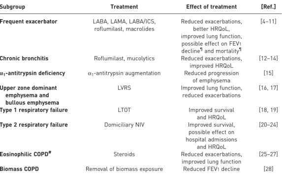
TABLE 1
Subgroups of chronic obstructive pulmonary disease (COPD) that currently have
specific treatments
Subgroup Treatment Effect of treatment [Ref.]
Frequent exacerbator LABA, LAMA, LABA/ICS, roflumilast, macrolides
Reduced exacerbations, better HRQoL, improved lung function,
possible effect on FEV1
decline¶and mortality
[4–11]
Chronic bronchitis Roflumilast, mucolytics Reduced exacerbations, improved HRQoL
[12–14] α1-antitrypsin deficiency α1-antitrypsin augmentation Reduced progression
of emphysema
[15] Upper zone dominant
emphysema and bullous emphysema
LVRS Improved lung function,
reduced exacerbations
[16, 17]
Type 1 respiratory failure LTOT Improved survival
and HRQoL
[18, 19] Type 2 respiratory failure Domiciliary NIV Improved survival,
possible effect on hospital admissions
and HRQoL
[20–24]
Eosinophilic COPD# Steroids Reduced exacerbations,
improved lung function
[25–27] Biomass COPD Removal of biomass exposure Reduced FEV1decline [28] Frequent exacerbator, chronic bronchitis and α1-antitrypsin deficiency are stable over time; the other subgroups may vary according to disease severity or evidence is not yet clear. LABA: long-acting β-agonists; LAMA long-acting muscarinic antagonists; ICS: inhaled corticosteroids; HRQoL; health-related quality of life; FEV1: forced expiratory volume in 1 s; LVRS: lung volume reduction surgery; LTOT:
long-term oxygen therapy; NIV: noninvasive ventilation. #: see “Subgroups shared between COPD and asthma” section;¶: effect only reported for LABA/ICS.
The only possible exception to this is osteoporosis, which is almost twice as common in subjects with
airflow obstruction compared to those without [48], is particularly associated with emphysema [40] and
has been shown to change with pulmonary treatment (e.g. lung volume reduction surgery (LVRS)). A
prospective cohort study [49] looking at the effect of LVRS on bone mineral density (BMD) in patients
with severe COPD concluded that surgery signi
ficantly improved BMD compared to pulmonary
rehabilitation. The increase correlated with residual volume, diffusing capacity of the lung for carbon
monoxide and fat-free mass, suggesting that restoration of respiratory dynamics, gas exchange and
nutritional status induced an improvement in bone metabolism and mineral content independent of
exercise. However, changes in BMD could occur with increased activity resulting in the bene
ficial effect of
LVRS and may have confounded results. Furthermore, most interventional studies of osteoporosis outside
COPD show more modest BMD improvement, generally in the range of 3
–8% [41], whereas LVRS
patients in this study improved by up 24%, suggesting that the result should be interpreted with caution.
The degree to which the relationship between osteoporosis and emphysema is confounded by prior steroid
treatment is not clear, but there are male patients with little or no steroid exposure who develop
osteoporosis, suggesting that it may not be entirely iatrogenic [50]. However, further studies are indicated
to understand mechanisms and determine optimal treatment.
Pulmonary hypertension Systemic inflammation Bronchiectasis Bacterial colonisation Reduced survival Increased exacerbation frequency
FIGURE 1Chronic obstructive pulmonary disease subgroups without clear treatment implications. Thefigure shows the subgroups with clear prognostic implications for patients that are clinically relevant, but where therapeutic strategies are as yet unclear. The directions of the arrows indicate the probable direction of the association, e.g. patients with more frequent exacerbations are less likely to survive. The direction of association of bacterial colonisation and bronchiectasis is not clear; thus, a bidirectional arrow is shown.
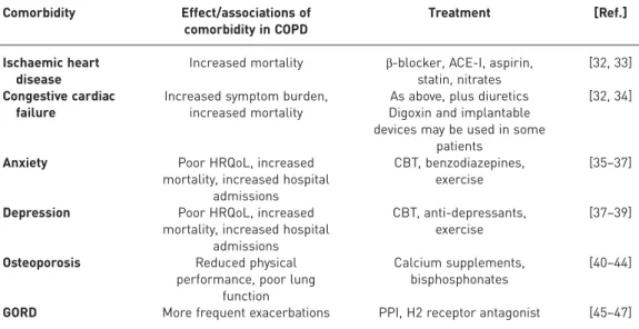
TABLE 2
Common comorbidities in chronic obstructive pulmonary disease (COPD)
Comorbidity Effect/associations of comorbidity in COPD
Treatment [Ref.]
Ischaemic heart disease
Increased mortality β-blocker, ACE-I, aspirin, statin, nitrates
[32, 33] Congestive cardiac
failure
Increased symptom burden, increased mortality
As above, plus diuretics Digoxin and implantable devices may be used in some
patients
[32, 34]
Anxiety Poor HRQoL, increased
mortality, increased hospital admissions
CBT, benzodiazepines, exercise
[35–37]
Depression Poor HRQoL, increased mortality, increased hospital
admissions
CBT, anti-depressants, exercise
[37–39]
Osteoporosis Reduced physical
performance, poor lung function
Calcium supplements, bisphosphonates
[40–44]
GORD More frequent exacerbations PPI, H2 receptor antagonist [45–47] This is not an exhaustive list of all conditions or treatments; where possible, references specific to COPD, major review articles or international guidelines on management have been cited rather than single studies. GORD: gastro-oesophageal reflux disease; ACE-I: angiotensin-converting enzyme inhibitor; HRQoL: health-related quality of life; CBT: cognitive behavioural therapy, PPI: proton-pump inhibitor.
Subgroups with implications for treatment
Frequent exacerbators, de
fined as those with more than two exacerbations a year, were definitively described
in the ECLIPSE (Evaluation of COPD Longitudinally to Identify Predictive Surrogate End-points) study [51].
Treatment options in this group include bronchodilators, long-acting
β-agonists (LABA)/inhaled
corticosteroids (ICS), roflumilast and azithromycin. In a number of trials, bronchodilation has been shown
to reduce exacerbation frequency. Long-acting muscarinic antagonists (LAMA) appear to exhibit this as a
class effect, e.g. tiotropium and glycopyrronium both reduced exacerbation frequency by >20% in the
GLOW2 trial and were not statistically different in this regard [5]. A meta-analysis of LABAs showed a
reduction in exacerbations that was close to 20%, although differences between them were reported;
formoterol only reduced exacerbations when used alongside ICS, whereas salmeterol had an effect when used
alone [6]. LAMA may be superior to LABA at reducing exacerbations, as shown in the POET (Prevention of
Exacerbations of COPD) study (tiotropium versus salmeterol) [11]. LAMA/LABA also reduce exacerbation
frequency, although the effect does not appear additive. For example, in the SPARK study (indacaterol and
glycopyrronium), the combination only reduced exacerbations by a further 12% over monotherapy [52].
LABA/ICS have been demonstrated to improve lung function and HRQoL and to reduce exacerbations by
∼25% in COPD patients [7]. Whilst most COPD trials have not focused on frequent exacerbators,
international guidelines recognise the impact of certain drug classes on exacerbation frequency, emphasising
the role of LABA/ICS [53]. Macrolides have become a hot topic throughout respiratory medicine, primarily
because of their potential anti-inflammatory effects. In COPD, the largest study to date used azithromycin
and prolonged time to
first exacerbation from 174 to 266 days, implying it might be useful in frequent
exacerbators [10]. Long-term safety issues include resistance and have been discussed elsewhere [54].
Chronic bronchitis occurs in 45% of COPD patients [55] and is linked to higher exacerbation frequency
and, hence, risk of decline [56]. Speci
fic therapies for patients with chronic bronchitis have been reviewed
elsewhere [57]. Roflumilast appears to prevent exacerbations best in COPD patients who have chronic
bronchitis [14], although utility may not be restricted to this group. Its use provided modest but
significantly improved forced expiratory volume in 1 s (FEV
1) compared with placebo and a reduction in
exacerbations. Mucolytics have also been used to reduce sputum viscosity and aid expectoration; those
most widely used are carbocysteine and N-acetylcysteine. Whilst trials have not been specific to chronic
bronchitis, their mechanism of action suggests they will work best in chronic sputum producers. The most
recent systematic review of clinical effectiveness of mucolytics included 30 studies and demonstrated a
small, but statistically signi
ficant, reduction in exacerbations in treated COPD patients [12].
α
1-antitrypsin de
ficiency (AATD) is the textbook case of a subgroup in COPD. It is associated with lower
zone dominant emphysema [58], although significant heterogeneity exists, possibly due to genetic
modi
fiers, as in usual COPD [59]. Augmentation therapy for AATD has been routinely used in some
countries since the
first trials were completed; a meta-analysis has demonstrated that it may reduce
emphysema progression [15]. Further trials, including inhaled
α
1-antitrypsin, are due to be published soon
and other specific therapies may become available in due course [60].
Upper zone dominant emphysema, as defined by visual appearance on chest computed tomography scans, was
speci
fically reported in the NETT (National Emphysema Treatment Trial) study, which demonstrated that
LVRS works best in this group of patients [16]. Further research using endobronchial valves as a less invasive
means of LVRS initially targeted this group [17]. Post hoc analyses suggest that whether
fissures are intact may
be more important than disease location or heterogeneity [61]. Patients with upper zone dominant emphysema
also exhibit different genetic risk factors, implying variation in pathogenesis [62]. Bullous emphysema is also
treated surgically, although patient selection and operative technique is much more individualised [63].
Overt or relative hypoxia occurs in COPD, especially in later stages. The basis for long-term oxygen
therapy (LTOT; oxygen for >15 h per day) is derived from two landmark placebo-controlled randomised
controlled trials, the NOTT (Nocturnal Oxygen Therapy Trial) study [18] and the MRC (Medical
Research Council) study [19], which demonstrated improved survival. The NOTT study also demonstrated
a decrease in mean pulmonary artery pressure. LTOT is indicated for stable patients who have an arterial
oxygen tension <7.3 kPa on two separate occasions or 7.3
–8.0 kPa in the presence of pulmonary
hypertension, nocturnal hypoxia or secondary polycythaemia. LTOT is not restricted to type 1 respiratory
failure; however, if used in type 2 patients, adverse effects on hypercapnia are generally excluded prior to
prescription. Aside from LTOT, two other modes of oxygen therapy exist, ambulatory and short burst;
both have been reviewed elsewhere [64]. Ambulatory oxygen is indicated in mobile patients who meet
LTOT criteria and is commonly considered in others who exhibit exertional desaturation to <90%.
Short-burst oxygen therapy criteria are poorly de
fined and no benefits have been reported [65].
Noninvasive ventilation (NIV) is used to treat type 2 respiratory failure and has a strong acute evidence
base in hospital [66] for COPD patients. The evidence concerning use of home NIV in COPD is
contradictory and concerns have been raised about whether potential benefits on incidence of severe
exacerbations and hospital admissions are appropriately balanced with poor HRQoL [20
–23]. More
recently, it has been shown to improve survival by 21% [24]; a systematic review is ongoing [67].
Eosinophilic COPD is a controversial area, mainly because of the issue of whether it is truly distinct from
asthma (overlap pathologies will be discussed later). Recent evidence suggests it may be identi
fied by sputum
cytokine profile [68]. Nevertheless, trials focussing on such patients have been published and suggest that
steroids are more bene
ficial in this group than in other COPD patients [25]. Several studies have reported
that use of sputum eosinophilia as a guide to the use of steroids was effective [26, 27, 69], and systemic
eosinophil counts may also be a useful guide to treating exacerbations with oral corticosteroids [70]. Recent
evidence suggests that blood eosinophil counts >2% predicted a response to ICS in several major COPD
trials [71], although these were post hoc analyses and further de
finitive work in this area is awaited.
Biomass fuel COPD is common in females, particularly in the developing world [72]. Airway predominant
phenotypes appear to be more common, with bronchial hyperresponsiveness (BHR) being a particular
feature in wood smoke exposure [73]. Consistent with this is the reported increased prevalence of an
overlap between asthma and COPD [74]. Systemic and pulmonary inflammation is similar to cigarette
smoke-induced disease [75, 76], with less emphysema [77] and less rapid FEV
1decline [78]. Patients
decline more slowly if the biomass exposure is reduced [28], but is not clear whether inhaled therapies
used for
“usual COPD” are of similar efficacy. The presence of BHR and overlap with asthma suggests that
ICS might be an effective strategy, though no clinical trials have been reported yet.
Subgroups with less clear implications for therapy
Pulmonary hypertension in COPD adversely affects survival and exercise capacity. Although oxygen
therapy protects against progression of pulmonary hypertension in patients with advanced COPD, its use
is limited to those meeting LTOT criteria. Vasoactive compounds used in primary pulmonary
hypertension (sildena
fil, bosentan and nitric oxide) have been investigated in COPD [79–83]. Some trials
showed worsening HRQoL in treated patients, hence they are not used routinely, although individual
patient trials are employed in selected patients by specialist centres [84].
Systemic in
flammation appeared to mark a subgroup of patients in the ECLIPSE study. This subgroup
comprised 16% of the whole group and had increased mortality and exacerbation rates compared to
patients without in
flammation [85]. It remains unclear whether this group should be treated differently;
future trials of therapies aimed at reducing exacerbations might target this group, or include them as a
pre-speci
fied subgroup analysis.
Stable state airway bacterial colonisation occurs in 30
–70% of COPD patients, and may relate to sputum
colour [86
–88]. It is defined as a significant pathogenic bacterial load (usually >1 × 10
6cfu·mL
−1) present
in sputum when the patient is well. Airway bacterial colonisation is associated with increased pulmonary
in
flammation [89] and increased frequency of exacerbations [90]; hence, it seems a logical subtype to
target. Differences between laboratories, local bacterial patterns and lack of consistency of culture results
over time may impact signi
ficantly on our ability to use this as a phenotype that directs therapy.
Moxi
floxacin has been tested to limited clinical benefit (exacerbation rates did not fall, although changes
in bacteriology were seen) [91], and a macrolide trial is ongoing [92].
There is growing recognition that bronchiectasis may occur in patients who have had COPD for some time,
but its prevalence varies widely (30
–70% of subjects), depending on the presence of AATD and the method
used to de
fine bronchiectasis on computed tomography [93, 94]. Again it is unclear whether treatment
should differ, even though there are implications for survival (COPD with bronchiectasis versus COPD
without bronchiectasis, hazard ratio 2.54) [95] and recovery time from exacerbations [96]. Exacerbation
frequency has not been found to differ [95, 96], although colonisation is more common [96].
Air
flow obstruction, aetiology of which is thought to be due to tuberculosis [97], is a poorly defined
phenotype as it is not clear whether the underlying prognosis is the same as classical smoking-related
COPD or whether treatment should differ. It is likely that pathogenesis differs, but this has not been
investigated fully. Many clinicians will adopt treatment approaches outside the guidelines for
“usual
COPD
”, but it remains important to emphasise to non-specialists that not all fixed airflow obstruction is
“usual COPD”. Early life events that impact on lung function [98] seem unlikely to be amenable to
therapy later in life, but this too remains uncertain.
Constancy of COPD subgroups
In general, COPD is regarded as a disease that is slowly progressive; thus, it should be relatively easy to
select a treatment at a given time-point that will remain useful for some time. However, there has been
little research into the progression of the subgroups we have proposed. It may be much harder to propose
a specific therapy for those that vary unless very detailed monitoring is undertaken or the duration of
therapy is
fixed.
There are a number of COPD subgroups that remain constant once present. The frequent exacerbator
subgroup was relatively stable in the ECLIPSE study [51], as was systemic inflammation [85]. Whilst
therapies that target exacerbations may reduce their frequency [4], this positive treatment effect does not
detract from the fact that patients on a range of treatments in the ECLIPSE study still exhibited stable
exacerbation frequency, enabling it to be considered a suf
ficiently stable feature to guide treatment.
Chronic bronchitis is defined as sputum production on most days for at least 3 months within at least
2 consecutive years [99], and consequently is probably constant enough to guide therapy. By de
finition,
AATD is a constant phenotype as it is determined by genotype.
Other subgroups appear to develop over time but are sufficiently constant to guide therapy decisions over
a few years. Upper zone dominant emphysema has not been studied longitudinally in usual COPD;
however, in AATD, it is an early event prior to the development of lower zone dominant emphysema
followed by homogenous disease [100]. This occurs over many years; thus, it is probably constant enough
to guide decision making on treatment such as chronic bronchitis. Longitudinal blood gas studies have not
been commonly carried out in COPD, although type 2 failure does develop over time, occurring in 24% of
patients who have had an acidotic hypercapnic exacerbation 5 years after the
first event [101]. Predictors
of hypoxia and hypercapnia include lung function and body composition [102]. What data are available
suggest that progression of blood gas changes occurs in COPD, but that changes are slow enough (outside
exacerbations) to enable gases to be used to guide therapy.
Phenotypes where variation occurs over time are also seen in COPD. Pulmonary hypertension usually occurs
late in disease [103], although it can occur alongside mild COPD [104]. Probably the only relevant clinical
feature to de
fine constancy is whether it is ever reported to disappear once it has developed; intuitively one
would imagine that it is as constant as the degree of respiratory failure though this is not supported by
published evidence. One small study has shown that pulmonary artery pressure varies with exacerbations and
exercise [105]; animal studies also imply phases of remission [106]. There have been few published studies
detailing longitudinal changes in sputum bacterial content in stable COPD. Data in AATD suggests that it does
not always stay the same [107], Moraxella catarrhalis and Pseuodomonas aeruginosa are capable of spontaneous
remission in COPD [108, 109], and serial cultures during clinical trials also demonstrate variability [10]. It
seems unlikely that colonisation can be defined on the basis of a single sputum culture when stable, but it
might become a suf
ficiently constant phenotype to guide therapy if the definition can be better clarified.
Clinically relevant subgroups in asthma
Bronchial asthma is a complex disease with many underlying mechanisms and, therefore, can be
considered a syndrome containing subgroups with important similarities but also differences caused by
variable underlying aetiologies [1]. Some subgroups described in the literature are based on clinical
features found in asthma databases and cluster analyses; molecular and genetic approaches have also been
widely used [110]. This merging of clinical and pathological features has been reviewed extensively
elsewhere [111], using the term endotype to delineate this way of defining disease. Within this review, we
have chosen to focus on clinically relevant subgroups rather than endotypes, as this may be more useful in
clinical practice, although discussion of some molecular elements is unavoidable.
Subgroups with treatment implications
A number of distinct asthma phenotypes have treatment implications and are summarised in table 3.
There is a degree of overlap in that aspirin-sensitive asthma (ASA), which is caused by increased cysteinyl
leukotriene production exacerbated by nonsteroidal anti-inflammatory drugs, is often accompanied by
severe eosinophilic rhinosinusitis and nasal polyps. Therefore, it has many features of atopic/allergic
asthma, which is often eosinophilic, and is associated with elevated IgE, exhaled nitric oxide fraction
(Fe
NO) and periostin levels [125]. Nonatopic, noneosinophilic asthma typically demonstrates lower airway
hyperresponsiveness [126, 127] and lower corticosteroid responsiveness [127].
Based on cluster analysis [128], age at disease onset is a key differentiating factor linked to underlying
genetic/molecular features, although it does not in
fluence treatment directly. Nevertheless, it is clear that
certain asthma subgroups, which vary with age, will require different treatment. Early-onset asthma is
often more atopic/allergic, and ORMDL3 polymorphisms exhibit a stronger association with childhood
and severe asthma [129, 130]. These observations suggest that there are different disease mechanisms in
younger patients, which have been partly proven by studies of airway cells. Four in
flammatory subgroups
are distinguishable in induced sputum: eosinophilic, neutrophilic, paucigranulocytic and mixed
granulocytic [131]. For the purposes of treatment, this tends to be divided into eosinophilic and
noneosinophilic disease. Supporting this simplified sub-division, molecular phenotyping showed a
T-helper (Th)2-high gene signature in airway epithelial tissue in 50% of mild asthmatics, and was
associated with more atopy and response to ICS therapy [132]. The remaining patients showed a Th2-low
gene signature similar to healthy controls, with less airway obstruction, lower hyperreactivity and lower
response to ICS. This implies that selection could be applied to the use of ICS and, in particular,
high-dose ICS, based on airway eosinophilia. Trials using this strategy have had some success [115], and it
is supported by the latest severe asthma guidelines [114]. Newer drugs targeting severe eosinophilic asthma
are also emerging; most are monoclonal antibodies that are likely to be used in highly selected populations.
Examples include mepolizumab (anti-interleukin (IL)-5) [115], and lebrikizumab (anti-IL-13), the latter
being useful in patients with elevated periostin [113]. However, the relationship between eosinophilia and
other features of asthma, such as airway hyperresponsiveness, is by no means clear cut; indeed, they may
be inherited separately [133], hence this cannot be the only way in which we de
fine our phenotypes and
design new treatments. Noneosinophilic disease has shown some response to macrolide therapy [116, 117],
although this is not a widely recommended treatment strategy [114].
Other clinical features can be used to identify patients who fall into subgroups that may in
fluence
management. For instance, early-onset allergic, exercise-induced asthma (EIA) and ASA phenotypes
belong to the Th2-high form of disease while neutrophilic asthma and smooth-muscle mediated
paucigranulocytic asthma belong to the non-Th2 group [110]. Sputum cell counts may be impractical for
routine phenotyping, particularly in primary care; hence, a detailed history (age of onset, atopy, aspirin
sensitivity, smoking, etc.) is key to identifying subgroups and tailoring treatment in clinical practice. In
allergic asthma, avoidance of triggers may be a strategy, although the utility of avoidance for house dust
mite allergy is debatable [134]. Omalizumab is a well-validated strategy for allergic asthma with high IgE,
which reduces corticosteroid use and exacerbation rates [135] and improves long-term control [136].
Patients with ASA should avoid aspirin and nonsteroidal anti-in
flammatory drugs. Although there is some
evidence that leukotriene inhibition is particularly effective in ASA, it is not consistent [123, 137] and
standard asthma management should be followed.
History also identi
fies occupational asthma, which is best diagnosed by the occupational asthma system
score on serial peak expiratory
flow rate testing or specific provocation challenge [138]. It is subdivided
into asthma precipitated by the exposure or pre-existing asthma that is worsened by exposure. Sputum
eosinophil counts and Fe
NOrelate reasonably well to speci
fic provocation challenge [139], although
distinct groups without Fe
NOelevation are identi
fiable [140]. Cessation or reduction of exposure is key, as
delineated in a systematic review by a European Respiratory Society Task Force [124]. Patients with
continued exposure tend to progress faster in terms of lung function decline and symptoms [141].
Fungal sensitisation in asthma is common with an estimated prevalence of up to 30% [142], covering a
spectrum from allergic bronchopulmonary aspergillosis (ABPA) through severe asthma with fungal
sensitisation (SAFS) and asymptomatic sensitisation. Diagnosis is made by examining serum IgE, and by
performing speci
fic radioallergosorbent tests to Aspergillus and Aspergillus precipitins. Both ABPA and
SAFS respond to antifungals [118, 119], although their ef
ficacy is debatable [114]. Susceptibility to fungal
infection and sensitisation may be genetically determined (e.g. human leukocyte antigen type [143] and
Toll-like receptors [144]).
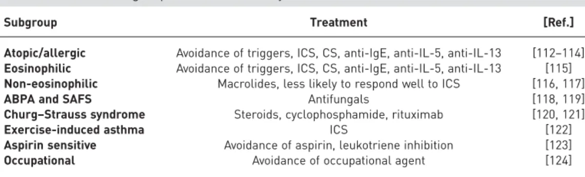
TABLE 3
Asthma subgroups and their stability over time
Subgroup Treatment [Ref.]
Atopic/allergic Avoidance of triggers, ICS, CS, anti-IgE, anti-IL-5, anti-IL-13 [112–114] Eosinophilic Avoidance of triggers, ICS, CS, anti-IgE, anti-IL-5, anti-IL-13 [115] Non-eosinophilic Macrolides, less likely to respond well to ICS [116, 117]
ABPA and SAFS Antifungals [118, 119]
Churg–Strauss syndrome Steroids, cyclophosphamide, rituximab [120, 121]
Exercise-induced asthma ICS [122]
Aspirin sensitive Avoidance of aspirin, leukotriene inhibition [123]
Occupational Avoidance of occupational agent [124]
Aspirin sensitive and occupational subgroups appear to stay the same over time; other subgroups may vary according to treatment or the clinical picture is unclear. In each subgroup, some of the treatment options and relevant references are shown (see text for further details). All have shown either symptomatic improvement, reduction in exacerbations or both. Bronchodilator agents are not listed and should be used in all subgroups when required or other therapies with equal efficacy should be used. ABPA: allergic bronchopulmonary aspergillosis; SAFS: severe asthma with fungal sensitisation; ICS: inhaled corticosteroids; CS: corticosteroid; IL: interleukin.
Churg–Strauss syndrome is a rare, progressive, systemic disorder characterised by eosinophilia,
extravascular necrotising granulomas, worsening asthma, lung in
filtrates and eventually antineutrophil
cytoplasmic antibody-positive systemic vasculitis [145]. Significant heterogeneity is seen at presentation
and longer term. Lower baseline eosinophil counts predict mortality [146]. Precise management differs
according to disease severity (e.g presence of glomerulonephritis), but in general involves aggressive
immunosuppression, often with cylophosphamide alongside corticosteroids [120]. Rituximab has also been
used with some success [121].
EIA is an acute transient airway narrowing that occurs during and, most often, after exercise. EIA is
de
fined as a decrease in FEV
1>10% from baseline measured up 30 min after exercise. A higher prevalence
of EIA has been reported among elite athletes, especially in endurance sports, such as rowing and
cross-country skiing, compared to the general population [147]. However, some elite athletes with EIA
have neither a history of childhood asthma nor a family history of asthma, suggesting that environmental
factors are more important than genetic inheritance [148]. The most effective therapy for EIA is regular
use of ICS [122]. Early use of leukotriene inhibition may be equally effective [149, 150].
Subgroups with less clear implications for therapy
Severe refractory asthma is an area of intense research, which many newer therapies are being targeted
towards. However, this is also heterogeneous with eosinophilic and noneosinophilic phenotypes [151],
implying that severity may not be the feature that drives speci
fic treatment. Nevertheless, at least part of
severe asthma has a different pathobiology compared to nonsevere disease. For example, innate immunity
in the airway is in
fluenced by glucocorticoids [152] and there have been recent insights into the role that
innate immunity plays in steroid refractory severe disease [153].
Neutrophilic asthma also exhibits some innate immune features [154], although whether this is a true
phenotype remains debatable. Neutrophilic asthma is associated with low FEV
1, air trapping [155] and
smoking [156], and thus may represent overlap with COPD, or indeed with other pro-inflammatory
trigger factors. For instance, two studies have reported clusters of older, obese, female, nonatopic
asthmatics [128, 157].
“Extensive remodelling asthma” is characterised by pronounced airway remodelling
and minimal in
flammation [158], increased airway smooth muscle mass may also occur. One cause may
be an intrinsic airway smooth muscle abnormality. Whether extensive remodelling has therapeutic
implications remains uncertain.
Reconciling variability and triggers with subgroup identi
fication
Unlike COPD, asthma is classically thought to be variable. This brings problems when defining subgroups,
as patients may change over time or in response to treatment. We suggest that identifying the current
subgroup is the most important factor when modulating therapy. Knowing the subgroup at initial
diagnosis may also help but past features should not preclude altering treatment based on current
presentation.
Triggers
Speci
fic trigger or detrimental factors are interesting elements of phenotyping. Classical triggers (allergens
and occupation) that change treatment are part of our subgroup de
finitions. Recognised triggers that do
not alter speci
fic asthma management include obesity, smoking, gastro-oesophageal reflux disease
(GORD), menstrual cycle and air pollution. Obesity is a risk factor for asthma, and asthma often causes
weight gain; there is no consensus about the exact relationship between the two. One cluster analysis
identified a noneosinophilic obese group of patients [128], another has reported a nonatopic obese group
who required multiple courses of corticosteroids [157]. However, a recent meta-analysis reported that body
mass index (BMI) was a key feature of asthma subgroups but, of the clusters that were predominantly
obese, clinical and in
flammatory differences occurred between them suggesting heterogeneity independent
of obesity [159]. Weight loss appears to help asthma control [160]; however, since weight loss has
pluripotent health bene
fits, we felt it did not represent specific asthma management. Smoking also
influences the course of asthma. Asthmatics who smoke show poor response to corticosteroid therapy,
more frequent exacerbations and worse lung function progressing to
fixed obstruction, but smoking might
make airways disease worse.
An association between asthma and GORD has been the subject of considerable investigation. There is no
doubt that GORD is more common in asthma [161] and this has led to the suggestion that re
flux
treatment may improve asthma control. Several large clinical trials [162, 163] and a Cochrane review [164]
have been published, demonstrating that treatment with high-dose proton-pump inhibitor improves cough
but has no effect on asthma symptoms, lung function or exacerbation rates.
17% of asthmatic females report worsening of symptoms near menses, and this perimenstrual asthma
phenotype is associated with higher BMI, lower forced vital capacity, GORD, aspirin sensitivity and poor
asthma control [165]. However, healthy females exhibit similar changes in pulmonary physiology across
their menstrual cycle to those with asthma [166], and multivariate analysis has shown that baseline asthma
characteristics can differ in those who report symptom changes across their cycle [167]. As such, it is likely
that sex hormones represent a trigger, and that perimenstrual asthma is not a truly different subtype.
Constancy of asthma subgroups
The constancy of asthma phenotypes over time, or after therapy implementation, is not as clear in COPD.
Asthma is variable, thus phenotypes ought to change over time, but few published data exist. Those that
appear to remain constant are occupational asthma and ASA. Occupational asthma progresses faster if
exposure continues [124], implying that the underlying disease process is unchanged. The risk of
precipitating severe exacerbations means that testing whether patients remain aspirin sensitive long-term
has rarely been performed. Case reports demonstrate that aspirin sensitivity can develop in patients who
have previously taken it without respiratory problems [168], but studies which have challenged known ASA
patients have shown that most remain sensitive [169, 170]. Asthma subgroups that clearly change with
treatment include ABPA, SAFS and Churg
–Strauss syndrome. Atopy and allergy may also remit, usually
over time but occasionally with treatment, such as desensitisation [171]. For example, 21% of children aged
<2 years with severe peanut allergy will be tolerant by 5 years of age [172]. Remission of asthma was equally
common in allergic/atopic and nonatopic patients in a large prospective cohort study of childhood-onset
asthma, averaging 65% of patients [173]. Even in adults, allergic responses vary over time, as demonstrated
by the fact that 39% of patients have different results on serial skin-prick testing (mostly gaining new
allergies), but in 13% of cases allergies were lost and in 4% allergies were gained and lost [174].
Published evidence is inconsistent regarding the constancy of asthmatic airway in
flammation. In adults,
50% of mild-to-moderate asthmatics have persistently noneosinophilic (often neutrophilic) disease and the
remainder have persistent or intermittent eosinophilia [175]. Intermittent and persistent groups exhibited
similar clinical characteristics, and hence may represent a milder form of the same population.
Therapeutically, the neutrophilic patients respond poorly to usual asthma therapy. Conversely, the sputum
in
flammatory phenotype proved inconsistent in asthmatic children [176]. This may also change with
treatment; for example, corticosteroids inhibit neutrophil apoptosis [177] such that neutrophilic asthma
could actually represent over-treated eosinophilic asthma. This hypothesis is supported by the observation
that corticosteroid withdrawal abolished neutrophilic subjects among moderate asthmatics [156].
Subgroups shared between COPD and asthma
When a patient presents with symptoms of increased variability of airflow alongside partially reversible
air
flow obstruction, it is known as the asthma–COPD overlap syndrome (ACOS) [178]. A consensus
conference has proposed that an ACOS patient must ful
fil two major criteria or one major and two minor
criteria from the following. 1) Major criteria: positive bronchodilator response (>400 mL and >15% FEV
1),
sputum eosinophilia or previous diagnosis of asthma. 2) Minor criteria: increased total serum IgE, history
of atopy or positive bronchodilator test (>200 mL and >12% FEV
1) on at least two occasions [179]. ACOS
typically includes patients with early-onset asthma and a long disease duration who then ful
fil criteria for
COPD with age, COPD patients with increased reversibility and smoking asthmatics who have
fixed
air
flow obstruction. Overall, 13–19% of patients with obstructive lung diseases have some overlap and this
increases with age [180, 181]. In the UPLIFT (Understanding Potential Long-term Impacts of Tiotropium)
trial, two-thirds of moderate-to-severe COPD patients exhibited bronchodilator responsiveness [182];
however, most clinical trials for either asthma or COPD exclude patients with features of the other disease,
implying that their results will be poorly generalisable in real-life. ACOS has been extensively reviewed
elsewhere [178, 179]. Our reasoning for including it here is to revisit the concept in light of the more
complex subgroups of both asthma and COPD that we have described. Its importance is that patients
exhibiting features consistent with ACOS are more likely to be frequent exacerbators [183], have more
respiratory symptoms, higher mortality [183], higher comorbidity rates [184], greater healthcare utilisation
[184] and worse HRQoL [181].
Overlap patients are generally thought to exhibit phenotypes part way between COPD and asthma. For example,
G
IBSONand S
IMPSON[178] reported prevalence of atopy that was highest in asthma (100%) intermediate in
ACOS (64%) and lowest in COPD (25%). There are some differences that may depend on the predominant
pathology. Positive bronchodilator response observed in COPD is associated with increased eosinophilic
in
flammation [185] whilst irreversible COPD more frequently exhibits neutrophilia. Smoking asthmatics
typically have inflammatory features that resemble COPD with increased neutrophilia and sometimes
airway remodelling [186]. Classical asthma drivers, such as occupation, are associated with
fixed airflow
obstruction after chronic exposure [187] and ABPA has also been reported in patients with COPD [188].
During exacerbations of ACOS, airway mucosal eosinophils increase more than neutrophils [189] explaining the
improvement with systemic corticosteroids or ICS. Conversely, analysis of volatile organic compounds in severe
persistent asthma compared to severe COPD showed that volatile organic compound patterns remained
different, suggesting singular pathophysiological mechanisms [190]. Taken together, the evidence suggests that
there may be aspects of shared pathogenesis that move treatment decisions to that of the subgroup irrespective
of the primary diagnosis. Shared subgroups are shown in
figure 2.
Examples of subgroups common to asthma and COPD
The evidence for treating overlap is far less extensive than for single pathology. In addition, it would be
beyond the scope of this article to review all shared subgroups comprehensively; thus, we have chosen a
few pertinent examples. One signi
ficant shared phenotype where evidence exists for management is
eosinophilic airways disease. The evidence that asthmatics with eosinophilia respond to steroids is
incontrovertible, and a study from Leicester, UK, has demonstrated that patients with COPD and relatively
high blood eosinophils (>2%) respond better to oral steroids in the context of an exacerbation than those
with lower levels [70]. Similar results have been reported for use of ICS [25
–27, 69].
Frequent exacerbations in asthma can be associated with risk factors such as severe nasal sinus disease,
GORD, recurrent respiratory infections and obstructive sleep apnoea [191]. Many, such as GORD, are
similar to the risk factors for frequent exacerbation in COPD patients in the ECLIPSE study [51]. Similar
strategies to manage risk factors are advocated in both diseases, alongside potential additional therapies
such as macrolides [10, 116, 192].
Messages for clinical practice
Characterising patients with chronic respiratory symptoms may be more important than giving a label of
asthma or COPD in predicting prognosis and response to treatment. A lot of phenotypes have been
de
fined in the literature but some of them do not yet have treatment or prognostic implications. Asthma
subgroups, unlike COPD, are likely to change over time and the current phenotype should be regarded as
the most important factor to guide treatment.
Acknowledgements
We would like to thank GSK for organising the European Respiratory Network of Excellence event that stimulated the debates leading to this review.
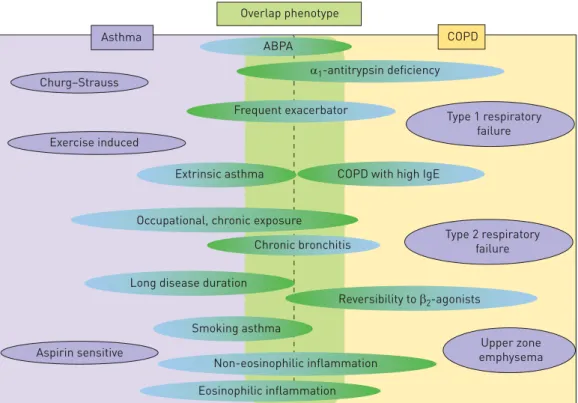
Overlap phenotype Churg–Strauss Type 1 respiratory failure Type 2 respiratory failure Upper zone emphysema Exercise induced Aspirin sensitive COPD Asthma ABPA Frequent exacerbator Extrinsic asthma
Long disease duration
Smoking asthma
Non-eosinophilic inflammation Eosinophilic inflammation
Reversibility to β2-agonists COPD with high IgE
Occupational, chronic exposure Chronic bronchitis
α1-antitrypsin deficiency
FIGURE 2 Schematic diagram of the shared subgroups between asthma and chronic obstructive pulmonary disease (COPD). The figure shows phenotypes that are recognised in both conditions. The placement/colour of the shapes indicates the degree to which they are recognised in asthma and COPD. ABPA: allergic bronchopulmonary aspergillosis.
References
1 Lotvall J, Akdis CA, Bacharier LB, et al. Asthma endotypes: a new approach to classification of disease entities within the asthma syndrome. J Allergy Clin Immunol 2011; 127: 355–360.
2 Han MK, Agusti A, Calverley PM, et al. Chronic obstructive pulmonary disease phenotypes: the future of COPD. Am J Respir Crit Care Med 2010; 182: 598–604.
3 Parr DG. Patient phenotyping and early disease detection in chronic obstructive pulmonary disease. Proc Am Thorac Soc 2011; 8: 338–349.
4 Wedzicha JA, Rabe KF, Martinez FJ, et al. Efficacy of roflumilast in the COPD frequent exacerbator phenotype. Chest 2013; 143: 1302–1311.
5 Kerwin E, Hébert J, Gallagher N, et al. Efficacy and safety of NVA237 versus placebo and tiotropium in patients with COPD: the GLOW2 study. Eur Respir J 2012; 40: 1106–1114.
6 Wang J, Nie B, Xiong W, et al. Effect of long-acting beta-agonists on the frequency of COPD exacerbations: a meta-analysis. J Clin Pharm Ther 2012; 37: 204–211.
7 Nannini LJ, Lasserson TJ, Poole P. Combined corticosteroid and long-actingβ2-agonist in one inhaler versus long-acting β2-agonists for chronic obstructive pulmonary disease. Cochrane Database Syst Rev 2012; 9: CD006829.
8 Celli BR, Thomas NE, Anderson JA, et al. Effect of pharmacotherapy on rate of decline of lung function in chronic obstructive pulmonary disease: results from the TORCH study. Am J Respir Crit Care Med 2008; 178: 332–338. 9 Calverley PM, Anderson JA, Celli B, et al. Salmeterol and fluticasone propionate and survival in chronic
obstructive pulmonary disease. N Engl J Med 2007; 356: 775–789.
10 Albert RK, Connett J, Bailey WC, et al. Azithromycin for prevention of exacerbations of COPD. N Engl J Med 2011; 365: 689–698.
11 Vogelmeier C, Hederer B, Glaab T, et al. Tiotropium versus salmeterol for the prevention of exacerbations of COPD. N Engl J Med 2011; 364: 1093–1103.
12 Poole P, Black PN, Cates CJ. Mucolytic agents for chronic bronchitis or chronic obstructive pulmonary disease. Cochrane Database Syst Rev 2012; 8: CD001287.
13 Fabbri LM, Calverley PM, Izquierdo-Alonso JL, et al. Roflumilast in moderate-to-severe chronic obstructive pulmonary disease treated with long-acting bronchodilators: two randomised clinical trials. Lancet 2009; 374: 695–703. 14 Rennard SI, Calverley PM, Goehring UM, et al. Reduction of exacerbations by the PDE4 inhibitor roflumilast –
the importance of defining different subsets of patients with COPD. Respir Res 2011; 12: 18.
15 Stockley RA, Parr DG, Piitulainen E, et al. Therapeutic efficacy of α-1 antitrypsin augmentation therapy on the loss of lung tissue: an integrated analysis of 2 randomised clinical trials using computed tomography densitometry. Respir Res 2010; 11: 136.
16 Fishman A, Martinez F, Naunheim K, et al. A randomized trial comparing lung-volume-reduction surgery with medical therapy for severe emphysema. N Engl J Med 2003; 348: 2059–2073.
17 Sciurba FC, Ernst A, Herth FJ, et al. A randomized study of endobronchial valves for advanced emphysema. N Engl J Med 2010; 363: 1233–1244.
18 Continuous or nocturnal oxygen therapy in hypoxemic chronic obstructive lung disease: a clinical trial. Nocturnal Oxygen Therapy Trial Group. Ann Intern Med 1980; 93: 391–398.
19 Long term domiciliary oxygen therapy in chronic hypoxic cor pulmonale complicating chronic bronchitis and emphysema. Report of the Medical Research Council Working Party. Lancet 1981; 1: 681–686.
20 Budweiser S, Hitzl AP, Jôrres RA, et al. Impact of noninvasive home ventilation on long-term survival in chronic hypercapnic COPD: a prospective observational study. Int J Clin Prac 2007; 61: 1516–1522.
21 Budweiser S, Jorres RA, Riedl T, et al. Predictors of survival in COPD patients with chronic hypercapnic respiratory failure receiving noninvasive home ventilation. Chest 2007; 131: 1650–1658.
22 McEvoy RD, Pierce RJ, Hillman D, et al. Nocturnal non-invasive nasal ventilation in stable hypercapnic COPD: a randomised controlled trial. Thorax 2009; 64: 561–566.
23 Funk GC, Breyer MK, Burghuber OC, et al. Long-term non-invasive ventilation in COPD after acute-on-chronic respiratory failure. Respir Med 2011; 105: 427–434.
24 Köhnlein T, Windisch W, Köhler D, et al. Non-invasive positive pressure ventilation for the treatment of severe stable chronic obstructive pulmonary disease: a prospective, multicentre, randomised, controlled clinical trial. Lancet Respir Med 2014; 2: 698–705.
25 Siva R, Green RH, Brightling CE, et al. Eosinophilic airway inflammation and exacerbations of COPD: a randomised controlled trial. Eur Respir J 2007; 29: 906–913.
26 Kitaguchi Y, Komatsu Y, Fujimoto K, et al. Sputum eosinophilia can predict responsiveness to inhaled corticosteroid treatment in patients with overlap syndrome of COPD and asthma. Int J Chron Obstruct Pulmon Dis 2012; 7: 283–289.
27 Brightling CE, Monteiro W, Ward R, et al. Sputum eosinophilia and short-term response to prednisolone in chronic obstructive pulmonary disease: a randomised controlled trial. Lancet 2000; 356: 1480–1485.
28 Zhou Y, Zou Y, Li X, et al. Lung function and incidence of chronic obstructive pulmonary disease after improved cooking fuels and kitchen ventilation: a 9-year prospective cohort study. PLoS Med 2014; 11: e1001621. 29 Divo M, Cote C, de Torres JP, et al. Comorbidities and risk of mortality in patients with chronic obstructive
pulmonary disease. Am J Respir Crit Care Med 2012; 186: 155–161.
30 Sundh J, Stallberg B, Lisspers K, et al. Co-morbidity, body mass index and quality of life in COPD using the Clinical COPD Questionnaire. COPD 2011; 8: 173–181.
31 Tan SL, Wood AM. Chronic obstructive pulmonary disease and co-morbidities: a review and consideration of pathophysiology. Panminerva Med 2009; 51: 81–93.
32 Cavailles A, Brinchault-Rabin G, Dixmier A, et al. Comorbidities of COPD. Eur Respir Rev 2013; 22: 454–475. 33 Montalescot G, Sechtem U, Achenbach S, et al. 2013 ESC guidelines on the management of stable coronary
artery disease: the Task Force on the management of stable coronary artery disease of the European Society of Cardiology. Eur Heart J 2013; 34: 2949–3003.
34 Yancy CW, Jessup M, Bozkurt B, et al. 2013 ACCF/AHA guideline for the management of heart failure: a report of the American College of Cardiology Foundation/American Heart Association Task Force on practice guidelines. Circulation 2013; 128: e240–e327.
35 Atlantis E, Fahey P, Cochrane B, et al. Bidirectional associations between clinically relevant depression or anxiety and chronic obstructive pulmonary disease (COPD): a systematic review and meta-analysis. Chest 2013; 144: 766–777.
36 Coventry PA, Gellatly JL. Improving outcomes for COPD patients with mild-to-moderate anxiety and depression: a systematic review of cognitive behavioural therapy. Br J Health Psychol 2008; 13: 381–400.
37 Coventry PA, Bower P, Keyworth C, et al. The effect of complex interventions on depression and anxiety in chronic obstructive pulmonary disease: systematic review and meta-analysis. PLoS One 2013; 8: e60532. 38 Yohannes AM, Alexopoulos GS. Depression and anxiety in patients with COPD. Eur Respir Rev 2014; 23:
345–349.
39 Yohannes AM, Connolly MJ, Baldwin RC. A feasibility study of antidepressant drug therapy in depressed elderly patients with chronic obstructive pulmonary disease. Int J Geriatr Psychiatry 2001; 16: 451–454.
40 Romme EA, Murchison JT, Edwards LD, et al. CT-measured bone attenuation in patients with chronic obstructive pulmonary disease: relation to clinical features and outcomes. J Bone Miner Res 2013; 28: 1369–1377. 41 Khosla S, Bilezikian JP, Dempster DW, et al. Benefits and risks of bisphosphonate therapy for osteoporosis. J Clin
Endocrinol Metab 2012; 97: 2272–2282.
42 Smith BJ, Laslett LL, Pile KD, et al. Randomized controlled trial of alendronate in airways disease and low bone mineral density. Chron Respir Dis 2004; 1: 131–137.
43 Nordin BE. The effect of calcium supplementation on bone loss in 32 controlled trials in postmenopausal women. Osteoporos Int 2009; 20: 2135–2143.
44 Li LS, Caughey GE, Johnston KN. The association between co-morbidities and physical performance in people with chronic obstructive pulmonary disease: a systematic review. Chron Respir Dis 2014; 11: 3–13.
45 Sakae TM, Pizzichini MM, Teixeira PJ, et al. Exacerbations of COPD and symptoms of gastroesophageal reflux: a systematic review and meta-analysis. J Bras Pneumol 2013; 39: 259–271.
46 Sigterman KE, van Pinxteren B, Bonis PA, et al. Short-term treatment with proton pump inhibitors, H2-receptor antagonists and prokinetics for gastro-oesophageal reflux disease-like symptoms and endoscopy negative reflux disease. Cochrane Database Syst Rev 2013; 5: CD002095.
47 Sasaki T, Nakayama K, Yasuda H, et al. A randomized, single-blind study of lansoprazole for the prevention of exacerbations of chronic obstructive pulmonary disease in older patients. J Am Geriatr Soc 2009; 57: 1453–1457. 48 Sin DD, Man JP, Man SF. The risk of osteoporosis in Caucasian men and women with obstructive airways
disease. Am J Med 2003; 114: 10–14.
49 Mineo TC, Ambrogi V, Mineo D, et al. Bone mineral density improvement after lung volume reduction surgery for severe emphysema. Chest 2005; 127: 1960–1966.
50 Duckers JM, Evans BA, Fraser WD, et al. Low bone mineral density in men with chronic obstructive pulmonary disease. Respir Res 2011; 12: 101.
51 Hurst JR, Vestbo J, Anzueto A, et al. Susceptibility to exacerbation in chronic obstructive pulmonary disease. N Engl J Med 2010; 363: 1128–1138.
52 Wedzicha JA, Decramer M, Ficker JH, et al. Analysis of chronic obstructive pulmonary disease exacerbations with the dual bronchodilator QVA149 compared with glycopyrronium and tiotropium (SPARK): a randomised, double-blind, parallel-group study. Lancet Respir Med 2013; 1: 199–209.
53 Global Initiative for Obstructive Lung Disease. Global Strategy for the Diagnosis, Management and Prevention of COPD. www.goldcopd.com Date last accessed: April 2014.
54 Simoens S, Laekeman G, Decramer M. Preventing COPD exacerbations with macrolides: a review and budget impact analysis. Respir Med 2013; 107: 637–648.
55 Izquierdo-Alonso JL, Rodriguez-Gonzalezmoro JM, de Lucas-Ramos P, et al. Prevalence and characteristics of three clinical phenotypes of chronic obstructive pulmonary disease (COPD). Respir Med 2013; 107: 724–731. 56 Donaldson GC, Seemungal TA, Bhowmik A, et al. Relationship between exacerbation frequency and lung
function decline in chronic obstructive pulmonary disease. Thorax 2002; 57: 847–852.
57 Kim V, Criner GJ. Chronic bronchitis and chronic obstructive pulmonary disease. Am J Respir Crit Care Med 2013; 187: 228–237.
58 Laurell CB, Eriksson S. The serum alpha-l-antitrypsin in families with hypo-alpha-L-antitrypsinemia. Clin Chim Acta 1965; 11: 395–398.
59 Wood AM, Simmonds MJ, Bayley DL, et al. The TNF-α gene relates to clinical phenotype in α-1-antitrypsin deficiency. Respir Res 2008; 9: 52.
60 Stockley RA, Turner AM.α-1-antitrypsin deficiency: clinical variability, assessment, and treatment. Trends Mol Med 2014; 20: 105–115.
61 Herth FJ, Noppen M, Valipour A, et al. Efficacy predictors of lung volume reduction with Zephyr valves in a European cohort. Eur Respir J 2012; 39: 1334–1342.
62 DeMeo DL, Hersh CP, Hoffman EA, et al. Genetic determinants of emphysema distribution in the national emphysema treatment trial. Am J Respir Crit Care Med 2007; 176: 42–48.
63 Greenberg JA, Singhal S, Kaiser LR. Giant bullous lung disease: evaluation, selection, techniques, and outcomes. Chest Surg Clin N Am 2003; 13: 631–649.
64 Stoller JK, Panos RJ, Krachman S, et al. Oxygen therapy for patients with COPD: current evidence and the long-term oxygen treatment trial. Chest 2010; 138: 179–187.
65 O’Neill B, Mahon JM, Bradley J. Short-burst oxygen therapy in chronic obstructive pulmonary disease. Respir Med 2006; 100: 1129–1138.
66 British Thoracic Society Standards of Care Committee.: Non-invasive ventilation in acute respiratory failure. Thorax. 2002; 57: 192–211.
67 Dave C, Turner A, Dretzke J, et al. Protocol for a systematic review and economic evaluation of the clinical and cost-effectiveness of non-hospital-based non-invasive ventilation (NIV) in patients with stable end-stage COPD with hypercapnic respiratory failure. Syst Rev 2014; 3: 32.
68 Ghebre MA, Bafadhel M, Desai D, et al. Biological clustering supports both“Dutch” and “British” hypotheses of asthma and chronic obstructive pulmonary disease. J Allergy Clin Immunol 2015; 135: 63–72.
69 Brightling CE, McKenna S, Hargadon B, et al. Sputum eosinophilia and the short term response to inhaled mometasone in chronic obstructive pulmonary disease. Thorax 2005; 60: 193–198.
70 Bafadhel M, McKenna S, Terry S, et al. Blood eosinophils to direct corticosteroid treatment of exacerbations of chronic obstructive pulmonary disease: a randomized placebo-controlled trial. Am J Respir Crit Care Med 2012; 186: 48–55.
71 Barnes N. Old data for a new paradigm. Eur Respir J 2014; 44: Suppl. 58, p1547.
72 Po JY, FitzGerald JM, Carlsten C. Respiratory disease associated with solid biomass fuel exposure in rural women and children: systematic review and meta-analysis. Thorax 2011; 66: 232–239.
73 Gonzalez-Garcia M, Torres-Duque CA, Bustos A, et al. Bronchial hyperresponsiveness in women with chronic obstructive pulmonary disease related to wood smoke. Int J Chron Obstruct Pulmon Dis 2012; 7: 367–373. 74 Golpe R, Sanjuan Lopez P, Cano Jimenez E, et al. Distribution of clinical phenotypes in patients with chronic
obstructive pulmonary disease caused by biomass and tobacco smoke. Arch Bronconeumol 2014; 50: 318–324. 75 Hu G, Zhou Y, Hong W, et al. Development and systematic oxidative stress of a rat model of chronic bronchitis
and emphysema induced by biomass smoke. Exp Lung Res 2013; 39: 229–240.
76 Aksu F, Capan N, Aksu K, et al. C-reactive protein levels are raised in stable chronic obstructive pulmonary disease patients independent of smoking behavior and biomass exposure. J Thorac Dis 2013; 5: 414–421. 77 Camp PG, Ramirez-Venegas A, Sansores RH, et al. COPD phenotypes in biomass smoke- versus tobacco
smoke-exposed Mexican women. Eur Respir J 2014; 43: 725–734.
78 Ramirez-Venegas A, Sansores RH, Quintana-Carrillo RH, et al. FEV1decline in patients with chronic obstructive pulmonary disease associated with biomass exposure. Am J Respir Crit Care Med 2014; 190: 906–1002
79 Vonbank K, Ziesche R, Higenbottam TW, et al. Controlled prospective randomised trial on the effects on pulmonary haemodynamics of the ambulatory long term use of nitric oxide and oxygen in patients with severe COPD. Thorax 2003; 58: 289–293.
80 Stolz D, Rasch H, Linka A, et al. A randomised, controlled trial of bosentan in severe COPD. Eur Respir J 2008; 32: 619–628.
81 Valerio G, Bracciale P, Grazia D’Agostino A. Effect of bosentan upon pulmonary hypertension in chronic obstructive pulmonary disease. Ther Adv Respir Dis 2009; 3: 15–21.
82 Rao RS, Singh S, Sharma BB, et al. Sildenafil improves six-minute walk distance in chronic obstructive pulmonary disease: a randomised, double-blind, placebo-controlled trial. Indian J Chest Dis Allied Sci 2011; 53: 81–85. 83 Lederer DJ, Bartels MN, Schluger NW, et al. Sildenafil for chronic obstructive pulmonary disease: a randomized
crossover trial. COPD 2012; 9: 268–275.
84 Hurdman J, Condliffe R, Elliot CA, et al. Pulmonary hypertension in COPD: results from the ASPIRE registry. Eur Respir J 2013; 41: 1292–1301.
85 Agusti A, Edwards LD, Rennard SI, et al. Persistent systemic inflammation is associated with poor clinical outcomes in COPD: a novel phenotype. PLoS One 2012; 7: e37483.
86 Miravitlles M, Marin A, Monso E, et al. Colour of sputum is a marker for bacterial colonisation in chronic obstructive pulmonary disease. Respir Res 2010; 11: 58.
87 Marín A, Monsó E, Garcia-Nuñez M, et al. Variability and effects of bronchial colonisation in patients with moderate COPD. Eur Respir J 2010; 35: 295–302.
88 Marin A, Garcia-Aymerich J, Sauleda J, et al. Effect of bronchial colonisation on airway and systemic inflammation in stable COPD. COPD 2012; 9: 121–130.
89 Sethi S, Maloney J, Grove L, et al. Airway inflammation and bronchial bacterial colonization in chronic obstructive pulmonary disease. Am J Respir Crit Care Med 2006; 173: 991–998.
90 Patel IS, Seemungal TA, Wilks M, et al. Relationship between bacterial colonisation and the frequency, character, and severity of COPD exacerbations. Thorax 2002; 57: 759–764.
91 Miravitlles M, Marín A, Monsó E, et al. Efficacy of moxifloxacin in the treatment of bronchial colonisation in COPD. Eur Respir J 2009; 34: 1066–1071.
92 Uzun S, Djamin RS, Kluytmans J, et al. Influence of macrolide maintenance therapy and bacterial colonisation on exacerbation frequency and progression of COPD (COLUMBUS): study protocol for a randomised controlled trial. Trials 2012; 13: 82.
93 O’Brien C, Guest PJ, Hill SL, et al. Physiological and radiological characterisation of patients diagnosed with chronic obstructive pulmonary disease in primary care. Thorax 2000; 55: 635–642.
94 Parr DG, Guest PG, Reynolds JH, et al. Prevalence and impact of bronchiectasis in alpha1-antitrypsin deficiency. Am J Respir Crit Care Med 2007; 176: 1215–1221.
95 Martinez-Garcia MA, de la Rosa Carrillo D, Soler-Cataluna JJ, et al. Prognostic value of bronchiectasis in patients with moderate-to-severe chronic obstructive pulmonary disease. Am J Respir Crit Care Med 2013; 187: 823–831.
96 Patel IS, Vlahos I, Wilkinson TM, et al. Bronchiectasis, exacerbation indices, and inflammation in chronic obstructive pulmonary disease. Am J Respir Crit Care Med 2004; 170: 400–407.
97 Ehrlich RI, Adams S, Baatjies R, et al. Chronic airflow obstruction and respiratory symptoms following tuberculosis: a review of South African studies. Int J Tuberc Lung Dis 2011; 15: 886–891.
98 Bush A. COPD: a pediatric disease. COPD 2008; 5: 53–67.
99 Definition and classification of chronic bronchitis for clinical and epidemiological purposes. A report to the Medical Research Council by their Committee on the Aetiology of Chronic Bronchitis. Lancet 1965; 1: 775–779. 100 Holme J, Stockley JA, Stockley RA. Age related development of respiratory abnormalities in non-index α-1
antitrypsin deficient studies. Respir Med 2013; 107: 387–393.
101 Costello R, Deegan P, Fitzpatrick M, et al. Reversible hypercapnia in chronic obstructive pulmonary disease: a distinct pattern of respiratory failure with a favorable prognosis. Am J Med 1997; 102: 239–244.
102 Saure EW, Eagan TM, Jensen RL, et al. Explained variance for blood gases in a population with COPD. Clin Respir J 2012; 6: 72–80.
103 Andersen KH, Iversen M, Kjaergaard J, et al. Prevalence, predictors, and survival in pulmonary hypertension related to end-stage chronic obstructive pulmonary disease. J Heart Lung Transplant 2012; 31: 373–380. 104 Hurdman J, Condliffe R, Elliot CA, et al. Pulmonary hypertension in COPD: results from the ASPIRE registry.
Eur Respir J 2013; 41:1292–1301.
105 Hanaoka M, Ideura G, Ito M, et al. Pulmonary haemodynamic changes in patients with severe COPD. Respirology 2008; 13: 919–922.