doi:10.1152/ajprenal.00413.2009
298:F454-F460, 2010. First published 2 December 2009;
Am J Physiol Renal Physiol
Stanislas Pauwels, Olivier Devuyst and François Jamar
François Jouret, Stéphan Walrand, Kleber S. Parreira, Pierre J. Courtoy,
tubule in conscious mice
(SPECT) for functional investigation of the proximal
Single photon emission-computed tomography
You might find this additional info useful...
29 articles, 13 of which can be accessed free at: This article cites
http://ajprenal.physiology.org/content/298/2/F454.full.html#ref-list-1
1 other HighWire hosted articles This article has been cited by
[PDF] [Full Text] [Abstract]
, December , 2010; 51 (12): 1962-1968.
J Nucl Med
Valkema, Otto C. Boerman, Eric P. Krenning and Marion de Jong
Marleen Melis, Jan de Swart, Monique de Visser, Saskia C. Berndsen, Stuart Koelewijn, Roelf
-Octreotate Radionuclide Therapy 3
Lu-Labeled Tyr 177
Dynamic and Static Small-Animal SPECT in Rats for Monitoring Renal Function After
including high resolution figures, can be found at: Updated information and services
http://ajprenal.physiology.org/content/298/2/F454.full.html
can be found at:
AJP - Renal Physiology
about Additional material and information
http://www.the-aps.org/publications/ajprenal
This infomation is current as of December 20, 2011.
American Physiological Society. ISSN: 0363-6127, ESSN: 1522-1466. Visit our website at http://www.the-aps.org/. (monthly) by the American Physiological Society, 9650 Rockville Pike, Bethesda MD 20814-3991. Copyright © 2010 by the respective cells and vasculature, as well as to the control of body fluid volume and composition. It is published 12 times a year
publishes original manuscripts on a broad range of subjects relating to the kidney, urinary tract, and their AJP - Renal Physiology
on December 20, 2011
ajprenal.physiology.org
Single photon emission-computed tomography (SPECT) for functional
investigation of the proximal tubule in conscious mice
François Jouret,1* Stéphan Walrand,2* Kleber S. Parreira,1Pierre J. Courtoy,3Stanislas Pauwels,2 Olivier Devuyst,1* and François Jamar2*
1Division of Nephrology,2Laboratory of Molecular Imaging and Experimental Radiotherapy, and3de Duve Institute,
CELL Unit, Université catholique de Louvain Medical School, Brussels, Belgium
Submitted 22 July 2009; accepted in final form 30 November 2009
Jouret F, Walrand S, Parreira KS, Courtoy PJ, Pauwels S, Devuyst O, Jamar F. Single photon emission-computed tomography (SPECT) for functional investigation of the proximal tubule in con-scious mice. Am J Physiol Renal Physiol 298: F454 –F460, 2010. First published December 2, 2009; doi:10.1152/ajprenal.00413.2009.— Noninvasive analysis of renal function in conscious mice is necessary to optimize the use of mouse models. In this study, we evaluated whether single photon emission-computed tomography (SPECT) us-ing specific radionuclear tracers can be used to analyze changes in renal proximal tubule functions. The tracers included99mTC-
dimer-captosuccinic acid (99mTc-DMSA), which is used for cortex imaging; 99mTc-mercaptoacetyltriglycine (99mTc-MAG3), used for dynamic
renography; and123I-
2-microglobulin, which monitors
receptor-me-diated endocytosis. 99mTc-DMSA SPECT imaging was shown to
delineate the functional renal cortex with a⬃1-mm spatial resolution and accumulated in the cortex reaching a plateau 5 h after injection. The cortical uptake of99mTc-DMSA was abolished in Clcn5 knockout
mice, a model of proximal tubule dysfunction. Dynamic renography with99mTc-MAG3 in conscious mice demonstrated rapid extraction
from blood, renal accumulation, and subsequent tubular secretion. Anesthesia induced a significant delay in the99mTc-MAG3 clearance.
The tubular reabsorption of123I-
2-microglobulin was strongly
im-paired in the Clcn5 knockout mice, with defective tubular processing and loss of the native tracer in urine, reflecting proximal tubule dysfunction. Longitudinal studies in a model of cisplatin-induced acute tubular injury revealed a correlation between tubular recovery and 123I-
2-microglobulin uptake. These data show that SPECT
imaging with well-validated radiotracers allows in vivo investigations of specific proximal tubule functions in conscious mice.
receptor-mediated endocytosis; tubular secretion; ClC-5; Dent’s dis-ease; radionuclear tracers
TRANSGENIC MOUSE STRAINS ARE widely used to investigate the
structure and function of developing and mature kidneys and to model human disorders. Thus far, functional studies into dis-ease mechanisms essentially require animal death and ex vivo investigations that are potentially limited by various factors including low fertility rates, gender effects (e.g., X-linked transmission), or limited life span in some models. These restrictions complicate the design of intervention studies and require the development of large colonies, increasing costs and raising ethical questions. To circumvent some of these limita-tions, efforts are being made to develop in vivo functional imaging techniques in mice. In particular, the possibility to perform quantitative and/or dynamic molecular imaging
stud-ies using high-resolution positron emission tomography (PET) or single photon emission-computed tomography (SPECT) has emerged recently (23).
Among its numerous applications, SPECT may be useful for noninvasive investigation of renal function in the mouse by means of dynamic imaging, parenchymal scanning, and quan-tification of filtration, secretion, and reabsorption events (8). In particular, the possibility to use different radiotracers for SPECT imaging provides the opportunity to investigate vari-ous transport processes taking place in the proximal tubule (PT), a segment that plays a paramount role in homeostasis (Fig. 1). Diseases causing PT dysfunction are associated with life-threatening complications, including electrolyte and acid-base disorders, protein and amino acid imbalance, as well as abnormal handling of numerous endogenous and exogenous compounds.
To evaluate the technical possibilities of dynamic molecular imaging in vivo, we have used a SPECT system (26) to investigate the transport functions of the PT including apical and basolateral uptake and apical secretion. We evaluated these parameters in control mice, mice with restricted kidney infarc-tion, and mice with congenital or acquired PT dysfunction (renal Fanconi syndrome). In particular, we used Clcn5 knock-out (KO; Clcn5Y/⫺) mice that are characterized by generalized
PT dysfunction and low-molecular-weight (LMW) proteinuria due to defective receptor-mediated endocytosis (7) and repre-sent a well-established model for Dent’s disease (28). We also used a mouse model of acquired, reversible PT injury induced by cisplatin (9) to evaluate the potential recovery over time.
To avoid the potential deleterious effects of general anes-thesia, SPECT imaging was performed in conscious mice, using three well-established renal radionuclear tracers (Fig. 1).
99mTc-mercaptoacetyltriglycine (99mTc-MAG3), which is
se-creted into the urine following basolateral uptake by PT cells, is regarded as the best available tubular marker for dynamic renography (12). Dimercaptosuccinic acid (99mTc-DMSA),
which accumulates in the renal cortex by a basolateral uptake in the epithelial cells lining the pars recta (S3 segment), is used for cortex imaging (16, 25). Finally,123I-
2-microglobulin is a
reference tracer for assessing the receptor mediated endocyto-sis of ultrafiltered LMW proteins by PT cells (7).
MATERIALS AND METHODS
Animals. Experiments were conducted on 12- to 20-wk-old C57/
BL6 (body wt: 25–30 g) and matched pairs of Clcn5 wild-type (Clcn5Y/⫹) and KO (Clcn5Y/⫺) littermates. The Clcn5Y/⫺mice have been extensively characterized (28) and had similar body weight, hematocrit, and plasma creatinine levels than wild-type littermates in this age group. Restricted kidney infarction was induced in C57 mice
* F. Jouret, S. Walrand, O. Devuyst, and F. Jamar contributed equally to this study.
Address for reprint requests and other correspondence: O. Devuyst, Div. of Nephrology, Université Catholique de Louvain Medical School, Ave. Hippocrate, UCL 54.30, B-1200 Brussels, Belgium (e-mail: olivier.devuyst@uclouvain.be). First published December 2, 2009; doi:10.1152/ajprenal.00413.2009.
on December 20, 2011
ajprenal.physiology.org
by clamping the superior pole of the organ for 30 min under anesthe-sia. A model of partial or full recovery of acute PT injury was obtained by a single injection of either vehicle (saline) or cisplatin (12 mg/kg BW ip) in C57 mice with follow-up for 2 wk (9). Mice were housed individually in metabolic cages to assess overnight urine flow rate and renal function during the cisplatin protocol. All procedures were performed in accordance with National Institutes of Health guidelines for the care and use of laboratory animals, and with the approval of the Committee for Animal Rights of the Université catholique de Louvain Medical School.
Radiotracers. Human 2-microglobulin (2M; Sigma, St. Louis,
MO) was iodinated with 123I-Na⫹ (GE Healthcare, Brussels,
Bel-gium) and Iodobeads (Pierce, Aalst, BelBel-gium) as previously described (7) (purity in excess of 92% trichloroacetic acid-precipitable). Dimer-captosuccinic acid (TechneScan DMSA) or mercaptoacetyltriglycine (TechneScan MAG3) from Tyco HealthCare (Mechelen, Belgium) were radiolabeled with 99mTc-Na-pertechnetate (Tyco HealthCare)
following the standard procedure. The physical half-life of123I is 13
h and that of99mTc is 6 h. Radiopharmaceutical purity was⬎99%, as
confirmed by instant thin-layer chromatography. All radiotracers were diluted in saline (NaCl 0.9%) and injected (lateral caudal vein of the tail iv) in a total volume of 150l.
SPECT system. The small-animal SPECT device (Linoview
Sys-tems, Amsterdam, The Netherlands) is made of four␥-detectors based on pixilated CsI(Na) scintillators (5-mm thickness, 2.44⫻ 2.44-mm crystal size). Detectors are fitted with a rake collimator equipped with
an adjustable slit aperture, made of two iridium square rods (2⫻ 2 ⫻ 60-mm) (Supplemental Fig. 1; all supplementary material for this article is available on the journal web site). The spatial resolution of the system after reconstruction is 0.35 mm (26).
Acquisition procedure. In a preliminary experiment, dynamic
99mTc-MAG3 SPECT imaging was performed before and after
anes-thesia (ketamine, 100 g/g, Merial, Brussels, Belgium; xylazine 10 g/g, Bayer, Antwerp, Belgium) in two pairs of C57 mice. Subse-quently, kinetic studies with the three radiotracers were performed on conscious Clcn5 mice placed in a plastic holder to reduce animal motion during imaging (Supplemental Fig. 1). The mice (pairs of
Clcn5 WT vs. KO) were studied over a total period of 8 wk, with three
sequential injections of DMSA, MAG3, and2M on the same mice, separated by a minimal interval of 5 days.
Static SPECT was obtained hourly (acquisitions of 5 min each) during 5 h after intravenous (iv) injection of 700Ci of99mTc-DMSA
(n ⫽ 4 pairs). At 6 h postinjection, a 30-min acquisition was per-formed under anesthesia to obtain high-resolution images. In two pairs of mice, the scanning was repeated 30 min after furosemide injection (10 mg/kg BW ip; final volume: 30l). Continuous dynamic SPECT (acquisitions of 15-s each) was achieved over 30 min after iv injection of 600 Ci of 99mTc-MAG3 (n ⫽ 3 pairs) or 200 Ci 123I-
2
-microglobulin (n⫽ 3 pairs), respectively. The linear motion of the detectors was set in such a way that the four-slit apertures would draw the narrowest rectangle possible around the animal. The distance between the actual contours and the collimator aperture was typically
Fig. 1. Transport of2-microglobulin (2M), mercaptoacetyltriglycine (MAG3), and dimercaptosuccinic acid (DMSA) along the proximal tubule (PT). The
epithelial cells lining the convoluted part (S1–S2 segments) of the PT efficiently reabsorb the low-molecular-weight2M from the primitive urine (glomerular ultrafiltrate) by receptor-mediated endocytosis after interaction with the multiligand receptors megalin and cubilin. After internalization and progression along the endocytic apparatus (E),2M is degraded in lysosomes (L). In addition, PT cells secrete MAG3 from the plasma into the urine. The basolateral uptake of MAG3 occurs in exchange for intracellular dicarboxylates, namely,␣-ketoglutarate (␣-KG), through the organic anion transporter OAT1 (SLC22A6). A-KG is a key intermediate in the Krebs cycle, which enters PT cells through a Na⫹-dicarboxylate cotransporter (NaDC; not formally identified as NaDC3; see below), whereas Na⫹is recycled through the Na⫹-K⫹-ATPase. The MAG3 secretion into the lumen is mediated by the multidrug resistance protein MRP2 inserted in the apical membrane of PT cells. The Na⫹-dependent phosphate transporter NPT1 may also be involved in MAG3 secretion. The epithelial cells lining the pars recta (S3 segment) of the PT are involved in the basolateral uptake of DMSA from peritubular capillaries, which is mediated by the Na⫹-dicarboxylate cotransporter NaDC3 (also named SLC13A3). A limited fraction of DMSA is filtered through the glomerulus and lost in the urine. Modified from Refs. 4 and 24.
F455
SPECT IMAGING IN MOUSE KIDNEY
AJP-Renal Physiol•VOL 298 • FEBRUARY 2010 •www.ajprenal.org
on December 20, 2011
ajprenal.physiology.org
⬃3 mm. The linear acquisition generates linograms forming a com-plete set of tomographic data allowing the reconstruction of the activity map (27).
Data processing. Reconstructions were performed using the
max-imum-likelihood expectation maximization algorithm with attenua-tion correcattenua-tion but without scatter correcattenua-tion or spatial resoluattenua-tion recovery (11). Time-activity curves were generated. 99mTc-MAG3
dynamic parameters were calculated using a three-compartment model, by fitting the curves to two exponentials.
The glomerular filtration rate (GFR) of123I-
2-microglobulin was
assessed by fitting all the organ curves (heart, kidney, thyroid, liver, spleen, bladder) by a multicompartmental model (Supplemental Fig. 2). The GFR can be calculated as GFR ⫽ kg
1
Vp (i), where kg 1
is the glomerular uptake rate of the 123I-
2-microglobulin, and Vp is the
plasma volume. Only the interval from 30 to 180 s postinjection was considered to avoid the effects of dilution after bolus injection, as well as 123I-
2-microglobulin tubular reabsorption and metabolization,
respectively. All kinetic parameters of renal uptake were calculated using SigmaPlot 2000 (Systat Software, Erkrath, Germany). The plasma volume of the mice was estimated based on blood volume (8% body wt) (20) and 0.45 as average hematocrit value for the Clcn5 mice.
Autoradiography. For autoradiography, kidneys were exsanguined
in situ, removed 7 min after iv injection of 620 ng/g of body weight of 125I-
2-microglobulin, and fixed for 6 h at 4°C in 4%
formalde-hyde. Ten-micrometer sections were overlaid, treated with emulsion
reagent (Ilford Scientific Product, Wilrijk, Belgium), and revealed after 1 mo.
Plasma and urine analyses. Urinary creatinine (modified Jaffé
method) and albumin (turbimetric method) and plasma creatinine (enzymatic determination) were measured on a Synchron CX5 ana-lyzer (Beckman Coulter, Brea, CA), following the manufacturer’s protocols (3). The urinary excretion of the LMW Clara cell protein (CC16, 16 kDa) was measured by radioimmunoassay (7).
Statistics. Results are expressed as means ⫾ SD. Comparisons
between Clcn5Y/⫹ and Clcn5Y/⫺ mice were made by Student’s unpaired t-tests assuming equal variance. The significance level was set at P ⬍ 0.05. Interassay variability was calculated as variation coefficient in 6 kidneys.
RESULTS
Imaging of the kidney cortex based on static 99mTc-DMSA
uptake.99mTc-DMSA is regarded as the ideal tracer for cortical
imaging of the kidney. After uptake through the basolateral membrane, 99mTc-DMSA remains firmly bound to PT cells.
We first investigated the spatial resolution of 99mTc-DMSA
SPECT imaging of the kidney in wild-type (C57) mice. At 5 h postinjection, 99mTc-DMSA SPECT clearly delineated the
functional renal cortex, whereas the medulla and the pelvis area (⬃1 mm) remained silent (Fig. 2A). The tracer uptake was
Fig. 2. Single photon emission-computed to-mography (SPECT) imaging of99mTc-DMSA
in mouse: perfusion (kidney infarction) and functional (Clcn5 inactivation) defects. A and
B:99mTc-DMSA SPECT (A) in wild-type mice
after apical infarction of the right kidney (R). Coronal (top) and transverse (bottom) slices show no activity in the infarction area (arrow-heads). Bars⫽ 3 mm. Histological examination after hematoxylin-eosin staining (B) confirms the infarction of the top right pole of the corre-sponding kidney (low magnification, top; high magnification, bottom). Bars⫽ 3 mm (top) and 100m (bottom). C: kinetics of99mTc-DMSA
uptake in the kidney cortex measured using consecutive SPECT acquisitions (n ⫽ 8 kid-neys from 4 mice). The asymptotic curve was fitted on the mean (red circle), and supports equilibrium (9.8⫾ 0.4% of injected dose) at 5 h postinjection in Clcn5Y/⫹mice. SD is repre-sented at each time point. Renal uptake of
99mTc-DMSA is abolished in Clcn5Y/⫺ mice
(n⫽ 8 kidneys, blue square). D and E: repre-sentative coronal (D) and transverse (E) sec-tions of 99mTc-DMSA SPECT imaging of
Clcn5Y/⫹and Clcn5Y/⫺kidneys (6 h postinjec-tion), before and after furosemide administra-tion (10 mg/kg body wt ip). In Clcn5Y/⫹ kid-neys, the tracer is concentrated over the cortex area and insensitive to furosemide administra-tion. In Clcn5Y/⫺mice, the99mTc-DMSA
sig-nal is minimal, exclusively located in the pelvis, and rapidly cleared after furosemide injection. Dotted circles represent the cortical contours in
Clcn5Y/⫺kidneys. Bars⫽ 3 mm.
on December 20, 2011
ajprenal.physiology.org
abolished over an apical area of experimental infarction, the boundaries of which correlated with the histopathological ex-amination (Fig. 2, A and B).
The kinetics of in vivo99mTc-DMSA renal uptake in wild-type
(Clcn5Y/⫹) mice increased progressively to a plateau level of
⬃10% of the injected dose 5 h after injection (Fig. 2C; Table 1). The interassay variation coefficient (5.6%) demonstrated highly reproducible uptake measurements. Coronal and transverse sec-tions evidenced the accumulation of99mTc-DMSA in the cortex
of Clcn5Y/⫹kidneys, insensitive to furosemide (Fig. 2, D and E). In contrast, the cortical uptake of99mTc-DMSA was abolished in
Clcn5Y/⫺ kidneys (Fig. 2, C and E; Table 1), with minimal activity in the pelvis that was cleared after furosemide injection. These results demonstrate that99mTc-DMSA SPECT
imag-ing is able to delineate the functional renal cortex in the mouse, with a ⬃1-mm spatial resolution and a kinetics of uptake similar to the asymptotic curve observed in humans. Defective
99mTc-DMSA cortical imaging is observed in cases of
defec-tive perfusion (kidney infarction) or generalized dysfunction of PT cells (Clcn5Y/⫺ mice).
Dynamic renography with 99mTc-MAG3. 99mTc-MAG3 is
considered as the best tracer for dynamic imaging of the kidney. The tracer is concentrated in PT cells by basolateral uptake, followed by secretion into the urine without intracel-lular metabolization. The effect of anesthesia on the 99m
Tc-MAG3 uptake by wild-type (C57) mice is shown on Fig. 3. Ketamine-xylazine anesthesia induced a significant delay in the
99mTc-MAG3 clearance, with a delayed and higher peak of
uptake, as well as a higher degree of variability than in conscious mice. The dynamic renography in conscious Clcn5 mice is shown on Fig. 4.
The SPECT imaging of99mTc-MAG3 handling by wild-type
Clcn5Y/⫹ kidneys is characterized by a rapid extraction from
blood (T1/2IN, 0.4 ⫾ 0.1 min), maximal renal accumulation
between 90 and 120 s after iv injection (⬃12% of injected dose/kidney), and subsequent tubular secretion (T1/2OUT, 4.7⫾
0.5 min). Of note, the kinetic parameters of 99mTc-MAG3
handling are not significantly affected in Clcn5Y/⫺mice (Fig. 4; Table 1).
Renal handling of 123I-
2-microglobulin in Clcn5 mice.
2-microglobulin is a 12-kDa protein that is ultrafiltrated and
completely reabsorbed via receptor-mediated endocytosis at the apical pole of PT cells. Monitoring the endocytic uptake of
2-microglobulin is thus considered as a faithful marker of the
activity of PT cells (7). The dynamic SPECT imaging of
123I-
2-microglobulin handling by wild-type Clcn5Y/⫹kidneys
showed a rapid glomerular filtration followed by a progressive accumulation in the cortex, with maximal activity (6.4⫾ 0.7% of injected dose/kidney) at 7 min postinjection (Fig. 5A; Table 1). The efficient breakdown of 123I-
2-microglobulin in PT
cells was reflected by 123I reflux into the blood. The 123I
Table 1. Quantification by SPECT of proximal tubule functions in Clcn5 mice
n Clcn5Y/⫹ Clcn5Y/⫺ P Value 99mTc-DMSA Uptake, %ID/kidney 8 9.8⫾ 0.4 0.4⫾ 0.1 ⬍0.01 99mTc-MAG3 T1/2IN, min 6 0.4⫾ 0.1 0.4⫾ 0.1 NS T1/2OUT, min 6 4.7⫾ 0.5 4.2⫾ 1.2 NS 123I- 2-microglobulin Tmax, min [6.75; 7.25] [1.75; 3] ⬍0.01
Uptake7 min, %ID/kidney 6 6.4⫾ 0.7 0.5⫾ 0.2 ⬍0.01
Values are means⫾ SD. n, No. of kidneys from 4 (3) pairs of Clcn5 mice. SPECT, single proton emission-computed tomography;99mTc-DMSA,99m
TC-dimercaptosuccinic acid; %ID, % of injected dose; 99mTc-MAG3, 99m
Tc-mercaptoacetyltriglycine; T1/2 IN, extraction from blood; T1/2OUT, tubular
se-cretion; Tmax, maximal renal accumulation; NS, not significant. Unpaired
Student’s t-test was used to compare Clcn5Y/⫺and Clcn5Y/⫹mice.
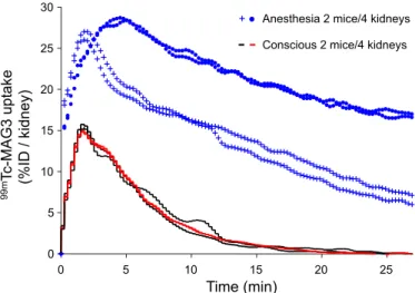
Fig. 3. Effect of anesthesia on99mTc-MAG3 uptake in C57 mice. Dynamic
SPECT imaging in 2 conscious mice (black and red lines matching the 4 kidneys) shows a rapid extraction of99mTc-MAG3 from blood (T
1/2IN, 0.4⫾
0.1 min), a maximal renal accumulation 120 s after iv injection (⬃15% of injected dose/kidney), and subsequent tubular secretion (T1/2OUT, 3.1⫾ 0.3
min). Variability coefficients for T1/2INand T1/2OUTare 6.6 and 9.3%,
respec-tively. In contrast, ketamine-xylazine anesthesia in 2 mice (blue symbols for the 4 kidneys) induces a significant delay in99mTc-MAG3 clearance (T
1/2OUT,
21.1⫾ 9.0 min), as well as a higher variability for T1/2IN(54.0%) and T1/2OUT
(45.2%).
Fig. 4. Dynamic SPECT imaging of99mTc-MAG3 uptake in Clcn5 mice. The
time-activity curve shows the rapid uptake of99mTc-MAG3 in the kidney
cortex (T1/2IN, 0.4⫾ 0.1 min; variability coefficient, 12.2%), resulting from the
blood flow and the concentration by PT cells. The peak (maximal accumula-tion: 11.7⫾ 1.6% of injected dose/kidney) occurs between 90 and 120 s after iv injection, i.e., when excretion overcomes influx. The subsequent down-sloping curve corresponds to the tubular secretion and elimination into the urine (T1/2OUT, 4.7⫾ 0.5 min; variability coefficient, 9.1%; n ⫽ 6 kidneys
from 3 pairs of Clcn5Y/⫺and Clcn5Y/⫹littermates). The spontaneous washout is slightly faster in Clcn5Y/⫺mice (blue squares) than in wild-type Clcn5Y/⫹ mice (red circles).
F457
SPECT IMAGING IN MOUSE KIDNEY
AJP-Renal Physiol•VOL 298 • FEBRUARY 2010 •www.ajprenal.org
on December 20, 2011
ajprenal.physiology.org
present in Clcn5Y/⫹ urine at 60 min postinjection was mostly
free iodine (92⫾ 2% of total urine activity, n ⫽ 3). In strong contrast, Clcn5Y/⫺ kidneys showed a severe defect in the
reabsorption of ultrafiltrated 123I-
2-microglobulin (Fig. 5A;
Table 1), with predominant urinary loss of intact 123I- 2
-microglobulin (62 ⫾ 4% of total urine activity, n ⫽ 3). The autoradiography of kidneys fixed 7 min after injection of
125I-
2-microglobulin, performed sequentially in the mice used
for in vivo SPECT imaging, showed a strong cortical signal in
Clcn5Y/⫹kidneys contrasting with the lack of tracer uptake in
Clcn5Y/⫺ kidneys (Fig. 5B). Importantly, the calculated glo-merular filtration rate of 125I-
2-microglobulin (0.22 ⫾ 0.02
ml/min), based on the first part of the curve (see Fig. 5A), was similar in both strains of Clcn5 mice. These data demonstrate that the congenital PT dysfunction caused by the lack of ClC-5 is adequately reflected by SPECT imaging of defective renal handling of2-microglobulin.
Renal uptake of 123I-
2-microglobulin: recovery after
cis-platin injury. We used a model of reversible tubular toxicity induced by cisplatin, to test the potential of SPECT to provide a longitudinal assessment of acquired PT dysfunction in indi-vidual mice. The PT dysfunction, evidenced by the urinary excretion of albumin (and that of CC16, data not shown), peaked at day 5 and showed progressive recovery thereafter (Fig. 6A). A similar profile was observed by sequential SPECT
Fig. 6. Cisplatin-induced acute kidney injury: albuminuria and123I- 2
-micro-globulin handling. Shown are serial determination of the urinary excretion of albumin (A) and repetitive SPECT imaging of123I-
2-microglobulin uptake in
mice treated with cisplatin (B). The urinary excretion of albumin (baseline: 0.06⫾ 0.04 g/mg creatinine) peaked at day 5 (165 ⫾ 70 g/mg creatinine) and subsequently decreased toward normal values at day 11 (2.5⫾ 1 g/mg creatinine) and day 13 (3.8⫾ 3.2 g/mg creatinine) (*P ⬍ 0.05, at least 4 values/time point). ns, Not significant. Representative sequential SPECT im-aging for123I-
2-microglobulin handling in control mice (red circle) and a
cisplatin-treated mouse reveals defective endocytic uptake at day 5 (blue inverted triangle), with partial recovery at day 12 (blue triangle). Note that the excretion defect (curve⬎7 min) persists at day 12.
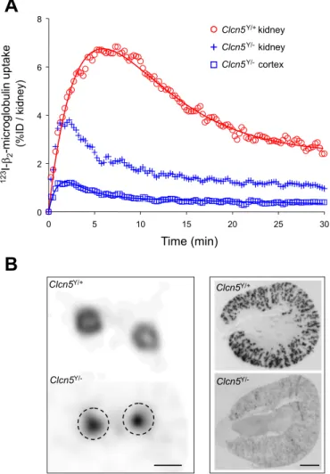
Fig. 5. Dynamic renal handling of 2-microglobulin in Clcn5 mice. The
time-activity curves show the progressive uptake of 123I-
2-microglobulin
following glomerular filtration and apical, receptor-mediated endocytosis by PT cells. The first part of the ascending curve corresponds to the progressive accumulation of the tracer in the urinary lumen (i.e., glomerular filtration), whereas accumulation beyond the first 3 min corresponds to apical endocytosis by PT cells. A: dynamic SPECT analysis123I-
2-microglobulin in Clcn5Y/⫹
(red circle) and Clcn5Y/⫺ (blue cross) whole kidneys, and Clcn5Y/⫺cortex (blue square). The curves show the average of 6 kidneys (3 pairs of mice) in each group. In Clcn5Y/⫹mice,123I-
2-microglobulin rapidly accumulates in
the cortex, with subsequent washout corresponding to breakdown and further release of free123I into the blood. In Clcn5Y/⫺mice,123I-
2-microglobulin
rapidly clears through the kidneys as a pure result of glomerular filtration.
B: representative distribution of iodine-labeled2-microglobulin in transverse
kidney sections at 7 min postinjection in Clcn5Y/⫹and Clcn5Y/⫺mice: in vivo SPECT imaging vs. autoradiography. In Clcn5Y/⫹kidney,2-microglobulin is
restricted to the cortex by SPECT, correlating with a segmental distribution along radial proximal tubules. In Clcn5Y/⫺mice, SPECT only detects activity in the pelvis without significant uptake in the cortex, as confirmed by the lack of autoradiographic signal. Dotted circles represent the cortical contours in
Clcn5Y/⫺kidney. Bars⫽ 5 mm (left) and 1 mm (right).
on December 20, 2011
ajprenal.physiology.org
imaging for the renal uptake of123I-
2-microglobulin in
indi-vidual mice, with a major defect of uptake at day 5 followed by partial recovery at day 12 (Fig. 6B).
DISCUSSION
This report demonstrates the feasibility and validity of both static and dynamic SPECT imaging to investigate specific PT functions in conscious mice. To the best of our knowledge, this is the first report of dynamic SPECT renography in conscious mice demonstrating the feasibility of continuous monitoring of drug handling from the time of iv injection.
Our studies of99mTc-MAG3 showed a delayed tracer
clear-ance and wider intra-assay variability in anesthetized mice, confirming the adverse impact of ketamine/xylazine on base-line renal function (14, 21, 22, 29). To circumvent the need for anesthesia, we used a simple plastic holder, which was suffi-cient to immobilize the mice without discomfort during SPECT acquisition (Supplemental Fig. 1). The imaging of conscious mice allowed us to sequentially use three radiotracers to assess distinct PT functions in the same mice: 1) uptake of 99m
Tc-DMSA to evaluate the functional cortex with high resolution; 2) dynamic99mTc-MAG3 SPECT to investigate the basolateral
capture and apical secretion; and 3) processing of 123I- 2
-microglobulin to assess apical receptor-mediated endocytosis. We took advantage of the Clcn5 KO mice (7, 18, 28) to provide a proof of concept for the use of SPECT, which showed a severe defect in both apical receptor-mediated endocytosis and basolateral anionic uptake, without significant alteration of the apical secretion by PT cells.
The renal uptake of 99mTc-DMSA showed a progressive
accumulation in mouse kidney cortex, matching standard curves reported in humans (16). At the plateau phase,99m
Tc-DMSA SPECT yields a millimetric delineation of the cortex, sufficient to identify a local area of parenchymal damage, as documented by the experimental infarction model (Fig. 2, A and B). The procedure has a low interassay variability, which supports its usefulness for repetitive and long-term biodistri-bution studies, as well as longitudinal studies (e.g., evaluation of toxicity following treatment) in the same mice.
The dynamic SPECT quantification of 2-microglobulin
renal uptake provided the first in vivo time course of PT receptor-mediated endocytosis in conscious mice. The radio-labeled LMW protein is freely filtered by the glomeruli, avidly captured in the renal cortex, and rapidly degraded with subse-quent release of the radiotracer and residual peptide/amino acids to peritubular capillaries (2). The compartmental analysis of123I-
2-microglobulin biodistribution (Supplemental Fig. 2)
enabled us to determine a GFR of 0.22⫾ 0.02 ml/min, similar to that reported previously by using FITC-inulin clearance (20). The usefulness of SPECT to evaluate PT function is further demonstrated by the Clcn5 KO mice, characterized by defective endocytosis due to the internalization of the multili-gand receptors, megalin and cubilin (7, 18). Our data show that the defective PT apical endocytosis previously evidenced by ex vivo counting (7) is confirmed by noninvasive in vivo SPECT of 123I-
2-microglobulin uptake. Despite similar GFR, the
tubular reabsorption of 123I-
2-microglobulin is strongly
im-paired in Clcn5Y/⫺mice, with altered metabolization in PT and
subsequent loss in the urine, mostly as native 123I-labeled
protein.
Similarly to2-microglobulin, the renal handling of99m
Tc-DMSA has been regarded as an indicator of PT dysfunction in human and animal models (1, 25). For example, treatment of rats with Na-maleate induces a typical renal Fanconi syn-drome, with marked reduction of99mTc-DMSA uptake (19). In
our study, SPECT quantification of 99mTc-DMSA capture
shows that inactivation of Clcn5 is associated with a lack of tracer accumulation in the renal cortex, suggesting a severe defect in PT basolateral function. It is likely that the residual signal detected in the pelvis of Clcn5Y/⫺ mice represents a
small glomerular filtration of99mTc-DMSA (17). The
basolat-eral uptake of 99mTc-DMSA from peritubular capillaries into
S3 PT cells is mediated by the Na⫹/dicarboxylate cotransporter NaDC3 (SLC13A3) (4, 13) (Fig. 1). Of note, the coexpression of nonconducting fragments of ClC-5 with NaDC transporters alters their translation and/or trafficking in Xenopus laevis oocytes, resulting in decreased surface expression (15). The severe deficit in both apical and basolateral SPECT uptake reported here further demonstrates that the functional loss of ClC-5 induces a generalized intracellular trafficking in PT cells (7, 18).
Renal scintigraphy with 99mTc-MAG3, a radiolabeled
ana-log of p-aminohippurate, can provide excellent image quality even with severe renal failure (10). For instance,99mTc-MAG3
imaging detected changes in renal function in anesthetized mice as early as 5 h following ischemia-reperfusion injury (22). A model for 99mTc-MAG3 secretion by PT cells has
recently been proposed, based on in vitro transport experiments in X. laevis oocytes (24) (Fig. 1). 99mTc-MAG3 is
intracellu-larly accumulated through the basolateral organic anion trans-porter OAT1 that is expressed in all PT segments (4). The molecular counterpart of 99mTc-MAG3 apical transport could
involve the Na⫹-dependent phosphate transporter NPT1 and/or the multidrug resistance protein MRP2 (5, 24). Our results demonstrate that PT dysfunction caused by the functional loss of ClC-5 is not associated with a defect in99mTc-MAG3 renal
handling. The contrast between the complete loss of 99m
Tc-DMSA basolateral uptake and the preserved 99mTc-MAG3
secretion in Clcn5 KO kidneys provides further evidence that these two pathways are functionally independent (24) and corroborates immunolocalization studies showing a distinct distribution of NaDC3 and OAT1 in PT segments (4). Addi-tional SPECT studies using mouse models invalidated for distinct organic anion transporters may help establish the metabolic pathways handling these particular radiotracers, as well as other drugs and metabolites.
In conclusion, SPECT analysis represents a new approach to characterize the renal PT function in conscious mice, based on acute and/or repetitive biodistribution studies using distinct radiolabeled tracers. Two limitations cannot be ignored. First, the spatial resolution of SPECT, being limited to the millimet-ric range, cannot compete with rapidly evolving live-micros-copy methods (6). Second, although SPECT offers a temporal resolution of 5–10 s for pharmacokinetic studies, this may not be sufficient to explore faster functions such as organelle activation or intracellular trafficking. Nevertheless, the devel-opment of creative approaches dedicated to small-animal im-aging will permit further functional investigations of transgenic mouse strains, with relevance for renal physiology or disease mechanisms.
F459
SPECT IMAGING IN MOUSE KIDNEY
AJP-Renal Physiol•VOL 298 • FEBRUARY 2010 •www.ajprenal.org
on December 20, 2011
ajprenal.physiology.org
ACKNOWLEDGMENTS
The authors thank H. Belge and M-F. van den Hove for helpful discussion; and V. Beaujean, Y. Cnops, M. De Bast, M. Helbo, Th. Lac, A. Saliez, and P. Van der Smissen for excellent technical assistance. The Clcn5 mice were kindly provided by W. B. Guggino and S. E. Guggino (Dept. of Physiology, Johns Hopkins University Medical School, Baltimore, MD).
GRANTS
F. Jouret was a Research Fellow of the Fonds de la Recherche Scientifique (FNRS). These studies were supported by the Belgian agencies FNRS and Fonds pour la Recherche Scientifique Médicale, the ‘Fondation Alphonse and Jean Forton’, a Concerted Research Action (05/10-328), an Inter-university Attraction Pole (IUAP P6/05), the DIANE project (Communauté Française de Belgique), and the EUNEFRON (FP7, GA 201590) program of the European Community.
DISCLOSURES
No conflicts of interest are declared by the authors.
REFERENCES
1. Anninga JK, Valdes Olmos RA, de Kraker J, van Tinteren H,
Hoefnagel CA, van Royen EA. Technetium-99m dimercaptosuccinic
acid and ifosfamide tubular dysfunction in children with cancer. Eur J
Nucl Med 21: 658 –662, 1994.
2. Birn H, Christensen EI. Renal albumin absorption in physiology and pathology. Kidney Int 69: 440 –449, 2006.
3. Biver S, Belge H, Bourgeois S, Van Vooren P, Nowik M, Scohy S,
Houillier P, Szpirer J, Szpirer C, Wagner CA, Devuyst O, Marini AM.
A role for Rhesus factor Rhcg in renal ammonium excretion and male fertility. Nature 456: 339 –343, 2008.
4. Burckhardt BC, Burckhardt G. Transport of organic anions across the basolateral membrane of proximal tubule cells. Rev Physiol Biochem
Pharmacol 146: 95–158, 2003.
5. Burckhardt G, Bahn A, Wolff NA. Molecular physiology of renal p-aminohippurate secretion. News Physiol Sci 16: 114 –118, 2001. 6. Caplanusi A, Parreira KS, Lima WR, Marien B, Van Der Smissen P,
de Diesbach P, Devuyst O, Courtoy PJ. Intravital multi-photon
micros-copy reveals several levels of heterogeneity in endocytic uptake by mouse renal proximal tubules. J Cell Mol Med 12: 351–354, 2008.
7. Christensen EI, Devuyst O, Dom G, Nielsen R, Van der Smissen P,
Verroust P, Leruth M, Guggino WB, Courtoy PJ. Loss of chloride
channel ClC-5 impairs endocytosis by defective trafficking of megalin and cubilin in kidney proximal tubules. Proc Natl Acad Sci USA 100: 8472– 8477, 2003.
8. de Jong M, Barone R, Krenning E, Bernard B, Melis M, Visser T,
Gekle M, Willnow TE, Walrand S, Jamar F, Pauwels S. Megalin is
essential for renal proximal tubule reabsorption of (111)In-DTPA-oct-reotide. J Nucl Med 46: 1696 –1700, 2005.
9. Hishikawa K, Marumo T, Miura S, Nakanishi A, Matsuzaki Y,
Shibata K, Ichiyanagi T, Kohike H, Komori T, Takahashi I, Takase O, Imai N, Yoshikawa M, Inowa T, Hayashi M, Nakaki T, Nakauchi H, Okano H, Fujita T. Musculin/MyoR is expressed in kidney side population
cells and can regulate their function. J Cell Biol 169: 921–928, 2005. 10. Itoh K.99mTc-MAG3: review of pharmacokinetics, clinical application to
renal diseases and quantification of renal function. Ann Nucl Med 15: 179 –190, 2001.
11. Lange K, Carson R. EM reconstruction algorithms for emission and transmission tomography. J Comput Assist Tomogr 8: 306 –316, 1984. 12. Maisey M. Radionuclide renography: a review. Curr Opin Nephrol
Hypertens 12: 649 –652, 2003.
13. Markovich D, Murer H. The SLC13 gene family of sodium sulphate/ carboxylate cotransporters. Pflügers Arch 447: 594 –602, 2004. 14. Mitchell SKI, Toal RL, Daniel GB, Rohrbach BW. Evaluation of renal
hemodynamics in awake and isoflurane-anesthetized cats with pulse-wave Doppler and quantitative renal scintigraphy. Vet Radiol Ultrasound 39: 451–458, 1998.
15. Mo L, Wills NK. ClC-5 chloride channel alters expression of the epithe-lial sodium channel (ENaC). J Membr Biol 202: 21–37, 2004.
16. Moretti JL, Rapin JR, Saccavini JC, Lageron A, Le Poncin M, Bardy
A. 2,3-Dimercaptosuccinic-acid chelates. 2. Renal localization. Int J Nucl
Med Biol 11: 275–279, 1984.
17. Müller-Suur R, Gutsche HU. Tubular reabsorption of technetium-99m-DMSA. J Nucl Med 36: 1654 –1658, 1995.
18. Piwon N, Gunther W, Schwake M, Bosl MR, Jentsch TJ. ClC-5 Cl⫺ channel disruption impairs endocytosis in a mouse model for Dent’s disease. Nature 408: 369 –373, 2000.
19. Provoost AP, Van Aken M. Renal handling of technetium-99m DMSA in rats with proximal tubular dysfunction. J Nucl Med 26: 1063–1067, 1985. 20. Qi Z, Whitt I, Mehta A, Jin J, Zhao M, Harris RC, Fogo AB, Breyer
MD. Serial determination of glomerular filtration rate in conscious mice
using FITC-inulin clearance. Am J Physiol Renal Physiol 286: F590 – F596, 2004.
21. Rao S, Verkman AS. Analysis of organ physiology in transgenic mice.
Am J Physiol Cell Physiol 279: C1–C18, 2000.
22. Roberts J, Chen B, Curtis LM, Agarwal A, Sanders PW, Zinn KR. Detection of early changes in renal function using99mTc-MAG3 imaging
in a murine model of ischemia-reperfusion injury. Am J Physiol Renal
Physiol 293: F1408 –F1412, 2007.
23. Rowland DJ, Cherry SR. Small-animal preclinical nuclear medicine instrumentation and methodology. Semin Nucl Med 38: 209 –222, 2008. 24. Shikano N, Kanai Y, Kawai K, Ishikawa N, Endou H. Transport of
99mTc-MAG3 via rat renal organic anion transporter 1. J Nucl Med 45:
80 –85, 2004.
25. van Luijk WH, Ensing GJ, Meijer S, Donker AJ, Piers DA. Is the relative 99mTc-DMSA clearance a useful marker of proximal tubular
dysfunction? Eur J Nucl Med 9: 439 –442, 1984.
26. Walrand S, Jamar F, de Jong M, Pauwels S. Evaluation of novel whole-body high-resolution rodent SPECT (Linoview) based on direct acquisition of linogram projections. J Nucl Med 46: 1872–1880, 2005. 27. Walrand S, van Dulmen A, van Rossem H, Pauwels S. Acquisition of
linograms in SPET: implementation and benefits. Eur J Nucl Med Mol
Imaging 29: 1188 –1197, 2002.
28. Wang SS, Devuyst O, Courtoy PJ, Wang XT, Wang H, Wang Y,
Thakker RV, Guggino S, Guggino WB. Mice lacking renal chloride
channel, CLC-5, are a model for Dent’s disease, a nephrolithiasis disorder associated with defective receptor-mediated endocytosis. Hum Mol Genet 9: 2937–2945, 2000.
29. Yang XP, Liu YH, Rhaleb NE, Kurihara N, Kim HE, Carretero OA. Echocardiographic assessment of cardiac function in conscious and anesthetized mice. Am J Physiol Heart Circ Physiol 277: H1967– H1974, 1999.
on December 20, 2011
ajprenal.physiology.org