HAL Id: hal-01869675
https://hal.archives-ouvertes.fr/hal-01869675
Submitted on 20 Jan 2021
HAL is a multi-disciplinary open access
archive for the deposit and dissemination of
sci-entific research documents, whether they are
pub-lished or not. The documents may come from
teaching and research institutions in France or
abroad, or from public or private research centers.
L’archive ouverte pluridisciplinaire HAL, est
destinée au dépôt et à la diffusion de documents
scientifiques de niveau recherche, publiés ou non,
émanant des établissements d’enseignement et de
recherche français ou étrangers, des laboratoires
publics ou privés.
Formation of UV-induced DNA damage contributing to
skin cancer development
Jean Cadet, Thierry Douki
To cite this version:
Jean Cadet, Thierry Douki. Formation of UV-induced DNA damage contributing to skin cancer
development. Photochemical & Photobiological Sciences , Royal Society of Chemistry, 2018, 17,
pp.1816-1841. �10.1039/c7pp00395a�. �hal-01869675�
Formation of UV-induced DNA damage contributing to skin cancer
development
Jean Cadet1*, Thierry Douki2*
1Département de Médecine Nucléaire et Radiobiologie, Faculté de Médecine, 3001 12e Avenue Nord, Université de Sherbrooke, Sherbrooke, Québec JIH 5N4, Canada
2Univ. Grenoble Alpes, CEA, CNRS, INAC, SyMMES/CIBEST, F-38000 Grenoble
*Corresponding authors:
e-mail (Jean Cadet): jean.cadet@usherbrooke.ca e-mail (Thierry Douki): thierry.douki@cea.fr
≠ Jan van der Leun commemorative issue
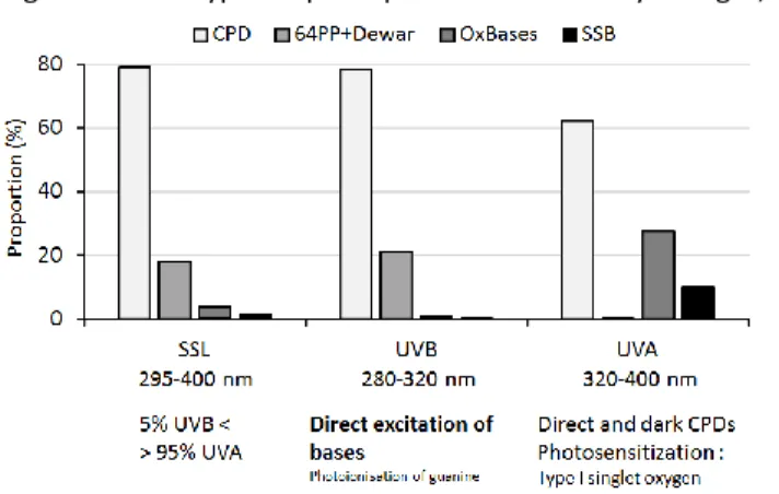
Abstract. UV-induced DNA damage plays a key role in the initiation phase of skin cancer. When left unrepaired or when damaged cells are not eliminated by apoptosis, DNA lesions express their mutagneic properties, leading to the activation of proto-oncogene or the inactivation of tumor supression genes. The chemical nature and the amount of DNA damage strongly depend on the wavelength of the incident photons. The most energetic part of the solar spectrum at the Earth’s surface (UVB, 280-320 nm) leads to the formation of cyclobutane pyrimidine dimers (CPDs) and pyrimidine (6-4) pyrimidone photoproducts (64PPs). Less energetic but 20-time more intense UVA (320-400 nm) also induces the formation of CPDs together with a wide variety of oxidatively generated lesions such as single strand breaks and oxidized bases. Among those, 8-oxo-7,8-dihydroguanine (8-oxoGua) is the most frequent since it can be produced by several mechanims. Data available on the respective yield of DNA photoproducts in cells and skin show that exposure to sunlight mostly induces pyrimidine dimers, wich explains the mutational signature found in skin tumors, with lower amounts of 8-oxoGua and strand breaks. The present review aims at describing the basic photochemistry of DNA and discussing the quantitative formation of the different UV-induced DNA lesions reported in the litterature. Additional information on mutagenesis, repair and photoprotection are briefly provided.
Introduction
Overexposure to sunlight, and in particular to the ultraviolet (UV) portion of its spectrum, is unambiguously linked to the onset of skin cancer 1, 2 as
well as photoaging and ocular pathologies. Solar UV has two main components classified as “Class I carcinogens” by IARC 3: UVB (290-320 nm) and UVA
(320-400 nm) emitted in an about 1:20 ratio that varies according to latitude and season. A causative relationship, at least in terms of mutation induction, has been established between UVB and the induction of non-melanoma skin cancers (NMSC) 4-6 that consist
of basal cell carcinoma (BSC) and squamous cell carcinoma (SCC).Evidence have also been provided for
a major role of sunlight in malignant melanoma (MM) 7
but the respective contribution of UVA and UVB remains less understood. Depletion of stratospheric ozone that is accompanied by an enhanced contribution of UVB radiation and concomitantly a higher incidence of both NMSC and MM, has been identified as another critical environmental factor 8.
Solar UV is not the only concern for skin cancer since artificial tanning equipments represent another risk. These devices emit intense UV radiations richer than sunlight in UVA and evidence is growing for their involvement in MM and to a lesser extent SCC 9-11.
UV radiation is a complete carcinogen. Its initiating properties arise from its ability to damage DNA and induce mutations in proto-oncogenes and tumor suppressor genes such as p53 12, 13. The main damaging
process involves direct absorption of UVB and to a lesser extent of UVA photons that trigger dimerization of pyrimidine bases 14-16. UVA radiation also damages
DNA in an oxygen-dependent mode that involves photosensitization 15, 17. The resulting reactive oxygen
and nitrogen species lead to base lesions such as 8-oxo-7,8-dihydroguanine (8-oxoGua) and single strand breaks (SSBs) 15, 18-20.. Efficient repair of DNA damage
before replication is a requisite to prevent carcinogenesis to occur. The importance of repair of DNA photoproducts is illustrated in the photosensitive xeroderma pigmentosum patients who in most cases suffer from a deficiency in the nucleotide excision repair (NER) mediated removal of overwhelming bipyrimidine photoproducts and exhibit an increase by up to ten thousand-fold of the risk of skin cancer on UV exposed zones together with a significantly reduced lifetime expectancy 21-23. The present survey is aimed at
critically reviewing the direct and sensitized photoreactions of cellular DNA induced by UVB, UVA and solar radiations. In addition information is also provided on several more biological end-points including DNA repair and photodamage prevention.
Pyrimidine dimers
Dimeric photoproducts involving adjacent pyrimidine bases are the most frequent UV-induced lesions in cellular DNA 18 . Cyclobutane thymine dimer
has been first identified almost 60 years ago 24, 25. Since
then, very large amounts of data have been gathered on the formation of other bipyrimidine photoproducts and their chemical properties 26, 27. Their relevance to
skin cancer has been clearly established by the observation of an overwhelming majority of mutations at bipyrimidine sites in specific genes of skin tumors, in particular in non-melanoma skin cancers 28, 29. Next
generation sequencing confirmed the accumulation of UV-specific mutations to a high rate in melanoma 30.
The biological significance of pyrimidine dimers in UV mutagenesis is further shown by the coincidence between hotspots for their formation and occurrence of mutations 31. While oxidatively generated DNA
lesions are produced by photosensitized processes, pyrimidine dimers arise mostly from direct absorption of UV photons. The maximal absorption of DNA is at 260 nm, namely in the UVC range. However, absorption of DNA remains significant in the UVB range with values of 20 and 3 % compared to 260 for 290 and 300 nm, respectively. UVA photons are much more weakly absorbed but may also induce the formation of cyclobutane pyrimidine dimers (CPDs), either by direct excitation or via photosensitization.
DNA photochemistry
In the last two decades, the spectacular advances in time-resolved spectroscopy and the development of novel theoretical approaches have drastically modified our perception of electronic excitation processes in DNA photochemistry. In particular, the role of stacking and interaction between bases as well as the dynamics of DNA were shown to play key roles. This is in particular revealed by the comparison between the fate of excited states of bases either in DNA or as monomers in aqueous solution. In the latter case, the * excited states produced after absorption of UV photons deactivate on a ps time scale back to ground state 32. A
similar rapid process also takes place in DNA but together with other pathways of longer time scale 33, 34,
some of them leading to photochemical reactions. The vast majority of the resulting DNA damage involves dimerization of adjacent pyrimidine bases. A role for C5-methylation of cytosine has also to be considered. Formation of minor DNA lesions such as adenine dimer
35, 36 or adenine-thymine dimer 37-39 has been reported
as minor photoreactions but will not be discussed here.
Cyclobutane pyrimidine dimers
One consequence of the formation of excited states is the formation of DNA photoproducts. Immediately after UV absorption by DNA, the excitation energy can be delocalized over a few bases into Frenkel excitons 40, 41 as the result of stacking between adjacent bases.
When the stacking is so strong that orbitals overlap, excited states acquire a charge transfer state character (CT states) 42. Both species can lead to DNA damage.
The Frenkel excitons were proposed on the basis of theoretical calculations to be involved in the formation of CPDs. These dimeric photoproducts arise from a [2+2] cycloaddition that takes place through an energy barrier-less process 43 on the ps time scale 44. In contrast
to what was long believed, triplet excited states seem to play a limited role in the formation of bipyrimidine photoproducts. An upper estimation of 10 % has been proposed. This is based on the observation that the quantum yield of CPDs is constant over a wide range of UVC and UVB wavelengths while the yield triplet state decreases 43. The limited role of triplet states in the
formation of CPDs has also been shown at the dinucleoside monophosphate level where they account only for approximately 3% of the CPDs 45. Another
interesting result of recent spectroscopic studies is the ability of UVA photons to directly induce excited states in DNA. Absorption of UVA photons by DNA bases does not take place in monomers but become significant in double stranded systems 46. Spectroscopic studies have
shown that the produced excited states are mostly CT states 47. The possibility of formation of CPDs from such
states is not documented. However, a combined spectroscopic and theoretical study has shown that vibrational motion can convert them into Frenkel excitons 48. The observation that dimer formation is
favored in double- compared to single-stranded DNA 47
could be explained by this property. It should though be stressed that the UVA photochemistry of DNA is quantitatively less significant than that of UVC and UVB since the molecular absorption coefficient of DNA in the UVA range in 1000 time lower than at 260 nm.
CPDs are formed at each of the four bipyrimidine doublets. The structure and the main properties of CPDs have been extensively studied in monomeric systems, dinucleoside monophosphates and isolated DNA. CPDs can be formed as several diastereoisomers which were isolated and characterized upon photolysis of isolated bases and nucleosides 16, 49, 50. However, the
geometrical constraints encountered in DNA restrict the formation of possible isomers (Fig. 1). First, only syn isomers, with the two pyrimidine rings in a parallel orientation can be produced. In double stranded B-DNA, the stacking of the bases also forces the bases to be located on the same side of the cyclobutane ring, in a cis isomery 51, 52. In single-stranded DNA or
destabilized double helix, trans isomers with the bases on opposite sides of the cyclobutane ring can be also produced 51, 52. It was also proposed that interstrand
anti isomers, with the bases in antiparallel orientation,
could be produced in quadruplexes 53. In B-form
double-stranded mammalian DNA, TT; TC, CT and CC
cis,syn CPDs are produced approximately in 100:50:25:10 ratios following exposure to UVC and UVB
51. These ratios are slightly affected by the irradiation
conditions such as the temperature or the ionic strength 52, 54, 55. However, the parameter that exhibits
the most drastic effect on the ratio between CPDs is the GC base pair content 56. In bacterial DNA with high
percentage of GC, TT CPD is a minor photoproduct. It can however be considered as the major UV-induced photoproducts upon UVB and UVC irradiation of isolated mammalian DNA 49, 51. This trend is even
stronger in UVA-irradiated isolated DNA where no 64PP is detected by highly specific chromatographic assays while TT CPD represents 90% of the CPDs 46.
CPDs exhibit several specific characteristics. In particular, although the modified pyrimidine bases no longer absorb UVB, CPDs keep a residual absorption in the UVC range with a maximum at 230 nm. At these wavelengths, absorption leads to the reversion of the CPD with a high efficiency 57, 58. As a consequence,
prolonged irradiation with UVC, typically the 254 nm light used in many experimental works, leads at high
dose to an equilibrium between formation and reversion of CPDs, especially for C-containing CPDs 51.
Pyrimidine (6-4) photoproducts and their Dewar valence isomers
Pyrimidine (6-4) pyrimidone photoproducts (64PPs) are not directly produced from the excited states. Time-resolved studies have shown that their spectroscopic signature appears after 4 ms 59, namely 1000 time more
slowly than CPDs 44. This is explained by the formation
of a cyclic reaction intermediate exhibiting an oxetane structure when the 3’-end pyrimidine is a thymine (Fig. 2). When the 3’-end base is a cytosine, the intermediate is an azetidine. Unambiguous evidence for the formation of a cyclic intermediate has been provided by the isolation and the characterization of a thietane as the precursor of the 64PP between thymine and 4-thiothymine 60. In all cases, the cyclic intermediates
decompose into the final photoproducts that bear a pyrimidone ring on the 3’-end and in which the C4 substituent of the 3’-end base is shifted to the 5 position of the 5’-end base. Involvement of a Paternò-Büchi reaction, namely a biradical process, has been proposed for the formation of the cyclic intermediate. More recently, involvement of a charge transfer excited states was proposed through either the triplet 61 or the
singlet channel 43. Interestingly, the latter theoretical
work predicted a high energy barrier that could explain why the quantum yield of 64PPs decreases over the UVC/UVB range and why 64PPs are produced in very low amount or not at all in the UVA range. In UVC- and UVB-irradiated double-stranded DNA, 64PPs are less frequent than CPDs 51, 62. They are produced in a 1:3 to
1:5 ratio. However, this value depends on the bipyrimidine sites. At TT, 64PP is ten times less frequent than CPDs 51. The ratio is even lower at CT where 64PP
is hardly detectable. CC 64PP is also much less frequent than the corresponding CPD and its formation has been reported mostly in isolated DNA. In contrast, TC 64PP is the most frequent 64PP 51, 63, produced in a yield almost
as large as that of TC CPD, making it the third most frequent photoproduct following UVB and UVC irradiation.
The most striking features of 64PPs are associated with the presence of the pyrimidone ring. First, 64PPs exhibit an absorption maximum in the UVB range (325 nm for TpT and dCpT 64PPs, and 315 nm for TpdC and dCpdC 64PPs). When excited at these wavelengths, 64PPs are fluorescent. For derivatives of dinucleoside monophosphates, which mimic some of the geometrical constraints encountered in DNA, the emission is maximal at 390-400 nm and the quantum yield is in the range of 1 to 3 % 64. Irradiation of 64PPs
photoisomerization into a Dewar valence isomer first characterized in TpT 65 (Fig. 3). The latter compound
arises from an intramolecular 4 electrocyclisation and takes place within 130 ps 66. Similar photoreactions
were also reported for C- and 5mC-containing 64PPs 67-69. The published quantum yields of photoisomerization
range between 2 58 and 8% 66 in model systems. This
value is lower in DNA because of the presence of adjacent bases, and in particular G 70. Dewars were
detected at the four bipyrimidine sites in isolated DNA exposed to UVB radiation 51. Large doses of UVB are
necessary to reach a high ratio between the yield of Dewars and that of 64PPs. In contrast, combination of UVB and UVA exposure leads to a much larger photoisomerization yield. This can be explained by the absorption properties of DNA and 64PPs 71. The
presence of a pyrimidone ring is also the basis of a recently proposed Trojan horse process. Pyrimidones have been shown to exhibit a high energy and well populated triplet state. When added to a solution of plasmid DNA, both 5-methylpyrimidone 72 and an
analog of TT 64PP 73 were found to favor the formation
of additional CPDs upon exposure to a lamp emitting within the 300-400 nm range. Theoretical works show that pyrimidone could be a sensitizer when incorporated within DNA 74. It remains to be shown
experimentally whether the photosensitizing properties of the pyrimidone moieties of 64PPs present within double-stranded DNA can be expressed before conversion into Dewars.
Photoproducts of 5-methylcytosine
5-Methylcytosine (5mC) is an important epigenetic factor mostly present in CpG islands in genomic DNA. These frequently methylated sites are UV mutational hotspots and have been shown, mostly by ligation-mediated polymerase chain reaction (LMPCR), to be also hot spots for the formation of CPDs 75-77. Dimeric
photoproducts involving 5mC and either T or C have been isolated in dinucleoside monophosphates 67, 78.
More recently, the effect of the methylation site of cytosine was investigated in a trinucleotide baring a TX (with X=C, 5mC or N4-methylC, a bacterial minor base) sequence flanked with a 3’-G 79. Compared to that of
TCG, photolysis of T5mCG leads to an increased formation of CPDs, which is more significant with UVB than UVC. Conversely, the yields of 64PPs and Dewars decrease as a consequence of methylation. Opposite trends were observed for TN4mCG 79. The results
obtained with T5mCG were explained by conformational changes rather by absorption properties of 5mC as often proposed 80.
Dimers by UVA and photosensitization
Other processes exist that lead in particular to the formation of CPDs. They involve absorption of the UV energy by other molecules with subsequent transfer to DNA that thus indirectly reaches an excited state. The most common of this photosensitization pathway is known as triplet-triplet energy transfer (TTET) 81. TTET
takes place with compounds absorbing UVA and exhibiting a large yield of intersystem crossing. In addition, the energy level of the first triplet excited state has to be higher than that of DNA components. Under these conditions, the triplet excited state of bases, and in particular thymine 82, can be populated,
opening the way to occurrence of photoreactions. As a matter of fact, it is well known that TTET, both in small model systems (bases, nucleoside, and dinucleoside monophosphates) and DNA, leads to the formation of CPDs but not of 64PPs and Dewars. TT CPD is the major photoproduct, representing at least 90% of the lesions, much more than following UVB and UVC irradiation where its proportion among CPDs is approximately 50% (Fig. 4). Two main classes of molecules have been studied for their potency at mediating TTET. The first one are the aromatic ketones such as acetophenone or benzophenone 83. This chemical structure is often
encountered in some non-steroidal anti-inflammatory drugs. The second and more recently investigated class of TTET photosensitizers are the fluoroquinolones 84.
These compounds are drugs used as anti-bacterial agents that have been shown to induce a strong photosensitivity in treated patients.
While the role of triplet state thymine is unquestionable in model systems 45, 85, recent findings
suggest that excitation of a single isolated thymine base may not be the only mechanism involved in the photosensitized formation of CPDs. First, photosensitization of DNA with different proportion of TT, TC and CT sites led to a constant over-reactivity of TT 86. A random excitation of thymine bases should
have resulted in a low yield of TT CPD in T-poor DNA, which is not observed. In addition, CC CPDs were also detected, although in low yield, which is not compatible with the energy level of the triplet state of this base. It is thus likely that, like for the direct excitation of DNA, collective effects are involved in the photosensitized formation of CPDs. A possibility is the occurrence of the Dexter energy transfer that would lead to a charge transfer state and explain the similarity with the formation of CPDs by UVA irradiation. This hypothesis remains yet to be investigated. Another work involving a series of benzophenone derivatives tethered to two thymines allowed proposing a role for delocalized triplet states in the TTET mechanism 87.
An alternative process of CPD formation has been recently described 88. It also involves energy transfer
but, unlike classical photosensitization, it occurs after rather than during UV exposure. UVA-irradiated melanocytes continue to accumulate CPDs after termination of irradiation as the result of a chemiexcitation process. CPD levels are maximal around two hours after UVA exposure and are thereafter decreased by DNA repair. This was observed for TT, TC and CT CPDs. interestingly, this process did not take place in keratinocytes, suggesting a role played by melanin. Accordingly, treatment of isolated DNA with irradiated precursors of melanin led to the formation of CPDs. Altogether, the proposed mechanism involves the formation of oxidation products of melanin and its precursors, likely by peroxynitrite, which may migrate to the nucleus. There, they decompose into small molecules generated in an excited state, for example activated carbonyls, that transfer their energy to DNA and induce the formation of CPDs through chemiexcitation 88. More chemical
studies are necessary to understand this novel pathway that raises new issues in terms of prevention and photoprotection.
Biological properties of pyrimidine dimers
The observation that most mutations in skin tumors are located at bipyrimidine sites points to strong mutagenic properties of pyrimidine dimers. A wide series of biochemical studies have been devoted to the unravelling of the underlying properties. In the case of CPDs, a major reaction to consider is deamination. This hydrolytic process converts cytosine and its derivatives into uracil. The reaction rate is very low for unmodified cytosine but is drastically increased when the C5-C6 bond is saturated like in CPDs. Recent DFT calculations have suggested that 2 water molecules are necessary to achieve the deamination process. A first mechanism, which can be extended to CT and TC CPDs, was established for 5,6-dihydrocytosine 89 (Fig. 5) while
another was proposed for the deamination of CC CPD
90. CPDs deaminate with half-reaction times of a few
hours in the case of the cis,syn diastereoisomers of the dinucleoside monophosphates TpdC and dCpT 91-94. In
DNA, the deamination rate is slightly lower 95-97 but
plays a major role in the mutagenic properties of cytosine-containing CPDs. The well-established scheme for mutagenesis is the initial formation of TC, CT and CC CPDs that deaminate into TU, UT and UU CPDs. During replication, U residues in CPDs code like T and lead to the incorporation of A 98-101 which in turns codes for T
in the next replication. The resulting transition mutations are the hallmarks of UV mutagenesis, namely TC at TC sites and the tandem mutation CCTT.
Mutations are CT sites are much less frequent, maybe as the result of the more efficient repair of this CPD 102.
In vitro experiments have also shown that TT CPD is
more a blocking than a mutagenic photoproduct 103,
mostly because the two thymine rings involved in the dimer keep their coding properties. Deamination of C-containing CPDs was shown to depend on the sequence, with an increased rate at TCG triplets 104.
Interestingly, this property explained the sequence specificity of the mutations found in UVC-irradiated mouse skin 105. In cells, deamination of CPDs is also
increased during transcription 106. Deamination of
5mC-containing photoproducts is slower than that of related unmethylated cytosine lesions. However, deamination is increased by a factor 25 when flanked by G, as in CpG islands 104, 107. This trend is increased by the
acceleration of deamination in nucleosome for both Tm5C and TC CPDs 108.
Like CPDs, 64PPs may undergo deamination. However, this reaction is not possible on the 3’-end of the photoproduct since the base is no longer a pyrimidine and the C4-amino group has migrated to the C5 position of the 5’-end base. Deamination of 5’-end cytosine thus remains possible in CC and CT 64PPs. Interestingly, it was found that the deamination rate of the CT 64PP was much lower than that of the corresponding cis,syn CPD 91. The most frequent 64PP,
TC 64PP, does not deaminate. In spite of this limited involvement of deamination in the fate of 64PPs, the latter photoproducts can be highly mutagenic. This is mostly explained by their major impact on the DNA structure that strongly modifies the network of hydrogen bonding during replication. In particular, bonding with G to the 3’-end moiety and A to 5’-end base of 64PPs is favored. As a result, TT 64PP is highly mutagenic and leads to TC transitions 109, while TC
64PP is much less. In contrast, more frequent incorporation of A opposite the pyrimidone moiety takes place in Dewars. Consequently, TT Dewar is much less mutagenic 110 than its TC analog which induces
CT transitions 111.
Knowledge of the mutagenic properties of the different classes of photoproducts is not enough to establish their respective contributions to solar mutagenesis. Repair has to be considered since it determines the lifetime of DNA damage in the genome and therefore the possibility to express their mutagenic potential. All investigations point to a much more efficient repair through the global nucleotide excision repair (NER) pathway of 64PPs and Dewars than CPDs
112-114. This is explained by the larger impact of the
former photoproducts on the DNA structure, making them more easily detectable by the repair machinery,
in particular XPC. The contribution of 64PPs to UV mutagenesis is thus expected to be lower than that of CPDs. Accordingly, expression of photolyase enzymes specific for either class of pyrimidine dimer in mammalian cells showed that removal of CPDs prevent mutagenesis while that of 64PPs has no impact 115. A
similar experiment with mice expressing CPD photolyase led to the conclusion that presence of CPDs is critical for skin cancer occurrence 116. These results
were recently emphasized by the observation that the most frequent 64PP, TC 64PP, is also the most efficiently repaired 117.
Studies have shown that the four CPDs are not repaired with the same rate 102, 118. TT CPD is the most
slowly repaired while CT CPD is the fastest. TC and CC CPDs exhibit an intermediate rate. Interestingly, these results are in line with mutagenesis data since mutations at CT sites are almost never observed. HPLC-MS/MS analyses made 2h after UVB irradiation of cultured keratinocytes did not show differences in the repair rate of TT and TC 64PPs. Recent results using next sequencing generation at earlier time point suggest that TC 64PP is more efficiently repaired in transcribed regions 119. An important finding in the repair of
UV-induced DNA damage is that UVA decreases the repair capacities of cells 112, 120. This observation was recently
explained, as least partly, by the oxidation of repair proteins such as PCNA and RPA 121, 122. Other potential
mechanisms such as modulation of regulation of specific pathways remain to be explored. Sunlight thus appears to exhibit two concomitant deleterious effects, namely the formation of mutagenic DNA damage and the decrease in the capacities of cells to remove them.
Formation of pyrimidine dimers in cells and skin
Because of the major role of bipyrimidine in the initiating step of skin cancer, a very large number of methods have been developed for their quantification in cellular DNA. These assays involve immunological detection, chromatographic techniques, [32
P]-postlabeling, LMPCR and more recently next generation sequencing of DNA repair lesions. The techniques involving spectrometric detection are usually excellent for the simultaneous and specific detection of the different photoproducts. However, they provide only average data on the overall genome. Sequencing techniques in contrast are less sensitive but provide information at the nucleotide level. LMPCR requires a preliminary treatment to convert CPDs into breaks. The recently developed approach 117 that involves
sequencing of the oligonucleotides generated during the NER process is more flexible providing data on both CPDs and 64PPs depending on the antibody used for the
immunoprecipitation step. Antibody-based detection is also widely used in immunofluorescence and radioimmunoassays. These techniques often provide only relative values and do not permit the individual quantification of the four derivatives of a same class of photoproducts. However, their ease of use makes them very popular, especially in the field of DNA repair. Antibodies constitute also relevant tools for the immunohistochemical detection of both CPDs and 64PPs in skin.
In spite of some minor differences in the determination of the respective yields of the different bipyrimidine photoproducts, all techniques show that DNA reacts in a similar way following exposure to UVB and UVC either as an isolated form in aqueous solution or in cells. The absolute yields are obviously different between the two systems because absorption is impacted by the cellular context. In cultured cells, the reported yield of CPDs ranges between 0.1 and 1 dimer per 105 bases per J/m2 for UVC (254 nm) lamps 123-128
(Table 1). The mean value is approximately 0.45 CPDs per 105 bases per J/m2. With UVB sources, the yield is
roughly one order of magnitude lower than at 254 nm with a mean yield of 0.05 CPDs per 105 bases per J/m2 112, 120, 125, 126, 129, 130. An interesting action spectrum has
been published that shows the strong wavelength dependence on the formation of CPDs in the UVB range
18, with a ratio of roughly 60 between the yields
determined at 290 and 310 nm. It was also observed that formation of CPDs in skin was 20-times less efficient in human skin explants than in the keratinocytes collected from the same donors, likely because of the protection afforded by melanin and the corneal layer 120. It may be added that
immunohistochemistry experiments have shown that the formation of CPDs is not uniform within the skin. For short wavelengths (<280 nm), the frequency of CPDs is lower in the deepest layer of the epidermis 131.
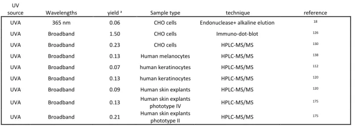
Pyrimidine dimers and in particular CPDs are also produced in cells and skin by UVA irradiation 18, 112, 126, 130, 132-138. Formation of 64PPs has been reported using
immunological approaches 126, 139 but not by more
specific chromatographic techniques120, 130, 140.
Quantitative data in the UVA range all point to a yield of CPDs at least 3 orders of magnitude lower in the UVA than the UVB range. A mean yield based on available quantitative data is approximately 0.3 CPDs per 1010
bases per J/m218, 112, 126, 130, 137, 138. Contamination of the
UVA sources by UVB as an explanation for these observations has been discarded by experiments involving either filters or the use of monochromatic laser pulses. Photosensitization has also been proposed. However, the observation that CPDs are
detected in UVA-irradiated isolated DNA and in a yield similar to that found in cells strongly suggests a direct mechanism triggered by the low but real UVA absorption of DNA 46, 126, 141. Interestingly, the fact that
TT CPD is present in much larger proportion compared to other CPDs after UVA than after UVB or UVC exposure is in line with spectroscopic observations of different excites states in the different wavelengths ranges 47. Because UVA is more than 20 time more
intense than UVB in sunlight, UVA-induced CPDs represent a few percent of the overall load of CPDs in skin. In spite of this low yield formation in cells and skin, the role of UVA-induced CPDs in solar genotoxicity cannot be ruled out. The fact that mutations induced by UVA occur mostly at bipyrimidine sites 142, 143 like with
UVB further supports the biological relevance of UVA-induced CPDs 144. The likely biological relevance of
UVA-induced CPDs is also shown by the observation that CPDs are produced in 3- to 5-fold larger amounts than 8-oxoGua 18, 120, 130, 140, the most frequent UVA-induced
oxidatively generated lesion. One exception are melanocytes where the ratio between the yields of CDPs and 8-oxoGua is 1.4 138, showing that this cell type
is more sensitive to oxidative stress 145. A study has
shown that the formation of CPDs by UVA is favored in the basal layer as the result of scattering phenomena
146, where melanocytes are the most frequent. This
observation raises some interesting question on the origin of melanoma that more significantly involves UVA than UVB as the critical UV domain 147.
In mammalian cells and skin, CPDs were found, using sequencing 148-150 and chromatographic
approaches 16, 51, to be produced in the following order
of frequency: TT > TC > CT > CC. Recent techniques involving new sequencing techniques also showed that TT CPD was the most frequent photoproduct. Excision-seq confirmed that the decreasing CPD frequency was TT>TC>CT>CC 63. CPD-seq led to ratios between the
frequencies of CPDs of 54:22:14:10 for TT, TC, CT and CC respectively 151, which are very close to those
determined by HPLC-MS/MS. The HS-damage-Seq assay also led to a distribution similar to HPLC-MS/MS. It should be stressed that some recent studies yielded distributions significantly different from all those mentioned above. Applying LMPCR to a large number of genes in UVB-irradiated cells, a more comparable yield of UVB-induced CPDs was determined at the four bipyrimidine sites 152. These results may be explained
by the fact that CC sites were more than twice as frequent as TT in the studied genes, and 1.4 times more than CT and TC. Using next generation sequencing, excision-seq and CPD-seq data show an inversion between the relative frequency of TC and CT CPDs
compared to the previously mentioned techniques 119.
This may be explained by the fact that excision-seq and CPD-seq are based on the sequencing of DNA excision repair products and that CT CPDs are repaired significantly faster than the other CPDs.
Ratio between CPDs and 64PPs in cells was found to range between 3 and 5 depending on the assay, a value similar to that determined in isolated DNA, at least for irradiations performed with UVB and UVC. In these wavelength ranges, the HPLC-MS/MS assay was able to detect TC and TT 64PPs in a 5:1 ratio, but could not detect CC and CT 64PPs in cells exposed to doses compatible with cell survival 51. Next generation
sequencing-based techniques confirmed the major formation of TC 64PP but also detected some 64PPs at CT and CC sites 119. At biologically relevant doses of UVB
and UVC, only tiny if any amounts of Dewars are detected 51, 126, 130. In contrast, Dewars have to be taken
into consideration when simulated or real sunlight is used. Based on immunological approaches, Dewars have been detected in significant yield in SSL-exposed human fibroblasts 153, rodent cells 126 and mouse skin 154. HPLC-MS/MS measurements confirmed these
results in cultured cells 130 and skin explants 155. In the
latter samples 30% of the 64PPs were photo-isomerized at 3 MED of SSL. In contrast, Dewars were not detected by the same technique in various models at biologically relevant doses of UVB. Immunological detection yielded a positive signal with UVB but much lower than with SSL 126. Altogether, Dewars are undoubtedly
biological relevant lesions for human exposure. The relative yields of bipyrimidine photoproducts discussed above are average values for the whole genome. However, the distribution of CPDs, 64PPs and Dewars is not homogeneous in the genome. Important parameters that modify the yield of photoproducts are related to the location of the pyrimidine sites with respect to nucleosome and chromatin. It is well known that the yield of CPDs is lower in the nucleosome than in the linker region 156, 157. A recent work provided
accurate information on the favored locations of CPDs within a rotationally phased nucleosome 158. The ratio
between 64PPs and CPDs is also affected by the nucleosome structure 159. More recent work extended
these in vitro data to yeast cells. A pattern of formation reflecting nucleosome position was observed 151.
Interestingly, a nucleosome has a strong impact on the deamination rate of cytosine dimers 108. Sequencing
approaches have also unambiguously shown that formation of photoproducts is greatly reduced by DNA condensation in heterochromatin 119, 160. In agreement
with previous works 161, these techniques also showed
formation of photoproducts either positively or negatively 119, 151. Telomeres are another specific
cellular DNA structure which were found to be more sensitive than the bulk of the genome to UV-induced damage 162. More local sequence effects have also been
observed. In vivo, CPDs are more frequent within pyrimidine tracks 152, 163. Both in vivo and in vitro data
showed that flanking bases of a bipyrimidine site drastically modify its photoreactivity. For example formation of CPD at GTTG is much less efficient than in tetrads containing a 5’- or 3’-end C 57, 148, 149. The
inhibitory effects of G could be explained by conformational factors and formation of charge transfer states leading to a fast deactivation of excited states 57, 164. Sequencing techniques showed a favored
formation of 64PPs downstream C and upstream A 63, 165.
Preventing pyrimidine dimer formation
Because of their relevance to skin carcinogenesis, protection against pyrimidine dimers is necessary. As shown above, the formation of this class of damage involves direct absorption of UV photons by DNA. The most efficient way to protect cutaneous cells is thus to decrease the UV dose reaching the genome. This is achieved by natural pigments such as melanin and UV absorbing molecules present in sunscreens. Systemic photoprotection that has also been proposed mostly relies on supplementation by antioxidants. This protection will not be further discussed here because it mostly targets inflammatory and immunological responses. One worth noting exception is the nicotinamide supplementation aimed at restoring cellular energy and enhancing DNA repair 166.
Natural photoprotection
Epidemiological studies have shown that the risk for NMSC in black skins is approximately 70-time lower than in fair skins 167. The ratio is around 20 for MM 167, 168. This is explained by the high content in melanin of
dark skins that behaves as a UV absorber, thereby preventing the formation of photoproducts in DNA and from other adverse effects of sunlight such as photoageing 169, 170. Yet, the yield of CPDs when the
whole skin is considered only differs by a factor 10 between the two skin types. This does not completely explain the very low incidence of NMSC observed in dark skin. A relevant immunohistochemical observation was that while CPDs-containing keratinocytes are roughly homogeneously distributed within the different epidermal layers in fair skin upon exposure to sunlight, DNA damage is almost absent from the basal layer in black skin 171, 172. The same observation was
reported for melanocytes 173 which are present only in
the basal layer. Since skin cancer involves mostly keratinocytes and melanocytes present in the basal layer, this targeted protection accounts for the drastically reduced carcinogenesis in dark skin.
Evidence was also provided for a protection afforded by pigmentation in Caucasian skins. The yield of formation of SSL or UVB-induced CPDs is larger in fair (phototypes I and II) than in darker skin (phototype IV)
171, 173-176. On the average, a factor of approximately 2 is
observed. Comparative studies on this issue are made difficult because some authors apply the same physical dose to all volunteers while others use multiples of the individual minimal erythemal dose. As a general trend, a better correlation between phototype and yield of CPDs is observed for the physical dose. Only limited information is available on the link between phototype and UVA-induced CPDs. However one work showed that the protection afforded by skin pigmentation is similar to that in the UVB range 175. A last question on
the protection against pyrimidine dimers in Caucasian skin is the effect of tanning. Several studies have shown that only a modest protection, by a factor 2 on average, is provided by tanning, irrespectively of the skin type 177-179. Furthermore, the UVA-induced tanning, which
involves pigment darkening rather than de novo melanogenesis, does not afford protection against DNA photoproducts formation 180.
Sunscreens
When natural photoprotection afforded by skin pigmentation is not sufficient, like in fair skin or in case of extended exposure during occupational or recreational activities, use of artificial photoprotection is required. Although adequate clothing is the most efficient strategy, it is not always possible or wished. In this case, application of sunscreens is a suitable alternative. By absorbing incident UV photons, filters present in sunscreens prevent the formation of CPDs and 64PPs, as shown in vivo 181-183 and ex-vivo in skin
explants 137, 155, 184, 185. Photoprotection of DNA was also
shown in several in vitro assays 139, 186. The protection
properties of sunscreens are provided by UV mineral and organic UV-filters, used in combination in order to cover a wide spectrum including UVA and to provide photostability to the mixture. Modern sunscreens also often contain antioxidants 187. A few studies have
shown a resulting decrease in the formation of pyrimidine dimers 188, 189. The underlying mechanisms
remain unclear but possibly involves absorption of UVA by sunscreen components 190.
The question of the extent of actual DNA protection afforded by sunscreens is still open, especially when compared to the Sun Protection Factor (SPF) used to assess the efficiency with which commercial products
at prevent erythema induction. Although the yield of DNA photoproducts and the onset of erythema depend on the applied UV dose, they differ in terms of mechanism. Formation of CPDs and 64PPs are formed by direct photochemical reactions whereas erythema is a complex inflammatory response which exhibits a threshold dose. Data on the comparison between SPF and DNA protection factor are sparse and slightly contradictory. While very similar values between the two endpoints were reported by some authors 181, 183,
other works suggested that DNA protection is slightly lower than the SPF 137, 155, 184, 185. Further investigation
is required but it should be emphasized that, in spite of lower values reported in the latter studies, sunscreens (especially those with a high SPF) confer very efficient DNA protection 191. The resulting decrease in skin
cancer is shown by interventional studies where the amount of sunscreen is controlled and applied on a regular basis 192, 193. Yet, results of epidemiological
works on sunscreen-based photoprotection are sometimes disappointing 194, 195. The possible
explanations are that i) sunscreen users increase their overall recreational exposure 196, 197 and ii) that the
amount of sunscreen applied is much lower than the 2 mg/cm2 used for the determination of SPF 198.
Oxidatively generated damage to DNA
The search for the formation of photo-induced oxidatively damage in cellular DNA has been hampered by the lack of accurate methods of measurement until the advent of high performance liquid chromatography coupled with electrochemical detection (HPLC-ECD) 199.
This has allowed as the two first relevant applications the detection of 8-oxoGua in the DNA of mammalian cells upon exposure to UVA radiation in the presence of riboflavin 200, 201. Since the mid 80’s mechanistic studies
involving mostly nucleosides have provided detailed information on the oxidative pathway of pyrimidine and purine bases triggered by UVA excited photosensitizers
26, 202.
Photo-induced oxidative reactions to isolated DNA and model compounds
Several mechanisms including photo-ionization, singlet oxygen (1O2) oxidation and hydroxyl
radical-mediated degradation of isolated DNA and its constituents 203-209 have been deciphered in relation
with their potential implication in the direct and photosensitized effects of UVB and UVA components of solar radiation.
Photo-ionization
Earlier investigations have shown that the base moiety of 2’-deoxyguanosine (dG) could be subject to oxidative degradation in aerated aqueous solution upon UVC irradiation 210. The main resulting non
identified decomposition product 211 was suggested to
arise from ionization of the guanine moiety 26, the most
susceptible base to one-electron oxidation in agreement with its lowest oxidation potential among nucleic acid components 212-214. Monophotonic
photoionization of double-stranded oligonucleotides by UVC photons was also confirmed by the detection of ejected electrons 215. Photoionization of guanine in DNA
received recent support from a detailed chemical study showing that guanine bases in isolated DNA could be converted into 8-oxoGua and also involved in a cross-link reaction with polyamines upon initial UVB-mediated one-electron oxidation reaction 216. Further
confirmation was provided by a detailed photophysical study that demonstrated that the radical cation of guanine is generated in telomere G-quadruplexes by direct low-energy UVB radiation and subsequent monophotonic ionization 217. The reported quantum
yield of 8-oxoGua formation, (3.2 ± 0.3) x 10-4, that is 1
order of magnitude higher than that reported for calf thymus DNA 216 remains however lower, by about
4-fold, than that of CPD induction 217. A large body of
information is available on the chemical reactions of the guanine radical cation (Gua°+) that is able in aqueous
solutions to undergo two main competitive conversion pathways 204, 218, 219. Deprotonation gives rise to a highly
oxidizing guanine radical (Gua-H)° 220 that is able to
react with superoxide anion radical (O2°-) and not with
molecular oxygen (O2).221. These reactions include
one-electron reduction and addition leading to chemical repair 221 and formation of
2,2,4-triamino-5-(2H)-oxazolone (Z) 222 respectively. Hydration of Gua°+ that
has been shown to be an efficient reaction in duplex DNA 219, 223 generates 8-hydroxy-7,8-dihydroguanyl
radical that through one-electron oxidation triggered by O2 is converted into 8-oxoGua whereas competitive one-electron reduction gives rise to 2,6-diamino-4-hydroxy-5-formamidopyrimidine (FapyGua) (Fig. 6). Gua°+ may be also involved in other nucleophilic
additions mediated by the free amino group of lysine and cytosine leading to the formation of DNA-protein adducts 224, 225 and interstrand cross-links respectively 226, 227. The formation of DNA intrastrand cross-links
between the C8 of Gua°+ and N3 of thymine has been
also reported 228, 229. Evidence has been also provided
showing that exposure of DNA to high intensity 266 nm nanosecond laser irradiation is able to ionize the four main DNA bases with similar efficiency according to a bi-photonic ionization process 230, 231. Interestingly
oxidation damage in double-stranded DNA 229, 232, 233.
This has been rationalized in terms of redistribution of initial radical damage through efficient charge transfer involving a multi-step hopping process along the oligonucleotide chain 234, 235. Preferential trapping by
guanine bases that act as sinks of positive holes gives rise to stable degradation products in a sequence dependent manner with the 5’-guanine base at GG doublets being a preferential target 236.
Type I and Type II photosensitization reactions
Photodynamic agents comprise various endogenous and exogenous sensitizers that absorb in the UVA and/or visible ranges 15, 237, 238. Once excited
they operate in aerated solutions by either reacting directly with a substrate or oxygen according to two mechanisms classified as Type I and Type II respectively
239, 240. The Type I photosensitization mechanism
involves predominantly a charge transfer reaction involving the nucleobases, guanine being the preferential target of the photosensitizer in its generally long-lived triplet excited state. The resulting unstable base radical cations triggered by UVA-excited riboflavin218, 237 and 2-methyl-1,4-naphthoquinone 241,
two typical Type I photosensitizers, generate in naked DNA a similar spectrum of degradation products than those induced by either mono- or bi-photonic ionization. These mostly include 8-oxoGua, Z, FapyGua in the decreasing order of their formation importance together with small amounts of several oxidized pyrimidine bases 218. An alternative mechanism consists
in hydrogen atom abstraction, involving in particular the methyl group of thymine as reported for benzophenone 83. This explains the relative increase
with respect to guanine of degradation lesions of 5-formyluracil 218, a typical methyl oxidation product of
thymine that arises from the initial formation of 5-(uracil)methyl radical 26, 204. In addition superoxide
anion radical (O2°-) or its corresponding conjugated acid
(HO2°) is also generated as a side reaction through the
O2-mediated oxidation of the radical anion or neutral
H-atom adduct of the photosensitizer 239, 240. O2°- which is
predominant in neutral aqueous solution due to its low pKa value shows a very low reactivity towards DNA
components at the exception of oxidizing (Gua-H)° 242.
O2°- is able to undergo dismutation leading to the
formation of hydrogen peroxide (H2O2) another poorly
reactive ROS in the absence of transition metals 242.
However the situation is likely to be different in cells particularly upon UVA irradiation that has been shown to enhance the release of free ferrous ion 243, 244. This is
likely to favor the formation of highly reactive hydroxyl radical (°OH) through the reduction of H
2O2 in a
Fenton-type reaction 245.
. Comprehensive degradation
pathways are available on the reactions initiated by °OH
with both the bases 202, 204, 206, 209 and the 2-deoxyribose 207, 208 of isolated DNA. Oxidatively generated damage
to pyrimidine bases that includes among the main decomposition products 5,6-dihydroxy-5,6-dihydrothymine, 5-formyluracil, 5-hydroxymethyluracil and 5-hydroxycytosine mostly arises from initial °OH
addition to 5,6-double bond of the pyrimidine moiety and hydrogen abstraction from the methyl group of thymine and 5-methylcytosine 246-248. The situation is
different for guanine since the formation of its main degradation products including 8-oxoGua and FapyGua is rationalized in terms of predominant implication of
°OH-mediated pyrimidine peroxyl radicals with vicinal
guanine 249-252. The sugar moiety of DNA is also an
excellent target for °OH reactions. Hydrogen
abstraction at C3 and C5 gives rise to single strand breaks whereas oxidized abasic sites are mostly generated subsequent to the initial formation of carbon centered 2-deoxyribose radical at C1 and C4 207, 208. An
interstrand cross-link that involves cytosine is initiated by C4 hydrogen atom abstraction from the opposite sugar moiety and subsequent cascade of reactions of the peroxyl radical thus formed by O2 addition 253, 254.
A second reaction of the triplet excited state of photodynamic agents such as rose bengal, methylene blue 26, 237 and polar
R]-1-[(10-chloro-4-oxo-3-phenyl-4H-benzo[a]quinolizin-1-yl) carbonyl]-2-pyrrolidine-methanol (Ro19-8022) 255in aqueous aerated solutions
consists in energy transfer to triplet molecular oxygen. This ensures the generation of singlet oxygen (1O2) in
the 1Δg delta excited state (E = 22.4 kcal mol-1)256
according to the Type II photosensitization mechanism. Evidence has been provided from the determination of the rate quenching of 1O2 by DNA components that only
guanine is an efficient target of 1O2 257-260. This received
confirmation from reactivity investigations on the main DNA bases that were based on HPLC-MS/MS analysis 261
and theoretical studies 262-264. It was also found that 1O2
readilyreacts with 4-thiouracil, a component of RNA, giving rise to uracil and uracil-6-sulfonate 265. Detailed
structural and mechanistic information is available on the main guanine oxidation products formed upon exposure of dG and isolated DNA to 1O2265-269. This has
been facilitated by the use of thermolabile N,N'-di(2,3-dihydroxypropyl)-1,4-naphthalenedipropanamide (DHPN)270 as a chemical source of 1O2 thatmay be [18
O]-labeled 271. The selective reaction of 1O2 with the
guanine moiety has been rationalized in terms of Diels-Alder [2 + 4] cycloaddition giving rise to an unstable 4,8-endoperoxide.Evidence for the occurrence of the latter reaction was provided by the 13C NMR characterization
photosensitization of a lipophilic derivative of 8-methylguanosine derivative in CD2Cl2 at low
temperature 272. This recently received further support
for computational studies indicating occurrence of a two step-pathway with an initial syn addition of 1O2 at
C8 of the guanine moiety that is followed by a cyclization reaction 273. In the case of duplex DNA the
resulting endoperoxide undergoes an exclusive degradation pathway that is initiated by the loss of H8 proton with the subsequent formation of 8-hydroperoxyguanine (8-OOHGua). Mild reduction of the hydroperoxide generates 8-hydroxyguanine that is in dynamic equilibrium with predominant 8-oxoGua, the 6,8-diketo tautomer (Fig. 6) 274. The situation is
more complex for isolated guanine nucleoside since a second major decomposition pathway of the endoperoxide that leads to a highly quinonoid intermediate 275, 276 through dehydration has been
identified. It may be pointed out that 1O2 is unable to
react with the 2-deoxyribose moiety and therefore to induce the formation of direct single strand breaks (SSBs) in isolated DNA 261-263. The previously reported
generation of SSBs in isolated DNA exposed to 1O2 277
may be explained in terms of over-oxidation of 8-oxoGua that may lead to relatively unstable guanine oxidation products such as Z 278, 279 and subsequent
formation of labile abasic sites.
UVC and UVB radiations are poor oxidizing agents of cellular DNA
It was reported that UVB photons generate 8-oxoGua in the DNA of hairless mouse epidermis 280
and several mammalian cells including rat epithelium cells 281, mouse keratinocytes 282, Chinese ovary (CHO)
cells 283, HeLa cells 284 and human keratinocytes 285-287.
However UVB-mediated DNA oxidation is inefficient since 8-oxoGua is present at only 1% of the level of the CPDs that are the major photoproduct. This is even more pronounced for UVC irradiation that gives rise to CPDs with an about 800-fold higher efficiency than 8-oxoGua 18, 283. Another relevant indication of the
poorly oxidizing ability of UVB radiation was provided by the low yield of strand breaks and/or alkali-labile sites (0.19 lesions per 106 nucleobases and per kJ/m2)
that was measured in the DNA of AS52 CHO cells 18
using the sensitive alkaline elution technique 286. This
has to be compared with the frequency of CPDs (77), and also Fpg- (0.31) and endo III-sensitive sites (0.18) that represent mostly 8-oxoGua and several oxidized pyrimidine bases respectively. As already discussed in the two previous sub-sections, the formation of 8-oxoGua may involve 3 main mechanisms. The implication of 1O2 that has been suggested to be
generated in model studies 288, 289 by the quenching of
the purine and pyrimidine bases in their triplet excited states by O2 could be only a minor process since 1O2 is
not able in contrast to UVB radiation to generate SSBs in cellular DNA 290. This remark applies as well to the
putative contribution of ionization reaction of the guanine base as shown in model studies that also does not trigger the cleavage of the DNA backbone. Therefore the most important contributor appears to be °OH as further supported by the generation of
endonuclease III-sensitive sites, likely indicators of oxidized pyrimidine bases that are produced in similar amounts than DNA nicks. The UVB-induced formation of °OH may be accounted for by initial generation of O2
°-from UVB excitation of catalase 291, cyclooxygenase and
NADPH 292, 293 before undergoing dismutation into H2O2
that is then involved in the Fenton reactions. It has also be shown that UVB photons has a stimulating effect on the expression of nitric oxide synthase that is accompanied by enhanced release of nitrite oxide (°NO)
in however a delayed process 294. The fast reaction of °NO with O2°- gives rise to peroxynitrite 295 that in
presence of CO2 is converted into
nitrosoperoxycarbonate with subsequent release of carbonate radical anion (CO3°-), an efficient
one-electron oxidant of guanine 296. There is a need of
further information on the nature and distribution of oxidized purine and pyrimidine bases generated either immediately after the UVB-generated or during the delayed post-irradiation process.
UVA-sensitized oxidatively generated DNA damage in cells and skin
UVA radiation is expected to oxidize DNA mainly through photosensitized reactions that would involve still not identified photodynamic agents 17, 297-299. In
addition O2°- and °NO have been shown to be generated
in cultured cells in response to UVA exposure in a way similar to UVB irradiation 300, 301. It has been also
reported that bystander effect through a signal response is able to induce the generation of ROS 302, 303.
Furthermore UVA-induced inflammation processes in tissues could be involved in the delayed formation of both oxidizing and nitrating species 300, 304. However the
role of the contribution of these secondary radicals to UVA-mediated cellular DNA oxidation reactions remains to be assessed. Therefore in the present review emphasis is placed on the identification and mechanism of immediate formation of the main oxidatively generated DNA damage upon UVA irradiation.
UVA sensitized DNA oxidatively generated DNA damage
Two main approaches, DNA repair-based and HPLC-based methods, have been used to investigate the
formation of the main DNA oxidation products. The data that were obtained using either gas chromatography coupled to mass spectrometry (GC-MS) or various forms of immunoassays for measuring oxidized bases including 8-oxoGua are not discussed due to a lack of reliability of the measurements 305.
The UVA-induced formation of 8-oxoGua in the DNA in various mammalian cells has been clearly demonstrated on the basis of HPLC-ECD measurements by several groups 283, 306-309 at the end of the 1990s
following the initial observation made by Rosen et al.
281. This was further supported using a similar analytical
approach by the detection of 8-oxoGua in the DNA of THP-1 human monocytes 20, human fibroblasts and
keratinocytes 140, Drosophila larvae 310 and human skin
explants 120 following UVA irradiation. HPLC-MS/MS has
been shown to be a relevant alternative method for assessing the UVA-induced generation of 8-oxoGua in UVL9 CHO cells 130, human fibroblasts and human
melanocytes 138. It was reported that the formation of
8-oxoGua in the DNA of both human keratinocytes and human fibroblasts was linear with the applied dose of UVA within the dose range (0 - 20 kJ m-2) 140 in
agreement with measurements of Fpg-sensitive-sites in the DNA of CHO cells 130, human keratinocytes and
human melanocytes 138. Furthermore, the presence of
Fpg-sensitive sites was detected at the nucleotide level in the genome of UVA-irradiated mouse fibroblasts using the LMPCR assay 132. Another relevant finding
concerns the higher susceptibility of the DNA of fibroblasts with respect to keratinocytes and human skin explants to be oxidized by UVA irradiation 120, 140.
This is illustrated by the yields of 8-oxoGua that were assessed as 2.58 ± 0.58, 1.51 ± 0.58 and 0.71 ± 0.25 per 106 normal bases and per kJ m-2 respectively. The
comparison was extended to the UVA-induced formation of TT-CPDs that shows a strong predominance of the latter bipyrimidine photoproducts over 8-oxoGua with the ratio TT-CPDs/8-oxoGua decreasing in the following order: human skin (9.4) > fibroblasts (6.3) > keratinocytes (3.2)120, 140.
Relevant information on other UVA-induced oxidatively generated lesions in cellular DNA was gained from the measurement of DNA strand nicks that include direct strand breaks and alkali-labile sites using the alkaline elution technique 238, the alkaline comet
assay 20 and alkaline gel electrophoresis 283. In addition
the same methods were used to detect oxidized pyrimidine bases that were revealed as newly generated strand breaks upon incubation with endonuclease III repair enzyme. In all cases the predominance of 8-oxoGua over strand breaks and oxidized pyrimidine bases was observed showing
however some differences in the relative distribution of the three main classes of oxidatively generated damage. The relative yields of the SSBs together with those of the sites sensitive to Fpg and endo III in UVA-irradiated CHO cells 130 and TPH-1 human monocytes 20
are reported in Table 3. No information is available so far on the nature of the lesions recognized by endo III despite an earlier HPLC-MS/MS attempt 130 that was
unsuccessful likely due to a lack of sufficient sensitivity for detecting low amounts of lesions.
It has also been reported that double strand breaks (DSBs) were generated in UVA-irradiated cells on the basis of several measurements including detection of γ-H2AX foci, and neutral comet assay analysis 311-313.
However it appears quite unlikely that DSBs, considered as hallmarks of molecular effects of ionizing radiation
314, may be generated by low energy UVA photons
through a direct event that would require two simultaneous °OH hits 315, 316. As a reasonable
alternative it has been suggested than unrepaired CPDs could be converted into DSBs during replication 317.
Mechanisms of DNA oxidative degradation by UVA radiation.
8-OxoGua, a ubiquitous DNA oxidation product, has been shown to be predominantly generated in cellular DNA over oxidized pyrimidine bases and SSBs upon UVA irradiation (Table 3). This pattern is different qualitatively and/or quantitatively from the DNA damage distribution that is induced upon exposure of cells to ionizing radiation, biphotonic ionization and 1O2.
It may be reminded that 1O2 oxidation of cellular DNA
has been shown to exclusively generate 8-oxoGua318 at
the exclusion of strand breaks and oxidized pyrimidine bases 290. The presence of SSBs in UVA-irradiated DNA
is strongly indicative of the implication of °OH that is the
only ROS capable of cleaving DNA strands through initial hydrogen atom abstraction. However the contribution of °OH to the oxidation reactions of DNA is
much higher in cells exposed to gamma–rays by comparison to UVA radiation 20. Thus the ratio
SSBs/oxidized purine bases is 2.7 in γ–irradiated cells that is mostly due to the major involvement of °OH. On
the other hand the ratio is 0.47 in cells exposed to UVA radiation. This may be rationalized in terms of a relative contribution of 80% for 1O2 and 20% for °OH in the
overall UVA sensitized oxidation of DNA 20. The
occurrence of one-electron oxidation of nucleobase through Type I photosensitization appears to be unlikely since a very close proximity between the photosensitizers and DNA is required in order to allow efficient charge transfer reaction. This condition has not to be fulfilled by Type II photosensitizers since the intracellular diffusion of released 1O2 has been