HAL Id: inserm-03079706
https://www.hal.inserm.fr/inserm-03079706
Submitted on 17 Dec 2020
HAL is a multi-disciplinary open access
archive for the deposit and dissemination of
sci-entific research documents, whether they are
pub-lished or not. The documents may come from
teaching and research institutions in France or
abroad, or from public or private research centers.
L’archive ouverte pluridisciplinaire HAL, est
destinée au dépôt et à la diffusion de documents
scientifiques de niveau recherche, publiés ou non,
émanant des établissements d’enseignement et de
recherche français ou étrangers, des laboratoires
publics ou privés.
MODULATING CYTOKINES AS TREATMENT FOR
AUTOIMMUNE DISEASES AND CANCER
Erwan Mortier, Averil Ma, Barbara Malynn, Markus F Neurath
To cite this version:
Erwan Mortier, Averil Ma, Barbara Malynn, Markus F Neurath. MODULATING CYTOKINES
AS TREATMENT FOR AUTOIMMUNE DISEASES AND CANCER. 11, pp.608636, 2020,
�10.3389/fimmu.2020.608636�. �inserm-03079706�
EDITED BY : Erwan Mortier, Averil Ma, Barbara A. Malynn and Markus Neurath
PUBLISHED IN : Frontiers in Immunology
MODULATING CYTOKINES AS
TREATMENT FOR AUTOIMMUNE
DISEASES AND CANCER
Frontiers in Immunology 1 November 2020 | Modulating Cytokines for Therapy
About Frontiers
Frontiers is more than just an open-access publisher of scholarly articles: it is a pioneering approach to the world of academia, radically improving the way scholarly research is managed. The grand vision of Frontiers is a world where all people have an equal opportunity to seek, share and generate knowledge. Frontiers provides immediate and permanent online open access to all its publications, but this alone is not enough to realize our grand goals.
Frontiers Journal Series
The Frontiers Journal Series is a multi-tier and interdisciplinary set of open-access, online journals, promising a paradigm shift from the current review, selection and dissemination processes in academic publishing. All Frontiers journals are driven by researchers for researchers; therefore, they constitute a service to the scholarly community. At the same time, the Frontiers Journal Series operates on a revolutionary invention, the tiered publishing system, initially addressing specific communities of scholars, and gradually climbing up to broader public understanding, thus serving the interests of the lay society, too.
Dedication to Quality
Each Frontiers article is a landmark of the highest quality, thanks to genuinely collaborative interactions between authors and review editors, who include some of the world’s best academicians. Research must be certified by peers before entering a stream of knowledge that may eventually reach the public - and shape society; therefore, Frontiers only applies the most rigorous and unbiased reviews.
Frontiers revolutionizes research publishing by freely delivering the most outstanding research, evaluated with no bias from both the academic and social point of view. By applying the most advanced information technologies, Frontiers is catapulting scholarly publishing into a new generation.
What are Frontiers Research Topics?
Frontiers Research Topics are very popular trademarks of the Frontiers Journals Series: they are collections of at least ten articles, all centered on a particular subject. With their unique mix of varied contributions from Original Research to Review Articles, Frontiers Research Topics unify the most influential researchers, the latest key findings and historical advances in a hot research area! Find out more on how to host your own Frontiers Research Topic or contribute to one as an author by contacting the Frontiers Editorial Office: researchtopics@frontiersin.org
Frontiers eBook Copyright Statement
The copyright in the text of individual articles in this eBook is the property of their respective authors or their respective institutions or funders. The copyright in graphics and images within each article may be subject to copyright of other parties. In both cases this is subject to a license granted to Frontiers.
The compilation of articles constituting this eBook is the property of Frontiers. Each article within this eBook, and the eBook itself, are published under the most recent version of the Creative Commons CC-BY licence. The version current at the date of publication of this eBook is CC-BY 4.0. If the CC-BY licence is updated, the licence granted by Frontiers is automatically updated to the new version. When exercising any right under the
CC-BY licence, Frontiers must be attributed as the original publisher of the article or eBook, as applicable. Authors have the responsibility of ensuring that any graphics or other materials which are the property of others may be included in the CC-BY licence, but this should be checked before relying on the CC-BY licence to reproduce those materials. Any copyright notices relating to those materials must be complied with. Copyright and source acknowledgement notices may not be removed and must be displayed in any copy, derivative work or partial copy which includes the elements in question. All copyright, and all rights therein, are protected by national and international copyright laws. The above represents a summary only. For further information please read Frontiers’ Conditions for Website Use and Copyright Statement, and the applicable CC-BY licence. ISSN 1664-8714 ISBN 978-2-88966-211-1 DOI 10.3389/978-2-88966-211-1
TREATMENT FOR AUTOIMMUNE
DISEASES AND CANCER
Topic Editors:
Erwan Mortier, Centre National de la Recherche Scientifique (CNRS), France Averil Ma, University of California, San Francisco, United States
Barbara A. Malynn, University of California, San Francisco, United States Markus Neurath, University Hospital Erlangen, Germany
Citation: Mortier, E., Ma, A., Malynn, B. A., Neurath, M., eds. (2020). Modulating Cytokines as Treatment for Autoimmune Diseases and Cancer.
Frontiers in Immunology 3 November 2020 | Modulating Cytokines for Therapy
05 Editorial: Modulating Cytokines as Treatment for Autoimmune Diseases and Cancer
Erwan Mortier, Averil Ma, Barbara A. Malynn and Markus F. Neurath
08 Engineered IL-21 Cytokine Muteins Fused to Anti-PD-1 Antibodies Can Improve CD8+ T Cell Function and Anti-tumor Immunity
Shanling Shen, Gail Sckisel, Anupama Sahoo, Almin Lalani, Doug Den Otter, Josh Pearson, Jason DeVoss, Jay Cheng, Stephanie C. Casey, Ryan Case, Melissa Yang, Ray Low, Mark Daris, Bin Fan, Neeraj J. Agrawal and Khaled Ali
22 Tolerizing Strategies for the Treatment of Autoimmune Diseases: From
ex vivo to in vivo Strategies Anje Cauwels and Jan Tavernier
32 IL-15 in the Combination Immunotherapy of Cancer
Thomas A. Waldmann, Sigrid Dubois, Milos D. Miljkovic and Kevin C. Conlon
42 Modulating Cytokine Production via Select Packaging and Secretion From Extracellular Vesicles
Betsy J. Barnes and Carter C. Somerville
53 Adipokines and Inflammation Alter the Interaction Between Rheumatoid Arthritis Synovial Fibroblasts and Endothelial Cells
Rebecca Hasseli, Klaus W. Frommer, Maria Schwarz, Marie-Lisa Hülser, Carina Schreiyäck, Mona Arnold, Magnus Diller, Ingo H. Tarner, Uwe Lange, Joern Pons-Kühnemann, Markus Schönburg, Stefan Rehart,
Ulf Müller-Ladner and Elena Neumann
64 Potential T Cell-Intrinsic Regulatory Roles for IRF5 via Cytokine Modulation in T Helper Subset Differentiation and Function
Zarina Brune, Matthew R. Rice and Betsy J. Barnes
76 Defining the Threshold IL-2 Signal Required for Induction of Selective Treg Cell Responses Using Engineered IL-2 Muteins
Aazam Ghelani, Darren Bates, Kip Conner, Min-Zu Wu, Jiamiao Lu, Yi-Ling Hu, Chi-Ming Li, Ashutosh Chaudhry and Sue J. Sohn
95 Regulation of Human Innate Lymphoid Cells in the Context of Mucosal Inflammation
Anja Schulz-Kuhnt, Stefan Wirtz, Markus F. Neurath and Imke Atreya
119 Single-Chain Soluble Receptor Fusion Proteins as Versatile Cytokine Inhibitors
Aurora Holgado, Harald Braun, Kenneth Verstraete, Domien Vanneste, Nico Callewaert, Savvas N. Savvides, Inna S. Afonina and Rudi Beyaert
130 Structural Understanding of Interleukin 6 Family Cytokine Signaling and Targeted Therapies: Focus on Interleukin 11
Riley D. Metcalfe, Tracy L. Putoczki and Michael D. W. Griffin
155 Modulation of Signaling Mediated by TSLP and IL-7 in Inflammation, Autoimmune Diseases, and Cancer
Iva Marković and Savvas N. Savvides
Séverine Bézie, Antoine Freuchet, Céline Sérazin, Apolline Salama, Nadège Vimond, Ignacio Anegon and Carole Guillonneau
190 Cluster Analysis of Dry Eye Disease Models Based on Immune Cell Parameters – New Insight Into Therapeutic Perspective
Editorial: Modulating Cytokines as
Treatment for Autoimmune Diseases
and Cancer
Erwan Mortier1*, Averil Ma2, Barbara A. Malynn2and Markus F. Neurath3,4 1University of Nantes, CNRS, Inserm, CRCINA, Nantes, France,2Department of Medicine, University of California, San Francisco, San Francisco, CA, United States,3University Hospital Erlangen, University Erlangen-Nürnberg, Erlangen, Germany,4Deutsches Zentrum Immuntherapie DZI, Erlangen, Germany
Keywords: cytokine, oncology, inflammation, inhibitor, therapy
Editorial on the Research Topic
Modulating Cytokines as Treatment for Autoimmune Diseases and Cancer
INTRODUCTION
Cytokines are key mediators in the regulation of the normal immune response. They are ambivalent molecules which can be either beneficial to the treatment of diseases, but can also be harmful and participate in pathogenesis. Indeed, despite regulatory controls at multiple levels, abnormal immune responses involving cytokines can occur and cause various pathologies, including autoimmunity and inflammation-induced cancer. For these reasons, it is crucial to continue efforts focused on understanding the different modes of action of cytokines with an eye toward the design of new selective drugs that modulate cytokine activities that direct beneficial immune responses.
Deregulation of cytokine expression has a complex role in disease pathogenesis and novel therapeutic agents that neutralize cytokines have been successfully translated into clinical practice. For instance, the use of monoclonal anti-TNF antibodies have greatly improved the health of patients suffering from diseases like inflammatory bowel diseases, rheumatoid arthritis, spondyloarthritis, or psoriasis. Furthermore, additional cytokine blockers such as anti-IL-6R antibodies, IL-12/IL-23 p40 inhibitors and IL-23 p19 blockers have been approved for various immune-mediated diseases. The use of effector cytokines (e.g., IL-2, IFNg) either alone or in combination with other therapeutic reagents, such as checkpoint inhibitors and emerging immunocytokines, is accelerating in cancer immunotherapy. While, IL-2 was approved by the Food and Drug Administration for the treatment of metastatic kidney cancer in 1992 and for metastatic melanoma in 1998, researchers are still working to improve IL-2 efficacy and reduce toxicity. Given the broad range of biological activities of cytokines, the side effects of biologic therapies need to be carefully assessed and warrant the development of new therapeutics with improved specificity of action. Thus, fundamental discoveries on structural features of cytokines in interaction with their different receptor chains could lead to the identification of cytokines with reduced toxicity and increased specificity.
In this Research Topic issue entitled “Modulating Cytokines as Treatment of Autoimmune Diseases and Cancer”, we have compiled 6 original research articles, 1 hypothesis and theory and 6 reviews. This collection is divided into four sections. Thefirst section presents recent knowledge for a better understanding of the mode of action and the structural features of the interaction between cytokines and their receptors. The second section describes new targets for the treatment of
Frontiers in Immunology | www.frontiersin.org 1 October 2020 | Volume 11 | Article 608636
Edited and reviewed by: Silvano Sozzani, Sapienza University of Rome, Italy *Correspondence: Erwan Mortier erwan.mortier@univ-nantes.fr
Specialty section: This article was submitted to Cytokines and Soluble Mediators in Immunity, a section of the journal Frontiers in Immunology Received: 21 September 2020 Accepted: 28 September 2020 Published: 15 October 2020 Citation: Mortier E, Ma A, Malynn BA and Neurath MF (2020) Editorial: Modulating Cytokines as Treatment for Autoimmune Diseases and Cancer. Front. Immunol. 11:608636. doi: 10.3389/fimmu.2020.608636 EDITORIAL published: 15 October 2020 doi: 10.3389/fimmu.2020.608636 5
autoimmune diseases. The third is devoted to the use of cytokines to induce tolerance and the last section presents different combinations between cytokines and other therapeutic agents for the treatment of cancer.
BETTER UNDERSTANDING OF CYTOKINE
STRUCTURE/FUNCTION AND THEIR
MODES OF ACTION
Increasing knowledge of the structural interactions between a cytokine and its receptor chains is fundamental for generating original selective reagents targeting cytokine’s action. In their review,Markovic and Savvidesfocused on the structure and the mode of signaling assemblies of two closely related cytokines that share the IL-7Ra chain, IL-7 and TSLP. The review ofMetcalfe et al. is devoted to the IL-6 family with a focus on IL-11. The authors of both reviews present structural overviews of the two cytokine systems and their involvements in pathological conditions. They also provide an overview of the broad array of potential therapeutic agents in autoimmune diseases to thwart overexpression of the targeted cytokines including monoclonal antibodies, chemical compounds, soluble receptors, and muteins. Along this line,Holgado et al.focused on an original strategy for inhibiting the action of cytokines by generating cytokine-traps. Their approach is based on the generation of molecules consisting of the fusion of receptors chains to form soluble heterodimers capable of capturing cytokines before interacting with their membrane-bound receptors. They are able to efficiently modulate either IL-33 alone by the IL-33-Trap or two cytokines simultaneously using the dual IL-14/13-Trap to inhibit experimental airway inflammation.
Cytokines are primarily described as soluble factors, but they can also be packaged within extracellular vesicles.
In their review,Barnes and Somervilleprovide a new vision of the action of cytokines and lead us to bear in mind that the production of both cytokines and extracellular vesicles play an important role in pathology. These properties could be translated into therapy by engineering extracellular vesicles to deliver immune modulators such as cytokines in pathological conditions.
LOOKING FOR NEW TARGETS
The research community is always on the lookout for new targets for the design of novel therapeutics. Thus, it is crucial to deepen our knowledge of relevant targets involved in pathology.Brune et al.focused on IRF5, a key transcriptional regulatory factor of type-I interferon. Hyperactivation of IRF5 has been identified as key factor in several autoimmune diseases, including systemic lupus erythematosus. In their review, Brune et al. provide an original perspective on the complex role of IRF5 and focus on T cell functions and polarization.
Innate lymphoid cells (ILC) are unique cell populations that play important roles in immune defense in response to chronic inflammatory and autoimmune diseases. Schulz-Kuhnt et al.
focused on recent knowledge of the functions of human ILCs and provide a comprehensive view of the major regulators, including cytokines, that selectively support the three ILC subpopulations. A better understanding of the regulation of human ILC functions should help researchers use ILCs and modulate their action under inflammatory conditions in the future.
Adipokines are cytokines produced by adipocytes. Among them, visfatin appears to play an important role in the pathogenesis of rheumatoid arthritis (RA) by increasing the adhesion of RA synovialfibroblasts to endothelial cells.Hasseli et al. draw attention to visfatin and other adipokines as potentially interesting targets in the search for RA therapeutics.
In generating new reagents, investigators need to assess their efficacy in relevant animal models. In their study, Lio et al. analyzed the literature that used dry eye disease models tofind cells and cytokines that could be targeted in this pathology. They show the involvement of Th1 cells as well as IL-1b and TNFa proinflammatory cytokines. This meta-analysis also prompts precaution when using animal models that do not fully recapitulate human pathology.
USE OF CYTOKINES TO INDUCE
TOLERANCE
Autoimmune diseases are characterized by the disruption of tolerance to self antigens. Different approaches have been designed by researchers to restore tolerance, including cell therapy by injecting tolerogenic dendritic cells (Tol-DC) or regulatory T (T-reg) cells. Another approach is to target tolerogenic cells directly in vivo.Cauwels and Tavernierproposed an original strategy to expand endogenous Tol-DC in vivo by the administration of AcTakine molecules. The latter consists of a targeting module (VHH is more commonly used) fused to a mutated cytokine with reduced affinity to its cognate receptor. They engineered a Tol-DC AcTaferon with IFN-I to induce tolerance in autoimmune diseases.
IL-2 is an important cytokine for the development of T-reg cells, which constitutively express IL-2Ra. The latter forms a trimeric receptor with IL-2Rb and the common gamma chain and binds IL-2 with a high affinity allowing T-reg cells to respond to low dose of IL-2. In their study,Ghelani et al.attempt tofind the threshold required for IL-2 to selectively expand T-reg cells into effector cells. To this end, they generated a series of IL-2 muteins and found that minimal IL-2 receptor signaling is required to fully expand regulatory T cells and support their immunosuppressive functions.
IL-34 is another cytokine with tolerogenic properties. In their study,Bézie et al.show that CD4 and CD8 FoxP3 regulatory T cells increase significantly when cultured in the presence of monocytes differentiated by IL-34. In addition, human CD8 regulatory T cells grown under these conditions suppress the immune response in a humanized model of acute GVHD by effectively increasing the survival of the graft after organ transplantation by acting on T-regs cells and monocytes. These
results demonstrate that IL-34 should also be considered for therapy with regard to its property for promoting the development of regulatory T cells.
CYTOKINES IN COMBINATION WITH
OTHER THERAPEUTIC AGENTS IN
CANCER
IL-15 is a cytokine that shares with IL-2 an important role in supporting the development and functions of effector cells, such as NK and CD8 T cells. Unlike IL-2, IL-15 does not support regulatory T cells. For these reasons, IL-15 was identified early as a potential candidate for use in cancer immunotherapy. However, IL-15 administrated as monotherapy did not show therapeutic efficacy despite a dramatic expansion of NK and CD8 T cells. In their review, Waldmann et al. present assays that use IL-15 in combination with therapeutic monoclonal antibodies such as CD40, CD20, CD52, anti-EGFR, anti-CTLA-4, anti-PD-1 and anti-PDL-1. Preliminary combination studies show better efficacy than individual agents alone and hold promise for the treatment of patients with metastatic malignancy.
Another way to combine cytokines and therapeutic monoclonal antibodies is to fuse them to generate immunocytokines (ICK). In their study,Shen et al.fused anti-PD-1 antibody to IL-21 muteins. The advantages of such molecules are numerous. They improve both the half-life and bioavailability of cytokines. Targeting cytokine therapies to specific cells may better replicate the paracrine activities of physiologically delivered cytokines. This approach can thus enhance efficacy and limit off-target effects.
To restrict IL-21 activity on targeted cells and slow down the clearance of the ICK, the investigators mutated the cytokine to decrease its affinity for its cognate receptor. Preliminary preclinical data reveal that the anti-PD1-attenuated-IL-21 immunocytokine shows promise for anti-cancer indications.
Collectively, the above studies highlight the potential of recombinant cytokines and their inhibitors to more effectively treat autoimmunity and cancer. These results open new avenues for research and may lead to improved therapeutic options in clinical therapy by precision editing of cytokine responses.
AUTHOR CONTRIBUTIONS
All authors listed have made a substantial, direct, and intellectual contribution to the work, and approved it for publication.
ACKNOWLEDGMENTS
We would like to thank all the authors for their contributions to this Research Topic and the reviewers for their insightful comments. Conflict of Interest: The authors declare that the research was conducted in the absence of any commercial orfinancial relationships that could be construed as a potential conflict of interest.
Copyright © 2020 Mortier, Ma, Malynn and Neurath. This is an open-access article distributed under the terms of the Creative Commons Attribution License (CC BY). The use, distribution or reproduction in other forums is permitted, provided the original author(s) and the copyright owner(s) are credited and that the original publication in this journal is cited, in accordance with accepted academic practice. No use, distribution or reproduction is permitted which does not comply with these terms.
Mortier et al. Editorial: Modulating Cytokines for Therapy
ORIGINAL RESEARCH published: 08 May 2020 doi: 10.3389/fimmu.2020.00832
Edited by: Erwan Mortier, INSERM U1232 Centre de Recherche en Cancérologie et Immunologie Nantes Angers (CRCINA), France Reviewed by: Moutih Rafei, Université de Montréal, Canada Rosanne Spolski, National Institutes of Health (NIH), United States *Correspondence: Khaled Ali khaleda@amgen.com Specialty section: This article was submitted to Cytokines and Soluble Mediators in Immunity, a section of the journal Frontiers in Immunology Received: 24 January 2020 Accepted: 14 April 2020 Published: 08 May 2020 Citation: Shen S, Sckisel G, Sahoo A, Lalani A, Otter DD, Pearson J, DeVoss J, Cheng J, Casey SC, Case R, Yang M, Low R, Daris M, Fan B, Agrawal NJ and Ali K (2020) Engineered IL-21 Cytokine Muteins Fused to Anti-PD-1 Antibodies Can Improve CD8+ T Cell Function and Anti-tumor Immunity. Front. Immunol. 11:832. doi: 10.3389/fimmu.2020.00832
Engineered IL-21 Cytokine Muteins
Fused to Anti-PD-1 Antibodies Can
Improve CD8+ T Cell Function and
Anti-tumor Immunity
Shanling Shen1, Gail Sckisel1, Anupama Sahoo1, Almin Lalani1, Doug Den Otter1, Josh Pearson2, Jason DeVoss1, Jay Cheng1, Stephanie C. Casey1, Ryan Case3, Melissa Yang4, Ray Low4, Mark Daris4, Bin Fan4, Neeraj J. Agrawal4and Khaled Ali1* 1Departments of Oncology Research, Amgen Research, South San Francisco, CA, United States,2Pharmacokinetics and Drug Metabolism, Amgen Research, South San Francisco, CA, United States,3Discovery Attribute Sciences, Amgen Research, South San Francisco, CA, United States,4Biologics Discovery, Amgen Research, Thousand Oaks, CA, United States
Inhibitors that block the programmed cell death-1 (PD-1) pathway can potentiate endogenous antitumor immunity and have markedly improved cancer survival rates across a broad range of indications. However, these treatments work for only a minority of patients. The efficacy of anti-PD-1 inhibitors may be extended by cytokines, however, the incorporation of cytokines into therapeutic regimens has significant challenges. In their natural form when administered as recombinant proteins, cytokine treatments are often associated with low response rates. Most cytokines have a short half-life which limits their exposure and efficacy. In addition, cytokines can activate counterregulatory pathways, in the case of immune-potentiating cytokines this can lead to immune suppression and thereby diminish their potential efficacy. Improving the drug-like properties of natural cytokines using protein engineering can yield synthetic cytokines with improved bioavailability and tissue targeting, allowing for enhanced efficacy and reduced off-target effects. Using structure guided engineering we have designed a novel class of antibody-cytokine fusion proteins consisting of a PD-1 targeting antibody fused together with an interleukin-21 (IL-21) cytokine mutein. Our bifunctional fusion proteins can block PD-1/programmed death-ligand 1 (PD-L1) interaction whilst simultaneously delivering IL-21 cytokine to PD-1 expressing T cells. Targeted delivery of IL-21 can improve T cell function in a manner that is superior to anti-PD-1 monotherapy. Fusion of engineered IL-21 variants to anti-PD1 antibodies can improve the drug-like properties of IL-21 cytokine leading to improved cytokine serum half-life allowing for less frequent dosing. In addition, we show that targeted delivery of IL-21 can minimize any potential detrimental effect on local antigen-presenting cells. A highly attenuated IL-21 mutein variant (R9E:R76A) fused to a PD-1 antibody provides protection in a humanized mouse model of cancer that is refractory to anti-PD-1 monotherapy. Collectively, our preclinical data demonstrate that this approach may improve upon and extend the utility of anti-PD-1 therapeutics currently in the clinic.
fimmu-11-00832 May 7, 2020 Time: 11:29 # 2
Shen et al. Engineered IL-21 Muteins for Cancer
INTRODUCTION
Antibodies, that block T cell inhibitory receptors support superior priming and allow dysfunctional T cells to reengage and eradicate established cancers, have transformed the treatment of cancer (1, 2). Despite the success of co-inhibitory receptor antagonists these treatments work for only a small subset of patients (3). PD-1 is a cell surface co-inhibitory receptor expressed on activated T cells (1,2,4,5). When engaged, PD-1 works to constrain T cell function by increasing the threshold for activation leading to diminished anti-tumor immune responses (1,2,4,5). Combinatorial approaches to immunotherapy that use two or more monotherapies can significantly extend the utility of immunotherapies in the clinic (3,6–9). Specific combinations of cytokine and co-inhibitory receptor agonists or antagonists have proven particularly efficacious in preclinical models of cancer and are now being tested in human trials (8, 10–15). However, this approach remains challenging because of the risks of exacerbated toxicity and the need for complex clinical trial design (6,7). For cytokine-based therapies numerous challenges exist including pharmacokinetic barriers and immunogenicity, there is also the potential for the activation of inhibitory feedback pathways that can lead to immune suppression, all of which requires careful consideration (16–18).
Interleukin-21 is a type I cytokine and a member of the common cytokine receptor gamma-chain (cg-chain) family that has emerged as a promising immune therapeutic for the treatment of cancer (8). IL-21 that is produced by activated CD4+ T cells and natural killer T (NKT) cells signals via a heterodimeric receptor complex comprised of a discrete IL-21 receptor (IL-21R) subunit together with the cg-chain (19). Activation of the IL-21R complex leads to the activation of the JAK/STAT signaling pathway (20). IL-21R is broadly expressed in hematopoietic cells including T and B lymphocytes, natural killer (NK) cells and myeloid cells (20). Although not an essential growth or differentiation factor, IL-21 is a potent mitogen and survival factor for both NK cells and activated T cells (19,20). IL-21 can support the differentiation of CD4 + T helper 17 (Th17) as well as follicular helper T cells (Tfh) and can antagonize regulatory T cell (Treg) differentiation. Additionally IL-21 augments the survival of CD8+ T cells resulting in a less activated but more persistent T cell phenotype that leads to enhanced tumor and viral control (8,19–25). In B cells, IL-21 induces proliferation or apoptosis in a contextual manner and is involved in class switch recombination and optimal plasma cell differentiation (19,20). A challenging facet of cytokine immunotherapy is that while activating immune cells to potentiate immune responses, the same cytokine can also activate counter-regulatory pathways as exemplified by IL-2 and IFNγ. These counter-regulatory pathways activate protective immune responses, regulatory T cell responses and inhibitory pathways such as PD-L1 (18,26–32). In dendritic cells (DCs), IL-21 inhibits both maturation and activation and can induce the apoptosis of conventional DCs and in mixed cultures, can potently inhibit the priming of T cells, and may play a role in the induction of tolerance (17,19,20).
In humans, IL-21 has been tested as a non-targeted free cytokine in several cancer indications, but despite the promising
preclinical data and early phase I clinical data, development of this approach has not progressed further than phase II testing (33,34). More recently in preclinical models, combination of recombinant IL-21 cytokine together with co-inhibitory receptor antagonists, namely anti-CTLA4 and anti-PD-1 have demonstrated that IL-21 can extend the efficacy of these treatments, and these combinations are now being tested in the clinic (35). However, given the challenges of using cytokines as immunotherapies, it is possible that the preclinical efficacy observed with such combinations may not translate into the clinic. For the reasons discussed above, we hypothesized that to harness the immune potentiating activity of IL-21 it may be prerequisite to address the liabilities of this cytokine, including short half-life and off-target immune suppression. Toward this goal we devised a strategy focusing on an immunocytokine approach that would allow for the delivery of an engineered IL-21 cytokine, in a targeted manner that would circumvent potential liabilities, thus enabling improved exposures and maximizing efficacy.
MATERIALS AND METHODS
Protein Production
The recombinant fusion molecules were produced using a process similar to the process as described by Shi S. Y. et al. (36). Briefly, these molecules were cloned into a pTT5 expression vector and transiently transfected into HEK293-6e suspension cells. Conditioned medium was harvested 6 days post-transfection by centrifugation and then the molecules were purified from conditioned medium using MabSelect SuRe (GE Healthcare) and SP (GE Healthcare) cation exchange chromatography, before formulated into 10 mM acetic acid, 9% sucrose, pH 5.2.
Human, Cynomolgus Monkey and Mouse
PD-1/IL21R Binding Affinity
Characterization
IL21R and PD-1 binding affinity were quantitated with ForteBio Octet RED384 and Octet HTX instruments using 384 well plates at 27◦
C. Unless noted, Octet sample buffer was used for all sample dilution, baselines, association and dissociation steps (10 mM Tris, pH 7.5, 150 mM NaCl, 1 mM CaCl2, 0.10 mg.ml BSA, 0.13%
(v/v) Triton X-100). IL21R Binding Affinity
Both monovalent IL21R-FLAG-His and bivalent IL21R-Fc recombinant reagents were tested but produced very similar results (within ∼2–3 fold). Human IL21R(1-232)-FLAG-His, cyno IL21R(1-232)-FLAG-His and mouse IL21R(1-236)-FLAG-His were minimally biotinylated (∼1–2 bn/mol) and captured on Streptavidin SAX biosensor tips to a 2.0 nm loading level. The biosensor tips were then incubated in wells containing the anti-PD-1 antibody x IL21 samples in a 3-fold serial dilution. For wildtype IL21 cytokine fusions, the top antibody fusion sample concentration was 10 nM, while for IL21 cytokine mutein fusions the top antibody fusion concentration was 300 nM. An
Shen et al. Engineered IL-21 Muteins for Cancer
association time of 20 min and a dissociation time of 1.5 h was used to maximize curvature in the active binding sensorgrams for more accurate kinetic fits.
PD-1 Binding Affinity
The anti-PD-1 × IL21 antibody fusions were immobilized on amine reactive AR2G biosensor tips through EDC-NHS activation (600 s) followed by immobilization (15 – 20 nM proteins at pH 6 for 2000 s) and then quenched (1 M Ethanolamine, 300 s). After immobilization, the biosensor tips were incubated in Octet running buffer for 300 s (baseline). The final immobilization level for the anti-PD-1 × IL21 antibody fusions was at least 2 nm. The immobilized biosensor tips were then incubated in wells containing a 3-fold serial dilution of the soluble, recombinant PD-1 receptors for human PD-1 (1-170)-FLAG-His, cynomolgus PD-1 (1-167)-FLAG-His or mouse PD-1(25-167)-His (R&D Systems catalog #9047-PD). In all cases, the top PD-1 concentration was 30 nM. Association for 300 s and dissociation for 500 s were used since they empirically produced enough curvature for accurate kinetic fits.
All ForteBio Octet raw data was processed using the ForteBio Data Analysis software v9, v10, or v11: (a) two reference tip curves which had immobilized target but no interaction (i.e., Octet buffer only) were averaged and subtracted from the remaining sample tips curves in the same column; (b) the association and dissociation curves were isolated and aligned to the Y axis; (c) the association and dissociation interstep were aligned; (d) Savitzky-Golay filtering was implemented to reduce the high-frequency noise and (e) the resulting set of association and dissociation curves for each sample-target interaction were globally fit with a 1:1 binding model to determine the measured values of the association rate constantka(units M−1sec−1) and
the dissociation rates constantskd(unit sec−1); the equilibrium
dissociation constantKD(units M) was calculated as a ration of
the dissociation and association rates constants (=kd/ka).
Subcutaneous CT26 Tumor Model
Eight-week-old female BALB/c mice (Charles River Laboratories, Hollister, CA, United States) were injected subcutaneously on the right hind flank with 3 × 10⁵ CT26 cancer cells (CRL-2639, ATCC) in 0.1 mL of RPMI media on study day 0. On day 12, tumor volumes were determined, mice were randomized into study groups of ten animals per group, and treatments were initiated: IgG1 isotype 300µg intraperitoneal (IP) Q3Dx3, anti-PD-1 300 µg IP Q3Dx3, rmIL-21 50 µg IP 3x weekly for 3 weeks, or a combination of rmIL-21 and anti-PD-1. Tumor volumes were measured twice per week. All experimental studies were conducted under protocols approved by the Institutional Animal Care and Use Committee of Amgen (IACUC). Animals were housed at Association for Assessment and Accreditation of Laboratory Animal Care (AAALAC) International-accredited facilities (at Amgen) in ventilated micro-isolator housing on corncob bedding. Animals had accessad libitum to sterile pelleted food and reverse osmosis-purified water and were maintained on a 12:12 h light:dark cycle with access to environmental enrichment opportunities.
Humanized Mouse Model Reconstituted
With Human CTLs
NOD.Cg-Prkdcscid Il2rgTM1Wjl/SzJ (Jax stock number 005557) were used at 6–8 weeks of age. On day 0, animals were reconstituted with 2.5 × 10⁶ freshly thawed CTLs in 100 µl in PBS by retro-orbital injection, 2 × 10⁵EU IL-2 (Peprotech,
catalog # 200-02-1 mg, lot# 11172) in 0.02% BSA in PBS in 100 µl by intraperitoneal injection, and 1 × 10⁶ CMV peptide-expressing luciferase-labeled SKMEL-30 melanoma cells (CMV-SKMEL30-Luc) tumor cells in 100µl in a 50:50 mixture of growth factor reduced Matrigel (Corning) and serum-free RPMI subcutaneously on the right hind flank. CMV-SKMEL30-luc cells were transduced with the CMV antigens pp65, IE1, and UL138 by lentiviral transduction and blasticidin resistance was used as a selection marker (lentivirus was generated by Applied Biological Materials). The cell line was then luciferase labeled using lentivirus and puromycin selection, MAP tested (IDEXX), and expanded for usein vivo. IL-2 was reconstituted according to manufacturer’s protocol. Animals received two additional boosters of IL-2 on d2 and d11. On day 17, tumor volumes were determined, mice were randomized into study groups of ten animals per group, and treatments were initiated: Isotype 300µg IP Q3Dx3 (BioXCell), anti-PD-1 mAb3 (chimera consisting of anti-human PD-1 variable region and mouse IgG1 constant region) 300 µg IP Q3Dx3, anti-PD-1 mAb3 x R9E:R76A (chimera consisting of anti-human PD-1 variable region, a mouse IgG1 constant region and a C-terminus fusion of human IL-21 variant R9E:R76A) fusion protein monomer 363µg IP Q3DX3. Tumor volumes were measured twice/week. All experimental studies were conducted under protocols approved by the Institutional Animal Care and Use Committee of Amgen. Animals were housed at Association for Assessment and Accreditation of Laboratory Animal Care International-accredited facilities (at Amgen) in ventilated microisolator housing on corncob bedding. Animals had access ad libitum to sterile pelleted food and reverse osmosis-purified water and were maintained on a 12:12 h light:dark cycle with access to environmental enrichment opportunities.
Cynomolgus Monkey Studies
Experimentally naïve cynomolgus monkeys, 2 to 5 years of age, and weighing 2.7 to 5.7 kg at the onset of the study, were assigned to dosing groups. Blood samples were drawn for pharmacokinetic analysis prior to the first dose and at 0.083, 0.25, 1, 24, 72, 120, 168, 240, and 336 h after a single dose. Serum was separated from blood samples and stored frozen at -80◦
C and the resulting cell pellet underwent red cell lysis. Serum samples were analyzed for intact drug and the following pharmacokinetic parameters were evaluated from the serum samples: the terminal half-life calculated from the terminal slope of the log concentration-time curve (t1/2), maximum concentration (Cmax), the time of peak
plasma concentration (Tmax), and area under the curve (AUC).
Cynomologus monkey studies were conducted under protocols approved by the Charles River Laboratories IACUC. Animals were housed at AAALAC-accredited facilities (Reno, Nevada).
fimmu-11-00832 May 7, 2020 Time: 11:29 # 4
Shen et al. Engineered IL-21 Muteins for Cancer
In vitro STAT3 Phosphorylation
HuT78 (ATCC, TIB-161) and HuT78 PD-1 stable cell lines are serum starved for 16 h. HuT78 parental and HuT78 PD-1 stable cell lines (transduced with human PD-PD-1) were then seeded onto separate plates at 40,000 cells per well in the presence of serially diluted antibodies in triplicate for 40 min at 37◦C., 5% CO2. pSTAT3 Tyr705 levels were measured using AlphaLISA Surefire Ultra pSTAT3 (Tyr705) Assay Kit (Perkin Elmer, #ALSU-PST3-A10K).
PD-1 Reporter Assay
GloResponse Jurkat NFAT-luc2/PD-1 stable effector cells (Promega, #CS187102) and the CHO PD-L1 stable cell line (Promega, #CS178103) were co-cultured at a ratio of 1.25:1 in the presence of serially diluted antibodies in triplicate for 6 h at 37◦C., 5% CO2. Luminescence was measured using Bio-Glo Luciferase Assay System (Promega, #G7940).
Mixed Lymphocyte Culture
Mismatched donor pair leukopaks were obtained from AllCells Inc., Donor’s T cells were isolated using Pan T-cell Isolation Kit (Miltenyi Biotec, # 130-096-535) and a mismatched donor’s monocytes were isolated using Pan Monocyte Isolation Kit (Miltenyi Biotec, #130-096-537). Monocytes were further matured for 10 days using CellXVivo Human Monocyte-Derived Dendritic Cell Differentiation Kit (R&D Systems, #CDK004). Pan-T cells were co-cultured with matured monocytes at a ratio of 10:1 in the presence of serially diluted antibodies in triplicate for 72 h at 37◦C., 5% CO2. Supernatant IL-2 levels were measured by ELISA (Mesoscale Discoveries, #K151QQD-4).
In vitro B Cell Stimulation
Frozen human peripheral blood mononuclear cells (PBMCs) from normal donors were obtained from AllCells, Inc. (Alameda, CA, United States). Frozen cynomolgus PBMCs were obtained from SNBL USA, Ltd. (Everett, WA, United States). To assess the phosphorylation of STAT3 in a mixed human or cynomolgus cell population in response to anti-PD-1-IL21 treatment, frozen human or cynomolgus PBMCs were gently thawed, washed and resuspended with HBSS buffer. Cells were plated onto 96-well round-bottom polypropylene plates at 3–5 × 10⁵cells/well and treated with various doses of anti-PD-1-IL21 or appropriate controls for 10 min at 37◦
C, 5% CO2. Cells were then washed
with cold staining buffer (PBS + 2% FBS) and labeled with Alexa Fluor 488-conjugated mouse αCD3 (SP34-2) (BD Biosciences #557705) followed by a fixable live-dead stain in accordance with the manufacturer’s recommended protocol. Intracellular staining was achieved by fixing the cells with 200µl of 1X Lyse/Fix Buffer (BD Bioscience #558049) per well for 10 min at 37◦
C, washing the cells twice with staining buffer, then permeabilizing with 200µl of cold Perm III Buffer (BD Bioscience #558050) for 30 min on ice. After washing with staining buffer, the cells were stained with PE-conjugated mouseαStat3 (pY705) (BD Bioscience #612569). Cells were then washed twice with staining buffer and then analyzed by flow cytometry.
In vitro Cytotoxic T Cell Assay
Expansion of Cytomegalovirus (CMV)
Antigen-Specific Cytotoxic T Lymphocytes (CTLs) Cytomegalovirus antigen-specific CTLs were isolated from PBMCs of CMV seropositive donors. Monocytes were enriched (EasySep Human monocyte isolation kit, Stem Cell Technologies) from the donors and differentiated into dendritic cells (DCs) using the Human Dendritic Cell Differentiation Kit (R&D Systems). The DCs were then matured in the presence of TNF-alpha (R&D Systems), IL-6 (R&D Systems), IL-1 beta (Peprotech), Prostaglandin E2 (Acros organics) and 5 µg/ml pp65 CMV peptide (AnaSpec). Mature DCs were co-cultured with autologous PBMCs in G-Rex flasks (Wilson Wolf) at a ratio of 10:1 PBMC to DC in RPMI + 10% heat-inactivated FBS (Gibco) + 1X sodium pyruvate (Gibco) + 1X non-essential amino acids (Gibco) + 1Xβ-mercaptoethanol (Gibco). For some experiments cell were primed with 100 nM PD-1 mAb (Amgen) or 100 nM PD-1 X R9E:R76A monomer (Amgen) on day 2 post coculture or left untreated. To determine antigen specificity of the CTLs following expansion, cells were stained with iTAg Tetramer/PE– CMV pp65 tetramer (MBL) 5 days post priming and analyzed by flow cytometry.
FACS Characterization of CTLs
Seven days post co-culture, cells were collected, washed and counted. Single-cell suspensions were then stained with fluorochrome-conjugated antibodies and immunofluorescence was analyzed on a FACS Symphony (BD Biosciences) using standard techniques. Antibodies used in this experiment were: anti-CD3(clone:SK7, BD Biosciences) anti-CD8(clone:SK1, BD Biosciences); CD28 (clone:CD28.2, BD Biosciences); anti-CD62L (clone: DREG56, Biolegend); anti-Ki67 (clone:B56, BD Biosciences); anti-CXCR5(clone:RF8B2, BD Biosciences) and anti-PD-1(Amgen).
CTL Killing Assay and IFN-Gamma Expression Nine days post coculture, CD8(+) T cells were enriched from the PBMC: DC cultures and CMV specific CTLs were FACS sorted using standard protocol. Sorted cells were resuspended in RPMI + 5% heat-inactivated FBS (Gibco) + 1X sodium pyruvate (Gibco) + 1X non-essential amino acids (Gibco) + 1X β-mercaptoethanol (Gibco) and rested overnight. The cells were then added into 96-well black-wall clear-bottom plates (Corning) containing pp65 CMV peptide-pulsed luciferase-labeled SKMEL-30 melanoma cells at an effector to target ratio of 2:1. After a 36-h incubation, specific lysis was assessed by adding Bio-Glo reagent (Promega) and reading the plates on the BioTek Synergy Neo2 plate reader (BioTek instruments) using standard luminescence. The supernatants from the above cultures were collected, and IFN-gamma levels were assessed according to manufacturer’s protocol (Meso Scale discovery). In brief, dilution series of controls (detection limit 20,000 pg/mL) and cell culture supernatant (25 µl per well) were transferred to pre-blocked (with 1%w/v solution of Blocker B in PBS) IFN-gamma capture antibody-coated plates and incubated for 2 h at RT, followed by addition of IFN-gamma detection antibody and further incubation of 2 h at RT. The plates were then washed thrice with
Shen et al. Engineered IL-21 Muteins for Cancer
PBS-0.05% Tween and after addition of read buffer T, the plates were read using a MESO SECTOR S600 (Meso Scale Discovery). Alternative CTL Killing Assay
1 × 10ˆ6 CMV-specific CTLs were washed and resuspended in X-VIVO 15 media (Lonza) then plated in 24-well TC-treated plates (Corning) that have been coated with 0.5 µg/ml anti-CD3 (BioLegend) and 2.5µg/ml anti-CD28 (BioLegend). Test molecules were added at a final concentration of 500 nM along with 10 U/ml IL-2 (R&D Systems). Following 7 days of incubation at 37◦
C/5% CO2, CTLs were washed, resuspended
in RPMI + 10% heat-inactivated FBS (Gibco) + 1X sodium pyruvate (Gibco) + 1X non-essential amino acids (Gibco) + 1X β-mercaptoethanol (Gibco), then titrated into 96-well black-wall clear-bottom plates (Corning) containing pp65 CMV peptide-pulsed luciferase-labeled SKMEL-30 melanoma cells beginning at an effector to target ratio of 20:1. After a 3 day incubation, specific lysis was assessed by adding Bio-Glo reagent (Promega) and reading the plates on the EnVision (PerkinElmer) using standard luminescence settings.
Statistical Analysis
Graphs were plotted, and statistical significance was established using GraphPad Prism version 7.04 (GraphPad Software, San Diego, CA, United States)1. For correlation analysis Pearson correlation co-efficient analysis was used. For comparison of survival curves log-rank (Mantel–Cox) test was used. A Non-linear curve fitting was done using variable slopes (four parameters) method on log-transformed data to establish half maximal effective concentration (EC50) values. Anova with a Tukey’s multiple comparison test was used to calculate statistical differences between groups in vitro studies and to compare tumor volumes between the treatment groups. p < 0.05 (∗
) taken as statistical significance (∗∗
p< 0.01,∗∗∗
p < 0.001, NS, non-significant).
RESULTS
Design of Anti-PD-1 and IL-21 Cytokine
Fusion Proteins
Recombinant free IL-21 provides modest protection in various preclinical cancer models that is further amplified upon combination with other immune therapies (19, 35, 37). Using a subcutaneous mouse model of colon cancer, we confirmed a combination of recombinant free IL-21 and anti-PD-1 antibody (mAb) dosed concurrently, extended survival, in an established tumor model (Supplementary Figure S1A). IL-21R is expressed broadly throughout the hematopoietic system which significantly impacts cytokine biodistribution and the half-life. Cytokines can be engineered to improve pharmacokinetic properties and therapeutic index; however, most engineered cytokines have only modest improvements in pharmacokinetic properties and often still manifest dose-limiting toxicity and therefore remain constrained to dosing regimens below that of antibodies 1http://www.graphpad.com
(15, 30, 38, 39). Moreover, in fusion proteins, the high affinity interaction between the cytokine and its cognate receptor can skew biodistribution away from the targeting antibody noted in previous studies (40). We assessed whether IL-21 could be targeted to PD-1-positive cells by generating antibody cytokine fusion proteins (anti-PD-1 mAb × IL-21) using an anti-PD-1 antibody and the unmodified IL-21 sequence. We avoided fusing the IL-21 cytokine to the N-terminus of the antibody heavy chain or the light chain since this could impact antibody binding to PD-1. We therefore decided to fuse IL-21 to the C-terminus of the antibody heavy chain to preserve bivalency and for optimal targeting. The lysine residue at the C-terminus of the antibody heavy chain was deleted to remediate any potential clipping (41). As depicted in Supplementary Figure S2A, we explored two different designs where the N-terminus of IL-21 was fused to the C-terminus of the antibody heavy chain either with or without (a glycine and serine) linker (GGGGS). In all cases the antibody Fc region was engineered to be devoid of interactions with FcgRs and C1q (SEFL2-2, Supplementary Figure S2A) (42). We confirmed that homodimer fusion proteins, both G4S-linker and linker free variants, could be expressed and we next proceeded to test the cell potency of the fusion molecules. For this we used an IL-21R expressing human T cell line (Hut78) or a variant of this cell line engineered to express PD-1 protein [Hut78 PD-1(+)]. Cells were stimulated with test articles and STAT3 transcription factor phosphorylation was monitored as a surrogate measure of IL-21 pathway activation. As expected, strong phosphorylation of STAT3 was observed in both Hut78 cell lines irrespective of PD-1 expression when they were stimulated with recombinant free WT IL-21 (Supplementary Figure S2B). For the fusion proteins, we observed mild but significant loss in potency and efficacy of STAT3 phosphorylation in the absence of PD-1 expression in the Hut78 parental cells (Supplementary
Figure S2B). In contrast, in cells engineered to express cell surface PD-1, we observed complete restoration of STAT3 signaling with evidence for a mild improvement in potency as compared to WT free cytokine (Supplementary Figure S2B). From these results, we determined that fusion of IL-21 to the C-terminus of an antibody can serve to partially attenuate cytokine activity in manner that can be restored by antibody mediated targeting of cell surface PD-1 antigen (Supplementary
Figure S2B) (43). Antibody cytokine fusion proteins are known to have altered pharmacokinetic (PK) properties as compared to monoclonal antibodies or recombinant free cytokines. To understand how fusion of IL-21 cytokine to a mAb domain can alter pharmacokinetic propertiesin vivo, we next examined PK properties of a fusion protein consisting of an anti-PD-1 mAb and WT IL-21 (anti-PD-1 × IL-21 WT, homodimer). Anti-PD-1 × IL-2Anti-PD-1 WT or mAb domain was dosed intravenously into cynomolgus monkeys (Supplementary Figure S3A). The results as shown in Supplementary Figure S3B, demonstrate that as compared to the parent anti-PD-1 mAb the fusion protein has significantly lower exposures and shortened half-life. We thus hypothesized the abundance of IL-21R positive cells, expressed broadly on hematopoietic cells, coupled with the high affinity of the cytokine domain for its cognate receptor is likely to
fimmu-11-00832 May 7, 2020 Time: 11:29 # 6
Shen et al. Engineered IL-21 Muteins for Cancer
be the primary determinant of biodistribution properties of the fusion protein.
Design and Characterization of Single
Amino Acid Substitution IL-21 Variants
To restrict cytokine activity to targeted cells and thereby further improve PK properties and therapeutic index, we decided to implement a strategy in which the affinity of the IL-21 cytokine for IL-21R was attenuated, our strategy is outlined in Supplementary Figure S4. It is expected that under these conditions cytokine activity can only be delivered in cis upon a stabilized interaction between cytokine and cognate receptor, which is enabled by binding of the antibody domain to the targeted cell surface protein. We next proceeded to generate a panel of IL-21 muteins fused to an anti-PD-1 mAb using the linker free homodimer format. Structure guided engineering was used to create a panel of 101muteins each having a single amino acid substitution in the IL-21 amino acid sequence (Table 1). We focused on key amino acid residues in IL-21 that are conserved across (human and cynomolgus monkey) species and mediate the interaction between IL-21 and IL-21R. For the identification of residues that could be mutated to attenuate IL-21 binding to the IL-21R, we utilized the published co-crystal structure of the IL-21: IL-21R complex (PDB ID: 3TGX) (44). Residues within the IL-21: IL-21R were identified and selected forin silico mutagenesis to generate a panel of muteins in which each of the selected residues was changed to one of sixteen alternate amino acid residues (except cysteine, phenylalanine and tryptophan), using MODELER tool (Biovia Discovery Studio) to optimize conformation. In order to quantify the probable impact of each mutation on the binding of IL-21 to IL-21R, 11Gmut (where
11Gmut is the difference between the calculated binding free
energy, 11Gbind, of the mutated structure and the wild type structure and11Gbindis the difference between the free energy
of the complex and the unbound proteins) was calculated by using the Biovia Discovery Studio software (45). Mutations that led to11Gmut >1 kcal/mol were selected for further analysis.
Further residues for mutation were also identified by visual inspection of the IL-21: IL-21R complex structure (PDB ID: 3TGX) and the unbound structure (PDB ID: 2OQP) of IL-21 (44). Additional residues were selected in region 56-83 (residues R65, I66, V69, S70, K72, K73, K75, R76, K77, and S80) of IL-21 which has previously been reported to exhibit partial helix and disorder forms, and is present in the IL-21R binding interface (46). Each of the selected residues within region 56–83 were mutated to glycine and proline residues with the goal of disrupting the helix structure of this region to disfavor the bound conformation of the IL-21 (Table 1). Biophysical and functional properties of the fusion proteins were determined, and for the IL-21 domain these attributes were compared to those of the WT free cytokine (Table 2 and Figure 1). Equilibrium dissociation constant (KD)
was determined for IL-21R for free WT IL-21 and for each of the fusion proteins (Table 2). Since mutations in the IL-21 receptor binding domain impinge on the affinity of the cytokine for IL-21R, it was not possible to assign an accurate KDin many of the
muteins (Table 2). TABLE
1 | IL-21 residues selected for substitution. IL-21 aa Position I16 I66 I8 K72 K73 K75 K77 L13 P78 Q12 Q19 R5 R65 R76 R9 S70 S80 V69 Y23 Substituion I16D I66D I8A K72D K73A K75D K77D L13D P78D Q12A Q19D R5A R65D R76A R9A S70E S80G V69D Y23D I16E I66G I8D K72G K73D K75G K77G P79D Q12D R5D R65G R76D R9D S70G S80P V69G I66P I8E K72P K73E K75P K77P Q12E R5E R65P R76E R9E S70P V69P I8G K73G Q12N R5G R76G R9G S70Y I8N K73H Q12S R5H R76H R9H I8S K73I Q12T R5I R76I R9I K73N Q12V R5K R76K R9K K73P R5L R76L R9L K73Q R5M R76M R9M K73S R5N R76N R9N K73V R5Q R76P R9Q R5S R76Q R9S R5T R76S R9T R5V R76T R9V R5Y R76V R9Y R76Y
Shen et al. Engineered IL-21 Muteins for Cancer
TABLE 2 | IL-21 mutein binding to human IL-21R.
IL21R-Fc IL21R-Fc
Variant KD (nM) Variant KD (nM)
rhlL-21 0.027 V69D 0.040
Anti-PD-1 mab 1 x IL-21 WT 0.079 V69G 0.21
R5D No binding V69P 2.0
R5E No binding S70E 0.95
R5G No binding S70G 0.52
R5G Weak binding S70P ∼10
R5I Weak binding K72D 0.24
R5K Weak binding K72G 0.25
R5L Weak binding K72P 9.0
R5M Weak binding K73A 0.053
R5N No binding K73D 0.44
R5Q 2.100 K73E 0.073
R5S Weak binding K73G 0.25
R5T Weak binding K73H 0.19
R5V Weak binding K73I 0.17
R5Y Weak binding K73N 0.074
I8A Weak binding K73P ∼2
I8D Weak binding K73Q 0.069
I8E No binding K73S 0.17
I8G weak binding K73V 1.1
I8N Weak binding K75D 20
I8S −4 K75G 0.16 R9A 6.836 K75P ∼1 R9D >100 R76A ∼11 R9E No binding R76D ∼12 R9G −40 R76E 18 R9H 0.084 R76G ∼2 R9I 2.2 R76H ∼2 R9K 2.0 R76I 0.32 R9L 1.6 R76K ∼0.2 R9M Weak binding R76L ∼0.2 R9N Weak binding R76M ∼0.6 R9Q Weak binding R76N 15 R9S Weak binding R76P ∼0.4 R9T Weak binding R76Q 0.77 R9V Weak binding R76S 1.1 R9Y 0.063 R76T 0.11 Q12A 0.23 R76V 1.8 Q12D 0.42 R76Y 0.27 Q12E 0.031 K77G 0.66 Q12N 0.38 K77P 2.1 Q12S 0.32 P78D 1.2 Q12T <0.26 P79D 0.32 Q12V <2.2 S80G 0.27 L13D 11 S80P 0.31 I16D 0.094 R5A 0.24 I16E 0.076 S70Y 0.24 Q19D 0.17 Y23D 1.7 R65D 0.088 R65G 0.13 R65P 0.90 I66D 0.68 I66G 2.6 I66P 7.1
We also tested thein vitro activity of the fusion molecules using our engineered Hut78 cell lines. We report that in agreement with the binding data, because of the high degree of attenuation for IL-21R, we observed attenuated STAT3 phosphorylation in the absence of PD-1 expression in the Hut78 parental cells. In contrast, in cells engineered to express cell surface PD-1, we observed significant restoration of STAT3 signaling, but signaling was still partially attenuated as compared to free wildtype cytokine (Figure 1B). For those muteins where we could measure both affinity and potency in Hut78 PD-1 expressing cells, we were able to confirm a positive correlation between cell activity and the affinity of the molecules (Figure 1C). To test the blocking activity of the PD-1 mAb arm of the fusion protein, we used a reporter gene assay (Promega) in which PD-1-expressing Jurkat effector cells are incubated with antigen presenting cells expressing PD-L1 in the absence or presence of PD-1 blocking antibodies. The results, shown for a subset of the fusion proteins, suggest that the fusion proteins retain the ability to bind and block the PD-1 pathway with similar potency to the parent anti-PD-1 mAb (Figure 1D). For a more detailed characterization of the impact of mutations that disrupt binding of IL-21 to IL-21R, we selected a single anti-PD-1 × IL-21 variant (R76E) which had preferred attributes of high attenuation of activity in PD-1 (−) but retained significant activity in PD-1 expressing cells. In addition, since improving pharmacokinetic properties is important for both sustained blockade of the PD-1 pathway as well providing a more prolonged IL-2PD-1 signal, and since it has previously been demonstrated that the valency of Fc-fusion proteins can significantly affect PK properties, we decided to test differences between a homodimer versus a monomeric IL-21 fusion proteins (47). Variant anti-PD-1 x R76E was cloned and expressed with IL-21 domain fused to each heavy chain resulting in a fusion molecule with two IL-21 domains (homodimer), and a configuration where the IL-21 domain was fused to only one of the heavy chains resulting in a fusion molecule with only one IL-21 domain (monomer). In the case of IL-21 monomer, to achieve a heterodimer consisting of a single IL-21 subunit and a bivalent mAb, charge pair mutations (cpm) in the Fc domain were used to drive heterodimeric association of the individual heavy chains of the mAb domain (Figure 2A) (43). As shown in Figure 2B, variant anti-PD-1 × R76E has attenuated IL-21 activity on 1 (−) cells which is restored upon PD-1 expression. Compared to a WT IL-2PD-1 fusion protein, the anti-PD-1 × R76E (monomer and homodimer) has a more attenuated activity. In addition, we find that the monomer variant of anti-PD-1 × R76E has a modest improvement in potency over the homodimer variant. We next wanted to understand using a more complex in vitro system the potential for off-target activation of non-off-targeted IL-21R expressing cells, as it is known that IL-21 can be immunosuppressive when exposed to antigen presenting cells, and can potently inhibit the alloresponse in mixed lymphocyte cultures (MLC, Figure 2C) (17). As shown in Figure 2C, we confirm that free IL-21 can potently suppress the alloresponse and that IL-21-mediated immune suppression is dominant when free-IL-21 and anti-PD-1 are combined as monotherapies. We also tested WT and anti-PD-1 × R76E variants of IL-2anti-PD-1 fusion proteins, and report that
fimmu-11-00832 May 7, 2020 Time: 11:29 # 8
Shen et al. Engineered IL-21 Muteins for Cancer
FIGURE 1 | Anti-PD-1 × IL-21 fusion proteins with attenuated IL-21 activity. (A) Schematic representation of fusion protein domain assembly. (B) IL-21 activity of fusion proteins (tested at 3.7 nM) monitored by STAT3 phosphorylation using AlphaLISA in variants of Hut78 T cells either parental PD-1 (–) or engineering PD-1 (+) cells. Activity of muteins expressed relative to WT recombinant human IL-21 as a measure of attenuation. (C) Correlation between potency monitored using STAT3 phosphorylation in PD-1 (+) Hut78 T cells (monitored by AlphaLISA) and binding (determined by Octet) to human IL-21R. P-value for c Pearson correlation co-efficient, P = 0.0034 and r = 0.4236. (D) Potency of single amino acid substitution IL-21 variants fused to anti-PD-1 mAb as homodimer and parental mAb in blocking PD-1/L1 interaction monitored using Promega PD-1/L1 bioassay.
FIGURE 2 | Characterization of IL-21 R76E single substitution variant with attenuated activity. (A) Schematic representation of monomer IL-21 (upper panel) and homodimer (lower panel) fusion proteins. (B) IL-21 activity of R76E variant or free WT IL-21, monitored using STAT3 phosphorylation (AlphaLISA) in Hut78 PD-1(–) cells (left panel) and engineered Hut78 PD-1 (+) cells (right panel). (C) Activity of free WT IL-21, fusion proteins or parental mAb in mixed lymphocyte cultures of alloreactive pan-T cells and dendritic cells. N = 2 cynomolgus monkeys/group in c dosed 5 mg/kg with single dose of homodimer or monomer IL-21 fusion proteins. (D) Mean plasma concentration-time profiles of anti-PD-1 × R76E monomer and anti-PD-1 × R76E homodimer fusion proteins (upper panels), with summary of pharmacokinetic parameters (lower panels).
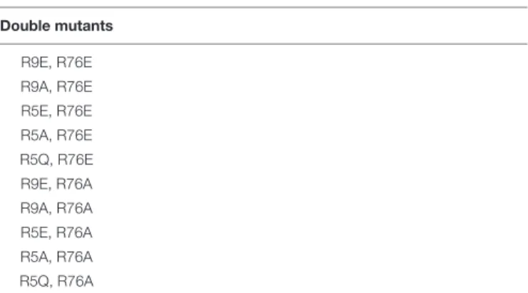
TABLE 3 | IL-21 residues selected for substitution and generation of double mutants. Double mutants R9E, R76E R9A, R76E R5E, R76E R5A, R76E R5Q, R76E R9E, R76A R9A, R76A R5E, R76A R5A, R76A R5Q, R76A
in the absence of any attenuation WT IL-21 fusion protein can also potently suppress the alloresponse response. When we examined the behavior of anti-PD-1 × R76E variant, we observe a subtle but significant suppression of the response at higher concentrations. To determine thein vivo characteristics
of the anti-PD-1 × R76E variants, pharmacokinetic parameters (PK) were determined using cynomolgus monkeys. As shown in Figure 2D, both monomer and homodimer variants exhibit distinct PK profiles, with the monomer showing superior exposures and half-life. To address potential liabilities relating to non-specific signaling that could translate into immune suppression mediated through the action on dendritic cells, and to further explore the potential for using a monomeric format to improve pharmacokinetic properties, we proceeded to generate more attenuated IL-21 variants.
Design and Characterization of Dual
Amino Acid Substitution IL-21 Variants
With Reduced Off-Target Signaling
To further reduce non-specific IL-21 signaling, a second panel of molecules was constructed (Table 3). Using the known IL-21/IL-21R structure to help guide selection, single amino acid substitution variants with the greatest degree of attenuation, as determined using cell and binding assays were combined to create a panel of double mutant variants fused as a monomer or
Shen et al. Engineered IL-21 Muteins for Cancer
TABLE 4 | Summary of in vitro attributes of anti-PD-1 x IL-21 double muteins.
Hut78 PD-1 (–) Hut78 PD-1(+) PD-1 reporter Mixed lymphocyte
Interpolated Interpolated Interpolated culture Interpolated hulL-21R cylL-21R huPD-1 cyPD-1
EC50 (nm) EC50 (nm) EC50 (nm) EC50 (nm) KD (nM)* KD (nM)* KD (nM)* KD (nM)*
IgGl – – – – – – – – rhlL-21 0.003 0.002 – – 0.029 0.044 – – PD-1 mAb 2 – – 0.487 0.161 – – 0.90 1.33 PD-1 mAb 2 x R5Q:R76E homodimer >1000 1.1 0.367 0.518 >300 >300 0.68 1.27 PD-1 mAb 2 x R5Q:R76E monomer >1000 0.28 0.809 0.249 >300 >300 0.56 1.28 PD-1 mAb 2 x R9E:R76A homodimer >1000 4.42 0.308 0.625 >300 >300 0.71 1.42 PD-1 mAb 2 x R9E:R76A monomer >1000 0.78 0.503 0.241 >300 >300 0.90 1.61 ∗
Sensograms are shown in Supplementary Figure S7.
FIGURE 3 | Characterization of IL-21 variants with dual amino acid substitutions. (A) Activity of fusion proteins and free WT IL-21 in Hut78 PD-1(–) cells (left panel) and engineered Hut78 PD-1 (+) cells (right panel) monitored using STAT3 phosphorylation (AlphaLISA) as a surrogate measure of IL-21 activity. (B) Potency of fusion proteins and parental mAb in blocking PD-1/L1 interaction monitored using Promega PD-1/L1 bioassay. (C) Activity of free WT IL-21, dual amino acid substation variants or parental mAb in mixed lymphocyte cultures of alloreactive pan-T cells and dendritic cells. (D–E) CTLs derived from PBMCs from Donor 1 under indicated priming conditions. Seven days post co-culture, proportions of (D) CMV antigen specific CTLs and (E) Ki67 (left-panel), PD-1 (middle-panel) and L-selectin (CD62L, right-panel) was analyzed by FACS analysis. CTLs primed under different conditions were isolated and cocultured with peptide pulsed melanoma cells for 36 h to examine (F) cytotoxicity against peptide-loaded melanoma cells, determined by measuring luciferase activity and (G) CTL IFN-gamma production. Experiments in (F) and (G) were conducted in triplicates and the error bars represent SEM. P-values were calculated using one-way Anova with a Tukey’s multiple comparison test. P-values: ***< 0.001, ** < 0.01.
homodimer to the C-terminus heavy chain of a bivalent anti-PD-1 antibody (Table 3 and Supplementary Figure S5A). A subset of the double mutant variants was evaluated for binding to IL-21R (Table 4 and Supplementary Figure S7). Consistent with the greater degree of attenuation, we were unable to establish KD values for the interaction between fusion protein(s) and
IL-21R, and we determined that these values are higher than the top concentration in the assay (300 nM), as such relative attenuation as compared with free WT cytokine is estimated to
be >1000 fold for these more attenuated molecules (Table 4 and Supplementary Figure S7). We next tested cell activity assays using a smaller subset of the double mutant constructs (Figure 3 and Supplementary Figures S5b,c). According to our hypothesis, cell association of fusion proteins in which IL-21/IL-21R association has been disrupted can be restored through binding of the mAb domain to a cell surface receptor allowing for the stabilized interaction between IL-21 and IL-21R. In line with our hypothesis, double muteins demonstrate a high degree
fimmu-11-00832 May 7, 2020 Time: 11:29 # 10
Shen et al. Engineered IL-21 Muteins for Cancer
of attenuation (>1000 fold as compared with free WT IL-21 cytokine) for STAT3 activation in cells devoid of PD-1 expression (Figure 3A and Supplementary Figure S6). Activity can be restored in cells engineered to express cell surface PD-1, but still partially attenuated as compared with free wildtype cytokine (Figure 3A and Table 4). We also confirmed that the fusion proteins consisting of the more attenuated IL-21 variants retain the ability to block the PD-1/PD-L1 interaction (Figure 3B and
Table 4). We next proceeded to test if additional attenuation could protect against non-specific activation of bystander IL-21R expressing APCs in a mixed culture system using alloreactive T cells that respond to antigen peptide complexes presented by dendritic cells (Figure 3C and Table 4). In contrast to recombinant WT IL-21 cytokine, which completely suppressed the alloresponse, the fusion proteins have similar activity to the parental anti-PD-1 mAb (Figure 3C). Our data suggest that in the absence of PD-1 expression the more attenuated fusion proteins fail to activate bystander cells expressing IL-21R in these conditions, and in the context of an alloresponse the fusion proteins have only limited signaling in trans allowing for the preservation of DC function. We next tested, the impact of PD-1 × IL-2PD-1 fusion proteins on the differentiation and effector function of cytotoxic T cells (CTL) derived from PBMCs. For this we tested the activity of PD-1 × IL-21 fusion protein using two CMV seropositive donors across four independent experiments (Figures 3D–G and a second independent donor Supplementary
Figure S8). Antigen specific CTLs were generated by co-culturing of peripheral blood mononuclear cells (PBMCs) and autologous peptide-loaded DCs in the presence of a PD-1 × IL-21 fusion protein and for comparison, PD-1 mAb or untreated cell were used as controls. After 7 days of co-culture with DCs, cell surface and intracellular markers of T cell proliferation and activation were monitored on antigen-specific CD8+ T cells. We report that T cell priming in the presence of PD-1 × IL-21 fusion protein gives rise to a mild but reproducible increase in the frequency of antigen-specific CD8+ T cells as compared to untreated control or PD-1 mAb treatment groups (Figure 3D and Supplementary Figure S8A). The increase in the frequency of antigen-specific cells was not correlated to increase in proliferation as the percentage of antigen-specific CD8+ T cells expressing Ki67 was equivalent across all treatment groups. We examined two further cell surface markers, namely PD-1 and L-selectin (CD62L), as markers of T cell activation and differentiation, respectively. Similar proportions of PD-1 positive T cells were observed across all treatment groups and was consistent across independent donors (Figure 3E and
Supplementary Figure S8A). We also monitored L-selectin a marker enriched on naïve and memory T cells. Previously it has been reported that IL-21 promotes the acquisition of alternative effector phenotype with increased L-selectin (48). We report that priming of T cells in the presence of PD-1 x IL-21 fusion protein leads to an increase in proportion of L-selectin (CD62L) positive CTLs as compared to PD-1 mAb and untreated treatment groups (Figure 3E and Supplementary Figure S8A). These data suggest that in the presence of PD-1 × IL-21 CTLs can acquire an effector phenotype but retain the naïve marker L-selectin. We next examined effector function of differentiated CTLs by
co-culturing CTLs together with peptide-loaded tumor cells. We report that CTLs conditioned with PD-1 × IL-21 fusion protein demonstrated superior cytotoxicity and IFN-gamma production as compared to untreated control or those primed together with a PD-1 mAb (Figures 3F–G and Supplementary Figure S8). We extended our studies to examine the effect of PD-1 × IL-21 fusion proteins on cytotoxicity of differentiated effector cells (Supplementary Figure S9). For these studies, to more faithfully mimic the clinical setting, in which fusion protein is expected to augment pre-existing immune responses, we used in vitro differentiated mature CTL lines. These were activated with a combination of CD3/28 beads (to mimic a chronic activation conditions) together with either anti-PD-1 mAb or fusion protein after which the CTLs were co-incubated with peptide pulsed PD-L1 (+) cancer cells. Our data suggest that under these conditions, CTLs treated with fusion protein have superior effector functions including cytotoxicity and IFN-gamma production versus anti-PD-1 mAb (Figures 3D–F and Supplementary Figures S8, S9).
In vivo Characterization of Dual Amino
Acid Substitution IL-21 Variants With
Improved Pharmacokinetic Properties
and Superior Efficacy in vivo in an
Anti-PD-1 Refractory Setting
We next wanted to extend our observations to understand pharmacokinetic properties of the more attenuated dual amino-acid substitution IL-21 fusion proteins. For this we used monomer fusions proteins because of their superior PK properties; groups of animals were dosed with fusion protein or parental mAb and PK parameters were calculated. The results as shown in Figure 4A suggest that attenuated cytokine variants have substantially improved PK properties as compared to first generation anti-PD-1 × R76E mutein (Figure 2). We extended our observations to explore in vivo activity of our fusion proteins. Since human IL-21 does not cross-react with mouse IL-21R and in the absence of an appropriate mouse surrogate molecule, we decided to implement a humanized mouse system; for this we used humanized mice, which were engrafted with human (PD-L1+) melanoma cells (SKMEL-30-Luc) engineered to express a model antigen (CMV-SKMEL-30-Luc, expressing peptide antigen derived from cytomegalovirus, CMV) and either a human-mouse chimeric PD-1 mAb, with a variable domain recognizing human PD-1 and a constant Fc-region from mouse IgG1, or a fusion protein consisting of the same parent PD-1 mAb and a monomeric variant of human IL-21 R9E:R76A (Figure 4B and Supplementary Table S1). On the same day as tumor engraftment, mice received adoptively transferred antigen (CMV)-specific CTLs, which we confirmed, demonstrate potent in vitro cytotoxicity against the antigen-expressing cancer cells (Figure 3F). In this model, the failure of tumor reactive CTLs to control cancer growth leads to development of progressive tumors which are palpable by day 17. Therapeutic administration (into mice with ∼100 mm3 established tumors) with an isotype control antibody or an anti-PD-1 mAb failed to resolve the disease or have any discernable impact on tumor growth, establishing this tumor model as both