Publisher’s version / Version de l'éditeur:
Vous avez des questions? Nous pouvons vous aider. Pour communiquer directement avec un auteur, consultez la première page de la revue dans laquelle son article a été publié afin de trouver ses coordonnées. Si vous n’arrivez pas à les repérer, communiquez avec nous à PublicationsArchive-ArchivesPublications@nrc-cnrc.gc.ca.
Questions? Contact the NRC Publications Archive team at
PublicationsArchive-ArchivesPublications@nrc-cnrc.gc.ca. If you wish to email the authors directly, please see the first page of the publication for their contact information.
https://publications-cnrc.canada.ca/fra/droits
L’accès à ce site Web et l’utilisation de son contenu sont assujettis aux conditions présentées dans le site LISEZ CES CONDITIONS ATTENTIVEMENT AVANT D’UTILISER CE SITE WEB.
International Journal of Immunopathology and Pharmacology, 32, 2018-07-17
READ THESE TERMS AND CONDITIONS CAREFULLY BEFORE USING THIS WEBSITE. https://nrc-publications.canada.ca/eng/copyright
NRC Publications Archive Record / Notice des Archives des publications du CNRC : https://nrc-publications.canada.ca/eng/view/object/?id=69aae75b-efa8-457f-b1c0-6e95b17e9793 https://publications-cnrc.canada.ca/fra/voir/objet/?id=69aae75b-efa8-457f-b1c0-6e95b17e9793
Archives des publications du CNRC
This publication could be one of several versions: author’s original, accepted manuscript or the publisher’s version. / La version de cette publication peut être l’une des suivantes : la version prépublication de l’auteur, la version acceptée du manuscrit ou la version de l’éditeur.
For the publisher’s version, please access the DOI link below./ Pour consulter la version de l’éditeur, utilisez le lien DOI ci-dessous.
https://doi.org/10.1177/2058738418787739
Access and use of this website and the material on it are subject to the Terms and Conditions set forth at
Bicyclic eremophilane-type petasite sesquiterpenes potentiate
peroxisome proliferator–activated receptor γ activator–mediated
inhibition of dendritic cells
https://doi.org/10.1177/2058738418787739 International Journal of
Immunopathology and Pharmacology Volume 32: 1–15
© The Author(s) 2018 Reprints and permissions:
sagepub.co.uk/journalsPermissions.nav DOI: 10.1177/2058738418787739 journals.sagepub.com/home/iji
Creative Commons Non Commercial CC BY-NC: This article is distributed under the terms of the Creative Commons Attribution-NonCommercial 4.0 License (http://www.creativecommons.org/licenses/by-nc/4.0/) which permits non-commercial use, reproduction and distribution of the work without further permission provided the original work is attributed as specified on the SAGE and Open Access pages (https://us.sagepub.com/en-us/nam/open-access-at-sage).
Background
Sesquiterpenes have been known to have anti-inflammatory activity in a variety of settings, showing inhibitory effects on nitric oxide produc-tion in lipopolysaccharide (LPS)-activated mouse macrophages.1–8 Some sesquiterpenes inhibit
inflammation by targeting dendritic cell (DC) mat-uration and activation. For example, a sesquiter-pene glycoside isolated from Kandelia candel
Bicyclic eremophilane-type petasite
sesquiterpenes potentiate peroxisome
proliferator–activated receptor
γ activator–mediated inhibition
of dendritic cells
Narcy Arizmendi1 , Chenjie Hou2, Fujiang Guo2, Yiming Li2 and Marianna Kulka1,3
Abstract
Dendritic cell (DC) activation induces expression of co-stimulatory surface molecules, as well as migration into secondary lymphoid organs, where they activate naïve T-cells. A family of plant derivatives, eremophilane-type petasite sesquiterpenes, can regulate the immune system through DC targeting due to their anti-inflammatory effects. Peroxisome proliferator– activated receptor gamma (PPARγ) is involved in inhibition of inflammatory responses and induction of DCs to acquire a mucosal phenotype. Since mucosal DCs are central in innate immune responses, we hypothesized that eremophilane-type petasite sesquiterpenes exerted their anti-inflammatory effects by inhibiting DC maturation and activation through PPARγ. This study assessed the bicyclic eremophilane-type petasite sesquiterpene compounds Fukinone and 10βH-8α,12-Epidioxyeremophil-7(11)-en-8β-ol (ZYFDC21 and ZYFDC22) in the maturation and activation of mouse DC. We measured surface expression of co-stimulatory molecules by flow cytometry and cell-free supernatant cytokine production upon lipopolysaccharide stimulation by enzyme-linked immunosorbent assays (ELISAs) in the presence or absence of PPARγ agonists. DCs were generated from C57BL/6 mice bone marrow cells and harvested. Cells were exposed to bicyclic eremophilane-type petasite sesquiterpenes ZYFDC21 or ZYFDC22 in the presence or absence of synthetic PPARγ agonists (GW1929 and TGZ) or the natural PPARγ ligand 15d-PGJ2, followed by overnight activation with LPS. We observed differences in the
upregulation of surface expression of CD86, along with TNF, IL-6, and IL-12p70 released by DCs stimulated with LPS, when using combinations of bicyclic eremophilane-type petasite sesquiterpenes ZYFDC21 or ZYFDC22, and PPARγ agonists, in particular the PPARγ ligand 15d-PGJ2. Our results indicate that bicyclic eremophilane-type petasite sesquiterpenes ZYFDC21
or ZYFDC22 inhibit maturation and activation of DC, and this activity is augmented upon PPARγ activation. Keywords
inflammation, peroxisome proliferator–activated receptor gamma, plant derivatives, transcription factor Date received: 5 December 2017; accepted: 6 June 2018
1 Nanotechnology Research Center, National Research Council Canada, Edmonton, AB, Canada
2 School of Pharmacy, Shanghai University of Traditional Chinese Medicine, Shanghai, China
3 Department of Medical Microbiology & Immunology, University of Alberta, Edmonton, AB, Canada
Corresponding author:
Marianna Kulka, Nanotechnology Research Center, National Research Council Canada, 11421 Saskatchewan Drive NW, Edmonton, AB T6G 2M9, Canada.
Email: Marianna.kulka@nrc-cnrc.gc.ca
inhibited pro-inflammatory cytokine production from LPS-stimulated bone marrow–derived DCs,9
and micheliolide, a sesquiterpene lactone, inhibits the production of interleukin-6 (IL-6) and tumor necrosis factor (TNF) from LPS-stimulated pri-mary DCs.10 While some examples of the
anti-inflammatory effects of sesquiterpene on DCs have been demonstrated, the molecular targets of spe-cific sesquiterpenes and their interactions with endogenous inflammatory signaling pathways are unknown.
One possible target of sesquiterpenes in many inflammatory cells is the peroxisome proliferator– activated receptor (PPAR) pathway, which plays an important role in several cellular functions, including maturation and differentiation. PPARs were initially identified as receptors that controlled physiological responses to dietary intake of fatty acids.11,12 Three
PPAR subtypes have been identified, alpha, delta and gamma, and are ligand-activated nuclear receptors which can be activated by polyunsaturated fatty acids, eicosanoids, and various synthetic ligands. PPAR gamma (PPARγ) is primarily expressed in adi-pose tissue and, to a lesser extent, in the colon, immune system, and the retina. PPARγ was first identified as a regulator of adipogenesis, but also plays an important role in cellular and adipocyte dif-ferentiation, insulin sensitization, glucose metabo-lism, atherosclerosis, and cancer.13 It has been shown
that PPARγ ligands have anti-inflammatory effects on mast cells, monocytes, macrophages, and DC, by modulating expression of co-stimulatory and adhe-sion molecules, altering their phenotype and leading to an impaired expression of pro-inflammatory cytokines/chemokine factors involved in T-cell acti-vation and recruitment.14–18
Several sesquiterpenes or terpenoid-like com-pounds have been shown to either directly activate PPARγ or to modify its response to other ligands. For example, odoratin, an undecanortriterpenoid from Chromolaena odorata, moderately activates PPARγ;19 tirotundin and tagitinin A, both
sesquit-erpene lactones, transactivate PPARγ-dependent promoters, including PPARγ response element (PPRE), small heterodimer partner (SHP), and
ABCA1 gene promoters in dose-dependent
man-ner,20 and artemisinic acid, the quintessential
ses-quiterpene, reduces expression of PPARγ in human adipose tissue-derived mesenchymal stem cells.21
Altogether, these data suggest that sesquiterpenes may similarly influence DC function through the
PPARγ pathway. Recently, our group isolated two novel eremophilane-type sesquiterpene com-pounds from Petasites tatewakianus Kitam.2 We
hypothesized that these novel sesquiterpenes would inhibit DC maturation and activation, and that this activity would be augmented in the pres-ence of a PPARγ agonist. In this study, we demon-strate, for the first time, that the novel bicyclic eremophilane-type petasite isolated sesquiterpenes have the ability to efficiently inhibit DC matura-tion and activamatura-tion, and this inhibimatura-tion is potentiat-edby the synthetic, as well as naturally occurring, nuclear peroxisome proliferator-activated receptor γ agonists.
Materials and methods
Plant material
Bicyclic sesquiterpenes Fukinone (ZYFDC21), and 10βΗ-8α,12-Epidioxyeremophil-7(11)-en-8β-ol (ZYFDC22), were isolated and purified from rhizome of P. tatewakianus, at the School of Pharmacy, Shanghai University of Traditional Chinese Medicine as previously described.22
Generation of bone marrow DCs from C57BL/6 mice
Female C57BL/6 mice (6–10 weeks old) were obtained from The Jackson Laboratory. All mice were treated according to protocols approved by the University of Alberta Animal Care and Use Committee. Bone marrow–derived DCs (BmDC) were generated using a standard protocol with little modification.23 Briefly, bone marrow was flushed
dispersed and collected from femurs and tibias of female C57BL/6 mice, passed through a 70 µm nylon mesh, and suspended in bone marrow–derived DC-complete media (RPMI 1640 containing 5 mM 4-(2-hydroxyethyl)-1-piperazineethanesulfonic acid (HEPES), 50 U Pen/Strep, 2 mM glutamine, 50 µM 2-ME, 50 mM gentamycin sulfate, and 10% fetal bovine serum (FBS)) in the presence of granulocyte-macrophage colony-stimulating factor (GM-CSF) and IL-4 (10 ng/mL; PeproTech, Rocky Hill, NJ, USA) and cultured in tissue culture dishes (Thermo Fisher, Carlsbad, CA, USA) in a humidified atmos-phere of 5% CO2 in air at 37oC. All media
compo-nents, except for GM-CSF and IL-4, were obtained from Gibco (Carlsbad, CA, USA). During culture, half of the media was replaced on days 3 and 6. On
day 8, BmDC were harvested, and their morphology was confirmed by optical microscopical analysis (Supplementary Figure 1).
Effect of sesquiterpenes and PPARγ agonists on BmDC
Initially, 0.2 × 10⁶BmDC/mL were deposited,
per well, in a 12-well plate and incubated with either eremophilane-type petasite sesquiterpene, ZYFDC21 (50 µM) and ZYFDC22 (25 µM), in the presence or absence of synthetic PPARγ ago-nists troglitazone (TGZ, 5 µM or 10 µM; Sigma Aldrich Canada, Oakville, ON, Canada) or N-(2-benzoylphenyl)-O-(2-(methyl-2-pyridinylamino) ethyl)-L-tyrosine (GW1929, 40 µM; Cayman Chemical, Ann Arbor, MI, USA) or the physio-logically relevant PPARγ natural ligand 15d- PGJ2-15deoxy-Δ12,14-Prostaglandin J2 (15d-PGJ2,
0.5 µM, or 5 µM; Cayman Chemical). Cells were then incubated for 20 h at 37°C and 5% CO2 and
viability was assessed by trypan blue exclusion (Gibco). Cells were exposed to the petasite ses-quiterpenes and synthetic and natural PPARγ ligands for 3 h and treated with LPS for 24 h, and BmDC were >90% viable after treatment (Supplementary Figure 2). In order to determine whether the PPARγ pathway and/or the petasite sesquiterpenes were involved in the maturation and activation of DCs, BmDC were treated with each petasite sesquiterpenes (ZYFDC21 or ZYFDC22) or PPARγ agonists for 3 h at 37°C and 5% CO2, with or without LPS (10 ng/mL)
over-night stimulation. BmDC stimulated with LPS or complete media alone were included as positive and negative controls, respectively. Cell-free supernatants from the different conditions were collected and stored at −20°C for cytokine analy-sis with commercial ELISAs. Cells were fixed for 5 min in 2% formaldehyde, suspended in cold 1% bovine serum albumin (BSA)-flow Buffer (0.05% sodium azide, 0.1% BSA in phosphate-buffered saline (PBS)), incubated overnight at 4°C, and analyzed by flow cytometry.
Flow cytometry of BmDC
After stimulation, 1 × 10⁵ BmDC were incubated
with their respective conjugated antibodies for 60 min at 4°C and washed twice. Data from 30,000 cells were collected by a CytoFlex flow cytometer
(Beckman Coulter, Brea CA, USA) and VersaComp antibody capture beads (Beckman Coulter, Brea CA, USA) were included to generate a compensa-tion matrix. Data analysis was performed using the FloJo V10 LLC software (Ashland, OR, USA). Gating was initially defined based on side scatter (SSC) versus forward scatter (FSC), BmDC posi-tive gating was determined using an APC-labeled Armenian Hamster anti-mouse CD11c (BD Pharmingen, San Diego, CA, USA). CD11c+ sub-population was then analyzed by the expression of CD80 and CD86 surface molecules with a FITC-Armenian Hamster IgG Anti-Mouse CD80 (Affymetrix eBioscience, Santa Clara, CA, USA) and APC-Rat anti-mouse CD86 antibodies (BD Pharmingen, San Diego, CA, USA) and compared to their respective isotype controls. Results were expressed as the median of fluorescence intensity (MFI) ± standard error of the median (SEM).
Cytokine release analysis
Levels of TNF, IL-6, and IL-12p70 released in the cell-free supernatants were quantified using com-mercial enzyme-linked immunosorbent assay (ELISA) according to the instructions of the ELISA Kits (Affymetrix eBiosciences). Results were expressed as means ± SEM.
Statistical analysis
Experiments were performed in triplicate, with BmDC obtained from at least three biological rep-licates (n ⩾ 3). Values are expressed as mean ± SEM. All statistical analyses were performed using GraphPad Prism statistical (GraphPad, Sand Diego, CA, USA). Statistical differences in the mean val-ues among treatment groups were determined by using a one-way analysis of variance (ANOVA) test with post hoc analysis with Tukey’s multiple comparison tests. In all cases, a value for P < 0.05 was considered statistically significant.
Results
PPARγ activation inhibits DC maturation
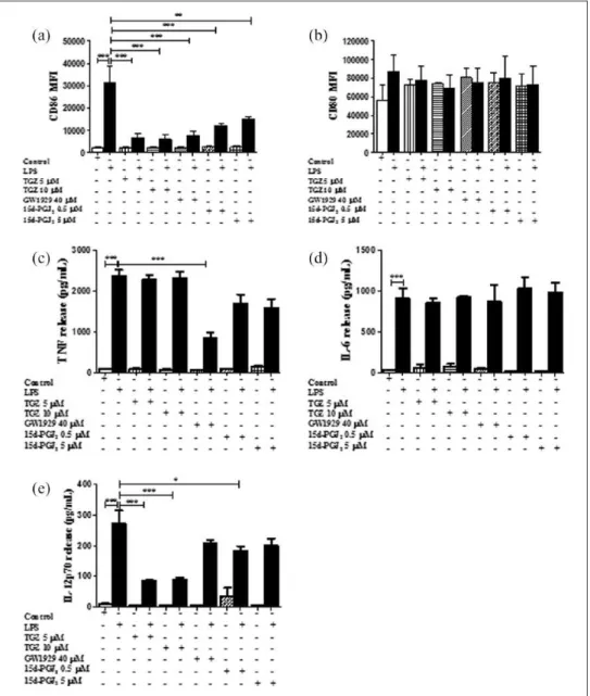
In order to determine whether PPARγ agonists modified the maturation of DC, we first analyzed the surface expression of the maturation markers CD80 and CD86 on BmDC by flow cytometry. Figure 1(a) shows that BmDC exposed to different
concentrations of synthetic PPARγ agonists TGZ (5 and 10 µM) or GW1929 (40 µM) presented CD86 MFI levels similar to control untreated cells; yet LPS stimulation induced upregulation of the co-stimulatory molecule CD86 on BmDC (MFI 31431 ± 7316, n = 5). Interestingly, when the BmDC were pretreated with the synthetic PPARγ agonists TGZ (5 or 10 µM) or GW1929 (40 µM) for 3 h fol-lowed by 20 h stimulation with LPS, there was a significant 75%–80% inhibition in the CD86 sur-face expression on BmDC compared to the LPS stimulation alone (MFI 6563 ± 1938 and MFI 5989 ± 2072 for TGZ 5 and 10 µM and 7459 ± 2317 for GW1929, respectively; n = 5). Expression of CD80- and CD86-positive BmDC after each treat-ment is shown in the suppletreat-mentary Figure 3 as percentage values. We also examined the expres-sion of CD80 after BmDC were exposed to the synthetic PPARγ agonists, followed by LPS over-night stimulation. We observed a 12%–20% inhibi-tion in the expression of CD80 when cells were pretreated with TGZ or GW1929 (Figure 1(b)).
Natural PPARγ ligand 15d-PGJ2 modulates DC maturation
The cyclopentenone metabolite of PGJ2,
15d-PGJ2, is a naturally occurring derivative of
pros-taglandin D2 (PGD2) and has been shown to
directly activate PPARγ24–26 BmDC were
preincu-bated with 15d-PGJ2 (0.5 or 5 µM) for 3 h, and as
shown in Figure 1(a), 15d-PGJ2 alone had no
effect on the expression of CD86 at either of the concentrations tested. 15d-PGJ2 treatment for 3 h
significantly decreased LPS-induced expression of CD86 by 60% and 50% (MFI 12,166 ± 1138 at 0.5 µM and 15147 ± 1376 at 5 µM; n = 3). CD80 surface expression did not reach statistical differ-ence (9%–17% for 0.5 and 5 µM; Figure 1(b), n = 3). BmDC stimulated with LPS or complete media were included as positive and negative controls, respectively.
PPARγ activation promotes the inhibition of BmDC cytokine secretion. We analyzed the effects of the activation of PPARγ on the cytokine secre-tion of TNF, IL-6, and IL-12p70 released in the cell-free supernatant of BmDC, after 3 h treatment with TGZ (5 or 10 µM) or GW1929 (40 µM) by commercial ELISAs. As shown in Figure 1(c), pre-treatment with GW1929 significantly inhibited release of TNF (about 65% ± 5% compared to
LPS). However, under the same conditions, BmDC release of IL-6 was unaffected by treatment with the PPARγ agonists, compared to LPS stimulation alone (Figure 1(d)). IL-12p70, the bioactive iso-form of the cytokine, was also evaluated in the cell-free supernatants of BmDC exposed to 5 and 10 µM TGZ with and without LPS stimulation. We found that TGZ significantly inhibited (68% ± 1% and 66% ± 2%, respectively) IL-12p70 production, as shown in Figure 1(e).
PPARγ ligation skews BmDC cytokine response
We were interested in studying the response of BmDC to the treatment with the natural PPARγ ligand 15d-PGJ2 (0.5 and 5 µM), and we found that
BmDC treated for 3 h with 15d-PGJ2 plus LPS
inhibited TNF release by 29% ± 9% and 33% ± 9% at 0.5 and 5 µM, respectively; however, this inhibi-tion was not statistically significant (Figure 1(c)). IL-12p70 showed a 33% ± 5% significant inhibi-tion at 0.5 µM (Figure 4(c)).
Bicyclic petasite eremophilane-type
sesquiterpenes potentiate the effects of PPARγ agonists on BmDC maturation and activation
Petasite sesquiterpenes have been shown to have anti-inflammatory activity in a variety of settings. We sought to assess the effects of two petasite ere-mophilane-type sesquiterpene compounds Fukinone (ZYFDC21) and 10βH-8α,12-Epidioxyeremophil-7(11)-en-8β-ol (ZYFDC22) isolated from the rhi-zome of P. tatewakianus on the maturation and activation of BmDCs. To evaluate the cytotoxic effects of the bicyclic compounds, we performed dose-response assays with several cell lines, using the XTT assay kit (Roche, data not shown). We selected sub-toxic doses of ZYFDC21 (50 µM) and ZYFDC22 (25 µM) and further evaluated their cyto-toxic effects on BmDC after 1, 3, 24 and 48 h incu-bation, measuring viability by trypan blue exclusion (supplementary Figure 2). BmDC viability was ⩾95% under all tested conditions, and therefore, these concentrations were used for all experiments.
There is evidence that some sesquiterpenes exert anti-diabetic, anti-carcinogenic, and anti-inflamma-tory effects, mediated by the PPARγ pathway.20 We
sought to identify whether the sesquiterpenes would inhibit BmDC maturation and activation and whether this inhibitory activity would be augmented by the
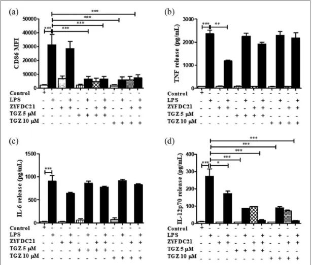
presence of a PPARγ synthetic agonist. For that pur-pose, BmDC were exposed to the synthetic PPARγ agonists TGZ (5 or 10 µM) or GW1929 (40 µM) in the presence or absence of the petasite sesquiterpe-nes ZYFDC21 (50 µM) or ZYFDC22 (25 µM) for 3 h, followed by the overnight LPS stimulation. First, we assessed the effects of bicyclic sesquiterpenes on BmDC maturation by flow cytometry. The presence of the sesquiterpenes ZYFDC21 (Figure 2(a)) and
ZYFDC22 (Figure 3(a)) alone induced a modest increase in CD86 expression (MFI 6985 ± 1825 and 6882 ± 1274, respectively) compared to control, untreated BmDC (MFI 2073 ± 510). Exposure to a combination of ZYFDC21 (50 µM) plus the syn-thetic PPARγ agonist TGZ (5 or 10 µM) or GW1929 (40 µM) followed by overnight LPS stimulation resulted in a significant downregulation in CD86 surface expression (up to 80% compared to LPS
lev-Figure 1. PPARγ activation inhibits DC maturation and cytokine secretion. CD86 surface expression in BmDC after 3 h
preincubation with PPARγ agonists TGZ (5 and 10 µM), GW1929 (40 µM), and 15d-PGJ2 (0.5 and 5 µM) followed by LPS overnight
stimulation was examined by flow cytometry. (a) CD86 results and (b) CD80 results are expressed as differences in MFI ± SEM between LPS activated-BmDC and PPARγ agonists ± LPS (n = 3–5; **P < 0.01 and ***P < 0.001). BmDC were incubated with PPARγ agonist TGZ (5 and 10 µM), GW1929 (40 µM), and 15d-PGJ2 (0.5 and 5 µM) for 3 h ± LPS overnight stimulation, and cell-free
supernatants were collected and tested for (c) TNF, (d) IL-6, or (e) IL-12p70 release by ELISA. Results are from cytokines released from LPS-activated BmDC and cells treated with PPARγ agonists ± LPS. Data are expressed as means ± SEM (n = 3–5; *P < 0.05 and ***P < 0.001).
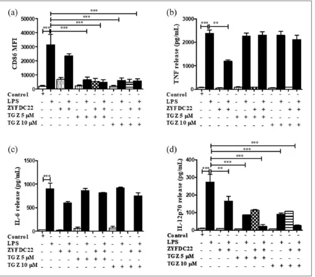
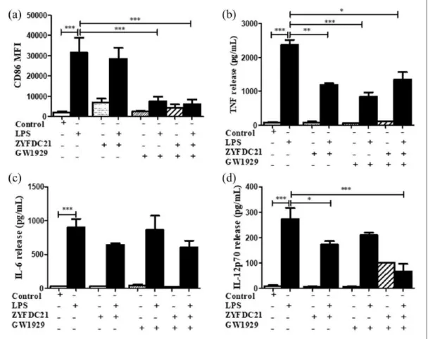
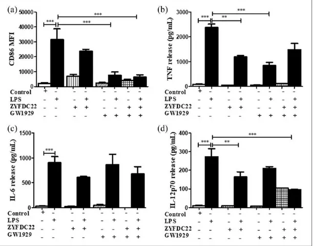
els alone; Figures 2(a) and 4(a)). The combination of ZYFDC22 (25 µM) with TGZ (5 or 10 µM) or GW1929 (40 µM) followed by overnight LPS activation resulted in a significant 85% and 80% inhibition, respectively, in CD86 surface expression (Figures 3(a) and 5(a)).
Second, we evaluated the activation of the immune response by the presence of the pro-inflammatory mediators TNF, IL-6, and IL-12p70 released in the cell-free supernatants of BmDC treated for 3 h with the synthetic PPARγ ago-nists (TGZ or GW1929), in combination with the petasite sesquiterpene (ZYFDC21 or ZYFDC22) and followed by LPS overnight stimulation. BmDC treated with the sesquiterpene ZYFDC21
and synthetic PPARγ agonist GW1929 and stimu-lated with LPS showed a significant 43% ± 9% inhibition on TNF release (Figure 4(b)). IL-6 released values showed that this cytokine was not significantly affected by any of the tested treatments.
In these studies, we found that IL-12p70, the bioactive isoform of IL-12, seems to be involved in the PPARγ/petasine sesquiterpene pathway. BmDC exposed for 3 h to the sesquiterpenes ZYFDC21 or ZYFDC22 in combination with synthetic PPARγ agonists, followed by overnight stimulation with LPS showed ≥ 90% inhibition for either sesquiter-pene in combination with TGZ (5 and 10 µM). The same was true when BmDC were treated with
Figure 2. Bicyclic eremophilane-type petasite sesquiterpene ZYFDC21 potentiates the effects of PPARγ agonists on BmDC
maturation and activation. BmDC were pretreated with the eremophilane-type sesquiterpene ZYFDC21 ± PPARγ synthetic agonist TGZ (5 or 10 µM) for 3 h, followed by LPS overnight stimulation, and DC were collected, fixed, and analyzed by flow cytometry. (a) Differences in the CD86 surface expression are represented as differences in MFI ± SEM between LPS activated-BmDC and the combination of ZYFDC21 + TGZ ± LPS (n = 5; ***P < 0.001). Cytokine release by BmDC treated with ZYFDC21 plus PPARγ agonist TGZ (5 and 10 µM) for 3 h ± LPS overnight stimulation, and cell-free supernatants were collected and tested for (b) TNF, (c) IL-6, or (d) IL-12p70 release by ELISA. Results are from cytokines released from LPS-activated BmDC, compared to BmDC cells treated with ZYFDC21, plus TGZ ± LPS. Data are expressed as means ± SEM (n = 5; *P < 0.05, **P < 0.01, and ***P < 0.001).
ZYFDC21 or ZYFDC22 in combination with GW1929, followed by stimulation with LPS, where we observed a substantial inhibition of IL-12p70 release (75%±10% for ZYFDC21 and 64±1% for ZFDC22) (Figure 4(d)).
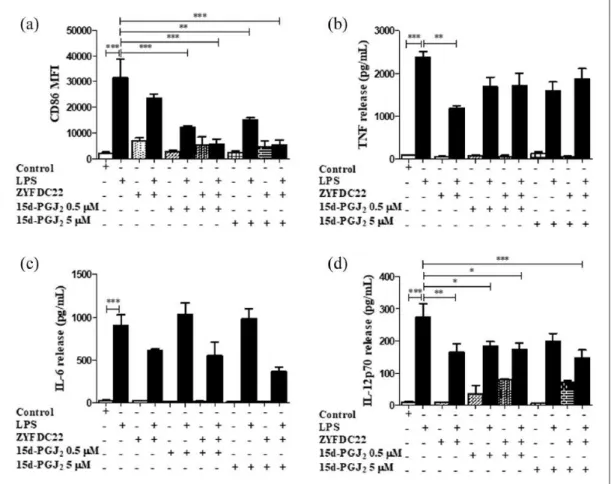
Petasite sesquiterpenes potentiate the effects of PGD2 metabolites on BmDC maturation and activation
BmDC were exposed to the natural PPARγ ligand (15d-PGJ2, 0.5 and 5 µM) for 3 h in combination
with eremophilane sesquiterpenes ZYFDC21 and ZYFDC22, followed by LPS overnight
stimulation. We observed a robust inhibition in the expression of the co-stimulatory molecule CD86. Cells incubated in the presence of 15d-PGJ2 and sesquiterpenes ZYFDC21 or ZYFDC22 plus LPS showed downregulation in more than 78% ± 6% and 82% ± 6% in the expression of CD86 with 0.5 and 5 µM of 15d-PGJ2 (Figures
6(a) and 7(a) respectively). Also, when BmDC were exposed to petasite sesquiterpenes ZYFDC21 or ZYFDC22 plus LPS, there was a 50% ± 2% inhibition in the TNF release (Figures 6(b) and 7(b)). In addition, the combination of the natural PPARγ ligand pre-treatment plus petasite sesquiterpenes and LPS overnight
Figure 3. Bicyclic eremophilane-type petasite sesquiterpene ZYFDC22 potentiate the effects of PPARγ agonists on BmDC
maturation and activation. BmDC were pretreated with the eremophilane-type sesquiterpene ZYFDC22 plus PPARγ synthetic agonist TGZ (5 or 10 µM) for 3 h, followed by LPS overnight stimulation, and DC were collected, fixed, and analyzed by flow cytometry. Differences in the (a) CD86 surface expression are represented as differences in MFI ± SEM between LPS activated– BmDC and ZYFDC22 + TGZ + LPS (n = 5; ***P < 0.001). Cytokine release by BmDC preincubated with ZYFDC22 and PPARγ agonist TGZ for 3 h ± LPS overnight stimulation, and cell-free supernatants were collected and tested for (b) TNF, (c) IL-6, or (d) IL-12p70 release by ELISA. Results are from cytokines released from LPS-activated BmDC, compared to cells treated with ZYFDC22 + TGZ ± LPS. Data are expressed as means ± SEM (n = 5; *P < 0.05, **P < 0.001, and ***P < 0.001).
stimulation promoted a modest IL-6 inhibition of 9% and 18% for ZYFDC21 (Figure 6(c)), but a solid 39, 60% IL-6 inhibition following a inhibitory trend with the PPARγ ligand (Figure 7(c)), not reaching statistical significance. The natural PPARγ agonist, 15d-PJG2, seems to have an additive inhibitory effect on the release of IL-12, which was significantly inhibited with the combination of sesquiterpene ZYFDC21 and 0.5 µM (54% ± 10%) or 5 µM (56% ± 10%) after LPS stimulation (Figure 6(d)). However, when we tested the sesquiterpene ZYFDC22 in combi-nation of 15d-PJG2, we found a 30% ± 10%
IL-12p70 inhibition at 0.5 µM and a 50% ± 10% IL-12p70 inhibition at 5 µM 15d-PJG2,
respec-tively (Figure 7(d)). Both sesquiterpenes were
able to significantly inhibit around 35% of IL-12 release after LPS stimulation.
Discussion
DCs are the most potent antigen-presenting cells (APCs) and are involved in initiating the adaptive immune responses. The expression of surface adhe-sion (CD40) and co-stimulatory (CD80 and CD86) and major histocompatibility complex (MHC) class-II molecules promote the contact between DCs and T-cells, while co-stimulatory molecules signal T-cells to proliferate and differentiate.27 In
the mouse, CD86 is the main activation marker of bone marrow–derived DC, being strongly upregu-lated after maturation, while CD80 expression is
Figure 4. Petasite sesquiterpene ZYFDC21 in combination with GW1929 inhibited CD86 and cytokine secretion on BmDC.
BmDC were pretreated with the eremophilane-type sesquiterpene ZYFDC21 plus PPARγ agonist GW1929 (40 μM) for 3 h, followed by LPS overnight stimulation, and DC were collected, fixed, and analyzed by flow cytometry. (a) Differences in the CD86 surface expression are represented as differences in MFI ± SEM between LPS-activated BmDC and ZYFDC21 + TGZ + LPS (n = 5; ***P < 0.001). Cytokine release by BmDC preincubated with ZYFDC21 plus GW1929 ± LPS overnight stimulation, and cell-free supernatants were collected and tested for (b) TNF, (c) IL-6, or (d) IL-12p70 release by ELISA. Results are from cytokines released from LPS-activated BmDC and compared to cells treated with ZYFDC21 plus GW1929 ± LPS. Data are expressed as mean ± SEM (n = 5; *P < 0.05, **P < 0.001, and ***P < 0.001).
less relevant for murine DC.27,28 In this context, our
studies demonstrated that LPS stimulation upregu-lates CD80 and CD86 expression on BmDC. We also confirmed that the use of synthetic (TGZ and GW1929), as well as natural (15d-PGJ2) PPARγ
ligands decreased the expression of CD86 after LPS stimulation. Furthermore, we observed a sig-nificant reduction on TNF cytokine release with the GW1929, while IL-12p70 production was attenu-ated by TGZ and PGJ2. These results were similar
to previous reports.14–16,29–31
Advances in the investigation of plant–derived chemicals used in alternative medicine for the treatment of several chronic diseases have shown that Petasite species from petasite sesquiterpenes possess anti-inflammatory properties.32–34 Due to
their anti-inflammatory effects mediated via leu-kotriene synthesis inhibition, sesquiterpenes have
been used for the treatment of inflammatory dis-eases such as arthritis, migraine, as well as asthma and allergy.35–37
The anti-inflammatory effect of Petasite ses-quiterpenes is based on their ability to block Ca2+ channels, decreasing intracellular Ca2+ concentration, inhibiting leukotriene B4 and cysteinyl leukotrienes synthesis in eosinophils and neutrophils.21,33,37–43 The active components
are sesquiterpene esters of the eremophilane type, and their bioactivity is attributed to peta-sine and isopetapeta-sine.44,45 Studies by Shimoda
et al.46 showed that the effective constituent in
the extract of Petasites japonicus was petasine, which had inhibitory effects on leukotriene syn-thesis39 and bronchoconstriction.47 Another
ere-mophilane-type sesquiterpene ketone, namely, Fukinones (1 and 3),48 exerted suppressive
Figure 5. Petasite sesquiterpene ZYFDC22 in combination with GW1929 inhibited CD86 and cytokine secretion on BmDC.
BmDC were pretreated with the eremophilane-type sesquiterpene ZYFDC22, plus GW1929 (40 µM) for 3 h, followed by LPS overnight stimulation, DC were collected, fixed and analyzed by flow cytometry. Differences in the CD86 surface expression (a), are represented as differences in MFI ± SEM between LPS activated-BmDC and ZYFDC22 + GW1929 + LPS (n = 5; ***P < 0.001). Cytokine release by BmDC preincubated with ZYFDC22 plus GW1929 ± LPS overnight stimulation, cell–free supernatants were collected and tested for TNF (b), IL-6 (c), or IL-12p70 (d) release by ELISA. Results are from cytokines released from LPS-activated BmDC, compared to cells treated with ZYFDC22 plus GW1929 ± LPS. Data are expressed as means ± SEM (n = 5; **P < 0.001 and ***P < 0.001).
mechanisms in a type I hypersensitivity model in rats and IgE-sensitized RBL-2H3 cells through inhibition of smooth muscle constriction and inhibition of degranulation, leukotriene release, and TNF production by mast cells.33,46 In this
context, Lee et al.33 also reported anti-allergic
and anti-inflammatory effects of several com-pounds obtained from plants of the petasites genus in an ovalbumin-induced asthma model; the molecule Bakkenolide B isolated from P.
japonicus inhibited the migration of eosinophils,
macrophages, and lymphocytes to the lungs. Previous studies from our lab showed that differ-ent extracts of petasites could inhibit type I and type IV hypersensitivity in mouse models of homogeneous and heterogeneous passive cuta-neous anaphylaxis.49
Elegant studies by Lin,20 evaluated the
agonis-tic activity of the sesquiterpene lactones tirotundin and targitining A, isolated from Tithonia
diversifo-lia against PPARs. For this, they used a transient
transfection reporter assay with HepG2 cells and found that tirotundin and targitining A transacti-vate PPARγ-dependent promoters, including PPRE (PPARγ response element), SHP, and ABCA1, and that both sesquiterpene lactones transactivate PPARγ by binding directly to the PPARγ ligand-binding domain (LBD). In this con-text, Zhang et al.19 showed that five isolated
com-ponents of C. odorata, another plant used in traditional medicine for their anti-inflammatory activities, had a transactivation effect on PPARγ. More recent studies by Wu et al.50 demonstrated
by luciferase reporter assay in HEK293 cells that
Figure 6. Petasite sesquiterpene ZYFDC21 potentiates the effects of PGD2 metabolites on BmDC maturation and activation.
BmDC were pretreated with the eremophilane-type petasite sesquiterpene ZYFDC21 ± the natural PPARγ ligand 15d-PGJ2 0.5
or 5 µM for 3 h, followed by LPS overnight stimulation, and BmDC cells were collected, fixed, and analyzed by flow cytometry. (a) Differences in the CD86 surface expression are represented as differences in MFI between LPS-activated BmDC and the combination of ZYFDC21 + 15d-PGJ2 + LPS (n = 5; **P < 0.01 and ***P < 0.001). BmDC were pretreated with ZYFDC21 ± 15d-PGJ2
(0.5 or 5 µM) for 3 h, followed by LPS overnight stimulation, and cell-free supernatants were collected and cytokine release was analyzed by ELISA. Differences in (b) TNF, (c) IL-6, or (d) IL-12p70 released are represented as differences between LPS activated-BmDC and ZYFDC21 plus 15d-PGJ2 ± LPS (n = 5; *P < 0.05, **P < 0.01, and ***P < 0.001).
the bicyclic sesquiterpene trans-caryophyllene aroma compound of plant foods and teas activates PPARα through direct interaction with the LBD of PPARα. However, trans-caryophyllene showed no binding affinity for or transactivation of PPARγ.
However, Adachi et al.45 demonstrated that
peta-sin derived from P. japonicus activates adenopeta-sine monophospahte–activated protein kinase (AMPK) in the liver, skeletal muscle, and adipose tissue of mice, via phosphorylation of AMPK. AMPK acti-vation enhanced the transcription of the prolifera-tor-activated receptor-γ coactivator-1α (PGC-1α), which regulates the genes involved in energy metabolism including mitochondrial biogenesis.
Our studies showed that the eremophilane-type sesquiterpenes ZYFDC21 and ZYFDC22 increased
CD80 and CD86 surface expression in non-stimu-lated BmDC. In contrast, when the cells were pre-treated with sesquiterpene ZYFDC21 (Fukinone) followed by LPS, we observed a decrease in CD86 surface expression. This inhibition was amplified up to 80% by the presence of the PPARγ agonists TGZ, GW1929, and 15d-PGJ2. The inhibitory
effect was also observed when we used ZYFDC22 (10βH-8α,12-Epidioxyeremophil-7(11)-en-8β-ol) in combination with TGZ, GW1929, or 15d-PGJ2, followed by LPS stimulation, where 85% of CD86 surface expression was significantly inhibited by the PPARγ agonists. The absence of co-stimulatory molecules, such as CD86, influences DC function, altering their maturation and varying the expres-sion of the necessary signals required for the
Figure 7. Petasite sesquiterpene ZYFDC22 potentiate the effects of PGD2 metabolites on BmDC maturation and activation.
BmDC were pretreated with the eremophilane-type petasite sesquiterpene ZYFDC22 ± the natural PPARγ ligand 15d-PGJ2 0.5
or 5 µM for 3 h, followed by LPS overnight stimulation, and BmDC cells were collected, fixed, and analyzed by flow cytometry. (a) Differences in the CD86 surface expression are represented as differences in MFI between LPS-activated BmDC and the combination of ZYFDC22 + 15d-PGJ2 + LPS (n = 5; **P < 0.01 and ***P < 0.001). BmDC were pretreated with ZYFDC22 ± 15d-PGJ2
(0.5 or 5 µM) for 3 h, followed by LPS overnight stimulation, and cell-free supernatants were collected and cytokine release was analyzed by ELISA. Differences in (b) TNF, (c) IL-6, or (d) IL-12p70 released are represented as differences between LPS-activated BmDC compared to ZYFDC22 plus 15d-PGJ2 ± LPS (n = 5; *P < 0.05, **P < 0.01, and ***P < 0.001).
activation and differentiation of naïve T-cells into type 1 (IL-12 and interferon gamma (IFNγ)) or type 2 (IL-4, IL-5, and IL-10) cytokine-producing cells. In this context, our studies showed that both sesquiterpenes ZYFCD21 and ZYFDC22 inhibited the secretion of the soluble factors TNF and IL-12p70 after LPS stimulation. These results are comparable to those obtained by Uchi et al.,51 who
demonstrated that the sesquiterpene lactone par-thenolide inhibited DC maturation and cytokine secretion induced by LPS.
The level of IL-12 secreted by DC induced by microbial pathogens, such as LPS, during the immunological synapse is a key factor in the out-come of immune responses. IL-12 is a critical Th1-skewing cytokine that elicits IFNγ production by T-cells and by natural killer (NK) cells,52 favoring
a Th2/Th3 response and inhibiting T cell recruit-ment.53 PPARγ is an important modulator on B and
T lymphocytes as well as DC14,54,55 and PPARγ
ligands include a class of antidiabetic drugs, thia-zolidinediones (TZD); as well as naturally pro-duced PGD2 and its metabolite 15-dideoxy-Δ PGJ2
(15d-PGJ2), which associate irreversibly to the
receptor through covalent binding, mediating their effects by activation of PPARγ-dependent and independent pathways.17,56 Prostaglandins’
pro-duction results in activation of PPARγ-mediated transcription, leading to the inhibition of differen-tiation, migration, and cytokine secretion by anti-gen-presenting cells, such as DC or macrophages, hence affecting the priming and effector functions of T lymphocytes.17
Our studies showed for the first time that DC exposed to the PPARγ ligands TGZ, GW1929, and 15d-PGJ2 in the presence of these novel isolated
bicyclic eremophilane-type petasite sesquiterpenes ZYFDC21 and ZYFDC22 followed by LPS stimu-lation exhibited a significant reduction (up to 95%) in the production of the bioactive isoform of IL-12 (IL-12p70). In this regard, it has been documented that 15d-PGJ2 abrogates IL-12 production by
directly inhibiting the function of IκB kinase (IKK), therefore preventing the translocation of nuclear factor-κB (NF-κB) to the nucleus.30,57–59 Our results
showed that sesquiterpenes reduced LPS-induced DC maturation and inhibited TNF and IL-6 release, as well as the production of the bioactive isoform of IL-12p70, presumably through the direct activation of PPARγ. Since it is well known that the transcrip-tion factor NF-κB plays a key role in the activatranscrip-tion
of PPARγ in the inflammatory response, it would be of interest to determine whether sesquiterpenes bind directly to the PPARγ receptors, thereby inhib-iting IKK, and to analyze the downstream signaling cascades that would prevent the translocation of NF-κB to the nucleus, interfering with the inflammatory response. In summary, our results suggest that the novel Fukinone and 10βH-8α,12-Epidioxyeremophil-7(11)-en-8β-ol sesquiterpenes derived from P. tatewakianus inhibit the maturation of DC, as well as the production of TNF, IL-6, and IL-12p70 after LPS stimulation. These events seem to be mediated and potentiated by the activation of PPARγ. Petasite sesquiterpenes are compounds with significant potential value for the treatment of inflammatory disorders.
Acknowledgements
The authors would like to thank Dr Ramses Ilarraza for his comments on the manuscript revision.
Declaration of conflicting interests
The author(s) declared no potential conflicts of interest with respect to the research, authorship, and/or publication of this article.
Funding
This study received financial support from the Canadian Institutes of Health Research (CIHR), the National Research Council of Canada (Intramural Funding), and the Natural Science Foundation of China (Canada-China Joint Health Research Initiatives Grant 81261120567).
ORCID iD
Narcy Arizmendi https://orcid.org/0000-0003-4562- 7432
References
1. Zhao JH, Shen T, Yang X, et al. (2012) Sesquiterpenoids from Farfugium japonicum and their inhibitory activ-ity on NO production in RAW264.7 cells. Archives of
Pharmacal Research 35: 1153–1158.
2. Hou C, Kulka M, Zhang J, et al. (2014) Occurrence and biological activities of eremophilane-type ses-quiterpenes. Mini-Reviews in Medicinal Chemistry 14: 664–677.
3. Qin ZB, Zhang J, Wu XD, et al. (2014) Sesquiterpenoids from Tussilago farfara and their inhibitory effects on nitric oxide production. Planta Medica 80: 703–709.
4. Cheng Z, Zhao J, Liu D, et al. (2016) Eremophilane-type sesquiterpenoids from an acremonium sp. fungus
isolated from deep-sea sediments. Journal of Natural
Products 79: 1035–1047.
5. Zhang M, Zhao JL, Liu JM, et al. (2016) Neural anti-inflammatory sesquiterpenoids from the endophytic fungus Trichoderma sp. Xy24. Journal of Asian
Natural Products Research 19: 651–658.
6. Zhao H, Peng Q, Han Z, et al. (2016) Three new ses-quiterpenoids and one new sesquiterpenoid derivative from Chinese eaglewood. Molecules 21: 281.
7. Wu XD, Ding LF, Tu WC, et al. (2016) Bioactive sesquiterpenoids from the flowers of Inula japonica.
Phytochemistry 129: 68–76.
8. Gao S, Xia G, Wang L, et al. (2017) Sesquiterpenes from Curcuma wenyujin with their inhibitory activi-ties on nitric oxide production in RAW 264.7 cells.
Natural Product Research 31: 548–554.
9. Dat le D, Thao NP, Tai BH, et al. (2015) Chemical constituents from Kandelia candel with their inhibi-tory effects on pro-inflammainhibi-tory cytokines produc-tion in LPS-stimulated bone marrow-derived dendritic cells (BMDCs). Bioorganic & Medicinal Chemistry
Letters 25: 1412–1416.
10. Qin X, Jiang X, Wang Y, et al. (2016) Micheliolide inhibits LPS-induced inflammatory response and pro-tects mice from LPS challenge. Scientific Reports 6: 23240.
11. Issemann I and Green S (1990) Activation of a mem-ber of the steroid hormone receptor superfamily by peroxisome proliferators. Nature 347: 645–650. 12. Guerre-Millo M, Gervois P, Raspe E, et al. (2000)
Peroxisome proliferator-activated receptor alpha activators improve insulin sensitivity and reduce adi-posity. Journal of Biological Chemistry 275: 16638– 16642.
13. Willson TM, Lambert MH and Kliewer SA (2001) Peroxisome proliferator-activated receptor gamma and metabolic disease. Annual Review of Biochemistry 70: 341–367.
14. Gosset P, Charbonnier AS, Delerive P, et al. (2001) Peroxisome proliferator-activated receptor gamma activators affect the maturation of human mono-cyte-derived dendritic cells. European Journal of
Immunology 31: 2857–2865.
15. Nencioni A, Grunebach F, Zobywlaski A, et al. (2002) Dendritic cell immunogenicity is regulated by peroxi-some proliferator-activated receptor gamma. Journal
of Immunology 169: 1228–1235.
16. Kock G, Bringmann A, Held SA, et al. (2011) Regulation of dectin-1-mediated dendritic cell acti-vation by peroxisome proliferator-activated receptor-gamma ligand troglitazone. Blood 117: 3569–3574. 17. Appel S, Mirakaj V, Bringmann A, et al. (2005)
PPAR-gamma agonists inhibit toll-like receptor-mediated activation of dendritic cells via the MAP kinase and NF-kappaB pathways. Blood 106: 3888–3894.
18. Zhang G, Yang J, Li P, et al. (2014) Rosiglitazone inhibits HMC-1 cell migration and adhesion through a peroxisome proliferator-activated receptor gamma-dependent mechanism. Iranian Journal of Allergy,
Asthma and Immunology 13: 11–18.
19. Zhang ML, Irwin D, Li XN, et al. (2012) PPARγ ago-nist from Chromolaena odorata. Journal of Natural
Products 75: 2076–2081.
20. Lin HR (2012) Sesquiterpene lactones from Tithonia
diversifolia act as peroxisome proliferator-activated
receptor agonists. Bioorganic & Medicinal Chemistry
Letters 22: 2954–2958.
21. Lee J, Kim MH, Lee JH, et al. (2012) Artemisinic acid is a regulator of adipocyte differentiation and C/EBP δ expression. Journal of Cellular Biochemistry 113: 2488–2499.
22. Hou CJ, Wang GY, Li YM, et al. (2016) Sesquiterpenes from the roots of Petasites
tatewa-kianus Kitam. Chinese Traditional Patent Medicine
38: 1970–1974.
23. Labeur MS, Roters B, Pers B, et al. (1999) Generation of tumor immunity by bone marrow-derived dendritic cells correlates with dendritic cell maturation stage.
Journal of Immunology 162: 168–175.
24. Ide T, Egan K, Bell-Parikh LC, et al. (2003) Activation of nuclear receptors by prostaglandins. Thrombosis
Research 110: 311–315.
25. Forman BM, Tontonoz P, Chen J, et al. (1995) 15-Deoxy-delta 12, 14-prostaglandin J2 is a ligand for the adipocyte determination factor PPAR gamma.
Cell 83: 803–812.
26. Scher JU and Pillinger MH (2005) 15d-PGJ2: The anti-inflammatory prostaglandin? Journal of Clinical
Immunology 114: 100–109.
27. Inaba K, Witmer-Pack M, Inaba M, et al. (1994) The tissue distribution of the B7–2 costimulator in mice: Abundant expression on dendritic cells in situ and during maturation in vitro. Journal of Experimental
Medicine 180: 1849–1860.
28. Inaba K, Inaba M, Romani N, et al. (1992) Generation of large numbers of dendritic cells from mouse bone marrow cultures supplemented with granulocyte/ macrophage colony-stimulating factor. Journal of
Experimental Medicine 176: 1693–1702.
29. Thieringer R, Fenyk-Melody JE, Le Grand CB, et al. (2000) Activation of peroxisome proliferator-activated receptor gamma does not inhibit IL-6 or TNF-alpha responses of macrophages to lipopolysac-charide in vitro or in vivo. Journal of Immunology 164: 1046–1054.
30. Faveeuw C, Gosset P, Bureau F, et al. (2003) Prostaglandin D2 inhibits the production of inter-leukin-12 in murine dendritic cells through multiple signaling pathways. European Journal of Immunology 33: 889–898.
31. Marion-Letellier R, Butler M, Dechelotte P, et al. (2008) Comparison of cytokine modulation by natu-ral peroxisome proliferator-activated receptor gamma ligands with synthetic ligands in intestinal-like caco-2 cells and human dendritic cells—Potential for die-tary modulation of peroxisome proliferator-activated receptor gamma in intestinal inflammation. American
Journal of Clinical Nutrition 87: 939–948.
32. Ziment I and Tashkin DP (2000) Alternative medi-cine for allergy and asthma. Journal of Allergy and
Clinical Immunology 106: 603–614.
33. Lee KP, Kang S, Park SJ, et al. (2013) Anti-allergic and anti-inflammatory effects of bakkenolide B iso-lated from Petasites japonicus leaves. Journal of
Ethnopharmacology 148: 890–894.
34. Sok DE, Oh SH, Kim YB, et al. (2006) Neuroprotection by extract of Petasites japonicus leaves, a traditional vegetable, against oxidative stress in brain of mice challenged with kainic acid. European Journal of
Nutritio 45: 61–69.
35. Johnson ES, Kadam NP, Hylands DM, et al. (1985) Efficacy of feverfew as prophylactic treatment of migraine. British Medical Journal 291: 569–573. 36. Heptinstall S, White A, Williamson L, et al. (1985)
Extracts of feverfew inhibit granule secretion in blood platelets and polymorphonuclear leucocytes. Lancet 1: 1071–1074.
37. Dong L, Qiao H, Zhang X, et al. (2013) Parthenolide is neuroprotective in rat experimental stroke model: Downregulating NF-κB, phospho-p38MAPK, and caspase-1 and ameliorating BBB permeability.
Mediators of Inflammation 2013: 370804.
38. Fiebich BL, Grozdeva M, Hess S, et al. (2005)
Petasites hybridus extracts in vitro inhibit COX-2 and
PGE2 release by direct interaction with the enzyme and by preventing p42/44 MAP kinase activation in rat primary microglial cells. Planta Medica 71: 12–19. 39. Bickel D, Roder T, Bestmann HJ, et al. (1994)
Identification and characterization of inhibitors of peptido-leukotriene-synthesis from Petasites hybridus.
Planta Medica 60: 318–322.
40. Thomet OA, Wiesmann UN, Schapowal A, et al. (2001) Role of petasin in the potential anti-inflamma-tory activity of a plant extract of Petasites hybridus.
Biochemical Pharmacology 61: 1041–1047.
41. Resnati M, Pallavicini I, Wang JM, et al. (2002) The fibrinolytic receptor for urokinase activates the G pro-tein-coupled chemotactic receptor FPRL1/LXA4R.
Proceedings of the National Academy of Sciences of the United States of America 99: 1359–1364.
42. Wu C, Chen F, Rushing JW, et al. (2006) Antiproliferative activities of parthenolide and golden feverfew extract against three human cancer cell lines.
Journal of Medicinal Food 9: 55–61.
43. Lee KP, Kang S, Noh MS, et al. (2015) Therapeutic effects of s-petasin on disease models of asthma and peritonitis. Biomolecules & Therapeutics 23: 45–52.
44. Chizzola R, Langer T and Franz C (2006) An approach to the inheritance of the sesquiterpene chemotypes within Petasites hybridus. Planta Medica 72: 1254– 1256.
45. Adachi Y, Kanbayashi Y, Harata I, et al. (2014) Petasin activates AMP-activated protein kinase and modulates glucose metabolism. Journal of Natural
Products 77: 1262–1269.
46. Shimoda H, Tanaka J, Yamada E, et al. (2006) Anti type I allergic property of Japanese butterbur extract and its mast cell degranulation inhibitory ingredi-ents. Journal of Agricultural and Food Chemistry 54: 2915–2920.
47. Ko WC, Lei CB, Lin YL, et al. (2000) Relaxant effects of petasins in isolated guinea pig trachea and their structure-activity relationships. Planta Medica 66: 650–652.
48. Naya Y and Kotake M (1967) The isolation of new oxetone derivatives from hop oil. Tetrahedron Letters 18: 1715–1716.
49. Zheng Q, Kong P, Wu X, et al. (2011) Experimental study of anti-allergy effects of bioactive fraction of Petasites japonicas. Journal of University of
Traditional Chinese Medicine 25: 79–82.
50. Wu C, Jia Y, Lee JH, et al. (2014) trans-caryophyllene is a natural agonistic ligand for peroxisome prolifer-ator-activated receptor-α. Bioorganic & Medicinal
Chemistry Letters 24: 3168–3174.
51. Uchi H, Arrighi JF, Aubry JP, et al. (2002) The sesquiterpene lactone parthenolide inhibits LPS- but not TNF-alpha-induced maturation of human monocyte-derived dendritic cells by inhibition of the p38 mitogen-activated protein kinase pathway.
Journal of Allergy and Clinical Immunology 110:
269–276.
52. Heufler C, Koch F, Stanzl U, et al. (1996) Interleukin-12 is produced by dendritic cells and mediates T helper 1 development as well as interferon-gamma production by T helper 1 cells. European Journal of Immunology 26: 659–668.
53. Asseman C, Mauze S, Leach MW, et al. (1999) An essential role for interleukin 10 in the function of regulatory T cells that inhibit intestinal inflamma-tion. Journal of Experimental Medicine 190: 995– 1004.
54. Gelman L, Fruchart JC and Auwerx J (1999) An update on the mechanisms of action of the peroxi-some proliferator-activated receptors (PPARs) and their roles in inflammation and cancer. Cellular and
55. Nencioni A, Wesselborg S and Brossart P (2003) Role of peroxisome proliferator-activated receptor gamma and its ligands in the control of immune responses.
Critical Reviews in Immunology 23: 1–13.
56. Shiraki T, Kamiya N, Shiki S, et al. (2005) Alpha,beta-unsaturated ketone is a core moiety of natural ligands for covalent binding to peroxisome prolifer-ator-activated receptor gamma. Journal of Biological
Chemistry 280: 14145–14153.
57. Ghosh S, May MJ and Kopp EB (1998) NF-kappa B and rel proteins: Evolutionarily conserved mediators
of immune responses. Annual Review of Immunology 16: 225–260.
58. Rossi A, Kapahi P, Natoli G, et al. (2000) Anti-inflammatory cyclopentenone prostaglandins are direct inhibitors of IkappaB kinase. Nature 403: 103– 108.
59. Straus DS, Pascual G, Li M, et al. (2000) 15-deoxy-delta 12,14-prostaglandin J2 inhibits multiple steps in the NF-kappa B signaling pathway. Proceedings of
the National Academy of Sciences of the United States of America 97: 4844–4849.