HAL Id: hal-03018741
https://hal-cnrs.archives-ouvertes.fr/hal-03018741
Submitted on 23 Mar 2021
HAL is a multi-disciplinary open access archive for the deposit and dissemination of sci-entific research documents, whether they are pub-lished or not. The documents may come from teaching and research institutions in France or abroad, or from public or private research centers.
L’archive ouverte pluridisciplinaire HAL, est destinée au dépôt et à la diffusion de documents scientifiques de niveau recherche, publiés ou non, émanant des établissements d’enseignement et de recherche français ou étrangers, des laboratoires publics ou privés.
in pancreatic beta cells and upregulated by stimulatory,
low concentrations of interleukin 1 β
Muhammad Saad Khilji, Danielle Verstappen, Tina Dahlby, Michala Cecilie
Burstein Prause, Celina Pihl, Sophie Emilie Bresson, Tenna Holgersen Bryde,
Phillip Alexander Keller Andersen, Kristian Klindt, Dusan Zivkovic, et al.
To cite this version:
Muhammad Saad Khilji, Danielle Verstappen, Tina Dahlby, Michala Cecilie Burstein Prause, Celina Pihl, et al.. The intermediate proteasome is constitutively expressed in pancreatic beta cells and upregulated by stimulatory, low concentrations of interleukin 1 β. PLoS ONE, Public Library of Science, 2020, 15 (2), pp.e0222432. �10.1371/journal.pone.0222432�. �hal-03018741�
The intermediate proteasome is constitutively
expressed in pancreatic beta cells and
upregulated by stimulatory, low
concentrations of interleukin 1
β
Muhammad Saad KhiljiID1,2, Danielle Verstappen1,3, Tina Dahlby1, Michala Cecilie Burstein Prause4, Celina Pihl
ID1, Sophie Emilie BressonID1, Tenna Holgersen BrydeID1, Phillip Alexander Keller Andersen1, Kristian Klindt1,
Dusan ZivkovicID5, Marie-Pierre Bousquet-Dubouch5, Bjo¨ rn Tyrberg6¤, Thomas Mandrup-PoulsenID1, Michal Tomasz MarzecID1*
1 Laboratory of Immuno-endocrinology, Inflammation, Metabolism and Oxidation Section, Department of
Biomedical Sciences, University of Copenhagen, Copenhagen, Denmark, 2 Department of Physiology, University of Veterinary and Animal Sciences, Lahore, Punjab, Pakistan, 3 Radboud Universiteit, Nijmegen, Netherlands, 4 Section for Beta-cell Biology, Department of Biomedical Sciences, University of Copenhagen, Copenhagen, Denmark, 5 Institut de Pharmacologie et de Biologie Structurale, Centre National de la Recherche Scientifique, Universite´ de Toulouse, Toulouse, France, 6 Department of Physiology, Institute of Neuroscience and Physiology, Sahlgrenska Academy, University of Gothenburg, Gothenburg, Sweden
¤Cardiovascular and Metabolic Diseases, Institute de recherches Servier, Suresnes, France
*Michal@sundk.ku.dk
Abstract
A central and still open question regarding the pathogenesis of autoimmune diseases, such as type 1 diabetes, concerns the processes that underlie the generation of MHC-presented autoantigenic epitopes that become targets of autoimmune attack. Proteasomal degrada-tion is a key step in processing of proteins for MHC class I presentadegrada-tion. Different types of proteasomes can be expressed in cells dictating the repertoire of peptides presented by the MHC class I complex. Of particular interest for type 1 diabetes is the proteasomal configura-tion of pancreaticβcells, as this might facilitate autoantigen presentation byβcells and thereby their T-cell mediated destruction. Here we investigated whether so-called inducible subunits of the proteasome are constitutively expressed inβcells, regulated by inflamma-tory signals and participate in the formation of active intermediate or immuno-proteasomes. We show that inducible proteasomal subunits are constitutively expressed in human and rodent islets and an insulin-secreting cell-line. Moreover, theβ5i subunit is incorporated into active intermediate proteasomes that are bound to 19S or 11S regulatory particles. Finally, inducible subunit expression along with increase in total proteasome activities are further upregulated by low concentrations of IL-1βstimulating proinsulin biosynthesis. These find-ings suggest that theβcell proteasomal repertoire is more diverse than assumed previously and may be highly responsive to a local inflammatory islet environment.
a1111111111 a1111111111 a1111111111 a1111111111 a1111111111 OPEN ACCESS
Citation: Khilji MS, Verstappen D, Dahlby T,
Burstein Prause MC, Pihl C, Bresson SE, et al. (2020) The intermediate proteasome is constitutively expressed in pancreatic beta cells and upregulated by stimulatory, low concentrations of interleukin 1β. PLoS ONE 15(2): e0222432.
https://doi.org/10.1371/journal.pone.0222432 Editor: Corentin Cras-Me´neur, University of
Michigan, UNITED STATES
Received: August 26, 2019 Accepted: January 21, 2020 Published: February 13, 2020
Peer Review History: PLOS recognizes the
benefits of transparency in the peer review process; therefore, we enable the publication of all of the content of peer review and author responses alongside final, published articles. The editorial history of this article is available here:
https://doi.org/10.1371/journal.pone.0222432 Copyright:© 2020 Khilji et al. This is an open access article distributed under the terms of the
Creative Commons Attribution License, which permits unrestricted use, distribution, and reproduction in any medium, provided the original author and source are credited.
Data Availability Statement: All relevant data are
within the manuscript and its Supporting Information files.
Introduction
The proteasome is a multi-subunit complex essential for the proteolytic degradation of cellular proteins and in the generation of specific sets of bioactive peptides [1] influencing a variety of cellular processes e.g. transcriptional regulation, signaling and the regulation of the cell cycle progression [2–5].
Proteasomal activity is executed by the proteolytic core, known as the 20S proteasome. It consists of a stack of four heptameric rings: two outerα and two inner β rings [1]. Theβ rings are composed of catalytically active subunits (β1, β2 and β5) that cleave peptide bonds at the C-terminal side of proteins [6] with caspase-, trypsin- and chymotrypsin-like activities, respec-tively [7,8]. The standard 26S proteasome contains a 19S regulatory cap that binds the polyu-biquitin chain, denatures the protein, and feeds it into the proteolytic core of the proteasome [9].
Standard proteasomes (s-proteasome) assembled withβ1, β2 and β5 subunits are ubiqui-tously expressed, but specialized proteasomes also exist and are constitutively expressed by e.g. immune cells [9], where they represent the dominant form. Formation of the proteolytic core of these specialized proteasomes involves substitution of the constitutively expressed catalytic β1, β2 and β5 subunits with the interferon (IFN)-γ-inducible β1i, β2i and β5i subunits (alterna-tively termed Psmb9/LMP2, Psmb10/MECL-1/LMP10 and Psmb8/LMP7, respec(alterna-tively) [6,10, 11]. The immune-proteasome (i-proteasome) has an alternative 20S catalytic core where all β-subunits are replaced by IFN-γ inducible β-β-subunits and where the 20S-associated 19S can be replaced by the 11S (also termed PA28αβ) proteasome regulator [9,12,13].
When standard and inducible subunits are present in cells, the latter are preferentially incorporated into newly produced 20S proteasomes [14,15]. Interestingly, co-expression of standard and inducibleβ subunits enables cells to assemble a variety of distinct 20S complexes, collectively referred to as intermediate proteasomes (int-proteasomes) [9]. The two most com-mon int-proteasomes are composed of two inner rings containing eitherβ1/β2/β5i or β1i/β2/ β5i. These int-proteasomes are not exclusive, as other combinations have been observed, including 20S proteasome with one constitutive (β1/ β2/ β5) and one immune (β1i/ β2i/ β5i) inner ring (also called asymmetric proteasomes, [16–18]).
Immune cells permanently and many other cells under conditions of oxidative stress, inflammation, cytokine stimulation, or viral and bacterial infection express and assemble i-and int-proteasomes [9,19]. Recently, induction of expression of such proteasomes upon exposure of human pancreatic islets and rat and mouse insulinoma cells to INFγ and β but not high concentrations of IL-1β, was reported [18,20]. Furthermore, int-proteasomes (but not i-proteasomes) are constitutively expressed in various cells, including liver, heart, kidney, lung or colon [16,21–24]. They constitute between 1% (heart) to 50% (liver) of the total proteasome pool [16,21,23,24].
The proteasomal composition in cells has broad implications, as proteasomes exhibit diverse substrate specificities. This affects the peptide repertoire generated for presentation on major histocompatibility complex (MHC) class I molecules [13,19,25], signal transduction via e.g nuclear factor kappa-light-chain-enhancer of activated B cells (NFκB) [26] and protein degradation e.g. of proinsulin [27].
The s-proteasome is known to improve glucose-stimulated insulin secretion [28], regulate intracellular proinsulin levels [27] or protect against lipotoxic endoplasmic reticulum stress [29]. However, the functions of i- and int-proteasomes are poorly defined. Importantly, consti-tutive expression of inducible proteasome subunits in pancreatic β cells has not been described,
but their induction upon INFγ and β treatment has been suggested to play a protective role against cytokine-induced apoptosis [20] and during antiviral responses [18].
Funding: This study was funded by The Punjab
Educational Endowment Fundhttps://www.peef. org.pk/(M.S.K), the Department of Biomedical Sciences at the University of Copenhagenhttps:// bmi.ku.dk/(T.D. and M.T.M.); the Augustinus Foundationhttps://augustinusfonden.dk/(T.D. and M.T.M.); EFSD/JDRF/Lilly European Programme in Type 1 Diabeteshttp://www.europeandiabetes foundation.org/, Vissing Fondenhttp://www.vissing fonden.dk/, Bjarne Jensen Fondenhttp://www. bjarnejensensfond.dk/, Poul og Erna Sehested Hansens Fond, no website, Eva og Hans Carl Holms Mindelegat https://haldguttenberg.dk/1-april/(M.T.M). The funders had no role in study design, data collection and analysis, decision to publish, or preparation of the manuscript.
Competing interests: The authors have declared
that no competing interests exist.
Abbreviations: i-proteasome, Immunoproteasome;
int-proteasome, Intermediate proteasome; s-proteasome, Standard proteasome;β1, beta subunit 1;β2, beta subunit 2; β5, beta subunit 5; β1i, inducible beta subunit 1; β2i, inducible beta subunit 2;β5i, inducible beta subunit 5; PSMB8, Proteasome subunit beta type-8 =β5i; PSMB9, Proteasome subunit beta type-9 =β1i; PSMB10, Proteasome subunit beta type-10 =β2i; HBSS, Hank’s balanced salt solution; IL-1β, Interleukin 1 β; INF, Interferon; MHC, Major histocompatibility complex.
Of special interest to type 1 and 2 diabetes pathogenesis is the constitutive profile of theβ cell proteasomes and their regulation. Type 1 diabetes (T1D) is an autoimmune disease, in which tolerance toβ cells is broken, with proinsulin serving as a major autoantigen. T1D is his-tologically characterized by pancreatic islet inflammation with increased levels of cytokines i.e. IL-1β, INF-γ/β and TNF-α, in the islet microenvironment [30]. Type 2 diabetes (T2D) arises when insulin secretion fails to meet demands mainly due to impaired insulin sensitivity, with β-cell oxidative and endoplasmic reticulum stress, lipotoxicity and glucotoxicity as conse-quences causing progressive loss ofβ cell functional mass [31]. All these cellular stresses induce an inflammatory response or are exacerbated by or associated with low-grade systemic inflam-mation via production of interleukin 1β (IL-1β) and IL-6 and recruitment and activation of innate immune cells [32,33]. As i- and int-proteasomes can modify e.g. signal transduction and MHC I peptide presentation, their constitutive and/or induced expression inβ cells by inflammatory cytokines is of high interest and therapeutic potential.
Here, we hypothesized thatβ cells constitutively express active non-standard proteasomes and that the expression is upregulated by innate inflammatory signals at low levels. We there-fore set out to analyze the composition of proteasomes in human and mouse islets as well as in the commonly usedβ-cell model INS-1E cell line in non-stimulated or cytokine-stimulated conditions. We report constitutive transcription and translation of inducible proteasome sub-units (β1i/ β2i/ β5i) in β-cells, albeit with lower expression levels compared to immune cell-lines. Of the inducible subunits,β5i is incorporated into active proteasomes in non-stimulated INS-1E cells, forming intermediate proteasomes that constitute 14% of total proteasomes in these cells. Furthermore, mRNA and protein expression of inducible subunits is upregulated bylow concentrations of IL-1β. β5i and β1i subunits were induced in all tested cellular models
whileβ2i was induced in mouse (but not human) islets and INS-1E cells. Consequently the composition and both constitutive and stimulated activity of proteasomes inβ cells has to be considered when investigating degradation mechanisms and antigen presentation on MHC I molecules of proinsulin and otherβ-cell proteins.
Materials and methods
Cell culture
The rat insulinoma INS-1E cell line, a gift from Claes Wollheim and Pierre Maechler, Univer-sity Medical Center, Geneva, Switzerland, was maintained as previously described [11]. The mouse insulinoma MIN6 cell line, was cultured in DMEM (Life Technologies, Nærum, Den-mark) with 25 mM glucose, supplemented with 10% FBS, 0.1% Penicillin/Streptomycin (P/S), 50 uMβ-mercaptoethanol and 2 mM L-glutamine. The mouse lymphocyte cell line A20, donated by Prof. Søren Buus, Department of Immunology and Microbiology, University of Copenhagen, Denmark, was cultured in RMPI-1640 (Life Technologies, Nærum, Denmark), containing 10% FBS, 1% P/S, 10 mM HEPES, 50 uMβ-mercaptoethanol and 4.5 g/L D-glu-cose. The human T lymphocyte cell line Jurkat, also from Prof. Buus, was cultured in RPMI-1640 with 10% FBS and 1% P/S. All cells were maintained at 37oC with 5% CO2. All cell-lines wereMycoplasma negative.
Animal Care
B6 C57BL/6NRJ mice were housed, handled and sacrificed according to Danish legislation for animal experimentation and with prior approval from the local animal ethics committee, issued by the Department of Experimental Medicine, University of Copenhagen. Animal han-dling and procedures were conducted by researchers with FELASA certification and super-vised by veterinarians.
Islet isolation and culture
Mouse islets were isolated by injection of LiberaseTM TL (Roche1, Hvidovre, Denmark) through the common bile duct to digest exocrine tissue. Islets were handpicked and either lysed immediately or cultured for 3–5 days in RPMI-1640 supplemented with 10% FBS and 1% P/S, at 37oC and 5% CO2. All data points represent separate islet collections (tested in tech-nical triplicates) and thus denote biological variability.
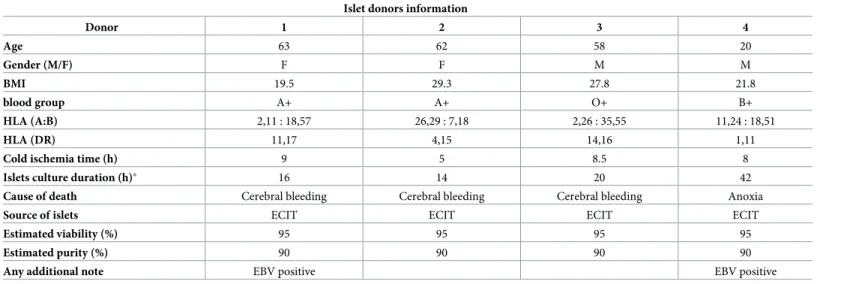
Human islets were isolated from healthy, heart-beating donors by the European Consor-tium for Islet Transplantation (ECIT) in Milan, Italy, with local ethical approval. The obtained islets were ~ 90% pure and no apparent difference in their quality was observed. Details on islet donors are included in theTable 1. Islets were cultured as previously described in [11].
Cytokine exposure
INS-1E cells were exposed to 10 ng/mL rat IFN-γ (R&D, Minnesota, USA) or 15 or150 pg/mL rat IL-1β (BD Bioscience, Lyngby, Denmark) or control medium for 24h. Human islets were exposed to 10 ng/ml human IFN-γ (BD Bioscience, New Jersey, USA) or 30 or 300 pg/ml rat IL-1β, while mouse islets were exposed to either 10 ng/mL rat IFN-γ or 50 or 300 pg/mL rat IL-1β or control medium, both for 24 hours prior to experiments.
Western blotting
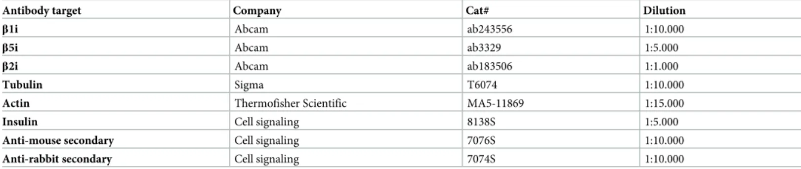
Prior to experiments cells or islets were lysed in lysis buffer, consisting of 100 mM Tris (pH 8.0), 30 mM NaCl, 10 mM KCl, 10 mM MgCl2, 2% NP-40, 20 mM iodoacetamide and protease inhibitor cocktail (Life Technologies, Nærum, Denmark). Protein concentrations were mea-sured using Bio-Rad Protein Assay Dye Reagent (Bio-Rad, Copenhagen, Denmark). Indicated amounts of proteins were loaded on Nu-Page 4–12% bis-tris gels (Thermo Fisher Scientific, Hvidovre, Denmark), and proteins were separated by SDS-PAGE. Gels were transferred to PVDF membranes using the iBLOT2 system (Thermo Fisher Scientific, Hvidovre, Denmark). Membranes were cut prior to incubation with primary antibodies (Table 2) overnight. Primary antibodies were diluted in 2% BSA in TBST (50 mM Tris pH 8, 150 mM NaCl, 0.1% Tween).
Table 1. Human islets donors information and islet preparations used during the investigation. Islet donors information
Donor 1 2 3 4 Age 63 62 58 20 Gender (M/F) F F M M BMI 19.5 29.3 27.8 21.8 blood group A+ A+ O+ B+ HLA (A:B) 2,11 : 18,57 26,29 : 7,18 2,26 : 35,55 11,24 : 18,51 HLA (DR) 11,17 4,15 14,16 1,11
Cold ischemia time (h) 9 5 8.5 8
Islets culture duration (h)� 16 14 20 42
Cause of death Cerebral bleeding Cerebral bleeding Cerebral bleeding Anoxia
Source of islets ECIT ECIT ECIT ECIT
Estimated viability (%) 95 95 95 95
Estimated purity (%) 90 90 90 90
Any additional note EBV positive EBV positive
�time from islets isolation to shipment
Membranes were blotted with appropriate secondary antibodies for 1 hour. Blots were devel-oped using chemiluminescence and captured using the Azure1Saphire Biomolecular Imager. Western blots were quantified using ImageJ software (v. 1.52a, [34]).
Proteasome activity
INS-1E, A-20 and Jurkat cells, human and mouse islets were plated in duplicates or triplicates in 96-well plates and treated with 50 nM ONX-0914, a selective inhibitor of theβ5i subunit activity (Selleck Chemicals, Rungsted, Denmark, IC50: ~10 nM forβ5i, [35]) or 2μM MG132, a broad proteasome inhibitor (Sigma-Aldrich, Søborg, Denmark) or control medium for 2 hours prior to experiments. Chymotrypsin-, trypsin- and caspase-like activity was measured through luminescent assay using commercially available Proteasome-GloTMAssay (Promega, Nacka, Sweden) according to the manufacturer’s protocol. Depicted data are averages of either technical duplicates or triplicates as indicated. The added trypsin-like, chymotrypsin-like and caspase-like activity is referred to as total proteasome activity.
Bulk mouse islet RNA Sequencing
Five hundred mouse islets were plated and exposed to IL-1β (50 pg/mL) for 10 days or left non-exposed for 10 days. Total mRNA was extracted from the islets by employing RNeasy 1 Micro Kit (Qiagen, Vedbæk, Denmark). Single-stranded, single-end sequencing libraries were generated using 35 ng of extracted RNA by means of TruSeq1 Stranded mRNA Library Prep (Illumina1, Copenhagen, Denmark), and library sequencing was done with the HiSeq 4000 System (Illumina1, Copenhagen, Denmark). Sequence files were drawn to the UCSC mouse genome NCB137/mm9. Further technical and analysis details in [36] and RNA-seq raw data are accessible here:https://www.ncbi.nlm.nih.gov/geo/query/acc.cgi?acc=GSE110691. In brief, expression levels of all genes were estimated by Cufflink (cufflinks v2.2.1,-p 6 -G $gtf_file— max-bundle-frags 1000000000) using only the reads with exact matches. Since specific mRNA levels were analyzed no correction for multiple testing was done. Results (RPKM) for the spe-cific genes of 3 independent experiments were analyzed by Student’s paired t-test, n = 3. RPKM for each gene is provided inTable 3.
Single-Cell RNA Sequencing of Pancreatic Islets
Each single-cell transcriptome was sequenced to *750,000 reads, sufficient for cell-type classi-fication. Islet cell subpopulations were analyzed forPSMB8, PSMB9 and PSMB10 genes
expression using published human islet single-cell sequencing data [37]. FastQ files were downloaded from ArrayExpress (accession: E-MTAB-5061). Data was analyzed with bcbio-nextgen (https://github.com/chapmanb/bcbio-nextgen), using the hisat2 algorithm [38] to
Table 2. Primary and secondary antibodies used during the investigation.
Antibody target Company Cat# Dilution
β1i Abcam ab243556 1:10.000
β5i Abcam ab3329 1:5.000
β2i Abcam ab183506 1:1.000
Tubulin Sigma T6074 1:10.000
Actin Thermofisher Scientific MA5-11869 1:15.000
Insulin Cell signaling 8138S 1:5.000
Anti-mouse secondary Cell signaling 7076S 1:10.000
Anti-rabbit secondary Cell signaling 7074S 1:10.000
align sequence reads to human genome version hg38 and uniquely aligned reads within RefSeq gene annotations were used to quantify gene expression with the Salmon algorithm [39]. Data is then expressed as log2 of counts per million (CPM). Only cells that passed the quality con-trol in the original study [37] were maintained for further analysis, and the cell type classifica-tion from the original study was also maintained.
Mass spectrometry for proteasome composition analysis
INS-1E cells were grown to 90% confluence in T175 flasks. The cells were washed with HBSS before incubation with pre-warmed culture media complemented with 0.1% formaldehyde for cross-linking for 15 minutes. Next, 125 mM glycine was added for 10 minutes at 37˚ C to quench the formaldehyde. The advantages of live cell cross-linking vs non-crosslinking step has been evaluated in [40]. The cells were then washed three times with HBSS and centrifuged, and pellets were stored at -80˚C for later proteasome composition analysis. Immuno-purifica-tion of the proteasomes from thein-vivo cross-linked lysates, was performed as previously
described [41]. Briefly, proteasomes were purified by incubating the lysates with CNBr sephar-ose beads (GE Healthcare) covalently bound to the antibody specific for theα2 subunit of the proteasome (MCP21) (100 mg of beads for 0.8 mg antibody), using 150 million cells per 50 mg of grafted beads. The supernatant was collected, and the beads were washed three times with 40 bead volumes of washing buffer (20 mM Tris-HCl pH 7.6, 1 mM EDTA, 10% glycerol, 150 mM NaCl, 0.1% NP-40, 2 mM ATP and 5 mM MgCl2). Finally, proteins were eluted with 0.5 ml of elution buffer (20 mM Tris-HCl pH 7.6, 1 mM EDTA, 10% glycerol, 3 M NaCl, 2 mM ATP and 5 mM MgCl2). Two additional cycles of purification were conducted, reincubating the collected supernatant with antibody-grafted beads. All fractions were pooled. LC-MS/MS analysis was performed as previously described [21,42]. Briefly, immuno-purified proteasome samples were precipitated with 20% trichloroacetic acid (TCA), washed with cold acetone and then denatured by boiling at 95˚C for 30 min in the Laemmli buffer, also reversing the cross-links [41]. Proteins were alkylated and concentrated on 12% acrylamide SDS-PAGE gel as a single band, which was cut and washed. Trypsin digestion was then performed overnight at 37˚C and the peptides were extracted from the gel. The digestion mixture was then dried in a Speed-Vac and resuspended with 2% acetonitrile, 0.05% trifluoroacetic acid. The peptide
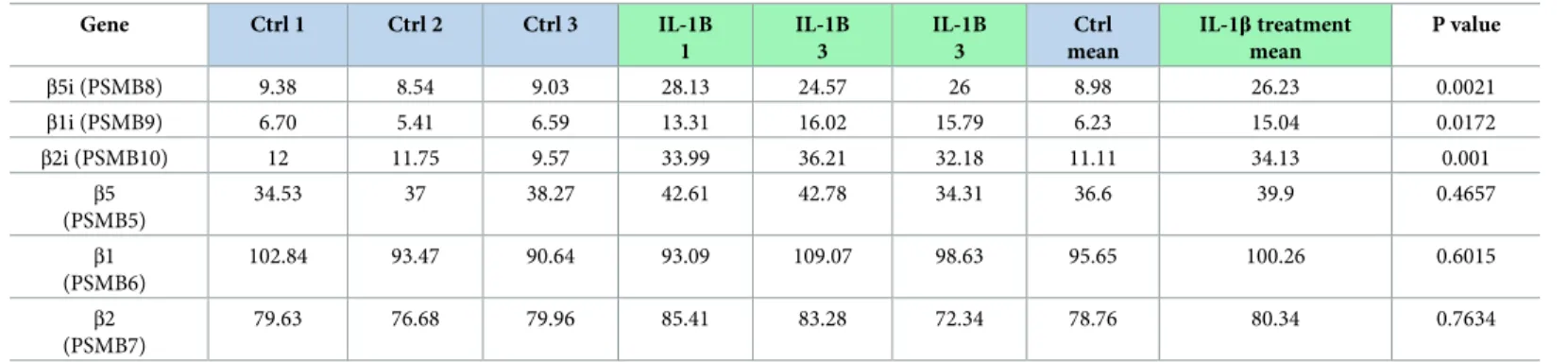
Table 3. Low concentrations of IL-1β induce β subunit mRNA expression in mouse islets. Gene Ctrl 1 Ctrl 2 Ctrl 3 IL-1B 1 IL-1B 3 IL-1B 3 Ctrl mean IL-1β treatment mean P value β5i (PSMB8) 9.38 8.54 9.03 28.13 24.57 26 8.98 26.23 0.0021 β1i (PSMB9) 6.70 5.41 6.59 13.31 16.02 15.79 6.23 15.04 0.0172 β2i (PSMB10) 12 11.75 9.57 33.99 36.21 32.18 11.11 34.13 0.001 β5 (PSMB5) 34.53 37 38.27 42.61 42.78 34.31 36.6 39.9 0.4657 β1 (PSMB6) 102.84 93.47 90.64 93.09 109.07 98.63 95.65 100.26 0.6015 β2 (PSMB7) 79.63 76.68 79.96 85.41 83.28 72.34 78.76 80.34 0.7634
Upregulation of inducible proteasome subunits upon prolonged, low-dose exposure to IL1-β. Five hundred mouse islets were cultured and exposed to IL-1β (50 pg/mL) for 10 days. Total mRNA was extracted, and bulk (whole pancreatic islets) sequenced and genes identified using the UCSC mouse genome NCB137/mm9. mRNA levels ofPSMB8, PSMB9 and PSMB10 for inducible subunits β5i, β1i and β2i, respectively, were significantly (p = <0.0005 each) upregulated by IL-1β exposure while PSMB5, PSMB6 and PSMB7 for corresponding standard proteasome subunits β5, β1 and β2 remained unchanged. Data presented as RPMK for individual islet collection
(biological replicates) in the respective conditions and their means. Results were analyzed by Student’s paired t-test, n = 3.
mixture was then analyzed by nano-LC-MS/MS using an UltiMate3000 system (Dionex) cou-pled to Orbitrap Fusion mass spectrometer (Thermo Fisher Scientific, Bremen, Germany). Proteins identification, validation and relative quantification were performed as previously published [41].
Statistical analysis
All samples were selected without bias and represent biological not technical variations. Distri-bution of islets, specifically, were randomized and independent of e.g. size and shape. As a result, samples should be homogenous and represent biological variation, and both protein expression and activity is therefore assumed to be normally distributed [43,44]. Furthermore, normality of all expression data was tested with a Shapiro-Wilk test and found normally dis-tributed and tested using a student t-test. Meanwhile proteasome activity and cell viability each data point is represented by a mean value of technical replicates, and as such should be normally distributed according to the central limits theorem [45]. Differences between two groups were assessed by a two-tailed Student’s t-test. All statistical analyses were done using GraphPad Prism (v. 6, La Jolla, CA). Data is represented as means± SD or SEM. P-values of �0.05 were considered significant.
Results
Inducible proteasome subunits are constitutively expressed in pancreatic
islets and
β-cell lines
To investigate whether proteasome inducibleβ subunits are expressed in non-stimulated β cells we re-analyzed RNA-sequencing data of single-cells dispersed from pancreatic islets from healthy individuals [37] and found that between 3.5 to 40% ofβ, α and δ cells express constitu-tively mRNA of all inducible subunits (Fig 1A and 1B).
Next, we lysed human and mouse islets, INS-1E (β-cell insulinoma), A20 (B cell lymphoma) and Jurkat (T cell leukemia) cells and analyzed their protein contents by SDS-PAGE and West-ern blotting. As expected, immune cell lines (A20 and Jurkat) showed high expression of the inducible subunits (Fig 1C–1E). Interestingly, we detected relatively low but consistent expres-sion of all three inducible subunits (β1i, β2i and β5i) in human and mouse islets and INS-1E cells (Fig 1C–1E).
INS-1E cells contain two major types of proteasomes
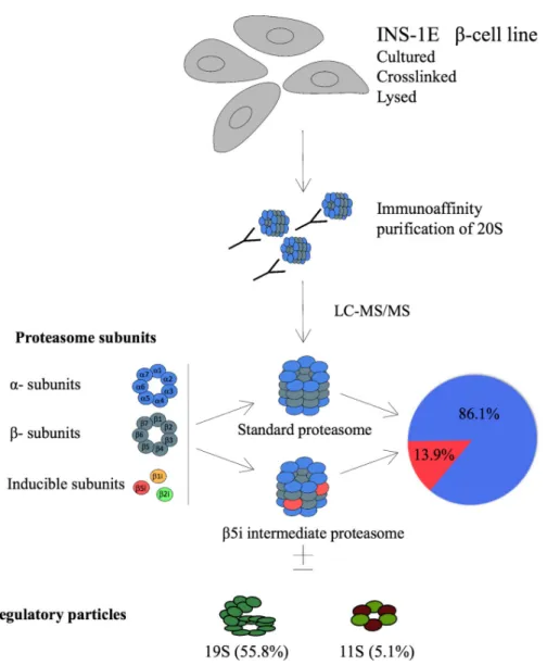
We next investigated the composition of proteasomes in INS-1E cells through immunoprecip-itation (IP) of the 20Sα2 proteasome subunit that is an obligatory member of all types of assembled proteasomes [46].
In vivo gently cross-linked INS-1E cells proteasomes were immunoprecipitated and
sam-ples analyzed by liquid chromatography coupled to tandem mass spectrometry (LC-MS/MS). About 86% of the total proteasomes were found to be s-proteasomes with enzymatically active rings composed ofβ1-β2-β5 subunits (Fig 2). However, almost 14% of the precipitated protea-somes containedβ5i replacing the standard β5 subunit, forming an intermediate proteasome β1-β2-β5i. We found neither β1i nor β2i in protein complexes precipitated with α2 subunit.
Finally, our MS data indicated the presence of proteasomal regulatory particles 19S and 11S (PA28αγ) within α2 proteasome complexes at the rate of 55.8% and 5.1%, respectively (Fig 2). However, with our experimental approach we cannot assign proteasome types to the detected specific regulatory particles. The remaining 39.1% of 20S corresponds to free (unactivated) proteasome [21,41].
Fig 1. Constitutive expression of proteasome inducible subunits in islets and cell lines. (A-B) Single cell RNA
sequencing analysis ofβ1i, β2i and β5i gene expression in human pancreatic islet alpha, beta and delta cells from healthy individuals (n = 6). The data is shown as means with SEM. (B) presents the percentage of cells with detectable levels of inducible subunit mRNA. (C-D) SDS-PAGE and Western blot analysis of basal expression of proteasome inducible subunits in immune cell lines A20 and Jurkat, in insulinoma beta cell line INS-1E, and human islets (H-islets) and mouse islets (M-islets). Values on top of the Western blots show the amount of protein loaded. C and D are representative blots of n = 3. (E) Quantification of relative expression levels of inducible proteasome subunits normalized to tubulin (C) or actin (D) in tested cell lines/islets (n = 3, biological replicates). The data is shown as means with SD.
The
β5i-selective small-molecule inhibitor ONX-0914 reduces
chymotrypsin-like activity of the
β cell proteasome
We next examined the profile of proteasome proteolytic activities: chymo, trypsin-and caspase-like. The proteolytic activities were tested in unstimulated live cells by addition of specific substrates to the medium (Cell-Based Proteasome-Glo™ Assay). As shown inFig 3A–
Fig 2. Identification of intermediate proteasomes in INS-1E cell line. Four x108cells were cross-linked, lysed, their
proteasomes immunoprecipitated with mAb MCP21 and analyzed by LC-MS/MS. The absolute quantities of each of the six catalytic subunits measured by the LC-MS/MS method were computed to calculate the stoichiometry of 20S proteasome subtypes and the fractions of regulatory particles associated with the 20S core particle, as detailed in Experimental Procedures. INS-1E cells were cultured at standard conditions and four biological replicates were analyzed.
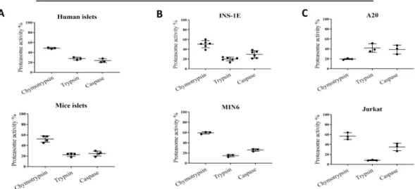
Fig 3. Total and proteolytic-specific enzymatic activities of proteasomes. (A) Proteasome activity in human (n = 3) and
mouse islets (n = 4), (B) beta cell lines: INS-1E (n = 6) and MIN6 (n = 3) and (C) immune cells: A20 and Jurkat (n = 3). All presented data points are represent biological replicates. Proteolytic-specific activities exhibited by proteasomes subunits, treated withβ5i subunit specific inhibitor ONX-0914 (2 h, 50 nM) or non-specific proteasome inhibitor MG132 (2 h, 2 μM), in
(D) human islets (n = 3), (E) mouse islets (n = 4) and (F) INS-1E cells (n = 4). Proteasome activity was evaluated in cultured
cells/islets using Promega Proteasome Glo assay. The data is shown as luminescence per islet/cell. Statistical analysis was performed by paired t-test of treatments versus control. The data is presented as means with SD.
3Cintact human and mouse islets, INS-1E, MIN6 and Jurkat cells exhibited strikingly similar profiles. Under basal conditions, chymotrypsin-like activity constituted 50–60% of total pro-teasome activity, with the remaining 40–50% of activity almost equally divided between tryp-sin- and caspase-like activities. In A20 cells, tryptryp-sin- and caspase-like activities constituted a larger part of the proteasome activity than did chymotrypsin-like activity, despite the fact that they expressed the highest amounts ofβ5i and β1i (both subunits have chymotrypsin-like activities).
We next probed what portion of the observed proteasome chymotrypsin-like activity can be attributed to the inducible subunitβ5i. We took an advantage of a selective and potent β5i subunit inhibitor, ONX-0914. Pretreatment of human and mouse islets and INS-1E cells with 50 nM ONX-0914 for 4 hours reduced chymotrypsin-like activity by 40%, indicating thatβ5i is enzymatically active in those islets and cells (Fig 3D–3F). At the same time, trypsin- and cas-pase-like activities were not affected by ONX-0914. As expected twoμM of MG-132 treatment (broad proteasome inhibitor) almost completely blocked all three types of enzymatic activities in all tested cells and islets.
Low concentrations of IL-1
β upregulate β1i, β2i and β5i subunit expression
in
β cells
Previous work has shown that theβ1i, β2i and β5i proteasome subunits are expressed in response to IFN-γ/β in cells other than that of hematopoietic origin including β-cells [20,21, 41] but their expression was not regulated by the high concentrations of IL-1β treatment [20]. Here we asked, if a similar to IFN-γ/β expression upregulation can be achieved by mimicking low-grade inflammation with the application of a low stimulatory concentration of IL-1β [36].
Mouse islets exposed to 50 pg/ml of IL-1β for 10 days exhibited a significantly higher mRNA expression ofβ5i (Psmb8), β2i (Psmb9) and β1i (Psmb10) compared to the untreated islets (Table 3). The mRNA levels forβ5 (Psmb5), β2 (Psmb6) and β1 (Psmb7) genes that encode standard subunits remained unchanged after the same exposure.
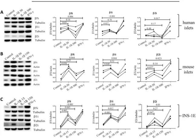
Furthermore, exposure of human and mouse islets, as well as INS-1E cells, to low concen-tration of IL-1β for 24 h (15 pg/ml for INS-1E, 30 pg/ml for human islets and 50 pg/ml for mouse islets) induced expression ofβ1i, β2i and β5i (Fig 4A–4C) with the exception ofβ2i in human islets. The high concentration of IL-1β (150 pg/ml for INS-1E and 300 pg/ml for human/mouse islets) further inducedβ1i and β5i expression (but not β2i) in INS-1E cells, but failed to induce upregulation in the subunit expression in human and mouse islets (Fig 4A and 4B). As expected, low IFN-γ treatment for 24 h induced expression of all inducible subunits (Fig 4A–4C). Concentrations of IL-1β in the low range are known to increase insulin biosyn-thesis [47,48]. Interestingly, induction of inducible proteasome expression by low concentra-tions of IL-1β or IFN-γ was associated with increased proinsulin expression levels in INS-1E cells while high concentrations of IL-1β diminished proinsulin expression (S1A andS1BFig). Furthermore, low concentration of IL-1β did not decrease the viability of mouse islets and INS1-E cells over the 24 h exposure to the cytokine (S1C and S1D Fig).
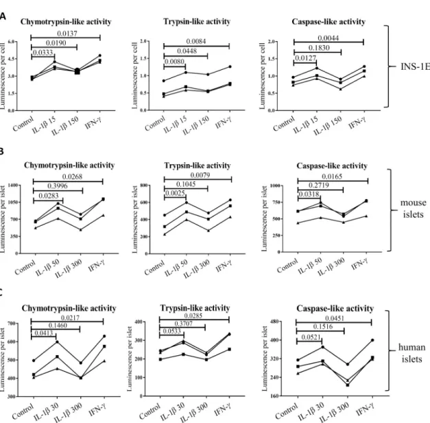
Low concentrations of IL-1β increase proteasome activity in β cells
INS-1E cells exposed to either low concentrations of IL-1β or IFN-γ showed significant increase in all three proteasome catalytic activities. Furthermore, high concentration of IL-1β increased chymotrypsin-like and trypsin-like activity compared to controls, although the increase was less significant than that observed for low concentration exposure (Fig 5A). Mouse and human islets showed a similar pattern of increasing chymotrypsin-, trypsin- and caspase-like activity, when exposed to low concentrations of IL-1β or IFN-γ (Fig 5B and 5C).
A high concentration of IL-1β did not have a significant effect on any of the proteasome-based catalytic activities in mouse and human islets.
Discussion
The present work shows that 1) inducible proteasome subunits are constitutively expressed in human and rodent islets and aβ-cell line, 2) β5i is incorporated into an active proteasome, forming int-proteasomes and 3) inducible subunit expression is upregulated by low IL-1β con-centrations. The cellular composition of proteasomes and their expressional regulation is of particular interest, because different types of proteasomes degrade proteins and peptides with different efficiency and specificity [49] influencing a variety of cellular processes including antigen presentation and thereby maintenance of peripheral tolerance or induction of autoim-munity [8,13].
The presence of int-proteasomes as normal constituents in different tissues has been estab-lished before as they have been reported to constitute up to 50% of the total proteasome pool, depending on the tissue [16,24], but a comprehensive investigation of proteasome composi-tion in primaryβ cells or β cell models in non-stimulated conditions has not been performed.
Fig 4. Cytokines induce upregulation of inducible proteasome subunits in islets/cells. Human (A, n = 4) and mouse islets (B, n = 3)
and INS-1E cells (C, n = 4) were exposed for 24 h to IL-1β at low (50 pg/ml for mouse islets and 15 pg/ml for INS-1E) or high dose (300 pg/ml for human/mouse islets and 150 pg/ml for INS-1E) or IFN-γ (10 ng/ml). Islets/cells were lysed and protein content analyzed by SDS-PAGE and Western blotting. Representative blots of four independent experiments (biological replicates) are shown (left) and quantification of inducible subunit bands relative to the tubulin (A and C) or actin (B) is presented (right). Statistical analysis was performed by paired t-tests of treatments versus control. Experiments done on individual islet donors (A and B) or biological cell replicates (C) are connected by lines.
The Human Protein Atlas RNA-seq data indicated constitutive expression of the inducibleβ1i, β2i and β5i subunits in human pancreas, with 10% of the RNA sequencing reads originating from the islets of Langerhans and 75% coming from exocrine glandular cells [50]. Immuno-staining of the islets for specific inducible subunits detectedβ5i protein by one of two employed antibodies, whileβ1i and β2i proteins were not detected [50]. To clarify this issue, we first investigated the expression of inducible proteasome subunits in unstimulated human dispersed islet cells. Re-analysis of the previously published data set of single-cell RNA sequencing [37] uncovered substantial subpopulations ofα-, β- and δ-cells that constitutively
Fig 5. Basal and cytokine induced activity of proteasome subunits in (A) INS-1E, (B) mouse and (C) human islets (all n = 3). Cells
and islets were exposed to low IL-1β dose (30 pg/ml for human islets, 50 pg/ml for mouse islets and 15 pg/ml for INS-1E for 24 hours), high IL-1β dose (300 pg/ml for human islets, 300 pg/ml for mouse islets and150 pg/ml for INS-1E for 24 hours), INF-γ (10 ng/ ml for human islets, 10 ng/ml for mouse islets and INS-1E for 24 hours) or control media. Statistical analysis was performed by paired t-tests of treatments versus control. Experiments done on individual islet donors (B and C) or biological cell replicates (A) are connected by lines.
express all inducible subunits (Fig 1A and 1B) placing human islets on par with other tissues that express those subunits constitutively [9,16]. When testingβ cell models, as well as human and mouse islets, we found that all three inducibleβ-subunits were detectable at the protein level without the need for cytokine stimulation (Fig 1C–1E), although the expression levels of each subunit varied substantially between tested groups.β cells can therefore potentially assemble i- and int-proteasomes containing one, two or three inducible subunits without immune-stimulation. It is however important to stress that the number ofβ1i-positive β cells is low in human islets (Fig 1B) thus substantially limiting the possibility to assemble i-protea-some. This observation should be taken into account while investigating proteasome function and composition in pancreaticβ cells.
The similar profile of expression of inducibleβ subunits in β, α and δ cells indicates that those subunits play parallel roles in degradation of the hormones abundantly handled by each cell type ER: insulin, glucagon and somatostatin, respectively. The cellular localization of the i-and int-proteasomes may also play a role in that process, asβ5i and β1i subunits are found in close proximity to the ER while the s-proteasomes are homogenously distributed in both nucleus and cytoplasm [51].
Next, we used mass spectrometry to identify the proteasome subtypes inβ cells. By immu-noprecipitating theα2 subunit from INS-1E cell lysates, we purified active proteasomes. Eighty-six % of total proteasomes contained standardβ subunits forming s-proteasomes (Fig 2). The remaining 14% contained onlyβ5i subunit, whereas β1i and β2i could not be detected. Therefore, INS-1E cells constitutively express two types of proteasomes, the s-proteasome and an int-proteasome, where at least oneβ- ring contains a β5i subunit. We neither detected i-proteasomes nor int-i-proteasomes with incorporation of inducible subunits other thanβ5i, despite their expression in unstimulated INS-1E cells. According to the rules of cooperative assembly,β1i cannot be incorporated without β5i but the opposite is feasible [52] and thus it is theoretically possible but biologically less plausible thatβ1i and β2i are expressed but do not participate in the formation of a pool of active proteasomes. Alternatively, sensitivity of the antibody used for the detection ofβ1i may be high relative to the sensitivity of the other anti-bodies used for subunits detection, distorting the evaluation of the intracellular stoichiometry of the inducible subunits. The MS data that failed to detectβ1i and β2i incorporation into pro-teasomes may thus be a more valid measure of the actual subunit stoichiometry in INS-1E cells. Furthermore, according to the rules of proteasome assembly, the lack of incorporation of β1i would prevent the incorporation of β2i subunit [52]. However, we cannot rule out thatβ1i andβ2i are incorporated but constitute the minor portion of active proteasomes, below the detection limit of our MS method.
The enzymatically active proteasomes are generally capped on one or both ends of the cen-tral 20S proteasomal core by regulatory particles 19S or 11S, but the method employed in our study does not distinguish which type of proteasome is associated with a given regulatory par-ticle. We have found that 56% of proteasomes in INS-1E cells contained the 19S particle known to associate with all types of proteasomes [21,46,53,54], while 5.1% proteasomes con-tained 11S that preferentially associates with int- and i-proteasomes [12,21,46,55]. This would indicate that about one third of all INS-1E int-proteasomes are bound to 11S particles while the other two third is associated with 19S particle, (hybrid int-proteasome) or not associ-ated with any regulatory particle and thus presumably not active. Regulatory particles dictate substrate availability and specificity with 19S recognizing client proteins marked by polyubi-quitin chains and 11S being involved in the degradation of short and non-ubipolyubi-quitinated pep-tides and antigen processing for MHC I presentation [56]. As a result, their presence within assembled and active proteasomes demonstrates that unstimulatedβ cells contain specialized and mixed populations of proteasomes, possibly reflecting functional specificity.
When profiles of proteasome substrate-specific activities were analyzed, we found that the islets and cell lines (with the exception of A20) all showed similar proteolytic profiles. Chymo-trypsin-like activity constituted between 50 and 60% of the total proteasome activity, while trypsin- and caspase-like activities were responsible for the remaining 40–50% (Fig 3A–3C). Immune cell lines have generally been reported to express a higher basal level of inducible sub-units, and the i-proteasome constitutes a dominant form of their proteasomes [8]. The fact that INS-1E cells and islets share a similar proteasomal catalytic activity profile indicates that inducible subunits codetermine the activity profiles not only in immune cells.
Off note, we have observed clear differences in the proteasomal catalytic activities in two tested immune cells models, Jurkat and A20. The latter cell profile indicates persistent if not dominant incorporation ofβ1 subunit (with caspase-like activity) but not β1i (chymotrypsin-like activity) and diminished activity ofβ5 and/or β5i subunits (chymotrypsin-like activity) that are obligatory part of active proteasomes [56]. The reasons for observed differences are not known but may indicate cancer-cell-specific adjustments, human (Jurkat) vs mouse (A20) divergence or reflect more physiologically important differences between T (Jurkat) and B (A20) cells. Finally, it is plausible that proteasome activity in A20 cells is additionally modified by e.g. post-translational modifications or altered transcription of proteasomal activators, as reviewed in [57].
We next pretreated islets and cells with aβ5i selective small-molecule inhibitor, ONX-0194, and found a 30 to 50% reduction in chymotrypsin-like activity, further indicating that theβ5i subunit is proteolytically active inβ cells (Fig 3D–3F).
Interpretation of the pathophysiological consequences of the proteasomes diversity inβ cells requires better understanding of factors influencing its expression and composition. The human genes coding forβ5i and β1i map to chromosome 6 precisely between the DNA sequences coding for human leucocyte antigen (HLA)-DQ, HLA-DM and Transporter 1 ATP Binding Cassette Subfamily B Member (TAP) 1 and 2 (S2 Fig), genes known to be major deter-minants of antigen presentation and predisposing to autoimmune diseases, including type 1 diabetes [58]. The promoter region ofβ5i contains binding sites for the NFκB transcription factor (S2 Fig), but high concentrations of IL-1β, a strong inducer of NFκB [59], do not increaseβ5i expression [20]. Accordingly, studies in neurons have shown that high concentra-tion of IL-1β induces Early growth response 1 protein (Egr1) that strongly inhibits transcrip-tional activity at theβ5i promoter [60]. At the same time, Freudenburg et al. speculated that since viral infections induce IL-1β synthesis, iNOS expression and nitric oxide production impeding on mitochondrial function, the resulting reduction in ATP levels would trigger i-proteasome activation and generation of altered peptides that may be immunogenic and enable killing of infected target cells as an appropriate host antiviral response [18]. How can these apparently disparate IL-1β functions be reconciled? We suggest that cytokine concentra-tion and/or duraconcentra-tion of exposure are a key determinants of cell fate. It has been reported that low IL-1β concentrations (0.01–0.1 ng/ml) are stimulating and protective for β cells e.g. they improve insulin biosynthesis and secretion and increaseβ cell proliferation, while higher con-centrations (5–20 ng/ml) can induce cell apoptosis and necrosis through e.g. induction of endoplasmic reticulum and mitochondrial stress [47,48]. These two outcomes employ differ-ent cellular pathways, the stimulatory pathway depending on PKC and phospholipases and the toxic pathway on NFκB signaling. We therefore used 10–100 fold lower IL-1β concentrations in our experiments compared to previous publications [20] as well as, in case of mouse islet used for bulk sequencing (Table 3), we extended the islet exposure to IL-1β up to 10 days in order to better mimic long-term low-grade inflammation. Indeed, we found that treatment of human and mouse islets and INS-1E cells with low concentrations of IL-1β increased the mRNA (Table 3) and protein expression of all inducible proteasomeβ subunits (except of β2i
in human islets,Fig 4), while it had no impact on mRNA levels of standard subunits (Table 3) and increased all substrate-specific proteolytic activities in human and mouse islets and INS-1E cells (Fig 5). Off note, 10 day mouse islets exposure to low concentrations of IL-1β did diminished their glucose induced insulin secretion but did not reduced islets insulin content nor induced endoplasmic reticulum stress or cell death as reported in Ibarra et al. Mol Cell Endocrinol. 2019.
Our results indicate that cytokine concentration is critical when evaluating the regulatory role of cytokines in proteasome expression and activity.
Proteasomes process proteins of both endogenous and exogenous origin and produce pep-tides that are complexed with MHC I. The shift in composition of proteasomes towards i-pro-teasomes, changes the peptide repertoire from non-immunogenic to immunogenic [18,61] and can contribute to the progression towards autoimmune diabetes [18,20,62,63]. Impor-tantly, the observed differences in cytokine action may reflect changing conditions in the islet microenvironment during inflammatory or metabolic stress. IL-1β is a central promoter of low-grade inflammation and protection against certain viral infections, including influenza [64]. One of the possible host protective mechanisms engaged by this cytokine could involve expression and assembly of int- and i-proteasomes that would result in an increased presenta-tion of viral antigens and/or modified self-antigens, thereby enabling T effector-cell dependent eradication of infected cells. Similarly, low grade inflammation and local IL-1β production in the islet microenvironment, could facilitate neoepitope presentation byβ cells through prefer-ential incorporation of inducible subunits to form int- or i-proteasomes. Interestingly, cells deficient inβ5i show lower MHC I expression and peptide presentation, and β5i pharmacolog-ical inhibition slows disease progression in mouse models of inflammatory diseases such as arthritis and lupus [35,65]. Furthermore,β5i has also been implicated in type 1 diabetes, and its inhibition has been shown to have a protective effect [35]. This could reflect the fact that the MHC I peptides repertoire is at least in part dependent upon the activity ofβ5i subunit in β cells expressing int- or i-proteasomes.
The role of immune- and, especially, intermediate proteasomes inβ cell pathophysiology remains to be uncovered in detail. The perspective that differential proteasome subunit expression dictates the repertoire ofβ-cell neoepitopes presented by MHC I deserves future investigation. Discoveries in this field could lead to targeted proteasome inhibition as treatment options in dis-eases with an autoimmune component. In this study, we lay the groundwork for such future investigations. For the first time, we show that int-proteasomes are constitutively expressed and active inβ cells and that inducible proteasome subunits can be upregulated in β cells in response to stimulatory low concentrations of IL-1β along with increases in total proteasome activities.
Supporting information
S1 Fig. Cytokine induced increase in proinsulin levels in INS-1E cells. Lysates of cells
exposed to IL-1β at low (15 pg/ml) and high concentration (150 pg/ml), IFN-γ (10ng/ml) and control medium, were run on SDS-PAGE and subjected to Western blotting in A) and proin-sulin band intensity normalized to tubulin in B). C) and D) Mouse islets and INS1-E cell via-bility was tested. Staining reagent (AlamarBlue) was added to the cell culture for 4 h, incubated at 37oC, and the resulting fluorescence was read on a plate reader. Statistical analysis was per-formed by paired t-tests of treatments versus control. The data is shown as means with SD. (TIF)
S2 Fig. Genetic localization ofβ5i and β1i genes. Genes for β5i (PSMB8) and β1i (PSMB9)
reside in the MHC-II region on human chromosome 6. Presented transcription factors were included in UCSC genome browser genome GRCh37 and visualized with integrated regulation
from ENCODE-track option. For clarity and in relevance to the current publication, only some transcription factors are presented.
(TIF)
Acknowledgments
The authors are thankful to JDRF award 31-2008-416 (ECIT Islet for Basic Research program) for providing human islets for this study. The authors also acknowledge C. Wollheim and P. Maechler (University Medical Centre, Geneva, Switzerland) for providing INS-1E cell line. Soren Buus, (Department of International Health, Immunology and Microbiology, Copenha-gen, Denmark) generously provided the A20 and Jurkat cells. We would like to thank Nils Bill-estrup for sharing and discussing his islet sequencing data that is presented in this manuscript.
Author Contributions
Conceptualization: Danielle Verstappen, Dusan Zivkovic, Marie-Pierre Bousquet-Dubouch,
Thomas Mandrup-Poulsen, Michal Tomasz Marzec.
Data curation: Muhammad Saad Khilji, Danielle Verstappen, Michala Cecilie Burstein Prause,
Phillip Alexander Keller Andersen, Dusan Zivkovic, Marie-Pierre Bousquet-Dubouch, Bjo¨rn Tyrberg, Thomas Mandrup-Poulsen.
Formal analysis: Muhammad Saad Khilji, Danielle Verstappen, Tina Dahlby, Michala Cecilie
Burstein Prause, Celina Pihl, Sophie Emilie Bresson, Tenna Holgersen Bryde, Phillip Alex-ander Keller Andersen, Dusan Zivkovic, Marie-Pierre Bousquet-Dubouch, Bjo¨rn Tyrberg, Thomas Mandrup-Poulsen, Michal Tomasz Marzec.
Funding acquisition: Michal Tomasz Marzec.
Investigation: Muhammad Saad Khilji, Danielle Verstappen, Tina Dahlby, Michala Cecilie
Burstein Prause, Sophie Emilie Bresson, Tenna Holgersen Bryde, Phillip Alexander Keller Andersen, Kristian Klindt, Marie-Pierre Bousquet-Dubouch, Bjo¨rn Tyrberg, Thomas Man-drup-Poulsen, Michal Tomasz Marzec.
Methodology: Muhammad Saad Khilji, Danielle Verstappen, Tina Dahlby, Michala Cecilie
Burstein Prause, Celina Pihl, Sophie Emilie Bresson, Tenna Holgersen Bryde, Phillip Alex-ander Keller Andersen, Kristian Klindt, Dusan Zivkovic, Marie-Pierre Bousquet-Dubouch, Bjo¨rn Tyrberg, Thomas Mandrup-Poulsen, Michal Tomasz Marzec.
Project administration: Michal Tomasz Marzec.
Supervision: Marie-Pierre Bousquet-Dubouch, Thomas Mandrup-Poulsen, Michal Tomasz
Marzec.
Validation: Muhammad Saad Khilji, Sophie Emilie Bresson, Marie-Pierre
Bousquet-Dubouch.
Visualization: Danielle Verstappen, Celina Pihl, Sophie Emilie Bresson, Tenna Holgersen
Bryde, Dusan Zivkovic, Marie-Pierre Bousquet-Dubouch, Bjo¨rn Tyrberg, Michal Tomasz Marzec.
Writing – original draft: Muhammad Saad Khilji, Danielle Verstappen, Tina Dahlby, Michala
Cecilie Burstein Prause, Celina Pihl, Sophie Emilie Bresson, Tenna Holgersen Bryde, Kris-tian Klindt, Dusan Zivkovic, Marie-Pierre Bousquet-Dubouch, Bjo¨rn Tyrberg, Thomas Mandrup-Poulsen, Michal Tomasz Marzec.
References
1. Bard JAM, Goodall EA, Greene ER, Jonsson E, Dong KC, Martin A. Structure and Function of the 26S Proteasome. Annu Rev Biochem. 2018; 87:697–724. Epub 2018/04/14.https://doi.org/10.1146/ annurev-biochem-062917-011931PMID:29652515; PubMed Central PMCID: PMC6422034.
2. Bhattacharyya S, Yu H, Mim C, Matouschek A. Regulated protein turnover: snapshots of the protea-some in action. Nat Rev Mol Cell Biol. 2014; 15(2):122–33.https://doi.org/10.1038/nrm3741PMID:
24452470; PubMed Central PMCID: PMC4384331.
3. Collins GA, Goldberg AL. The Logic of the 26S Proteasome. Cell. 2017; 169(5):792–806.https://doi. org/10.1016/j.cell.2017.04.023PMID:28525752; PubMed Central PMCID: PMC5609836.
4. Finley D. Recognition and processing of ubiquitin-protein conjugates by the proteasome. Annu Rev Bio-chem. 2009; 78:477–513.https://doi.org/10.1146/annurev.biochem.78.081507.101607PMID:
19489727; PubMed Central PMCID: PMC3431160.
5. Goldberg AL. Functions of the proteasome: from protein degradation and immune surveillance to can-cer therapy. Biochem Soc Trans. 2007; 35(Pt 1):12–7.https://doi.org/10.1042/BST0350012PMID:
17212580.
6. Kasahara M, Flajnik MF. Origin and evolution of the specialized forms of proteasomes involved in anti-gen presentation. Immunoanti-genetics. 2019; 71(3):251–61. Epub 2019/01/25.https://doi.org/10.1007/ s00251-019-01105-0PMID:30675634; PubMed Central PMCID: PMC6377343.
7. Unno M, Mizushima T, Morimoto Y, Tomisugi Y, Tanaka K, Yasuoka N, et al. Structure determination of the constitutive 20S proteasome from bovine liver at 2.75 A resolution. J Biochem. 2002; 131(2):171–3. Epub 2002/02/01.https://doi.org/10.1093/oxfordjournals.jbchem.a003084PMID:11820928.
8. Ferrington DA, Gregerson DS. Immunoproteasomes: structure, function, and antigen presentation. Prog Mol Biol Transl Sci. 2012; 109:75–112.https://doi.org/10.1016/B978-0-12-397863-9.00003-1
PMID:22727420; PubMed Central PMCID: PMC4405001.
9. Morozov AV, Karpov VL. Biological consequences of structural and functional proteasome diversity. Heliyon. 2018; 4(10):e00894.https://doi.org/10.1016/j.heliyon.2018.e00894PMID:30417153; PubMed Central PMCID: PMC6218844.
10. Koizumi S, Hamazaki J, Murata S. Transcriptional regulation of the 26S proteasome by Nrf1. Proc Jpn Acad Ser B Phys Biol Sci. 2018; 94(8):325–36. Epub 2018/10/12.https://doi.org/10.2183/pjab.94.021
PMID:30305478; PubMed Central PMCID: PMC6275327.
11. Ghiasi SM, Dahlby T, Hede Andersen C, Haataja L, Petersen S, Omar-Hmeadi M, et al. Endoplasmic Reticulum Chaperone Glucose-Regulated Protein 94 Is Essential for Proinsulin Handling. Diabetes. 2019; 68(4):747–60.https://doi.org/10.2337/db18-0671PMID:30670477; PubMed Central PMCID: PMC6425875.
12. de Graaf N, van Helden MJ, Textoris-Taube K, Chiba T, Topham DJ, Kloetzel PM, et al. PA28 and the proteasome immunosubunits play a central and independent role in the production of MHC class I-bind-ing peptides in vivo. Eur J Immunol. 2011; 41(4):926–35.https://doi.org/10.1002/eji.201041040PMID:
21360704; PubMed Central PMCID: PMC3100532.
13. Baldeon ME, Neece DJ, Nandi D, Monaco JJ, Gaskins HR. Interferon-gamma independently activates the MHC class I antigen processing pathway and diminishes glucose responsiveness in pancreatic beta-cell lines. Diabetes. 1997; 46(5):770–8.https://doi.org/10.2337/diab.46.5.770PMID:9133543.
14. Kunjappu MJ, Hochstrasser M. Assembly of the 20S proteasome. Biochimica et biophysica acta. 2014; 1843(1):2–12.https://doi.org/10.1016/j.bbamcr.2013.03.008PMID:23507199; PubMed Central PMCID: PMC3752329.
15. Heink S, Ludwig D, Kloetzel PM, Kruger E. IFN-gamma-induced immune adaptation of the proteasome system is an accelerated and transient response. Proceedings of the National Academy of Sciences of the United States of America. 2005; 102(26):9241–6.https://doi.org/10.1073/pnas.0501711102PMID:
15944226; PubMed Central PMCID: PMC1166598.
16. Guillaume B, Chapiro J, Stroobant V, Colau D, Van Holle B, Parvizi G, et al. Two abundant proteasome subtypes that uniquely process some antigens presented by HLA class I molecules. Proc Natl Acad Sci U S A. 2010; 107(43):18599–604. Epub 2010/10/13.https://doi.org/10.1073/pnas.1009778107PMID:
20937868; PubMed Central PMCID: PMC2972972.
17. Klare N, Seeger M, Janek K, Jungblut PR, Dahlmann B. Intermediate-type 20 S proteasomes in HeLa cells: "asymmetric" subunit composition, diversity and adaptation. Journal of molecular biology. 2007; 373(1):1–10.https://doi.org/10.1016/j.jmb.2007.07.038PMID:17804016.
18. Freudenburg W, Gautam M, Chakraborty P, James J, Richards J, Salvatori AS, et al. Reduction in ATP levels triggers immunoproteasome activation by the 11S (PA28) regulator during early antiviral response mediated by IFNbeta in mouse pancreatic beta-cells. PloS one. 2013; 8(2):e52408.https:// doi.org/10.1371/journal.pone.0052408PMID:23383295; PubMed Central PMCID: PMC3562186.
19. Kniepert A, Groettrup M. The unique functions of tissue-specific proteasomes. Trends Biochem Sci. 2014; 39(1):17–24. Epub 2013/11/30.https://doi.org/10.1016/j.tibs.2013.10.004PMID:24286712.
20. Lundh M, Bugliani M, Dahlby T, Chou DH, Wagner B, Ghiasi SM, et al. The immunoproteasome is induced by cytokines and regulates apoptosis in human islets. The Journal of endocrinology. 2017; 233 (3):369–79.https://doi.org/10.1530/JOE-17-0110PMID:28438776; PubMed Central PMCID: PMC5501413.
21. Fabre B, Lambour T, Garrigues L, Ducoux-Petit M, Amalric F, Monsarrat B, et al. Label-free quantitative proteomics reveals the dynamics of proteasome complexes composition and stoichiometry in a wide range of human cell lines. Journal of proteome research. 2014; 13(6):3027–37.https://doi.org/10.1021/ pr500193kPMID:24804812.
22. Pelletier S, Schuurman KG, Berkers CR, Ovaa H, Heck AJ, Raijmakers R. Quantifying cross-tissue diversity in proteasome complexes by mass spectrometry. Mol Biosyst. 2010; 6(8):1450–3.https://doi. org/10.1039/c004989aPMID:20498902.
23. Visekruna A, Joeris T, Schmidt N, Lawrenz M, Ritz JP, Buhr HJ, et al. Comparative expression analysis and characterization of 20S proteasomes in human intestinal tissues: The proteasome pattern as diag-nostic tool for IBD patients. Inflamm Bowel Dis. 2009; 15(4):526–33.https://doi.org/10.1002/ibd.20805
PMID:19067411.
24. Menneteau T, Fabre B, Garrigues L, Stella A, Zivkovic D, Roux-Dalvai F, et al. Mass Spectrometry-based Absolute Quantification of 20S Proteasome Status for Controlled Ex-vivo Expansion of Human Adipose-derived Mesenchymal Stromal/Stem Cells. Mol Cell Proteomics. 2019; 18(4):744–59.https:// doi.org/10.1074/mcp.RA118.000958PMID:30700495; PubMed Central PMCID: PMC6442357.
25. Heemels MT, Ploegh H. Generation, translocation, and presentation of MHC class I-restricted peptides. Annu Rev Biochem. 1995; 64:463–91. Epub 1995/01/01.https://doi.org/10.1146/annurev.bi.64. 070195.002335PMID:7574490.
26. Opitz E, Koch A, Klingel K, Schmidt F, Prokop S, Rahnefeld A, et al. Impairment of immunoproteasome function by beta5i/LMP7 subunit deficiency results in severe enterovirus myocarditis. PLoS Pathog. 2011; 7(9):e1002233.https://doi.org/10.1371/journal.ppat.1002233PMID:21909276; PubMed Central PMCID: PMC3164653.
27. Kitiphongspattana K, Mathews CE, Leiter EH, Gaskins HR. Proteasome inhibition alters glucose-stimu-lated (pro)insulin secretion and turnover in pancreatic {beta}-cells. J Biol Chem. 2005; 280(16):15727– 34. Epub 2005/02/12.https://doi.org/10.1074/jbc.M410876200PMID:15705591.
28. Brand MD, Parker N, Affourtit C, Mookerjee SA, Azzu V. Mitochondrial uncoupling protein 2 in pancre-atic beta-cells. Diabetes Obes Metab. 2010; 12 Suppl 2:134–40.https://doi.org/10.1111/j.1463-1326. 2010.01264.xPMID:21029310.
29. Litwak SA, Wali JA, Pappas EG, Saadi H, Stanley WJ, Varanasi LC, et al. Lipotoxic Stress Induces Pan-creatic beta-Cell Apoptosis through Modulation of Bcl-2 Proteins by the Ubiquitin-Proteasome System. J Diabetes Res. 2015; 2015:280615.https://doi.org/10.1155/2015/280615PMID:26064977; PubMed Central PMCID: PMC4438180.
30. Rabinovitch A, Suarez-Pinzon WL. Role of cytokines in the pathogenesis of autoimmune diabetes melli-tus. Rev Endocr Metab Disord. 2003; 4(3):291–9.https://doi.org/10.1023/a:1025160614313PMID:
14501180.
31. Nowlin SY, Hammer MJ, D’Eramo Melkus G. Diet, inflammation, and glycemic control in type 2 diabe-tes: an integrative review of the literature. J Nutr Metab. 2012; 2012:542698.https://doi.org/10.1155/ 2012/542698PMID:23316349; PubMed Central PMCID: PMC3536332.
32. Duncan BB, Schmidt MI, Pankow JS, Ballantyne CM, Couper D, Vigo A, et al. Low-grade systemic inflammation and the development of type 2 diabetes: the atherosclerosis risk in communities study. Diabetes. 2003; 52(7):1799–805.https://doi.org/10.2337/diabetes.52.7.1799PMID:12829649.
33. Zhou W, Sailani MR, Contrepois K, Zhou Y, Ahadi S, Leopold SR, et al. Longitudinal multi-omics of host-microbe dynamics in prediabetes. Nature. 2019; 569(7758):663–71.https://doi.org/10.1038/ s41586-019-1236-xPMID:31142858.
34. Rueden CT, Schindelin J, Hiner MC, DeZonia BE, Walter AE, Arena ET, et al. ImageJ2: ImageJ for the next generation of scientific image data. BMC Bioinformatics. 2017; 18(1):529.https://doi.org/10.1186/ s12859-017-1934-zPMID:29187165; PubMed Central PMCID: PMC5708080.
35. Muchamuel T, Basler M, Aujay MA, Suzuki E, Kalim KW, Lauer C, et al. A selective inhibitor of the immunoproteasome subunit LMP7 blocks cytokine production and attenuates progression of experi-mental arthritis. Nat Med. 2009; 15(7):781–7.https://doi.org/10.1038/nm.1978PMID:19525961.
36. Ibarra Urizar A, Prause M, Wortham M, Sui Y, Thams P, Sander M, et al. Beta-cell dysfunction induced by non-cytotoxic concentrations of Interleukin-1beta is associated with changes in expression of beta-cell maturity genes and associated histone modifications. Mol Cell Endocrinol. 2019; 496:110524.
37. Segerstolpe A, Palasantza A, Eliasson P, Andersson EM, Andreasson AC, Sun X, et al. Single-Cell Transcriptome Profiling of Human Pancreatic Islets in Health and Type 2 Diabetes. Cell Metab. 2016; 24(4):593–607.https://doi.org/10.1016/j.cmet.2016.08.020PMID:27667667; PubMed Central PMCID: PMC5069352.
38. Kim D, Langmead B, Salzberg SL. HISAT: a fast spliced aligner with low memory requirements. Nature methods. 2015; 12(4):357–60.https://doi.org/10.1038/nmeth.3317PMID:25751142; PubMed Central PMCID: PMC4655817.
39. Patro R, Duggal G, Love MI, Irizarry RA, Kingsford C. Salmon provides fast and bias-aware quantifica-tion of transcript expression. Nature methods. 2017; 14(4):417–9.https://doi.org/10.1038/nmeth.4197
PMID:28263959; PubMed Central PMCID: PMC5600148.
40. Bousquet-Dubouch MP, Baudelet E, Guerin F, Matondo M, Uttenweiler-Joseph S, Burlet-Schiltz O, et al. Affinity purification strategy to capture human endogenous proteasome complexes diversity and to identify proteasome-interacting proteins. Mol Cell Proteomics. 2009; 8(5):1150–64.https://doi.org/ 10.1074/mcp.M800193-MCP200PMID:19193609; PubMed Central PMCID: PMC2689779.
41. Fabre B, Lambour T, Delobel J, Amalric F, Monsarrat B, Burlet-Schiltz O, et al. Subcellular distribution and dynamics of active proteasome complexes unraveled by a workflow combining in vivo complex cross-linking and quantitative proteomics. Mol Cell Proteomics. 2013; 12(3):687–99.https://doi.org/10. 1074/mcp.M112.023317PMID:23242550; PubMed Central PMCID: PMC3591661.
42. Bertrand Fabre TL, David Bouyssie´ , Thomas Menneteau, Bernard Monsarrat, Odile Burlet-Schiltz, Marie-Pierre Bousquet-Dubouch. Comparison of label-free quantification methods for the determination of protein complexes subunits stoichiometry. EuPA Open Proteomics. 2014; 4:82–6.https://doi.org/10. 1016/j.euprot.2014.06.001.
43. Krishna S, Banerjee B, Ramakrishnan TV, Shivashankar GV. Stochastic simulations of the origins and implications of long-tailed distributions in gene expression. Proceedings of the National Academy of Sci-ences of the United States of America. 2005; 102(13):4771–6.https://doi.org/10.1073/pnas.
0406415102PMID:15772163; PubMed Central PMCID: PMC555697.
44. Freer DE, Statland BE, Johnson M, Felton H. Reference values for selected enzyme activities and pro-tein concentrations in serum and plasma derived from cord-blood specimens. Clin Chem. 1979; 25 (4):565–9. PMID:38019.
45. Forthofer RN, Lee, Eun, Hernandez, Mike. Biostatistics: A Guide to Design, Analysis and Discov-ery2006. 177 p.
46. Fabre B, Lambour T, Garrigues L, Amalric F, Vigneron N, Menneteau T, et al. Deciphering preferential interactions within supramolecular protein complexes: the proteasome case. Mol Syst Biol. 2015; 11 (1):771.https://doi.org/10.15252/msb.20145497PMID:25561571; PubMed Central PMCID: PMC4332148.
47. Spinas GA, Mandrup-Poulsen T, Molvig J, Baek L, Bendtzen K, Dinarello CA, et al. Low concentrations of interleukin-1 stimulate and high concentrations inhibit insulin release from isolated rat islets of Lang-erhans. Acta endocrinologica. 1986; 113(4):551–8.https://doi.org/10.1530/acta.0.1130551PMID:
3538741.
48. Bendtzen K, Mandrup-Poulsen T, Nerup J, Nielsen JH, Dinarello CA, Svenson M. Cytotoxicity of human pI 7 interleukin-1 for pancreatic islets of Langerhans. Science. 1986; 232(4757):1545–7.https://doi.org/ 10.1126/science.3086977PMID:3086977.
49. Vigneron N, Van den Eynde BJ. Proteasome subtypes and regulators in the processing of antigenic peptides presented by class I molecules of the major histocompatibility complex. Biomolecules. 2014; 4 (4):994–1025.https://doi.org/10.3390/biom4040994PMID:25412285; PubMed Central PMCID: PMC4279167.
50. Uhlen M, Fagerberg L, Hallstrom BM, Lindskog C, Oksvold P, Mardinoglu A, et al. Proteomics. Tissue-based map of the human proteome. Science. 2015; 347(6220):1260419.https://doi.org/10.1126/ science.1260419PMID:25613900.
51. Brooks P, Murray RZ, Mason GG, Hendil KB, Rivett AJ. Association of immunoproteasomes with the endoplasmic reticulum. Biochem J. 2000; 352 Pt 3:611–5. PMID:11104664; PubMed Central PMCID: PMC1221495.
52. Griffin TA, Nandi D, Cruz M, Fehling HJ, Kaer LV, Monaco JJ, et al. Immunoproteasome assembly: cooperative incorporation of interferon gamma (IFN-gamma)-inducible subunits. J Exp Med. 1998; 187 (1):97–104. Epub 1998/01/31.https://doi.org/10.1084/jem.187.1.97PMID:9419215; PubMed Central PMCID: PMC2199179.
53. Huang X, Luan B, Wu J, Shi Y. An atomic structure of the human 26S proteasome. Nat Struct Mol Biol. 2016; 23(9):778–85.https://doi.org/10.1038/nsmb.3273PMID:27428775.
54. Brooks P, Fuertes G, Murray RZ, Bose S, Knecht E, Rechsteiner MC, et al. Subcellular localization of proteasomes and their regulatory complexes in mammalian cells. The Biochemical journal. 2000; 346 Pt 1:155–61. PMID:10657252; PubMed Central PMCID: PMC1220835.
55. Cascio P. PA28alphabeta: the enigmatic magic ring of the proteasome? Biomolecules. 2014; 4(2):566– 84.https://doi.org/10.3390/biom4020566PMID:24970231; PubMed Central PMCID: PMC4101498.
56. Tanaka K. The proteasome: overview of structure and functions. Proc Jpn Acad Ser B Phys Biol Sci. 2009; 85(1):12–36. Epub 2009/01/16.https://doi.org/10.2183/pjab.85.12PMID:19145068; PubMed Central PMCID: PMC3524306.
57. Kors S, Geijtenbeek K, Reits E, Schipper-Krom S. Regulation of Proteasome Activity by (Post-)tran-scriptional Mechanisms. Frontiers in Molecular Biosciences. 2019; 6(48).https://doi.org/10.3389/fmolb. 2019.00048PMID:31380390
58. Noble JA, Valdes AM. Genetics of the HLA region in the prediction of type 1 diabetes. Curr Diab Rep. 2011; 11(6):533–42. Epub 2011/09/14.https://doi.org/10.1007/s11892-011-0223-xPMID:21912932; PubMed Central PMCID: PMC3233362.
59. Eizirik DL, Mandrup-Poulsen T. A choice of death—the signal-transduction of immune-mediated beta-cell apoptosis. Diabetologia. 2001; 44(12):2115–33.https://doi.org/10.1007/s001250100021PMID:
11793013.
60. James AB, Conway AM, Morris BJ. Regulation of the neuronal proteasome by Zif268 (Egr1). J Neu-rosci. 2006; 26(5):1624–34.https://doi.org/10.1523/JNEUROSCI.4199-05.2006PMID:16452686.
61. Huber EM, Basler M, Schwab R, Heinemeyer W, Kirk CJ, Groettrup M, et al. Immuno- and constitutive proteasome crystal structures reveal differences in substrate and inhibitor specificity. Cell. 2012; 148 (4):727–38. Epub 2012/02/22.https://doi.org/10.1016/j.cell.2011.12.030PMID:22341445.
62. Skowera A, Ellis RJ, Varela-Calvino R, Arif S, Huang GC, Van-Krinks C, et al. CTLs are targeted to kill beta cells in patients with type 1 diabetes through recognition of a glucose-regulated preproinsulin epi-tope. J Clin Invest. 2008; 118(10):3390–402.https://doi.org/10.1172/JCI35449PMID:18802479; PubMed Central PMCID: PMC2542849.
63. Unger WW, Velthuis J, Abreu JR, Laban S, Quinten E, Kester MG, et al. Discovery of low-affinity pre-proinsulin epitopes and detection of autoreactive CD8 T-cells using combinatorial MHC multimers. J Autoimmun. 2011; 37(3):151–9.https://doi.org/10.1016/j.jaut.2011.05.012PMID:21636247.
64. Schmitz N, Kurrer M, Bachmann MF, Kopf M. Interleukin-1 is responsible for acute lung immunopathol-ogy but increases survival of respiratory influenza virus infection. J Virol. 2005; 79(10):6441–8.https:// doi.org/10.1128/JVI.79.10.6441-6448.2005PMID:15858027; PubMed Central PMCID: PMC1091664.
65. Ichikawa HT, Conley T, Muchamuel T, Jiang J, Lee S, Owen T, et al. Beneficial effect of novel protea-some inhibitors in murine lupus via dual inhibition of type I interferon and autoantibody-secreting cells. Arthritis Rheum. 2012; 64(2):493–503.https://doi.org/10.1002/art.33333PMID:21905015; PubMed Central PMCID: PMC4584406.