© Emad Tahir, 2019
Determinants of iron status and anemia, and the
associations between iron status and divalent metals
among children aged 3 to 19 years old from four First
Nations communities in Quebec
Mémoire
Emad Tahir
Maîtrise en santé communautaire - avec mémoire
Maître ès sciences (M. Sc.)
Determinants of iron status and anemia, and the associations between
iron status and divalent metals among children aged 3 to 19 years old
from four First Nations communities in Quebec
Mémoire
Emad Tahir
Sous la direction de:
Mélanie Lemire, directrice de recherche
Pierre Ayotte, codirecteur
iii RÉSUMÉ
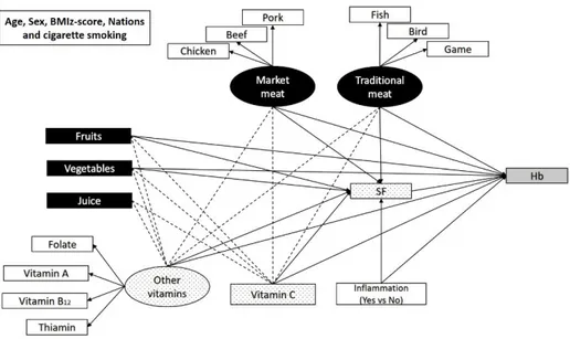
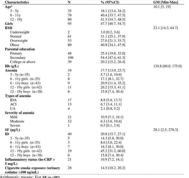
Contexte : L’anémie et la carence en fer sont fréquentes chez les enfants des Premières Nations et ont plusieurs effets néfastes sur la santé. Le fer est un métal divalent qui partage des voies d’absorption intestinale communes avec le cadmium (Cd), le cobalt (Co), le manganèse (Mn), le plomb (Pb) et le zinc (Zn) ; une déficience en fer augmente l’absorption de ces métaux ainsi que leur possible toxicité chez les humains. La présente étude examine la prévalence de l’anémie et la carence en fer, leurs déterminants ainsi que les associations entre la carence en fer et les concentrations sanguines de métaux divalents chez des jeunes des Premières Nations. Méthodologie : En 2015, l’étude pilote Jeunes Environnement et Santé (JES! - YEH!) a été menée chez des enfants et des adolescents (3 à 19 ans, n = 198) de quatre communautés de Premières Nations du Québec. Les concentrations d’hémoglobine, de ferritine sérique, et de métaux sanguins ainsi que de Mn dans les cheveux, de cotinine urinaire et de hs-CRP plasmatique ont été mesurées. Des mesures anthropométriques ont aussi été recueillies. Des déterminants (y compris la consommation d’aliments traditionnels et du marché) ont été évalués à l’aide d’un questionnaire administré par un assistant de recherche qui a ensuite servi pour le calcul des apports en nutriments. Une analyse descriptive a été réalisée et des modèles d’équations structurelles ont été utilisés pour tester les associations. RÉSULTATS : La prévalence respective de l’anémie et de la carence en fer était élevée (17.7% et 20.8% respectivement) dans la population d’étude. Les consommations de la viande traditionnelle, de fruits et de jus de fruits (naturel et en poudre) - par l’intermédiaire de leur association positive avec l’apport en vitamine C - étaient les variables alimentaires positivement associées à la ferritine sérique. Le sexe masculin était également associé à une ferritine sérique plus élevée. Le statut inflammatoire était inversement associé à l’hémoglobine, alors que la ferritine sérique était positivement associée à l’hémoglobine. Comme pour la ferritine, la consommation de fruits et de jus était positivement associée à l’hémoglobine via l’apport en vitamine C et la ferritine sérique, et ce, bien que la plupart des participants présentaient un apport suffisant en fer et en vitamine C. Les niveaux de Mn sanguin étaient significativement plus élevés que ceux des enfants des mêmes groupes d’âge rapportés dans l’enquête canadienne de mesures de santé. La ferritine sérique était inversement associée au Mn et au Co sanguins. Encore une fois, la consommation de fruits et de jus était inversement associée au Mn et au Co sanguins via l’apport en vitamine C et la ferritine sérique. Aucune association significative entre la ferritine sérique et d’autres métaux divalents n’a été observée. CONCLUSIONS : Nos résultats suggèrent que des interventions réduisant l’inflammation et favorisant des environnements alimentaires plus sains ainsi qu’une augmentation de la consommation de viande traditionnelle et d’aliments naturellement riches en vitamine C, qui est connue pour améliorer l’absorption du fer, pourraient contribuer à contrer l’anémie et la déficience en fer et à restaurer l’homéostasie du Mn et du Co dans l’organisme.
Mots-clés: Anémie de l'enfance; carence en fer; Premières Nations; vitamine C; inflammation; manganèse; cobalt
iv SUMMARY
Context: In First Nations communities, anemia and iron deficiency (ID) are frequent pediatric conditions with diverse adverse health outcomes. Iron is a divalent metal that shares absorptive pathways with cadmium (Cd), cobalt (Co), manganese (Mn), lead (Pb) and zinc (Zn) in the gastrointestinal tract; ID upregulates their uptake and likely their toxicity in humans. The present study examines the prevalence of anemia, ID and their determinants as well as study associations between ID status and other divalent metals among First Nations youth. METHODS: The 2015, First Nation Youth Environment and Health (JES!-YEH!) pilot study was conducted among children and teenagers (3 to 19 y, n = 198) from four First Nations communities in Quebec. Blood, hair, urine samples and anthropometric measurements were collected. Hemoglobin, serum ferritin (SF), blood Cd, Pb, Mn, and Co, plasma Zn and hs-CRP, hair Mn and urinary cotinine levels were measured. Determinants (including traditional and market food consumption) were assessed using an interview-administered questionnaire, based on which nutritional intakes were calculated. Descriptive analyses were performed, and structural equation models were used to test associations. RESULTS: The prevalence of anemia and ID was elevated (17.7% and 20.8% respectively) in JES!-YEH! study participants. Traditional meats, fruit and fruit juice consumption (natural and powdered) - via their positive association with vitamin C intake – were the food variables positively associated with SF. Male sex was also associated with higher SF. The inflammatory status was associated with lower hemoglobin, while higher SF was in turn associated with higher hemoglobin. As for SF, fruit and juice consumption were positively associated with hemoglobin, via vitamin C intake and SF, and this although, most participants presented sufficient iron and vitamin C intakes. Blood Mn was significantly higher than in the Canadian Health Measures Survey of the same age groups, and SF was inversely associated with blood Co and Mn. Again, fruits and juice consumption were inversely associated with blood Mn via vitamin C intake and SF. No significant association between SF and other divalent metals was found. CONCLUSIONS: Our findings suggest that interventions fighting inflammation and fostering healthier food environments as well as higher consumption of traditional meats and foods naturally rich in vitamin C, which is known to enhance iron absorption, could decrease anemia and ID and ultimately, restore blood Mn and Co homeostasis.
Key words: Childhood anemia; iron deficiency; First Nations; vitamin C; inflammation; manganese; cobalt
v Table of contents
RÉSUMÉ ... iii
SUMMARY ... iv
Table of contents ... v
List of tables ... viii
List of figures ... ix
List of abbreviations and acronyms ... x
Acknowledgments ... xii
Foreword ... xiii
Introduction ... 1
CHAPTER 1: LITERATURE REVIEW ... 4
1.1. Health, social disparities and childhood anemia among First Nations in Canada ... 4
1.2. Anemia ... 5
1.2.1. Definition and pathophysiology ... 5
1.2.2. Types of anemia among children and young adults ... 7
1.2.3. Health effects of anemia among children and young adults and its public health significance ... 9
1.3. Iron deficiency and iron deficiency anemia ... 9
1.3.1. Childhood iron deficiency manifestation and common causes according to life stage 9 1.3.2. Iron homeostasis ... 10
1.3.3. Iron intake and absorption ... 10
1.3.4. Iron storage, transport and iron biomarkers... 12
1.4. Anemia of chronic inflammation (ACI) ... 16
1.5. Other types of anemia ... 17
1.5.1. Lead (Pb) exposure ... 17
1.5.2. Vitamins A deficiency ... 18
1.5.3. Folate and vitamin B12 deficiency ... 18
1.5.4. Vitamin C deficiency (ascorbic acid) ... 19
1.5.5. Vitamin D deficiency... 20
1.5.6. Other vitamins and micronutrients deficiencies ... 20
1.6. Other determinants of anemia and ID in First Nation context ... 20
1.6.1. Housing conditions and overcrowding ... 21
1.6.2. Food insecurity, traditional activities and food transition ... 22
1.7. Divalent metals and their interactions with iron, iron deficiency and/or iron deficiency anemia ... 23
1.7.1. Cadmium exposure and health-related outcomes ... 24
vi
1.7.3. Manganese exposure and health-related outcomes ... 25
1.7.4. Lead exposure and health-related outcomes ... 27
1.7.5. Zinc exposure and health-related outcomes ... 28
CHAPTER 2: RESEARCH PROBLEM AND OBJECTIVES ... 29
2.1. Research hypothesis and objectives ... 29
2.2. The research questions guiding the present project are: ... 29
2.3. The underlying hypotheses for this project are: ... 29
2.4. The specific objectives are: ... 30
CHAPTER 3: METHODOLOGY ... 31
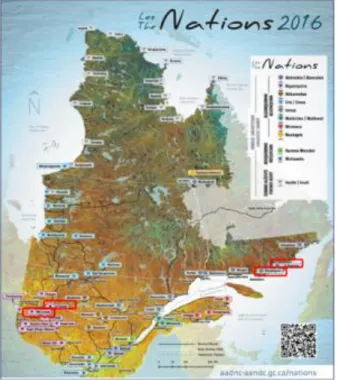
3.1. Study design and areas ... 31
3.1.1. Participant recruitment ... 32
3.1.2. Data collection ... 34
3.2. Dataset and measurements ... 35
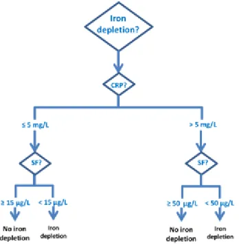
3.2.1. Assessment of anemia and iron deficiency... 35
3.3. Ethical considerations ... 37
CHAPTER 4. MANUSCRIPT ... 38
4.1. Résumé ... 40
4.2. Abstract ... 42
4.3. Introduction ... 44
4.4. Materials and Methods ... 46
4.5. Statistical analysis ... 51 4.6. Results ... 54 4.7. Discussion ... 62 4.8. Conclusion ... 67 4.9. Supplementary materials ... 68 General conclusion ... 76
Summary of the main findings and contributions of this research ... 76
Strength and limitations of the study ... 77
Perspective for future research ... 80
References ... 82
Appendix A-JES! - YEH! project fact sheet ... 90
Appendix B-Consent forms ... 94
vii
Appendix D-Total population, recruitment targets and number of participants recruited per age, sex and community ... 201 Appendix E-Table showing study variables, measurement method... 202 Appendix F-Letters of communication ... 204
viii List of tables
Table 1: Childhood anemia in Canadian and Indigenous contexts ... 5
Table 2:Cut-off points of hemoglobin (Hb), packed cells volume (PCV) and mean corpuscular... 6
Table 3: Morphological types and etiologies of anemia... 8
Table 4:Cut-off reference values for iron status assessment ... 14
Table 5: Characteristics of the study participants (n=193) ... 55
Table 6: Geometrics means blood and plasma metal concentrations by age groups and their comparison with available data from the Canadian Health Measures Survey cycle (CHMS) ... 57
Table 7: Daily food items consumption and dietary intakes and proportions of participants with adequate dietary intakes for all study participants and by nations (n=191) ... 59
Table S1: Determinants of serum ferritin (SF) and hemoglobin (Hb) tested in the structural equation model and variables included in the final models presented in Figure 2 and 3 ... 68
Table S2: Characteristics of the study participants by study nations (n=193) ... 69
Table S3: Geometric's means metal concentrations by age groups and by study nation ... 70
Table S4:Daily food items consumption and dietary intakes and proportions of participants with adequate dietary intakes by study nations (n=191) ... 71
Table S5: Significant direct and indirect associations between sociodemographic, dietary and physiological determinants of serum ferritin (SF) and hemoglobin (Hb) for all study participants (n=191) ... 72
Table S6: Direct and indirect associations between sociodemographic, dietary and physiological determinants of ferritin (SF) and hemoglobin (Hb) stratified by sex (n=191) ... 73
Table S7: Significant direct and indirect associations between sociodemographic, dietary and physiological determinants of serum ferritin (SF) and hemoglobin (Hb) for participants stratified by nations (N=191) ... 74
Table S8: Significant indirect associations between sociodemographic, dietary and physiological determinants of blood and plasma metal concentrations (n=188) ... 75
ix List of figures
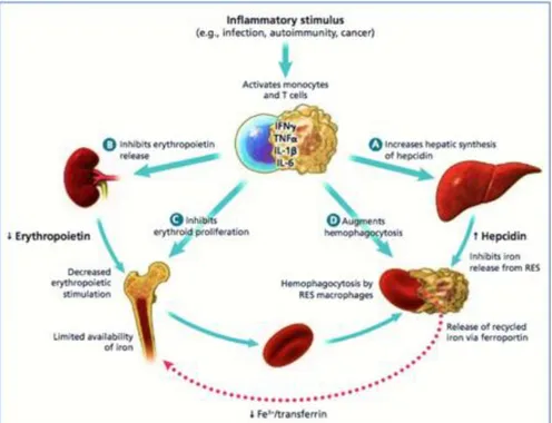
Figure 1: Diagram showing the mechanism of anemia in chronic inflammation or disease 16
Figure 2: Quebec First Nations - study areas highlighted in red rectangles ... 31
Figure 3: Algorithm of diagnosis of iron deficiency (ID) ... 36
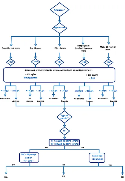
Figure 4: Algorithm for classification of anemia ... 50
Figure 5: SEM model 1 and 2. ... 53
Figure 6: SEM model 3 ... 53
Figure 7: Significant direct associations (not standardized coefficients) between dietary and non-dietary determinants of SF and Hb (Models 1 and 2) for all study participants (n=191) ... 59
Figure 8: Significant direct associations (not standardized coefficients) between sociodemographic, dietary and other determinants of SF and blood and plasma metals (Model 3) for all study participants (n=188). ... 61
x List of abbreviations and acronyms
ACI: Anemia of Chronic Inflammation
Cd: Cadmium
CDC: Center for Disease Control
CHMS: Canadian Health Measures Survey
CHU de Québec : “Centre Hospitalier Universitaire de Québec — Université Laval” CHUL: “Centre hospitalier de l’Université Laval”
CINE : Center for Indigenous Peoples’ Nutrition and Environment CNF : Canadian Nutrient File
Co: Cobalt
CTQ: “Centre de toxicologie du Québec” DNA: Deoxyribonucleic acid
FAO: Food and Agriculture Organization of the United Nations FFQ: Food Frequency Questionnaire
FNBI: First Nations Biomonitoring Initiative
FNQL: First Nations of Quebec and Labrador
FNQLHSSC: First Nations of Quebec and Labrador Health and Social Services Commission
g: Gram
Hb: Hemoglobin
hs-CRP: highly sensitive—C-Reactive Protein
IARC: International Cancer Research: Cancer Epidemiology and Genetic ID: Iron Deficiency
IDA: Iron Deficiency Anemia
INFOODS: International Network of Food Data Systems INSPQ: “Institut national de santé publique du Québec” IOM: Institute of Medicine
IQ: Intellectuel quotient
IUCPQ: “Institute universitaire de cardiologie et pneumologie de Québec” KNHANES: Korean National Health and Nutrition Examination Survey
L: Liter
MCH: Mean corpuscular hemoglobin MCV: Mean corpuscular volume
Mn: Manganese
NHANES: National Health and Nutrition Examination Survey (US)
Pb: Lead
RBCs: Red Blood Cells
SAS: Statistical Analysis System SF: Serum Ferritin
SI: Serum iron
sTfR: Soluble transferrin receptor TIBC: Total Iron Binding Capacity TSAT: Transferrin saturation UA: Unexplained Anemia
UIBC: Unbound Iron Binding Capacity WHO: World Health Organization
Zn: Zinc
ZPP: Zinc protoporphyrin
xi
To my dearest spouse Thowiba Mansour and my little girl Rfaa as well as to the soul of my beloved mother Halima, and the souls of my dear uncles Ibrahim and Elagib. May Allah bless all of us.
xii Acknowledgments
I, hereby, would like to express, in the first place, my countless gratitude to my supervisor Dr. Melanie Lemire who showed great support and compassion in every single step of my master’s thesis project. She was always there to provide help, discuss, guide and direct with great scientific rigor and proficiency. While working with Melanie, her door is always open for us, and she is the first one I always refer to whenever I had difficulties. I would equally thank my co-supervisor Dr. Pierre Ayotte for being a source of inspiration not only to me, but also to the entire team. Pierre is unlimited assets, highly dedicated to his work. I am so proud to have the mentorship of both Melanie and Pierre and to be part of their wonderful team and I humbly admit that with their support, I had not only acquired research related knowledge, but I had also gained valuable experiences added to my personal life as well as my future professional career.
I would also like to acknowledge with gratefulness the Youth Environment and Health (JES!-YEH!) study participants, participants’ parents and legal guardians and community partners as well as the research team who helped in study designing and collecting the data that I used for my research project; without them this work could not have been successfully accomplished. Also, not to forget recognizing our team members: Julie Ducrocq who always keeps answering my endless questions and reviewing my works; Annie Turgeon, Ginette Desbiens, and Denis Guillette who helped in my integration settling-up and logistic assist; Stéphanie Jodoin, Laura Atikesse and Maude Bradette-Laplante for their passionate participation and valued input in JES!-YEH! data quality. Additional special thanks go to Louise Johnson-Down, Caty Blanchette and finally Elhadji Anassour-Laouan-Sidi who was actively involved in the dietary intake calculation, statistical analysis, interpretation and validation of the study results.
Finally, and with much appreciation, I had to thank study fund providers: Health Canada and Nasivvik Research Chair in Ecosystem Approaches to Northern Health, and “CHU de Quebec” for providing me with a full bursary to be able to purse my studies, and also to thank Gisèle Groleau for her efforts of steering my academic orientation.
xiii Foreword
This document is a thesis intended to the attainment of a master’s degree in community health. It is fully authored by me “Emad Tahir” and conforms to the prerequisite of the Faculty of Graduate and Postdoctoral Studies of Laval University. It takes the form of a thesis with an inserted article. The article presented in chapter 4, for which I am the first author entitled “Determinants of anemia and iron status, and their associations with divalent metals among children aged 3 to 19 years old from four First Nations communities in Quebec” is in the process of submission to a peer-reviewed journal, the Canadian Journal of Public Health. To avoid repetition only one bibliography is presented in the reference section after chapter 5. For the concerns of enhanced quality and as the English is the preferred language for the author, this document is entirely written in English instead of French which is the main language of instruction in Laval University. For this purpose, a pre-authorization was obtained from the direction of the program of the master of community health which is also approved the protocol plan and methods.
Following a structured literature review of anemia, the divalent metals and ID and their determinants in the First Nations and Indigenous context, the high-quality data collected for the pilot study JES! -YEH! was used to evaluate the research objectives. Several of the co-authors of this article were responsible for the design of study JES!-YEH! including: Richard E. Bélanger1,4; Michel Lucas1,2; Donna Mergler5; Matthew Little1,2; Elhadji A. Laouan Sidi2; my research supervisor Mélanie Lemire1,2; and co-supervisor Pierre Ayotte1,2,3. We also included the Community of Winneway—Long Point First Nation, the Community of Lac Simon, the CSSS Tshukuminu Kanani of Nutashkuan, the Community of Unamen Shipu and Nancy Gros-Louis McHugh6 as co-authors. I conducted the descriptive statistics for anemia, iron deficiency, their determinants and metals. I equally interpreted the structural equation modeling which was conducted in collaboration with Elhadji A. Laouan Sidi2. All co-authors provided their feedback regarding the inserted article and their comments have been a valuable source of information._____________________________
1 Axe santé des populations et pratiques optimales en santé, Centre de recherche du CHU de Québec — Université Laval, QC, Canada ; 2 Département de médecine sociale et préventive, Université Laval, Québec, QC, Canada ;
3 Institut national de santé publique du Québec, Québec, QC, Canada ; 4 Département de pédiatrie, Université Laval, Québec, QC, Canada ; 5CINBIOSE, Université du Québec à Montréal, Montréal, QC, Canada
1 Introduction
Anemia results in impaired tissue oxygenation due to a decrease in the number of circulating red blood cells (RBCs) and/or hemoglobin (Hb) (Lutter, 2008; WHO, 2001). Globally, anemia is considered as one of the ten most prevalent health problems worldwide. It affects approximately 2.2 billion people, among which 47% are preschool children and approximately 25% are teens and adolescents (De Benoist et al. 2008; Mclean et al. 2008). Moreover, Anemia is also responsible for more than 68 million years lived with disability in 2010 (Kassebaum et al., 2014).
Childhood anemia is associated with growth, developmental, cognitive and psychomotor impairments (Jáuregui-lobera, 2014). It may also cause a reduction in the exercise span, muscle weakness, claudication and easy fatigability, cold sensitivity, reduced immunity, fainting and anorexia. Older female children and adults may suffer from menstrual irregularities and loss of libido (Balarajan, Ramakrishnan, Özaltin, Shankar, & Subramanian, 2011). If treatment is delayed, anemia will result in permanent sequelae (Cooper, Greene-Finestone, Lowell, Levesque, & Robinson, 2012).
The etiology of anemia is very complex and multifactorial, but generally, any factor causing an imbalance between RBCs production and turnover results in anemia. Commonly, a decrease in marrow stimulation due to iron and micronutrient deficiencies, inflammation from: chronic diseases, infections and obesity, lead (Pb) exposure or genetic diseases result in impaired RBCs and Hb synthesis, and in turn, anemia (Hammond, 1977; Mclean et al., 2008; Weiss & Goodnough, 2005).
Iron deficiency (ID) is the commonest nutritional deficiency; its stages start with depleted iron stores, iron deficiency erythropoiesis and iron deficiency anemia (IDA) in severe cases (WHO, 2001). IDA is the most common cause of anemia. Solely, it accounts for more than half of childhood anemia that is estimated to affect 750 million children around the world (de Benoist et al., 2008; Mclean et al., 2008; WHO, 2001). In industrialized countries, it only affects 6% of children, whereas the percentage is especially higher in other countries facing important nutritional issues (WHO, 2007).
2
Canada is among countries with the lowest prevalence of anemia. In 2009, only 3% of Canadian teens (6-11 years) and adolescent females (12-19 years) were known to be anemic, whereas the prevalence of anemia in adolescent males (12 -19 years) and preschool children (3-5 years) was as low as 1% (Cooper et al., 2012). However, anemia prevalence tends to be invariably higher among Indigenous People all over North America, where anemia is often rated as a moderate to severe public health concern (Christofides, Schauer, & Zlotkin, 2005). In addition to the dietary shifting from traditional foods (largely composed of wild meats and fish) to poor-quality market foods (lower in iron and other nutrients), Indigenous populations are still facing precarious housing conditions and higher rates of infections including Helicobacter pylori (Jamieson & Kuhnlein, 2008). Furthermore, vitamin deficiencies, food insecurity and prolonged breastfeeding with inadequate complementary feeding are common in Inuit and First Nations contexts and known as major risk factors for anemia and ID (Christofides et al., 2005; Gessner, 2009; Jamieson, Weiler, Kuhnlein, & Egeland, 2012). Iron is a divalent metal that is provided entirely through diet. However, its intestinal absorption is enhanced by the concurrent presence of iron absorption enhancers (i.e. vitamin C, beta-carotenes, and meat, poultry and fish factor (i.e. proteins) but decreased by the iron absorption inhibitors such as tannins, phytate, calcium and polyphenols (Jamieson & Kuhnlein, 2008). Iron is known to interact with other metals with a similar valence such as cadmium (Cd), cobalt (Co), manganese (Mn), Pb and zinc (Zn) and possibly predispose to their toxicity (Anne et al., 2012; Yangho Kim & Lee, 2011; Yeni Kim et al., 2009; Kwong, Friello, & Semba, 2004; WHO, 2001).
Pb and Cd are xenobiotics known to cause various health problems, including neurocognitive issues in case of exposure to Pb, whereas skeletal problems (inadequate bone mineralization), cardiovascular, reproductive and hematological side effects are related to Cd exposure (Järup, Berglund, & Elinder, 1998; Satarug & Moore, 2004). Conversely, Mn, Zn and Co are nutrients essential for the metabolism of lipids, vitamin B12, proteins and carbohydrates (WHO, 1996). These metals share common absorptive pathways with iron (Flanagan, Haist, & Valberg, 1980). Therefore, a state of ID may trigger the upregulation of intestinal divalent metal transporters, thereby increasing divalent metals’ intestinal absorption, concentration in
3
circulation and possibly, their toxicity (Anne et al., 2012; Meltzer et al., 2010). This has a particular concern in Indigenous context, where ID and elevated exposure to environmental contaminants are common and often simultaneously found (Fenge & Downie, 2003; Muckle, Ayotte, Dewailly E, Jacobson, & Jacobson, 2001; Plante, Blanchet, Rochette, & O’Brien, 2011).
Data on anemia and ID from First Nations’ children and young adults are sparse since First Nations children and youth living on-reserve are not included in the ongoing Canadian Health Measures Surveys (CHMS) (St-Amand, 2017), and in 2010, the First Nations Biomonitoring First Initiative only targeted adults (FNBI) (La Corte & Wuttke, 2012). Moreover, the very few existing studies on anemia and ID in First Nations are confined to preschool children and adults (Christofides et al., 2005). Furthermore, data regarding the interaction of iron status with several divalent metals (Cd, Co, Mn, Pb and Zn) are limited in Canada and elsewhere. This cross-sectional study sought to document the prevalence, types and severity of anemia and ID, and to explore their determinants as well as to study the association between the iron status and other divalent metals in blood or plasma among First Nations children from 3 to 19 years old.
4
CHAPTER 1: LITERATURE REVIEW
1.1. Health, social disparities and childhood anemia among First Nations in Canada First Nations are the largest group of Indigenous People (65%), the native inhabitants of Canada. Collectively they represent 4% of the Canadian population (Kelly-Scott & Smith, 2015). With almost 46% younger than 25 years of age, First Nations are a relatively younger population when compared with non-Indigenous Canadians (Kelly-Scott & Smith, 2015). First Nations and other Indigenous groups in Canada, namely Inuit and Métis, have very distinct diets, sociocultural realities, and lifestyle (Government of Canada, 2002). Conversely, all three groups share a common unresolved historical grief as a result of colonial assimilation during the past century (Brave Heart & DeBruyn, 1998). The repercussion of this assimilation, presumed to be transmitted from one generation to the next, includes higher rates of unemployment, poor health and unfavorable socioeconomic status as well as high rates of health issues including anemia (Kelly-Scott & Smith, 2015).
According to Christofides et al. (2005), anemia is a moderate-to-severe public health problem among preschool children of First Nations and Inuit children from northern Ontario and Nunavut, where the prevalence of anemia among children less than two years of age is 36%. This rate is eight times higher than rates on preschool non-Indigenous Canadian children (Christofides et al., 2005). It is likewise the case for depleted iron stores which affect 53% of those children, of whom 27% had IDA (Christofides et al., 2005).
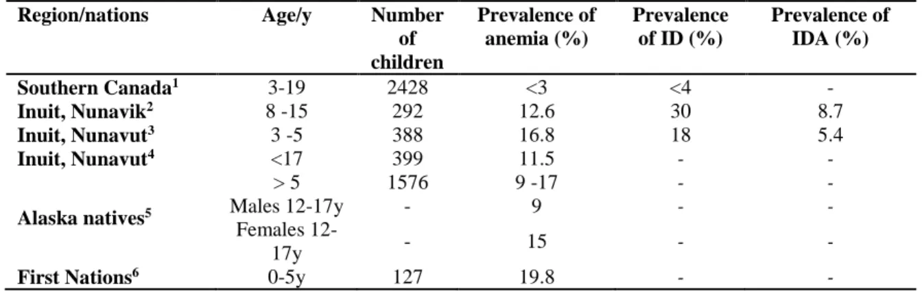
To our knowledge, few studies have been done to assess anemia and ID among children aged three years and older among First Nations communities. Until today, only four studies have documented the prevalence of anemia and ID among Indigenous children counting the age of 3 to 19 years, and most of the available studies were conducted among Inuit children from different circumpolar regions. Table 1 below summarizes the main studies among Indigenous children and youth populations of North America in comparison to the data from the southern Canadian population.
5
Table 1: Childhood anemia in Canadian and Indigenous contexts
Region/nations Age/y Number of children Prevalence of anemia (%) Prevalence of ID (%) Prevalence of IDA (%) Southern Canada1 3-19 2428 <3 <4 - Inuit, Nunavik2 8 -15 292 12.6 30 8.7 Inuit, Nunavut3 3 -5 388 16.8 18 5.4 Inuit, Nunavut4 <17 399 11.5 - - Alaska natives5 > 5 1576 9 -17 - - Males 12-17y - 9 - - Females 12-17y - 15 - -
First Nations6 0-5y 127 19.8 - -
1Cooper et al. (2012); 2Pirkle et al. (2014) ; 3Pacey (2009); 4Thika R, Moffatt ME (1994); 5Petersen et al. (1996); 6Whalen, Caulfield, & Harris, (1997)
1.2. Anemia
The following sections introduce relevant basic knowledge of anemia (types, classification and related health effects) and iron metabolism, followed by a brief description of determinants of anemia and iron status in the Indigenous context.
1.2.1. Definition and pathophysiology
According to the World Health Organization (WHO), anemia is defined as Hb concentration below specific threshold levels (Table 2) for age and sex (WHO, 2001). Hb is the protein that gives the red color of the blood and exclusively found in RBCs. It is structurally formed of four dissimilar protein chains, two alpha subunits and two beta subunits, called globins (Winslow, 2006). In the center of each globin, there is an iron atom attached to a protoporphyrin ring and responsible for the oxygen-carrying characteristic of Hb (Winslow, 2006).
6
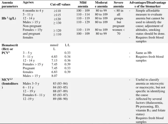
Table 2:Cut-off points of hemoglobin (Hb), packed cells volume (PCV) and mean corpuscular
volume (MCV) to diagnose anemia
1 Hb cut-off values to be adjusted for smoking as shown in Figure 4 WHO (2001) 2 NHANES (1999); 3 MCV = Hct %*10/RBCs count
Originally, RBCs are differentiated from the hemopoietic progenitors stem cells (Hall, 2006). Both, RBCs and Hb are synthesized mainly in the bone marrow of the long and flat bones (Turkoski, 2003). Unlike other cells in the human body, RBCs have special structural and metabolic particularities (Turkoski, 2003). First, in order to increase their capacity to transport Hb, RBCs are the only identified cells in the human body known to lose their nucleolus while they mature. In fact, Hb constitutes 96% of the dried weight of a mature RBC (Hall, 2006; Turkoski, 2003). Second, RBCs have a unique biconcave shape formed and maintained by the integrity of the cell membrane, intracellular proteins and intracellular enzymes. This particular shape, increases their surface area and flexibility for effective exchange and easy passage through small blood vessels (Hall, 2006). Third, since the nucleolus is a house of many components responsible for cell replication and energy
Anemia parameters
Age/sex
Cut-off values Mild
anemia Moderat e anemia Severe anemia Advantages/Disadvantage s of the biomarker Hb 1 (g/L) 6 months to 4 y 5 - 11 y 12 - 14 y Males > 15 y Non-pregnant Females > 15y and pregnant females 110 115 120 ≥ 130 ≥ 120 ≥ 110 100 – 109 110 – 114 110 – 119 110 – 129 110 – 119 100 – 109 80 to 99 80 to 109 80 to 109 80 to 109 80 to 109 80 to 99 < 80 in all groups but pregnant women < 70 - Simple affordable measure to evaluate anemia but cannot be used to identify the cause; adjustment of altitudes, smoking status should be done. - Requires fresh blood
samples Hematocrit (Hct) or PCV1 3 – 5 y 5 - 11 y 12 - 14 y Females > 15 y Pregnant females Males > 15 y mmol/ L 6.83 7.13 7.45 7.45 6.83 8.07 L/L 0.33 0.34 0.36 0.39 0.33 0.39 - Same as Hb - Requires fresh blood
samples MCV2,3 (femtoliters ) Males 3–5 y 6 – 11 y 12 – 19 y Females 6 - 11 y 12 -19 y 83 (83–84) 84 (83–85) 88 (87–89) 85 (83–87) 89 (88–90) - Useful to classify anemia as microcytic or macrocytic, but not specific in identifying the cause
- Affected by several factors (thalassemia, Pb poisoning, ID, vitamin B12 and folate
status)
- Requires fresh blood samples
7
production, RBCs are unable to replicate and have a relatively short half-life of only 120 days. At the end of this period, senescent RBCs fail to maintain their biconcave shape and are rapidly removed from the circulation (Hall, 2006; Turkoski, 2003).
For previously mentioned reasons, RBCs and Hb must continually be produced and released into the circulation (Balarajan et al., 2011). The rate of RBC synthesis is steady and tightly controlled by erythropoietin and the availability of the essential elements (iron, vitamins and amino-acids) required for Hb synthesis and RBC maturation. Thus, factors causing an imbalance between RBC synthesis, maturation or turnover results in a decrease in the circulating RBCs and Hb, and eventually anemia. The health effects and classification of anemia rely on these factors with varying clinical and public health importance for diagnosis, management and/or follow-up (Balarajan et al., 2011).
1.2.2. Types of anemia among children and you ng adults
Clinically, anemia is classified based on severity according to the Hb concentrations as mild, moderate or severe anemia. Another useful indicator of anemia other than Hb is the hematocrit concentration or packed cell volume, which is a simple measure that estimates the volume percentage of RBCs in whole blood but is less commonly used to assess severity (WHO, 2001). Table 2 is showing cut-off values for Hb and hematocrit, identifying severity of anemia by age groups and sex according to the WHO (2001) and NHANES (1999). Because erythropoietin production increases in response to the relative hypoxia produced by smoking and living at high altitude, which in turn enhances RBCs and Hb production, it is important to adjust for smoking status and altitude while using the Hb measures (Aitchison & Russell, 1988).
Based on the RBC size – reported as mean corpuscular volume (MCV) – anemia is also classified into normocytic, macrocytic and microcytic anemia (Fishman, Christian, & West, 1999; Mclean et al., 2008). The size of RBCs varies according to underlying causes of anemia, as outlined in Table 3. Equally, according to the RBC content of Hb, known as mean corpuscular hemoglobin (MCH), anemia can also be classified as normochromic or
8
hypochromic anemia with or without a decrease in the total number of RBCs (Fishman et al., 1999).
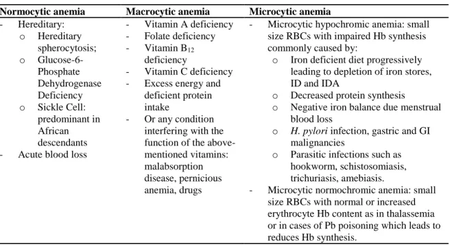
Table 3: Morphological types and etiologies of anemia
Normocytic anemia Macrocytic anemia Microcytic anemia
- Hereditary: o Hereditary spherocytosis; o Glucose-6-Phosphate Dehydrogenase Deficiency o Sickle Cell: predominant in African descendants - Acute blood loss
- Vitamin A deficiency - Folate deficiency - Vitamin B12
deficiency
- Vitamin C deficiency - Excess energy and
deficient protein intake
- Or any condition interfering with the function of the above-mentioned vitamins: malabsorption disease, pernicious anemia, drugs
- Microcytic hypochromic anemia: small size RBCs with impaired Hb synthesis commonly caused by:
o Iron deficient diet progressively leading to depletion of iron stores, ID and IDA
o Decreased protein synthesis
o Negative iron balance due menstrual blood loss
o H. pylori infection, gastric and GI malignancies
o Parasitic infections such as hookworm, schistosomiasis, trichuriasis, amebiasis.
- Microcytic normochromic anemia: small size RBCs with normal or increased erythrocyte Hb content as in thalassemia or in cases of Pb poisoning which leads to reduces Hb synthesis.
Fishman, Christian, & West, 1999; Mclean et al., 2008
Finally, an etiological classification more centered on the identification of the inherent causes of anemia in order to assign appropriate pharmacological treatment is commonly used in clinical practices. However, in a population-based research, blood tests requiring fresh blood samples such as MCV are costly and not often available. In such a context, anemia is classified using multiple blood iron and inflammation biomarkers, to categorize anemia into three main types: IDA, anemia due to chronic inflammation (ACI) and unexplained anemia (UA) (Cash & Sears, 1989; Patterson, Brown, & Roberts, 2001) as also proposed by (Plante et al., 2011). An iron deficient diet leads to ID, and ultimately, to IDA. In the absence of ID, chronic inflammation resulting from infections or obesity also leads to impaired Hb synthesis and then ACI (WHO, 2001). The very large category of UA includes other micronutrient deficiencies such as vitamins and amino-acid deficiencies that are also prevailing causes of anemia in children (WHO, 2011). This classification is also useful for easy planning of public health interventions and prevention plans.
9
1.2.3. Health effects of anemia among children and young adults and its public health significance
Anemia presents with variable symptoms depending on the severity, the underlying cause and physical activity. Individuals with a mild acute form of anemia may exhibit subtle or no symptoms during normal daily activities. Those with moderate to severe acute anemia may show fatigability, shortness of breath (dyspnea), palpitations, dizziness and muscle weakness even at rest. Very severe cases of acute anemia can progress to heart failure and even death (Turkoski, 2003).
Long-standing or chronic anemia in children is a public health concern. According to the WHO (2001), a prevalence of chronic anemia beyond 5% in a specific group is considered abnormal and immediate action should be undertaken. When anemia prevalence is between 5 and 19 %, it is defined as a mild public health problem, whereas between 20% to 39.9%, a moderate public health problem, and if it affects more than 40 % of a specific population, then anemia is considered as a severe public health problem (WHO, 2011).
1.3. Iron deficiency and iron deficiency anemia
1.3.1. Childhood iron deficiency manifestation and common causes according to life stage
In addition to anemia, childhood ID may manifest with specific symptoms such as reduced memory and attention, defective thermoregulatory mechanisms and increased susceptibility to infections. However, other symptoms, related to rapid epithelial-cell turnover such as dry and rough skin, hair loss, soft and spoon-shaped finger nails, cracked lips skin and the skin in the angle of the mouth and loss of tongue papillae, may occur (Lopez, Cacoub, Macdougall, & Peyrin-Biroulet, 2016). Less frequently documented, ID state is also known to increase the absorption of some metals such as cadmium (Cd), cobalt (Co), manganese (Mn), Pb and zinc (Zn) and possibly predispose to their toxicity (Anne et al., 2012; Yangho Kim & Lee, 2011; Yeni Kim et al., 2009; Kwong et al., 2004; WHO, 2001).
10
increased iron requirements in developing children and puberty in adolescents. Young women are particularly vulnerable and more often affected by ID due to negative iron balance caused by menstruation (IOM, 2001). Although not common in Canada, ID can also result from gastrointestinal parasitic infestation (Coad & Conlon, 2011; WHO, 2001).
1.3.2. Iron homeostasis
Iron is an essential element that is only supplied through diet and extremely important for the proper functioning of almost all mammalian cells to accomplish their metabolic functions of energy production and utilization (Wang & Pantopoulos, 2011). The iron attached to Hb in RBCs has a key role to carry oxygen from the lungs to muscles and other tissues (CDC, 1998). The total body contents of iron in the human body are greatly stable and estimated to be around 3.8 grams (g) in an adult male and 2.3 g in an adult female. In children, these values vary with age and child’s body weight (Health Canada, 2005). Iron status in the human body is a dynamic process involving iron intake from diet, absorption at intestinal level, storage in the liver, muscle and intracellular ferritin, and iron loss due to skin and mucosal cells turnover and menstruation (WHO, 2001). Overall, 70% of body iron is functional iron, the rest is either stored or circulating iron (IOM, 2001).
1.3.3. Iron intake and absorption
Iron bioavailability is the amount of ingested iron readily available for absorption (Hurrell & Egli, 2010). Factors affecting iron bioavailability are the types of iron, dietary composition of meals, and gastric acidity that increases the solublilization of iron and then facilitates its absorption at the intestinal level (Hurrell & Egli, 2010; IOM, 2001).
Dietary iron is found in two forms. First, the heme or ferrous iron exclusively found in animal sources of diet, in which the protoporphyrin ring and iron are derived from Hb or muscle cells myoglobin (Hurrell & Egli, 2010; IOM, 2001). Second, the non-heme iron, which is mainly found in plants and iron-fortified foods and supplements but also in diet from animal sources (CDC, 1998; Wang & Pantopoulos, 2011). Non-heme iron absorption is influenced
11
by gastric acidity and dietary composition of meals such as the presence of iron absorption-enhancers or inhibitors.
Meals rich in iron absorption-enhancers such as vitamin C, beta-carotenes (vitamin A) as well as meat, poultry and fish factor increases the availability of non-heme iron for absorption, whereas tannins, phytate, calcium and polyphenols decrease its bioavailability (Jamieson & Kuhnlein, 2008). Dietary vitamin C is one of the most potent iron absorption-enhancer, which increases the absorption of dietary non-heme iron up to four-fold (if consumed within 30 minutes of meals), and also enhances later iron mobilization and storage (Fishman et al., 1999; WHO, 2001). On the other hand, polyphenols (in some vegetables, berries, black tea, coffee, and spices such as cinnamon), phytates (in whole grains and legumes), calcium in dairy products as well as egg proteins are major non-heme iron absorption-inhibitors; thus a diet rich in these elements will lower the iron bioavailability (Balarajan et al., 2011; Hurrell & Egli, 2010). In contrast to non-heme iron, the bioavailability of the heme iron is less influenced by the presence of iron absorption-enhancers or inhibitors; consequently, its absorption rates are two to three times higher than that of the non-heme iron (Jackson, Williams, McEvoy, MacDonald-Wicks, & Patterson, 2016; Pantopoulos, Porwal, Tartakoff, & Devireddy, 2012).
The bioavailable iron is then taken up by an energy-dependent process involving a metal transporter (divalent metal transporter [DMT1] protein) located in the apical cellular membrane of the duodenal cells (IOM, 2001). The quantity of DMT1 is inversely proportional to the iron content of enterocytes during their early development in the crypts of Lieberkuhn (IOM, 2001). The absorbed iron is then transported through the enterocyte to be delivered to their basolateral surface with the help of the Feroportin-1 transporter, where it binds to plasma transferrin, and then transported to all body cells (IOM, 2001; Weiss & Goodnough, 2005). The concentration of Feroportin-1 is tightly controlled by the hepcidin hormone and the iron status of the organism (IOM, 2001). After a brief period of functionality, the turnover of the enteric mucosal cells takes place between 48 and 72 hours where they are shed into the lumen together with their contents of iron that entered the cells but had not yet been excreted to the plasma (IOM, 2001).
12
1.3.4. Iron storage, transport and iron biomarkers
Iron state is a spectrum of ranges from iron overload, to normal iron stores, and ID. Iron overload is rare to occur and usually related to internal homeostatic failure and chronic hemolytic anemias (IOM, 2001). The spectrum of ID starts with negative iron balance and iron depletion, followed by iron-deficient erythropoiesis, and finally IDA (Cooper et al., 2012). Negative iron balance and iron depletion are referred to a progressive decreased in iron stores in liver and spleen, despite the normal circulating serum iron (SI) concentration. SI constitutes a very small fraction of the total body iron pool (less than 1%) in which iron is transit in the circulation and then rapidly transported from absorption sites to sites of storage and utilization in bone marrow and other cells. Therefore, SI is characterized by a large postprandial and diurnal variability (CDC, 1998). The stage of iron-deficient erythropoiesis happens when the tissue functions are impaired because of prolonged negative iron balance. It is characterized by the diminution of iron carried by serum transferrin and the increase of tissue transferrin receptors (WHO, 2001).
Knowing that free iron is highly toxic to human cells, following its absorption from the intestine, iron must be transported attached to plasma proteins, and mainly transferrin (IOM, 2001). Transferrin normally carries approximately 3 mg of iron on its two binding sites (Pantopoulos et al., 2012). With its turnover rate of more than ten times per day, this makes it the most dynamic iron pool in the human body. It delivers iron to hepatocytes, bone marrow, reticuloendothelial cells and other body cells (IOM, 2001). Iron usually occupies one third of all serum transferrin binding sites; this proportion is an indicator of iron status and known as transferrin saturation (TSAT) (Pantopoulos et al., 2012). Conversely, the unbound iron binding capacity (UIBC) is defined as the amount of iron that can bind to vacant transferrin receptors (Åsberg, Thorstensen, & Borch-Iohnsen, 2012). This must not be mistaken with the total iron binding capacity (TIBC), obtained by adding UIBC and SI, which is the maximum amount of iron that can be carried bound to transferrin receptors. A TSAT percentage less than 15% has a specificity of 93% in estimating ID. TSAT is calculated by dividing the SI concentration by TIBC (μg/L) and then multiplying by 100, as it is usually expressed as a percentage (CDC, 1998).
13
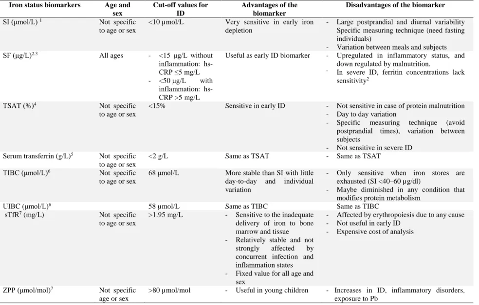
Iron can also bind to an intracellular protein called ferritin that may also be found in small amounts in the circulation and known as serum ferritin (SF). The ferritin is the main iron storage protein; a decrease in the level of intracellular ferritin is accompanied by a decrease in SF in early ID (stage of iron-deficient erythropoiesis). Levels of SF vary according to sex, age, and inflammatory condition (Table 3). A concentration of 1 μg/L of SF is roughly equivalent to 10 mg of stored iron (CDC, 1998; Yangho Kim & Lee, 2011). Generally, SF measurement is a sensitive and specific iron status biomarker (CDC, 1998). However, a careful interpretation of SF is needed in cases of severe ID and/or of infection and inflammation, where this measure is rather insensitive (Engle-Stone & Nankap, 2013). Indeed, measuring only SF in the case of inflammation will underestimate the iron status since SF is an acute phase reactant and inflammation always lead to an increased release of SF in the circulation (Cooper et al., 2012; Gibson, 2005; WHO, 2001). Another iron biomarker with an increasingly popularity in population-based studies is soluble transferrin receptor levels (sTfR) because it is less influenced by inflammatory status (WHO, 2001). Other less commonly used iron status biomarkers are Zn-protoporphyrin (ZPP), erythrocyte protoporphyrin and hepcidin levels. Details on the cut-off reference values, advantages and disadvantages associated with all these iron status biomarkers are summarized in Table 4.
14
Table 4:Cut-off reference values for iron status assessment
Iron status biomarkers Age and sex
Cut-off values for ID
Advantages of the biomarker
Disadvantages of the biomarker
SI (μmol/L) 1 Not specific
to age or sex
<10 µmol/L Very sensitive in early iron depletion
- Large postprandial and diurnal variability Specific measuring technique (need fasting individuals)
- Variation between meals and subjects SF (μg/L)2.3 All ages - <15 μg/L without
inflammation: hs-CRP ≤5 mg/L - <50 μg/L with
inflammation: hs-CRP >5 mg/L
Useful as early ID biomarker - Upregulated in inflammatory status, and down regulated by malnutrition.
- In severe ID, ferritin concentrations lack
sensitivity2
TSAT (%)4 Not specific
to age or sex
<15% Sensitive in early ID - Not sensitive in case of protein malnutrition - Day to day variation
- Specific measuring technique (avoid postprandial times), variation between subjects
- Not sensitive in severe ID Serum transferrin (g/L)5 Not specific
to age or sex
<2 g/L Same as TSAT - Same as TSAT
TIBC (μmol/L)6 Not specific
to age or sex
68 μmol/L More stable than SI with little day-to-day and individual variation
- Only sensitive when iron stores are exhausted (SI <40–60 µg/dl)
- Maybe diminished in any condition that modifies protein metabolism
UIBC (μmol/L)6 58 µmol/L Same as TIBC Same as TIBC
sTfR7 (mg/L) Not specific
to age or sex
>1.95 mg/L - Sensitive to the inadequate delivery of iron to bone marrow and tissue - Relatively stable and not
strongly affected by concurrent infection and inflammation states - Fixed value for all age and
sex
- Affected by erythropoiesis due to any cause - Not useful in early ID
- Expensive cost of analysis
ZPP (μmol/mol)7 Not specific
age or sex
>80 µmol/mol - Useful in young children - Increases in ID, inflammatory disorders, exposure to Pb
15
1Patterson, Brown, & Roberts(2001); 2D. I. Thurnham & McCabe (2010); Turgeon O’Brien, Blanchet, Gagné, Lauzière, & Vézina (2016); Guyatt et al.
(1992);3Turgeon O’Brien et al. (2016); 4Engle-Stone & Nankap (2013); 5Gibson (2005); 6 Plante et al. (2011); 7WHO (2001);8Pantopoulos et al. (2012)
- Very useful test in the absence of infection
- Less sensitive to early ID
- Not helpful to detect daily variation - Complex and costly procedure Erythrocyte protoporphyrin7 <5 years:
≥ 5 years: >70 μg/dL RBCs >2.6 μg/g Hb >61 mmol/mol Hb >80 μg/dL RBCs >3.0 μg/g Hb >70 mmol/mol Hb
- Useful in young children - Very useful test in the
absence of infection
- Increases in ID, inflammatory disorders, exposure to Pb
- Less sensitive to early ID
- Not helpful to detect daily variation - Complex and costly procedure
Hepcidin8 - Sensitive measure for
depleted iron stores
- Methods and interpretation of results are under development
16 1.4. Anemia of chronic inflammation (ACI)
Inflammation may impair RBCs production and disturb iron metabolism via several mechanisms as shown in Figure 1. First, excessive inflammatory cytokines release reduces erythropoietin production as well as the sensitivity of the bone marrow to erythropoietin, and consequently, disturb RBCs production in bone marrow (Jamieson et al., 2012; Weiss & Goodnough, 2005). Moreover, this condition provokes oxidative stress in RBCs resulting in their deformation and their premature turnover (Zarychanski & Houston, 2008).
Figure 1: Diagram showing the mechanism of anemia in chronic inflammation or disease. Reprinted from Zarychanski & Houston (2008) © Canadian Medical Association
Second, chronic diseases and inflammation are known to provoke a functional state of ID, by causing a sequestration of iron in its stores and preventing its transport and utilization, and thus preventing the integration of iron into Hb and then leading to the development of anemia (Zarychanski & Houston, 2008). Inflammation also leads to the up-regulation of SF and down-regulation of the ST in response to the inflammatory process. Conversely, it stimulates the production of hepcidin from the liver, which is an acute-phase reactant with antimicrobial activity that degrades Feroportin-1, and thus reduces the rates of intestinal absorption of iron (Anne et al.,
17
2012; Weiss & Goodnough, 2005). Third, obesity is now considered as a chronic inflammatory condition due to its association with adipose tissue necrosis and a potent contributor to ACI (Nead, Halterman, Kaczorowski, Auinger, & Weitzman, 2004). Obesity is prevalent in FN, and reaching an alarming rate in children (Public Health Agency of Canada, 2018).
These series of inflammatory mediated metabolic changes due to chronic diseases collectively lead to ID and IDA in absence of dietary iron deficiency or result in microcytic hypochromic anemia very similar to IDA in approximately 25% of individuals. This usually leads to a diagnostic dilemma in population-based studies (Zarychanski & Houston, 2008). An inflammatory workup using acute-phase reactants, such as the hs-CRP and alpha1-acid glycoprotein may not completely distinguish ACI from IDA (Subramaniam & Girish, 2015). The gold standard method mainly used by the clinician to differentiate IDA from ACI in such a case is a bone marrow biopsy, which is invasive and of limited use in population-based studies (Turgeon O’Brien et al., 2016; WHO, 2007).
1.5. Other types of anemia
Other types of anemia are those that can be explained by neither ID nor chronic diseases and inflammation. It includes all other forms of anemia such as those related to hereditary causes, elevated Pb exposure or nutritional issues other than ID such as vitamin A, vitamin C, vitamin B12, folate and vitamin D (WHO, 2001). In epidemiological studies, these are often referred as Unexplained Anemia (UA) since most epidemiological studies does not dispose of sufficient biomarker measurements needed to explain the causes of anemia other than ID and inflammation (Plante et al., 2011).
1.5.1. Lead (Pb) exposure
At higher levels of exposure, Pb is known to cause anemia. Elevated Pb exposure decreases heme synthesis and Hb production by inhibiting the activity of the delta-aminolevulinic acid dehydratase and ferrochelatase enzymes, thus generating microcytic anemia similar to IDA (Bellinger & Bellinger, 2006; Counter, Buchanan, Ortega, Rifai, & Shannon, 2007). These enzymes are
18
responsible for the incorporation of iron into the protoporphyrin ring. Furthermore, due to the pro-oxidant activity of Pb, it alters the RBCs membrane architecture and predisposes to a premature removal of these abnormal RBCs from the blood (Levander, 1979).
1.5.2. Vitamins A deficiency
Vitamin A is a fat-soluble vitamin essential for normal vision, gene expression and reproduction as well as growth and immunity in humans (IOM, 2001). Its deficiency is among the commonest vitamin deficiencies that affect iron metabolism and cause anemia (IOM, 2001).
The mechanism by which anemia occurs in vitamin A-deficient children is not completely understood. However, it was observed that vitamin A-deficient individuals have disrupted hematopoiesis and a defective proliferation and differentiation of the pluripotent hemopoietic cells (Fishman et al., 1999). Furthermore, it is also suggested that vitamin A deficiency decreases erythropoietin synthesis and release from kidneys, which may lead to RBCs underproduction and anemia. This type of anemia manifests as hypochromic anemia similar to IDA because of the alteration in iron supply to bone marrow, reduced iron mobilization and its accumulation in the form of hemosiderin in the spleen (Fishman et al., 1999; Jamieson & Kuhnlein, 2008).
Vitamin A deficiency occurs as a result of inadequate dietary intake. In general, animal-derived foods are rich in vitamin A (Kuhnlein, Receveur, Soueida, & Egeland, 2004), particularly the liver, whereas red palm oil and dark colored vegetables and fruits contain significant concentrations of vitamin A precursor (provitamin A carotenoid), which needs to be activated into vitamin A following ingestion (IOM, 2001).
1.5.3. Folate and vitamin B12 deficiency
Chronic folate (B9) and vitamin B12 deficiencies are both possible causes of macrocytic anemia. Chronic folic acid deficiency impairs thymidine formation, which further impair erythropoiesis and delays RBCs maturation. As a result, abnormal large-sized nucleated RBCs (megaloblasts) are released into the circulation. The removal of these abnormal cells in circulation by bone marrow macrophages is high, and possibly leads to macrocytic hypochromic anemia (Fishman et al., 1999;
19 WHO, 2001).
Folic acid is found in leafy green vegetables, while vitamin B12 is provided only via animal food sources. Vitamin B12 is stored in large quantities sufficient to accomplish body functions for several years (3 to 4 years) of non-intake. Its deficiency is common in exclusive vegetarians or in case of a defective absorption resulting from reduced or lack of intrinsic factor secretion (a glycoprotein needed for B12 transport across the intestinal wall) (Fishman et al., 1999; Kuhnlein et al., 2004). Among Indigenous communities, several traditional foods are known to be high in vitamin B12 but deficient in folate. But nowadays, since nutritional policies in Canada recommend the addition of folic acid to almost all cereals and grains from the market, folic acid deficiency is not common either in non-Indigenous or Indigenous context (Health Canada, 2005; Kuhnlein et al., 2004).
1.5.4. Vitamin C deficiency (ascorbic acid)
Human beings are lacking the enzyme (L-gulono-γ-lactone oxidase) responsible for vitamin C (ascorbate) synthesis. Therefore, ascorbic acid must be entirely supplied through diet (Lane & Richardson, 2014). Sources of vitamin C in Indigenous context include traditional foods such as wild fruits (berries) as well as market fruits and vegetables, primarily green and red pepper, citrus and tomatoes (Food and Agriculture Organization-United Nations [FAO], 2009; IOM, 2001b). Vitamin C deficiency is uncommon in children but it can be seen in children exclusively fed on cow milk (Fishman et al., 1999).
Vitamin C is known to enhance the absorption of iron from the gut, and especially for iron with reduced bioavailability such as non-heme iron (Lane & Richardson, 2014). This effect is mainly conveyed by its ability to reduce iron from ferric state to the more soluble and more absorbable ferrous form (Fishman et al., 1999). However, the action of vitamin C on iron is not limited to the gut since it was also identified that vitamin C modulates the iron intake and metabolism by mammalian cells (Lane & Richardson, 2014). Indeed, ascorbate stimulates ferritin synthesis and inhibits its degradation by the lysosomal ferritin that decreases cellular iron efflux (Lane & Richardson, 2014). Furthermore, it was observed that in iron non-deficient individuals, ascorbic acid increased the uptake of iron by the low-molecular-weight iron-citrate complexes in some
20
areas such as the brain (Lane & Richardson, 2014). In addition, the antioxidant activity of vitamin C prevent premature oxidative damage of RBCs (Lane & Richardson, 2014).
1.5.5. Vitamin D deficiency
Vitamin D is essential for bone marrow health, the site of erythropoiesis, because of its important role in maintaining normal bone structure and mineralization (Souberbielle et al., 2010). Moreover, it is suggested that vitamin D increases the sensitivity of bone marrow to erythropoietin. A recent study revealed up to 2-fold increase in risk of anemia associated with vitamin D deficiency (Sim et al., 2010).
In addition to fish and meats which are rich in vitamin D, other Indigenous traditional diet contains variable amounts of vitamin D (Center for Indigenous Peoples’ Nutrition and Environment [CINE], 2005). However, all milk and cereals sold in Canada are fortified with vitamin D (IOM, 2006). The intrinsic synthesis of vitamin D led by sun light is often suboptimal in Indigenous people because of their relatively darker color of skin compared to individuals with fare-colored skin. This most likely predisposes them to vitamin D deficiency related anemia (Libon, Cavalier, & Nikkels, 2013).
1.5.6. Other vitamins and micronutrients deficiencies
Vitamin E (alpha tocopherol), thiamine, niacin, and vitamin B6 are essential antioxidants that help maintain the cellular integrity of RBCs by preventing the oxidation of its cellular membrane by free radicals. Hence, they prevent premature RBC destruction and anemia (Fishman et al., 1999; Jamieson & Kuhnlein, 2008).
1.6. Other determinants of anemia and ID in First Nation context
Anemia in First Nations children is a complex and multifactorial issue, in which biological, individual lifestyle, social and community factors as well as sociodemographic cultural and environmental factors are interrelated. To understand how these factors interact, we are proposing a holistic framework composed of three zones inspired by the model of social determinants of health ( Whitehead & Dahlgren (1991, p. 11).
21
In the central zone of this framework lies biological and individual characteristics of First Nations’ children (age, sex and inherited factors) which may explain the individual vulnerability of certain children to develop anemia or ID. This is followed by an intermediate zone comprising individual lifestyle factors (exposure to cigarette smoke and dietary habits), the social and community factors (social and community networks and the living and working conditions including education, built and food security and traditional activities, etc.) that continuously interfere with each other and directly or indirectly with the biological and lifestyle factors in the central zone as well as with the external outermost zone. The outer zone represents the general sociocultural, political and economic as well as natural environments, and health services at regional (Public Health directions) and federal (Health Canada) levels. All previously mentioned factors are tightly governed by factors in the outermost zone of the framework which are the community sociocultural issues, politico-economical (provincial and federal) and even international issues.
The present master thesis in public health focuses on the determinants of anemia and ID related to individual lifestyles and socio-community factors, since factors in the inner zone are not modifiable and usually dealt by doctors in clinical practice to barely withstand complications, and factors in the outer zone are out of our scope of this study. The next section outlines the socio-community determinants of anemia and ID with a specific focus on First Nation children context.
1.6.1. Housing conditions and overcrowding
Data from Canadian Aboriginal Health Survey in 2012 report poorer rates of socioeconomic indicators among First Nations and other Indigenous population compared to other Canadian population (Rotenberg, 2012). First Nations and other Indigenous populations tend to have less education attainment, higher unemployment and lower incomes than the rest of the Canadian population (Statistics Canada, 2011). These rates are reflected on the overall poor health and an average of 10 years less in life expectancy of populations compared to their counterparts of the non-Indigenous Canadian population (Rotenberg, 2012). Indigenous populations also often live in overcrowded dwellings and have poorer housing and water supplies conditions (Statistics Canada, 2011). In 2011, 43% of the First Nations claimed living in poor housing conditions that need
22
important repairs compared to only 7% of non-Indigenous Canadians (Statistics Canada, 2011). Moreover, while 27% of First Nations were living in overcrowded dwellings compared to only 4% among non-Indigenous Canadians (Statistics Canada, 2015). Inadequate housing with overcrowding increase the risk of transmission of bacterial infection including Helicobacter pylori, viral and parasitic infections, which contribute to higher inflammatory rates and eventually higher prevalence of ACI (WHO, 2004).
1.6.2. Food insecurity, traditional activities and food transition
In Indigenous communities, food insecurity is estimated to be three times higher than in non-Indigenous context (Rotenberg, 2012). Food insecurity is an important determinant of anemia and ID since it means lower food intake, which results insufficient iron and other macro- and micronutrients required to meet the demands for Hb and RBCs synthesis (Balarajan et al., 2011; Pirkle et al., 2014).
The concept of food security is large and may encompass food and water access, quality, availability and preferences (Jamieson & Kuhnlein, 2008). Indigenous Peoples in the past were dependent on natural resources from the land, the rivers and the sea (traditional foods) via hunting, fishing, trapping and gathering of wild fruits (Institut national de santé publique du Québec [INSPQ], 2015). These activities provide households with traditional foods rich in heme and non-heme iron and other essential dietary nutrients. Heme iron from animal sources include fish, seafood as well as the wild bird’s meat and eggs, caribou, moose, deer, bear, hare and beaver meat (Jamieson et al., 2012; Kuhnlein, Receveur, Soueida, & Egeland, 2004). Several other essential elements (proteins, vitamins, magnesium, zinc, Mn, phosphorus, potassium, copper, and selenium) are also provided via consumption of traditional meats and wild fruits (INSPQ, 2015). Thus, the presence of a hunter/trapper/fisherman in a household was shown to be positively associated with household food security and adequate iron intake in men from 36 Inuit communities form Canadian arctic (Jamieson et al., 2012).
Nowadays, unfortunately, traditional foods are often replaced by a cereal-based market diets, especially among younger generations (INSPQ, 2015). This is largely related to the arrival of the European colonialism, where urbanization promotes market foods, including foods lower in