Université de Montréal
Proscillaridin A Effects on Histone Acetylation and
C-MYC Degradation in Acute Lymphoblastic Leukemia
Par: Gregory Armaos
Département de Pharmacologie, Université de Montréal Faculté de Médecine
Mémoire présenté
en vue de l’obtention du grade de Maîtrise en Sciences en Pharmacologie
juin 2016
Résumé
La leucémie lymphoblastique aiguë (LLA) représente environ 25% des cancers pédiatriques diagnostiqués chaque année. Dans 80 % des cas, une rémission complète est observée. Cependant, les patients résistants aux traitements ainsi que les patients en rechute présentent un mauvais pronostique. Les altérations épigénétiques sont des facteurs essentiels dans le développement et la progression de la maladie, ainsi qu’à la résistance aux traitements. Lors d’un criblage de médicaments approuvés par la FDA, nous avons découvert des molécules ayant des caractéristiques anticancéreux et épigénétiques. Pour évaluer l’activité de ces molécules, nous avons procédé à un criblage secondaire sur plusieurs lignées cellulaires leucémiques. Nous avons découvert qu’une de ces molécules, un glucoside cardiotonique appelé la proscillaridine A, avait une activité anticancéreuse spécifique pour des cellules leucémiques. Nous faisons donc l’hypothèse que la proscillaridine A pourrait avoir des effets épigénétiques et anticancéreux dans des modèles précliniques de LLA. Pour tester cette hypothèse, nous avons traité deux lignées cellulaires de LLA Nalm-6 (LLA pre-B) et Molt-4 (T-LLA) in vitro pendant 2 à 96 heures à des doses pertinentes sur le plan clinique. Nous avons alors pu observer une inhibition de croissance qui était dépendante de la dose administrée dans les deux lignées cellulaires, avec des valeurs de 50% d’inhibition de croissance (CI50) de 3.0 nM pour les Nalm-6 et de et 2.3 nM pour les Molt-4. De plus, nos
études sur le cycle cellulaire par BrdU démontrent un arrêt en phase G2/M. Nous avons également détecté par immunobuvardage de type western des baisses significatives de l’acétylation de résidus de l’histone 3. Les niveaux d’expression des enzymes responsables de cette acétylation, les histones acétyltransférases CBP, P300 et TIP60 ainsi que de l’oncogène
ii
C-MYC étaient également diminuées. Par des analyses de séquençage de l’ARN, nous avons observé une augmentation de l’expression des gènes impliquées dans les processus d’apoptose et de différentiation cellulaire, ainsi qu’une diminution des gènes impliqués dans la prolifération cellulaire comme en particulier les gènes cibles de C-MYC. Ces résultats prometteurs suggèrent le potentiel prometteur de la proscillaridine A comme nouvelle thérapie pour les patients atteints de LLA.
Abstract
Acute lymphoblastic leukemia (ALL) represents approximately 25% of all pediatric cancers diagnosed every year. In about 80% of cases, pediatric patients will attain a 5-year event-free survival. Unfortunately, patients who are resistant to treatment or who relapse have a poor prognosis. Hence, novel therapeutic approaches are necessary to increase survival rates. Epigenetic alterations, such as DNA methylation and histone modifications, are involved in disease development, progression, and in particular, resistance to treatment. These reversible alterations represent novel targets in ALL. We recently discovered candidate epigenetic drugs in FDA-approved drug libraries. We performed a secondary screen to test the activity of these drugs in a panel of cancer cell lines. We found that a cardiac glycoside, called proscillaridin A, had anticancer specificity against pediatric leukemia cell lines. Thus, we hypothesize that proscillaridin A has some drug repositioning potential in pediatric ALL. To characterize its epigenetic mechanism of action, we treated two ALL cell lines Nalm-6 (pre-B ALL) and Molt-4 (T-ALL) in vitro for different time points (2-96h) with clinically relevant concentrations of proscillaridin A and analyzed cell growth, cell cycle, gene expression and chromatin modifications. We observed dose-dependent growth inhibition in both cell lines, where 50% of growth inhibition (IC50) was obtained at 3.0 and 2.3 nM in Nalm-6 and Molt-4,
respectively. Our results using BrdU staining indicate a cell cycle block in the G2/M phase. By western blot, we detected significant decreases in histone 3 acetylation levels (H3K14ac, H3K9ac, and H3K27ac). Decreases in histone acetylation were associated with a significant reduction in histone acetyltransferase expression (CBP, P300 and TIP60) as well as the C-MYC oncogene. By RNA sequencing and gene set enrichment analysis, we observed an upregulation of apoptosis and cell differentiation genes, as well as a decrease in cell
iv
proliferation and C-MYC target genes. These promising results illustrate the potential of using the cardiac glycoside proscillaridin A as a novel drug in treatment of relapsed or refractory ALL.
Table of Contents
Résumé ... i
Abstract ... iii
List of tables... vii
List of figures ... viii
List of initials ... ix List of abbreviations ... x Acknowledgements ... xi Chapter 1: Introduction ... 1 1.0 Leukemia ... 1 2.0 Pediatric Leukemia ... 5
2.1 Pediatric ALL, Current Treatments, and Relapse ... 6
2.1.1 Biology of ALL ... 6
2.1.2 Treatment of pediatric ALL ... 10
2.1.3 Personalized Treatments for ALL ... 12
2.2.4 Relapsed and Resistant pediatric ALL ... 19
2.2 Pediatric AML, Current Treatments, and Relapse ... 24
2.2.1 Biology of pediatric AML ... 24
2.2.2 Treatment of pediatric AML ... 26
2.2.3 Relapse of pediatric AML ... 27
3.0 Epigenetics ... 28
3.1 DNA Methylation ... 31
3.2 Histone Post-Translational Modifications ... 34
3.2.1 Histone Acetyltransferases ... 38
3.2.2 Histone Deacetylases ... 41
4.0 The C-MYC Oncogene in Leukemia ... 42
5.0 Epigenetic Aberrations in Cancer ... 47
5.1 Epigenetic Aberrations in Pediatric Leukemia ... 48
5.1.1 Epigenetic Aberrations in Pediatric ALL ... 49
5.1.2 Epigenetic Aberrations in Pediatric AML ... 54
6.0 Epigenetic Therapies ... 56
6.1 DNA Methyltransferase Inhibitors as Epigenetic Therapy ... 57
6.2 Histone Modifying Drugs As Epigenetic Therapy ... 58
6.2.1 Histone Deacetylase Inhibitors (HDACi) ... 58
6.2.2 Histone Acetyltransferase Inhibitors (HATi) ... 60
6.3 Combinational Epigenetic Therapies ... 61
7.0 Background on Project ... 66
7.1 Screening FDA approved drugs for epigenetic and anticancerous properties ... 67
7.2 Cardiac Glycosides ... 68
vi
Chapter 2: Article ... 73
1. Introduction ... 73
2. Materials and Methods ... 77
3. Results ... 80
4. Discussion ... 105
Chapter 3: Discussion and Future Considerations ... 110
References ... i
List of tables
Table 1: The French, American, and British (FAB) classifications of AML ... 24
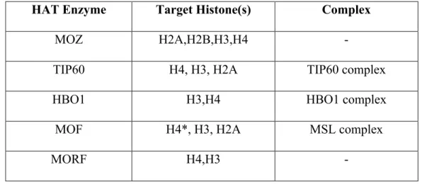
Table 2: The MYST HAT family and their target histone subunits. ... 39
Table 3: The GNAT HAT family and their target histone subunits. ... 39
Table 4: The CBP and P300 HATs and their target histone subunits. ... 40
Table 5: Approved epigenetic drugs. ... 56
Table 6: Selected clinical trials testing epigenetic drugs in combination with immunotherapy treatment. ... 62
Table 7: Selected clinical trials testing epigenetic drugs in combination with cytotoxic or other epigenetic drugs. ... 64
viii
List of figures
Figure 1: Normal and abnormal hematopoiesis. ... 4
Figure 2: Bone marrow accumulation of lymphoblasts in a pediatric ALL patient. ... 5
Figure 3: Pediatric B-ALL subtypes. ... 8
Figure 4: Pediatric T-ALL subtypes. ... 9
Figure 5: The three classes of epigenetic modifications. ... 29
Figure 6: Examples of epigenetic readers, writers, and erasers. ... 30
Figure 7: Differences in DNA methylation levels between normal and cancerous tissues. ... 33
Figure 8: Nucleoprotein complex of DNA and histone forms chromatin. ... 34
Figure 9: Histone tail modifications. ... 37
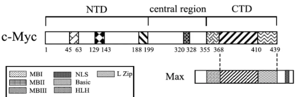
Figure 10: Structure of the human C-MYC oncogene. ... 42
Figure 11: Molecular structure of proscillaridin A ... 71
Figure 12: Proscillaridin A treatments decreases histone 3 acetylation levels and expression of HATs TIP60 and CBP in Molt-4 and Nalm-6 leukemic cell lines. ... 89
Figure 13: Proscillaridin A treatment decreases in the C-MYC oncogene and C-MYC target genes in Nalm-6 and Molt-4 leukemic cell lines. ... 91
Figure 14: Proscillaridin A downregulates genes involved in oncogenic pathways while upregulating genes involved in cellular differentiation. ... 93
Figure 15: Proscillaridin A induces growth inhibition and cell cycle arrest in Nalm-6 and Molt-4 leukemic cell lines. ... 96
Figure 16: Secondary drug screen on ALL and AML cell lines. (Supplementary figure 1). ... 98
Figure 17: Proscillaridin A induces epigenetic effects in Molt-4 and Nalm-6 cell lines. (Supplementary figure 2). ... 100
Figure 18: Expression of P300, CBP, and GCN5 HATs remain stable after proscillaridin A treatmentin Nalm-6 and Molt-4 cell lines. (Supplementary figure 3). ... 102
Figure 19: Proscillaridin A treatment induces changes in gene expression in Molt-4 cell line. (Supplemental figure 4). ... 104
List of initials
FAB: French, American and British WHO: World Health Organization FDA: Food and Drug Administration COG: Children’s Oncology Group
x
List of abbreviations
CLL: Chronic lymphoblastic leukemia CML: Chronic myeloid leukemia ALL: Acute lymphoblastic leukemia AML: Acute myeloid leukemia MLL: Mixed lineage leukemia JAK: Janus kinase
mTOR: Mammalian target of rapamycin HSCT: Hematopoietic stem cell transplantation CNS: Central nervous system
CSF: Cerebral spinal fluid MRD: Minimal residual disease
MeCP2: Methyl CpG binding protein 2 NTD: N-terminal domain
CTD: C-terminal domain MBI/II/III: Myc- box I/II/III MAX: MYC associated factor X HSC: Hematopoietic stem cell DNMT: DNA-methyltransferase
MGMT: O-6-methylguanine-DNA methyltransferase HAT: Histone acetyltransferase
MBP: Methyl binding protein MBD: Methyl binding domain HMT: Histone methyltransferase HDM: Histone demethylase HDAC: Histone deacetylase
DNMTi: DNA methyltransferase inhibitor HDACi: Histone deacetylase inhibitor DHAC: Dihydro-5-azacytidine
FCDR: 5-Fluoro-2’-azacytidine
HATi: Histone acetyltransferase inhibitor GFP: Green fluorescent protein
CMV: Cytomegalovirus ROS: Reactive oxygen species
BET: Bromo domain and extraterminal domain
BETi: Bromo domain and extraterminal domain inhibitors GSEA: Gene set enrichment analysis
MB-3: α-methylene-γ-butyrolactone 3 SAM: S-adenosyl-L-methionine ATM: Ataxia telangiectasia mutated STIM1/2: stromal interaction molecule1/2 SOCE: store-operated calcium entry
Acknowledgements
Over the course of the passed two and a half years, I have worked with many people in the Centre de recherche du CHU Sainte-Justine. I have met many inspiring, talented, generous, caring individuals and each one of them has helped me grow to become the person I am today. For this reason, I would like to take some time to thank a few in particular.
First, my research project would have never been accomplished without Dr. Noël Raynal giving me the opportunity to work in his lab. Without your help, I would not be here today writing the acknowledgement section of my Master’s thesis. I would also like to thank you for helping me complete the research I have conducted since January 2014; without you guiding me to answer the many questions we have asked ourselves, I would probably still be learning how to use a pipette. With your help, I have become a better scientist; especially with all those journal club meetings, presentations, and scientific conventions I have attended since starting to work in your lab. I can’t thank you enough for teaching me what science is about: asking questions and trying to figure out the answers.
I would also like to thank Simon Jacques-Ricard for being the best person in the lab with whom I was able to create interesting decoctions and conduct other non-scientific experiments. Using dry ice, curdled milk, agarose gels, and pretty much anything else we could get our hands on in the lab, we were able to create some very remarkable experiments over the passed two years. I would also like to thank you for supporting Gaspard; I know it was hard for you to always destroy a part of him as you vortexed your tubes, but you did a great job nonetheless.
xii
I would also like to thank Elodie Da Costa for being an awesome workbench partner and a great friend. Du coup, merci de m’avoir appris à parler français quoi! Thank you for having a great sense of humor, making me laugh, and joking around with me on a regular basis. Thank you for letting me borrow your bench top, pipette tips, pens, and western blot trays when you were either not in the lab or not paying attention. I know I could be a little bit annoying when I was invading your bench with papers and books on a daily basis, so I thank you again for not complaining too much. On a more serious note, I would also like to thank you for giving me advice and helping me out with my research; without that, I would not be where I am today. I will always remember that Spanish language we invented: Bueno lucko Elodita aveco tono projecto de researcho!
Next, I want to thank Annie Beaudry for teaching me how to properly conduct different lab experiments and procedures that helped me complete my research project. Thank you for being patient with my many questions about how to make solutions and how to properly run western blots. I would also like to thank you for supporting my ridiculous sense of humor and my constant use of the word “jerky” along with another word I cannot write in this manuscript (I am confident we all know what the word I’m referring to actually is).
Last but not least, I would like to thank my buddy Gaspard. Although you were silent most of the time, smelled like beta-mercapto, and took up literally half my workbench, I really loved having you around.
Chapter 1: Introduction
1.0 Leukemia
Leukemia is a malignant disease affecting normal developmental and maturation processes of white blood cells and lymphocytes [1, 2]. It is described as a progressive loss in normal cell maturation and an increase in cell proliferation, leading to abnormally high levels of undifferentiated cells in the bone marrow and blood stream [1, 2]. The accumulation of undifferentiated white blood cells and lymphocytes also disrupts the bone marrow niche, and consequently affects the maturation and production of other hematopoietic progenitor cells [1-3].
Depending on the blood cell affected, leukemia is separated into two major groups: lymphoblastic and myeloid. Whereas lymphoblastic leukemia only affects T- or B-lymphocytes, referred to as T-ALL or B-ALL, respectively, myeloid leukemia affects all other white blood cells, such as basophils, neutrophils, eosinophils, and monocytes [2]. Leukemia can also be classified as being either acute or chronic, depending on the maturity of the cell affected and the rate of disease progression. In cases of acute leukemia, patients are diagnosed with a rapidly progressing disease [2]. On the other hand, patients diagnosed with chronic leukemia are portrayed as having a slower rate of disease development and progression [2]. There are four main classes of leukemia: chronic lymphoblastic leukemia (CLL), chronic myeloid leukemia (CML), acute lymphoblastic leukemia (ALL), and acute myeloid leukemia (AML) [2, 4].
2
Pathophysiology of each leukemia subtype varies drastically from one main class to another [2, 4]. Hence, each leukemia subtype will have different biological characteristics and some will be more prevalent in certain age groups compared to others [2]. For instance, CLL is most frequently observed in elderly patients and affects cells in the bone marrow compartment and blood stream [2]. Patients can either be diagnosed with an aggressive or a dormant form of CLL, the former requiring therapeutic intervention while the latter does not [2]. Differences in cell biology, such as cell surface markers and chromosomal aberrations, can help classify patients in either the dormant or aggressive CLL categories [2]. In addition, presence of certain mutations or disorders in important tumor suppressor genes – like p53 for instance – are linked to the aggressive CLL subtype and result in a poor prognosis [2, 5].
On the other hand, CML only affects myeloid cell development in the bone marrow, which eventually leads to higher concentrations of undifferentiated myeloid cells in the blood stream [2]. Peak incidence for CML patients is observed between 50 and 60 years of age [2]. The most common chromosomal aberration involved in development of CML is a translocation between chromosomes 9 and 22, also known as the Philadelphia chromosome, forming the BCR-ABL fusion gene [2, 6, 7]. This translocation is observed in approximately 90% of CML patients [2, 6, 7]. There are three main phases in CML that correlate to disease progression and influence prognosis as well as overall survival: a chronic, an intermediate, and a blastic phase [2, 8]. As disease progresses from the chronic phase to the intermediate and blastic phases, there are increasing numbers of immature white blood cells in the bone marrow and blood stream [8]. Eventually, patients are placed into the blastic phase category upon the occurrence of white blood cell infiltration into different organs or greater than 30% blast
counts in their blood stream or bone marrow [8]. The blastic phase is deadly in most cases, usually leading to patients succumbing from their disease within 6 months [2, 8].
Alternatively, ALL is a malignant blood disorder affecting solely T- and B- lymphocytes [2, 9, 10]. ALL essentially alters development of T- and B- cells by inducing an increase in the cell proliferation and survival pathways [2, 10]. Interestingly, ALL is very rarely diagnosed in adults but has very high prevalence in children [2]. Like chronic leukemia subtypes, ALL is associated with numerous chromosomal translocations leading to formation of fusion proteins playing critical roles in disease initiation and progression by affecting important cellular pathways [2, 9, 11].
Like the other leukemia subtypes, AML is described as having an interruption in cell differentiation leading to accumulations of AML blast cells in the bone marrow that will eventually spill into the blood stream [2, 11]. AML is a disease affecting primarily adults, but in rare cases affects children as well [2, 11]. Pediatric cases of AML are associated with many chromosomal abnormalities, the most common one being translocations encompassing the mixed lineage leukemia (MLL) protein [2, 11].
4 Figure 1: Normal and abnormal hematopoiesis.
Differentiation of hematopoietic stem cells into either the myeloid or lymphoid lineages. Cells affected in myeloid and lymphoblastic leukemia are myeloblasts and lymphoblasts, respectively. HSC: hematopoietic stem cell, MSC: myeloid stem cell, LSC: lymphoid stem cell, Ne: neutrophil, Mo: monocyte, Eo: eosinophil, Ba: basophil. [12]
HSC$ MSC$ LSC$ Lymphoblast$ T2Cell$ B2Cell$ Myeloblast$ Megakaryoblast$ Megakaryocyte$ Ne$ Mo$ Eo$ Ba$ Platelet$
2.0 Pediatric Leukemia
Leukemia is the most common pediatric cancer diagnosed every year, representing approximately 30% of all newly diagnosed cases in pediatric patients around the world [11]. The two most common leukemia subtypes diagnosed in pediatric patients are ALL and AML [11]. Approximately 85% and 15% of acute leukemia cases in children are ALL and AML, respectively [13, 14].
The following sections describe the biology behind pediatric ALL and AML development and current treatment methods for each leukemia subtype.
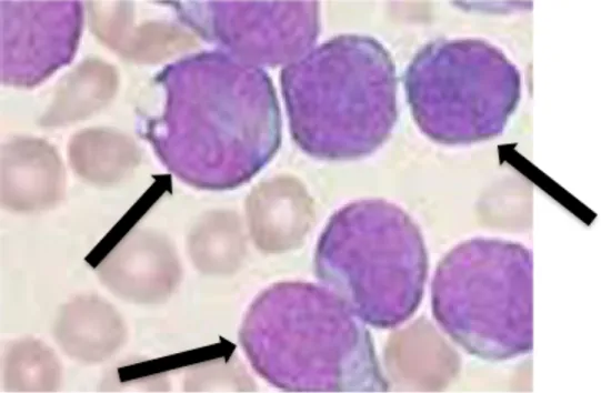
Figure 2: Bone marrow accumulation of lymphoblasts in a pediatric ALL patient.
In patients with ALL, there is an accumulation of leukemic blasts in the bone marrow, which disrupts development of other blood cells. Leukemic blasts are described as having very large nuclei, as shown by the black arrows.
6
2.1 Pediatric ALL, Current Treatments, and Relapse
2.1.1 Biology of ALL
Acute lymphoblastic leukemia (ALL) is characterized as a blood malignancy that induces a decrease in differentiation and an increase in proliferation in lymphoid progenitor cells [15, 16]. These events lead to an overpopulation of the bone marrow compartment and blood stream with immature leukemic cells, also called “blasts”. As a consequence, accumulation of blast cells affects normal blood cell development and impairs hematopoiesis [15, 16]. Depending on the lymphoid progenitor affected, ALL can be subdivided into two main categories: B- and T-ALL, the most common form in children being B-ALL [16, 17]. ALL accounts for approximately 25% of all pediatric cancers diagnosed every year with incidence peaking in children between the ages of 2 and 5 [18, 19]. Although current treatment methods cure over 85% of pediatric patients diagnosed with ALL in developed countries worldwide, patients who either relapse or experience resistance to treatment have a very poor outcome portrayed by an overall survival of approximately 25% [10, 20]. Hence, because of its prevalence, ALL is the leading cause of death by disease in children.
Molecular mechanisms underlying B- and T-ALL include inactivation of tumor suppressor genes and irregular activation of oncogenes and signal transduction pathways [21-23]. These genetic aberrations affect key regulatory processes, producing highly proliferative blast cells with unlimited self-renewal and cell-survival properties [21-23]. Chromosomal lesions, such as abnormal chromosomal numbers and structural abnormalities – especially translocations – are often identified in the pediatric ALL population [21-23]. Evidently, these chromosomal lesions differ between the two ALL subtypes.
The most common chromosomal translocation observed in pediatric B-ALL is the translocation between ETS variant 6 (ETV6), located on chromosome 12, and Runt-related transcription factor 1 (RUNX1), located on chromosome 21, termed the ETV6-RUNX1 fusion protein [24]. This translocation accounts for roughly 25% of all pediatric B-ALL cases [24]. In normal cells, both proteins act as transcription factors required in hematopoiesis and normal cell differentiation [24]. Fortunately, patients with the ETV6-RUNX1 translocation have an elevated survival rate [25]. The TCF3-PBX1 fusion protein, made up transcription factor 3 (TCF3) and pre-B cell leukemia homeobox 1 (PBX1), accounts for approximately 5% of B-ALL cases, and is associated with a good prognosis, despite the slight risk in CNS relapse [26]. For this reason, patients diagnosed with TCF3-PBX1 leukemia are given higher doses of CNS preventative therapies [26]. Next, translocations involved in forming the BCR-ABL1 fusion protein, formed from the fusion between breakpoint cluster region protein (BCR) and Abelson murine leukemia viral oncogene homolog 1 (ABL1), account for about 3% of cases and are correlated with good patient outcomes due to the recent introduction of imatinib in their treatment regimens [27]. Finally, translocations involving the mixed lineage leukemia (MLL) protein, which occur at a much higher frequency in patients under two years old, account for about 5% of B-ALL cases [19, 24, 28].
8 Figure 3: Pediatric B-ALL subtypes.
Pie chart of pediatric B-ALL subtypes.
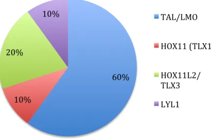
Chromosomal translocations in T-ALL are not as well characterized as their B-ALL counterparts. Pediatric patients are classified into four categories, each of which is associated with upregulation of different oncogenes [24, 29]. The four major oncogenes present in T-ALL are HOX11, LYL1, TAL-LMO2, and HOX3 [24, 29]. Translocations between the T-cell receptor and the HOX11 oncogene represent a distinct subset of patients part of the HOX11 category. Other than these noticeable chromosomal translocations, T-ALL can also be illustrated by abnormal NOTCH1 signaling that impairs T-cell differentiation in approximately half of T-ALL cases [30].
Hyperdiploidy ETV6-‐RUNX1 Trisomy 4 and 10 TCF3-‐PBX1 MLL-‐AF4 BCR-‐ABL1 Hypodiploidy
Figure 4: Pediatric T-ALL subtypes.
Pie chart of the four major oncogene subtypes in pediatric T-ALL.
In addition, chromosomal and genetic alterations also take part in development of acute leukemia in children. For example, Down’s syndrome (trisomy 21) increases the risk to develop pediatric ALL or AML significantly [31]. Patients with Down syndrome are frequently associated with ETV6-RUNX1 chromosomal translocation and are often categorized as high-risk mostly because of frequent relapse rates and lower overall survival [32].
Gain or loss of entire chromosomes, termed hyperdiploidy and hypodiploidy, respectively, are also frequently observed in pediatric patients [19, 24]. Chromosomes X, 4, 6, 10, 14, 17, 18, and 21 are the ones most often gained in ALL patients with hyperdiploidy and make up approximately 30% of all cases [19, 27]. These patients are associated with positive outcome [27]. Hypodiploidy, on the other hand, is much less frequent, observed in approximately 1% of patients, and is correlated to a poor prognosis [27].
60% 10% 20% 10% TAL/LMO HOX11 (TLX1) HOX11L2/ TLX3 LYL1
10 2.1.2 Treatment of pediatric ALL
Because ALL is such a diverse disease, the diagnostic of the correct subtype is critical in determining which treatment methods a patient should undergo and whether or not a given treatment will fail [33]. Hence, at the time of diagnosis, risk stratification for a patient is established and is an important aspect in attaining overall survival [33, 34]. Therefore, prior to starting treatment, patients are labeled as standard, intermediary, or high-risk [11]. Two important clinical characteristics considered at the time of diagnosis are age and white blood cell count [11, 33-35]. Favorable outcomes are observed in patients between the ages of 1 and 10, whereas unfavorable outcomes are observed in patients under 1 year of age and older than 10 years of age [33]. Also, white blood cell counts greater than 50,000 cells per µL of blood are predicted to have an unfavorable outcome [33].
Another important characteristic examined is leukemic cell penetration into sanctuary sites, defined as areas in the body that are difficult to target with conventional chemotherapy. Two common sanctuary sites in leukemia are the central nervous system (CNS) and testis [11, 33]. Penetration of leukemic cells into these organs is correlated with an unfavorable outcome and is usually involved in patient relapse [11, 33].
After establishing risk stratification, patients undergo treatment according to their risk category [11, 33]. Generally speaking, current treatment methods include three main stages [11, 33]. The first step in ALL treatment is induction therapy, lasting between 4-6 weeks; the goal of induction therapy is to eliminate all leukemic blasts from the bone marrow [11, 33]. Four medications are used in induction therapy: vincristine, prednisone, L-asparaginase, and an anthracycline (either doxorubicin or daunorubicin) [11, 33]. The usual two-drug regimen consists of vincristine followed by prednisone, which leads to remission in about 85% of
patients [11]. Addition of a third drug – either L-asparaginase or an anthracycline – further increases the remission rate to approximately 95% [11]. In high-risk patients, a fourth drug, usually an anthracycline or methotrexate, is added to the regimen in order to attain remission [11].
Since ALL penetration into the CNS is correlated with increased rates of patient relapse, CNS preventative therapy has also been established in treating the disease [11]. CNS preventative therapy includes intrathecal methotrexate injections followed by low to moderate cranial irradiation to eliminate any leukemic cell that crossed the blood-brain barrier [11]. CNS preventative therapy is very efficient, and leads to good prognosis in a majority of cases with minimal cytotoxicity if started at the same time as chemotherapy regimens [11, 36]. Unfortunately, some patients develop secondary cancers due to side effects of treatment, such as non-Hodgkin lymphoma and cancers affecting the brain and thyroid organs [37].
Induction therapy is followed by the second phase in ALL treatment called consolidation therapy. The goal of consolidation therapy is to eliminate any remaining leukemic cell population from the patient’s bone marrow and blood stream in order to prevent any further disease progression or relapse [11, 33]. Treatment protocols differ in length and intensity, usually lasting 6-9 months, but can last longer if a patient is in the high-risk category [33]. During this phase, patients receive higher doses of drugs formerly used in induction chemotherapy. Different classes of cytotoxic drugs not previously used during induction therapy, such as mercaptopurine, etoposide, cyclophosphamide and cytarabine, can also be added to treatment regimens in order to prevent resistance [11, 33].
The third and final stage of ALL treatment is maintenance therapy, where patients receive low-dose of chemotherapy drugs for approximately 2-3 years in order to prevent
12
disease relapse [11, 33]. During this phase, treatment protocols include weekly methotrexate and mercaptopurine administrations, in addition to prednisone and vincristine on a monthly basis [11, 33].
2.1.3 Personalized Treatments for ALL
Nowadays, personalized approaches exist taking patient risk stratification into account. Depending on chromosomal aberration identified in ALL patients, treatments can be administered to specifically target cancer cells with minimal effects on healthy cells. Several classes of these targeted therapies exist for ALL, with a few of these currently undergoing clinical trials [9, 27, 34, 38].
First, monoclonal antibodies can be used in treating specific ALL cases having cell surface receptors or markers that can potentially be targeted [9, 38]. A monoclonal antibody can either be conjugated or unconjugated; the former being attached to a radioactive or cytotoxic compound whereas the latter is not [9, 38]. An unconjugated monoclonal antibody identifies a target on the surface of the cell and initiates either antibody-dependent cell mediated toxicity or complement-dependent cytotoxicity [9]. On the other hand, conjugated monoclonal antibodies identify and bind onto their target cell and get internalized along with their conjugate. This phenomenon enables the release of the cytotoxic compound into the cell, causing cell death [38]. Numerous examples using unconjugated monoclonal antibodies exist in treating ALL. For instance, rituximab, a chimeric murine and human monoclonal antibody, specifically targets CD20 surface antigen present on B-lymphocytes enabling its use as a target in B-ALL cases [38]. CD20 expression is observed in over 50% of pediatric B-ALL
cases, and is usually upregulated after chemotherapy treatment [9, 39, 40]. The monoclonal antibody alemtuzumab specifically targets CD52 surface antigen present on T- and B-ALL cells and can be used in relapsed or refractory cases of pediatric ALL [9, 38]. Although response rates for alemtuzumab are generally very low, patients with the t(12;21) translocation have a slightly higher sensitivity to treatment compared to patients without this translocation [9, 41]. Another popular monoclonal antibody is epratuzumab. Epratuzumab targets CD22 surface antigen present primarily on pre-B cells, and can therefore be used in treating pediatric pre-B ALL [9, 38]. Recent studies suggest it also has promising effects on relapsed B-ALL cases in combination with chemotherapy, inducing a complete response in the majority of cases [9, 38, 42, 43].
Compared to unconjugated monoclonal antibodies, conjugated monoclonal antibodies appear to be more efficient in treating pediatric ALL [38]. Inotuzumab ozogamicin is a monoclonal antibody attached to calicheamicin, an anti-tumor antibiotic showing promising results in preliminary tests on patients with refractory ALL [38, 44]. Its mode of action specifically targets CD22-positive cells, which incorporate calicheamicin leading to cellular toxicity [38]. Additionally, SAR3419 is a monoclonal antibody linked to the natural cytotoxic compound dregeanin DM4 [45]. SAR3419 is used to target CD19, a cell surface marker expressed on all immature lymphocytes and can therefore be used in treating pediatric B-ALL cases [9, 45]. It was also recently identified as having synergistic effects when administered in combination with chemotherapy drugs presently used in induction therapy in pediatric ALL [46].
Second, another important group of compounds used in targeted ALL treatments are tyrosine kinase inhibitors. Because constitutive expression of tyrosine kinase inhibitors plays
14
an important role in development of leukemia and eventually resistance to treatment, targeting these receptors is a great way to also target development and progression of the disease [9, 34]. Specific molecules can target the two major groups of tyrosine kinase receptors in pediatric ALL: the BCR-ABL (in Philadelphia positive ALL, referred to Ph+ ALL) and the FLT3 tyrosine kinases [9, 34].
The recent discovery and approval of imatinib in 2013, a BCR-ABL tyrosine kinase inhibitor, greatly changed the overall long-term survival of pediatric patients with Ph+ leukemia from approximately 40% to 85% [9, 47]. In fact, when administered with conventional chemotherapy, patients treated with imatinib had similar long-term survival as patients undergoing hematopoietic stem cell transplantation [48]. Unfortunately, like many other types of treatments for pediatric ALL, resistance to treatment develops [49, 50]. Although very rare in children, point mutations of the ABL domain appear to be one of the leading causes of resistance to treatment and eventually causing death of the patient [9, 34, 49, 51]. After discovery of imatinib, development and approval of dasatinib and ponatinib, second and third generation BCR-ABL tyrosine kinase inhibitors respectively, quickly went underway in order to counter the resistant ALL cases [34, 51-53]. These two drugs show promising results in recent clinical trials on targeting BCR-ABL resistant cells. In addition to being more effective against Ph+ ALL, they are more efficient at crossing the blood-brain barrier and targeting leukemic cells in the central nervous system to prevent any potential relapse [9, 54].
FMS-like tyrosine kinase 3 – also known as FLT3 – is a tyrosine kinase receptor usually present in CD34+ pre-B progenitor cells that is essential in normal hematopoiesis [9, 34, 55]. However, when abnormally expressed by activating mutations, FLT3 upregulates proliferation pathways leading to B-ALL development [9, 34]. Although FLT3
gain-of-function mutations are rare in pediatric ALL cases, therapies targeting mutant forms of the protein consist mainly of lestaurtinib in combination with chemotherapy are administered to patients diagnosed with FLT3-driven ALL [9, 56]. Although this combination has shown promising results in certain cases, it still needs to be improved in order to attain better complete response rates on a regular basis [9].
Third, drugs that target nucleic acid synthesis are also used in treating certain cases of pediatric ALL. Initially, this drug family was rarely used in treating pediatric ALL since they are not specific and extremely toxic, but recent improvements have enabled use of these compounds in either high-risk or relapse cases [9, 57]. Clofarabine, a molecule inhibiting DNA polymerase and DNA repair among other things, is sometimes used in combination with conventional chemotherapy in relapse or resistant cases [58]. In other instances, clofarabine is used as a monotherapy in patients that already received high doses of cytotoxic therapy [59-61]. More recently, the Children’s Oncology Group (COG) is evaluating clofarabine administration in combination with drugs used during induction therapy for high-risk pediatric ALL patients [9]. Another compound in this category is the prodrug nelarabine; once it enters a cell, it is metabolized to a nucleoside analog and rapidly gets phosphorylated [9]. It is then inserted into the DNA sequence, prevents DNA synthesis, and induces apoptosis [9, 62]. Interestingly, since nelarabine is more potent in T-cell malignancies compared to B-cell malignancies, it is more often used on T-ALL patients who either relapse or are unresponsive to treatment [9, 63]. It is currently undergoing a clinical trial on a potential addition in induction therapy for pediatric T-ALL cases [9].
Fourth, compounds that inhibit serine and threonine kinases, responsible for irregular growth pathways, are also used in targeted therapy against pediatric ALL. Overexpression of
16
the serine/threonine kinases leads to cell proliferation and development of leukemia [9]. There are several types of serine/threonine kinases that can potentially be targeted for therapy in pediatric ALL [9, 34]. First, the Janus kinase (JAK) family of tyrosine kinases are great targets in high-risk pediatric ALL patients in which the JAK-STAT pathway is aberrantly expressed [9, 34]. The JAK pathway is usually overexpressed in high-risk B-ALL patients, as well as in children with ALL and Down syndrome [9, 34, 64]. Unfortunately, not much is known about the efficacy of JAK-STAT inhibition in pediatric ALL since clinical trials are currently underway [9, 34].
Another type of serine/threonine kinase that is studied in pediatric ALL cases is the mammalian target of rapamycin (mTOR). Aberrant activation of the PI3K/Akt/mTOR pathway is involved in accelerated cell growth and proliferation, which leads to development of leukemia [9, 34, 65]. Hence, targeting the mTOR pathway using mTOR inhibitors is an efficient way to target ALL with a hyperactive mTOR signaling pathway. In recent years, preclinical and clinical studies have determined a synergistic effect between mTOR inhibitors and chemotherapy drugs used in induction and maintenance therapies, such as cyclophosphamide, methotrexate, doxorubicin, etoposide, asparaginase, prednisone, and vincristine [66-70]. Several mTOR inhibitors exist in treating pediatric ALL. mTOR inhibitors, such as rapamycin or any of its analogs, have been greatly studied in vitro on ALL cell lines – especially pre-B ALL – and seem to have positive effects [34, 71]. In addition, combination studies in Ph+ leukemic cell lines demonstrate synergistic effects when rapamycin is combined with daunorubicin in vitro [72]. Not only did the combinational approach demonstrate a higher cytotoxicity in these cell lines, but it also increased autophagy and blockage of the cell cycle when compared to daunorubicin monotherapy [72].
Development of steroid resistance in pediatric ALL patients is one of the leading causes of treatment failure [68]. Interestingly, rapamycin treatment can actually sensitize steroid-resistant cells, which can potentially lead to countering steroid-related resistance [68]. These preclinical results provided a rationale to pursue clinical trials in pediatric ALL patients. Hence, ongoing clinical trials in combining rapamycin or its analog temsirolimus with cytotoxic compounds have been studied in relapsed or resistance pediatric ALL (NCT01403415) [73].
Fifth, targeting a cancer cell’s survival ability is another suitable approach for chemotherapy. In normal cells, there is a balance between apoptosis and cell survival mechanisms. In cancer cells, however, the balance is shifted towards an interruption of the apoptotic pathways and upregulation of survival genes and their downstream effectors [9]. Since there are many genes involved in pro-survival mechanisms in leukemic cells, several agents can be administered to patients depending on their gene expression profiles [9, 34]. Bortezomib, a proteasome inhibitor, prevents ubiquitin-dependent proteasome degradation, which subsequently inhibits the action of NF-κB and activates pro-apoptotic and cell cycle regulators such as p53, p21, p27, and Bax [9, 34]. The overall effect of bortezomib treatment is to increase effects of chemotherapy drugs by lowering the apoptosis threshold of leukemic cells [9, 34, 74-76]. Treatment of ALL cell lines with bortezomib has shown positive effects, prompting its use in numerous clinical trials in pediatric ALL patients [77]. As a monotherapy, bortezomib has been shown to be inefficient. As demonstrated in a phase I combination clinical trial with reinduction chemotherapy drugs, it has very promising effects in resistant pediatric ALL [9, 34, 78, 79]. The subsequent phase II part of this trial further demonstrated
18
positive effects of bortezomib combination with reinduction cytotoxic treatment in both B- and T-ALL resistant patients [34, 79].
Another pro-survival target in pediatric ALL are heat shock proteins (HSPs) – more specifically Hsp90. These proteins are involved with leukemia cell formation by trafficking and stabilizing oncogenes required in development of a malignant proliferative phenotype with abnormal survival properties [9, 80, 81]. In addition, elevated amounts of Hsp90 in patients are correlated with poor prognosis; hence, targeting these proteins in high-risk cases can potentially increase overall survival [81]. In preclinical studies, tanespimycin, an Hsp90 inhibitor, repressed growth of ALL cells in vitro and has even been shown to synergize with imatinib in Ph+ ALL cell lines [9, 82]. More recently, Akahane and colleagues demonstrated that NVP-AUY922, a much stronger inhibitor for the Hsp90 protein, promotes apoptosis in T-ALL cells [83]. This product is already in clinical trials for solid tumors and can potentially be an interesting candidate drug in treating T-ALL pediatric patients [84]. Despite recent advances in Hsp90 inhibitor development, these drugs are not frequently used in clinic since a therapeutic window has not yet been established [83, 85].
Other anti-apoptotic proteins frequently overexpressed in pre-B-ALL and T-ALL are members of the Bcl-2 family. Not only are these proteins important in regulating apoptosis, but they also play key roles in resistance to treatment during induction therapy [86]. Development of navitoclax and obatoclax, two Bcl-2 family inhibitors, was necessary in order to treat patients with highly resistant forms of ALL [87]. Both inhibitors show promising effects in vitro on pediatric ALL cell lines in addition to clinical studies on adult patients suffering from ALL and AML [88-91]. However, clinical trials have yet to be conducted on
pediatric patients [88-91]. Thus far, preclinical anti-Bcl-2 therapy has been shown to be more effective on t(4;11) ALL cell lines compared to all other forms of the disease [92].
2.2.4 Relapsed and Resistant pediatric ALL
While the majority of pediatric ALL patients are cured with current chemotherapy regimens, approximately 20% experience a relapse, with boys relapsing more frequently than girls [9, 11, 34]. Research on relapsed ALL patients has identified leukemic blast populations that have acquired chromosomal aberrations and genetic mutations, making them more resistant to chemotherapy treatment compared to blasts isolated at initial diagnosis [93-98]. Furthermore, genomic studies on blast cells present at relapse have determined an overexpression of the DNA repair and cell survival gene pathways compared to blasts at diagnosis [93, 99].
ALL relapse is primarily caused by expansion of a clonal population leading to treatment resistance [93]. Recent studies on ALL relapse have concluded it is a clonal disease. In fact, studies by Mullighan and colleagues have determined that cells present at relapse are also present at initial diagnosis at a very low concentration. This cell population then undergoes clonal expansion, populate the bone marrow and sanctuary sites in the body, and drive disease relapse [93, 95].
There are many factors that determine how clonal expansion affects the development of chemoresistance. Mutations and deletions of tumor suppressor genes may appear in the relapse blast cells that were not present in the initial cell population [93]. For example, a study by Hof and colleagues determined that p53 tumor suppressor gene, deleted in over 10% of the
20
relapse population, was correlated to a poor prognosis [100]. Deletions of the zinc finger protein IKZF1 were commonly identified in patients with relapsed Ph+ B-ALL [101, 102]. In these studies, IKZF1 deletions were correlated with a negative prognosis compared to patients diagnosed with Ph+ B-ALL in which IKZF1 was still present [93, 101, 102]. Resistance to prednisone and mercaptopurine treatment was also linked to deletions of MSH6, a gene in the mismatch repair pathway, leading to overall poor patient outcomes [103, 104]. Additionally, prednisone resistance has been linked to mutations of glucocorticoid receptors in the relapse population [95, 104, 105]. In general, a patient’s response to prednisone administration is a determining factor in development of treatment resistance or disease relapse [68]. Hence, patients having a decreased response to prednisone treatment also have higher rates of resistance and relapse after initial induction therapy [68]. In other cases, epigenetic mechanisms play critical roles in development of resistance to treatment [106].
At the time of relapse, protocols are implemented for risk stratification [33, 93, 107]. Time since complete remission is the most important factor in determining a positive or negative prognosis [33, 93, 107]. Patients can be placed into one of three different categories depending on the time between complete remission and relapse [33, 107]. Generally speaking, patients who relapse less than 36 months after remission are considered the most difficult to treat and have the worst prognosis with an overall survival rate of less than 30% [33, 93, 107, 108]. On the contrary, patients relapsing after three years are easier to treat, as demonstrated by an overall survival rate exceeding 50% [33, 93, 107]. Another risk factor taken into consideration are the sites of relapse [33]. Most commonly, isolated marrow relapse occurs in approximately 60% of the time and is correlated to a negative prognosis [33, 109]. The majority of remaining extramedullary relapse cases are located in the CNS and testicles,
comprising approximately 25% and 5%, respectively [33, 109]. In some rare situations, a combination of bone marrow and extramedullary relapse can occur, faring a negative prognosis [33, 109]. Furthermore, because patients diagnosed with T-ALL have a greater chance of relapsing early compared to patients with B-ALL, they are more often correlated with a poor outcome [93, 109, 110]. Unfortunately, between 7-23% of patients with T-ALL relapse will have a 5-year overall survival, indicating the need for new types of therapies [93, 109, 110].
With the development of novel chemotherapy drugs that are capable to reduce the incidence of CNS and testicular relapse, isolated extramedullary relapse – or relapse occurring in sites other than the bone marrow – is uncommon nowadays [111]. Patients with isolated CNS relapse are defined as having leukemic blasts in the cerebral spinal fluid (CSF) without any blasts present in the medullary compartment of the bone marrow [111]. Similarly, patients with isolated testicular relapse do not have blast cell accumulations in the bone marrow but will experience enlargement of one or both testicles due to blast infiltration [111]. According to the majority of research on relapse patients, the main reason why these two organs are ideal sanctuary sites for leukemic blasts is the inability for chemotherapeutic agents to penetrate the blood-brain and blood-testes barriers [111]. Other studies on testicular relapse cases points towards a cooler temperature in the testes compared to the rest of the body as another potential reason of relapse, which may lead to decreased efficacy of certain chemotherapy drugs at that location [112].
Some important factors at the time of diagnosis can actually predict which patients are predisposed to isolated extramedullary CNS or testicular relapse [111]. For instance, in patients with T-ALL, certain chromosomal translocations such as t(4;11) forming the
MLL-22
AF4 fusion protein and t(9;22), and presence of blast cells in the CNS at the time of diagnosis are all predictors of CNS relapse [113, 114]. In addition, patients diagnosed at an older age and who have a delayed response to induction therapy are also more prone to isolated CNS relapse [113, 115]. Moreover, patients diagnosed with T-ALL during the early puberty years are more prone to developing an isolated testicular relapse [116].
Once risk stratifications have been established, there are multiple ways to go about treating patients experiencing a relapse. Depending on their risk stratification, most patients undergo strict treatment regimens that will decrease blast counts in their sites of relapse. For example, treatment schedules for high-risk patients will include multi-drug chemotherapy combinations prior to undergoing hematopoietic stem cell transplantation (HSCT) once second remission is achieved [73, 117-120]. On the other hand, low and intermediate risk patients will undergo intensive rounds of chemotherapy without HSCT [73, 117, 118]. Importantly, the cytotoxic drugs used in relapse reinduction were ones not previously used in the initial induction phase [93]. Unfortunately, finding the best drug combinations for relapse patients is extremely difficult and not much progress has been made over the past 20 years in increasing overall survival [93]. In cases of isolated CNS and testicular relapse, intensive chemotherapy regimens using high-doses of methotrexate, cytarabine, steroids and asparaginase are undertaken [121, 122]. In some testicular relapse cases, removal of the affected enlarged testicle is sometimes performed [118, 123]. Usually, unilateral or bilateral testicular irradiation is also completed to prevent any subsequent disease [118, 123].
Unfortunately, even after breakthrough discoveries in pediatric ALL treatment, many patients experience multiple relapses. In addition, their chances of overall survival decrease for every ensuing relapse from approximately 44% to 27% in second and third relapses,
respectively [93, 124]. Another point that raises concern is the inability to treat some patients adequately after first relapse since they experience high toxicity and many devastating side-effects due to treatment [93, 125, 126]. Hence, novel treatment methods need to be developed and implemented in order to achieve better overall survival rates for pediatric ALL patients, preferably using less cytotoxic compounds.
24
2.2 Pediatric AML, Current Treatments, and Relapse
2.2.1 Biology of pediatric AML
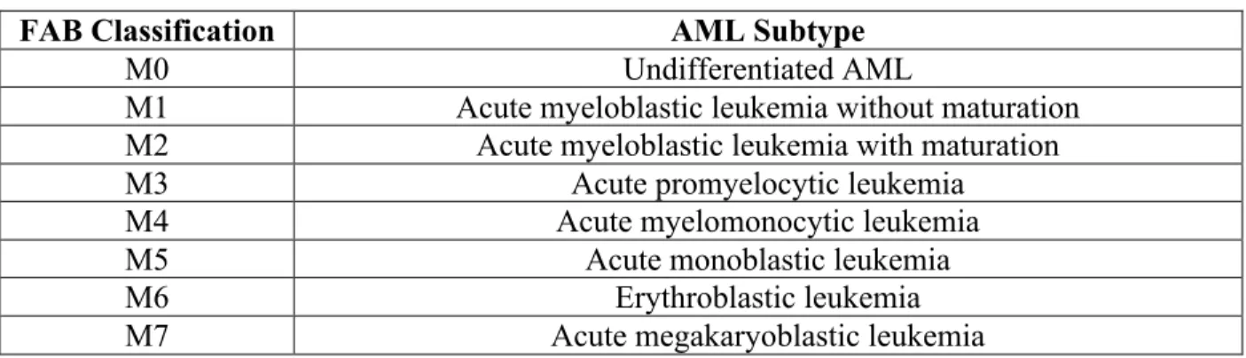
Acute myeloid leukemia, also known as acute non-lymphoblastic leukemia, is a heterogenous malignant disease affecting myeloid, erythroid, and megakaryocyte cell precursors [11, 127, 128]. Overall, long-term survival in pediatric AML cases has dramatically improved over the years, reaching roughly 50-65% in developed countries worldwide [11, 27]. AML can be subdivided into seven different classes in adults and children according to the French, American, and British (FAB) and World Health Organization (WHO) classification systems depending on the blood cell progenitor affected and its level of differentiation [129]. For a detailed list of AML subtypes according to FAB classifications, see table 1.
Table 1: The French, American, and British (FAB) classifications of AML
FAB Classification AML Subtype
M0 Undifferentiated AML
M1 Acute myeloblastic leukemia without maturation
M2 Acute myeloblastic leukemia with maturation
M3 Acute promyelocytic leukemia
M4 Acute myelomonocytic leukemia
M5 Acute monoblastic leukemia
M6 Erythroblastic leukemia
M7 Acute megakaryoblastic leukemia
AML is a multifactorial disease involving cooperative mechanisms between several cellular pathways in order to trigger disease onset [130, 131]. Development and progression of AML requires two important genetic abnormalities classified as being either type 1 or type 2 mutations [14, 132, 133]. Type 1 mutations affect genes involved in the signal transduction pathways which increases cell proliferation and inhibits apoptosis, whereas type 2 mutations
include chromosomal rearrangement of transcription factors resulting in formation of fusion proteins that impair differentiation processes [14, 134]. Consequently, when both type 1 and 2 mutations are present at a given time, the resulting groups of cells acquire a high self-renewal capacity and an uncontrollable proliferative ability leading to onset of AML [134].
Like ALL, there are numerous predispositions associated with AML. For instance, peak age incidence for pediatric patients is either in the first four weeks after birth (also referred to as congenital leukemia) or during adolescence, with boys being more prone to developing the disease [11]. In addition, children with Down syndrome have a higher risk of developing AML; however, these patients are very well treated, with overall survival attaining 85% [135, 136].
After diagnosing pediatric AML, patients undergo risk stratification assessments in order to determine appropriate treatment methods to attain long-term survival [11, 27]. The most important risk consideration is age: adolescent patients have a worse prognosis compared to patients below two years of age [11]. In addition, obese patients tend to be negatively correlated with long-term survival [11]. Patients with M0 and M7 subtypes are usually correlated to prominent treatment responses [11]. Other characteristics like the t(8;21) and t(15;17) chromosomal translocations are generally correlated with positive outcomes [11]. Another important risk factor taken into consideration prior to beginning treatment is high blast concentrations at diagnosis, which is usually indicative of a resistant disease associated with very high death rates [137]. Because onset of AML is linked to many mutated genes, one way to improve treatment methods is to identify and target these mutations prior to beginning chemotherapy. The ability to detect chromosomal aberrations and genetic mutations in AML is critical since they are also found to play important roles in relapsed and resistant AML
26
[127]. For example, high levels of AML1-ETO or CBFB-MYH11 fusion proteins are known to be associated with decreases in both patient survival and treatment responses [127, 138]. Hence, by specifically targeting these fusion proteins in personalized therapies, patients can potentially achieve better outcomes.
2.2.2 Treatment of pediatric AML
AML treatment consists of induction and consolidation therapies. First, induction therapy specifically aims at decreasing leukemic cell concentrations in order to attain remission [11, 139]. Induction therapy in AML involves combining cytosine arabinoside with either doxorubicin or daunorubicin [11, 139]. For higher risk AML patients, additional drugs are administered, such as etoposide and thioguanine [11, 139]. Similarly to ALL therapy, CNS directed therapy is also given to patients in order to prevent leukemic blast homing into the CNS [127].
Once remission is attained, the second phase of treatment, termed consolidation therapy, is initiated. Consolidation therapy involves administration of cytosine arabinoside and etoposides at very high doses [11, 139]. In some patients, certain cytogenetic aberrations are correlated with negative disease outcomes. Hence, in order to increase their quality of life and overall survival, hematopoietic stem cell transplantation is conducted immediately after induction therapy [11, 93, 127]. Furthermore, in cases where minimal residual disease (MRD) is elevated at the end of the induction phase, patients undergo hematopoietic stem cell transplantation (HSCT) in order to prevent any occurrence of relapse [73, 93, 101, 140].
2.2.3 Relapse of pediatric AML
Despite recent treatment advances, approximately 35-45% of pediatric patients diagnosed with AML will experience a relapse [9, 141-143]. The two most common relapse sites are the bone marrow and CNS, the former being much more prevalent than the latter [127]. Patients who experience a relapse immediately undergo reinduction therapy. In AML, drugs used in treating relapsed patients include cytarabine, fludarabine, and anthracycline [144, 145]. In addition to these drugs, patients also receive CNS targeted therapy to limit leukemic cell infiltration into sanctuary sites [127]. Ideally, once second remission is attained, patients receive HSCT [127].
3.0 Epigenetics
Epigenetics is the study of any cellular event that changes gene activity or expression without changing the actual DNA sequence [146]. These changes in gene expression can then be passed down to daughter cells during meiosis and mitosis [146-149]. Many types of epigenetic modifications exist in order to conduct cellular epigenome reprogramming that are regulated by a variety of different enzymes [146, 147, 149, 150]. Other than methylation and acetylation – the two most studied types of modifications – epigenetic regulation can also be directed by phosphorylation, ubiquitination, and sumoylation [146, 147, 149, 150].
The field of epigenetics can be separated into three main classes: DNA methylation, chromatin modifications, and microRNAs [146, 151]. Overall, epigenetic changes are necessary for normal cell development, differentiation, and survival; hence, any deregulations in epigenetic pathways may have drastic consequences on the cell’s phenotype [146, 147, 149, 150]. Unlike somatic mutations, changes to a cell’s epigenome are actually reversible and therefore constitute interesting therapeutic targets [152, 153].
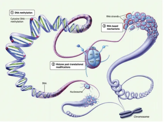
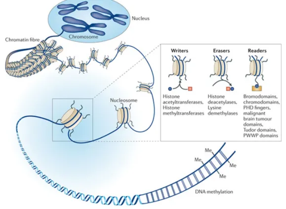
Figure 5: The three classes of epigenetic modifications.
(1) DNA methylation affects cytosine residues (red circles) of CpG sites on primarily on promoter regions. (2) Post-translational histone modifications occur at different histone tail residues. H3 and H4 subunits are most studied regarding gene expression patterns. (3) RNA-based mechanisms by short noncoding RNA sequences, such as micro-RNAs also regulate gene expression levels. These sequences are transcribed from DNA but do not undergo translation [154].
Epigenetic proteins can be functionally characterized as being writers, erasers, or readers [155, 156]. Epigenetic writers are enzymes that transfer epigenetic marks onto histones or DNA residues [155, 156]. Popular epigenetic writers are histone and DNA methyltransferases (DNMTs) and histone acetyltransferases (HATs) [156]. On the contrary, epigenetic erasers remove epigenetic marks from histones and DNA (i.e. histone and DNA demethylases and histone deacetylases) [156]. Finally, epigenetic readers are proteins that bind onto these epigenetic marks, thereby transmitting an epigenetic signal by either activating or silencing gene transcription through recruitment of other proteins [156]. There exist many
30
types of epigenetic readers that bind to the multitude of epigenetic marks previously mentioned. For instance, the bromodomain proteins recognize histone residue acetylation, whereas Methyl CpG binding protein 2 (MeCP2) binds onto methylated DNA residues [156].
Figure 6: Examples of epigenetic readers, writers, and erasers.
Depending on their function, epigenetic enzymes can be classified as being writers, erasers, or readers [157].
3.1 DNA Methylation
DNA methylation refers to covalent addition of a methyl group by a DNMT enzyme to cytosine residues located primarily on CpG dinucleotides. The methyl group donor for this reaction is S-adenosyl-L-methionine (SAM) [158]. Areas in the genome are enriched in CpG sites, which are refered to as CpG islands. These genomic DNA sequences are defined as stretches of approximately 200 base pairs comprising of more than 50% cytosine and guanine repeats [159]. CpG islands are scattered throughout the human genome, but are more concentrated in repetitive sequences, imprinted genes, and over half of gene promoters [160-162]. In normal cells, DNA methylation is required for maintaining a given gene expression pattern when genetic material is passed from mother cell to daughter cells [151]. It is also necessary in genetic imprinting and X chromosome inactivation [160, 163-165]. In addition, CpG island methylation is necessary for cell differentiation and normal cell development [162]. It has also been shown to play important roles in expressing tissue-specific genes [162]. There are four main types of DNMTs: DNMT1, DNMT2, DNMT3a, and DNMT3b. DNMT1 is required for maintaining a cell’s methylation patterns as it divides into two daughter cells thereby transmitting epigenetic information from one generation of cells to the next [160, 166, 167]. Hence, as a cell divides, DNMT1 uses the parental strand as a template to appropriately conserve methylation patterns [160, 165]. On the other hand, DNMT3a and DNMT3b are enzymes responsible for addition of new methyl groups to cytosine residues that were not previously methylated, also known as “de novo” DNA methylation. Interestingly, DNMT2 does not methylate DNA sequences, but targets cytosine residues in transfer RNA (tRNA) instead [168].
32
DNA methylation can lead to gene silencing by a variety of different mechanisms. First, DNA methylation acts as a direct transcriptional silencer mainly by recruiting DNA methylation readers, also known as methyl-binding domains (MBD) and other chromatin repressor complexes onto CpG islands of gene promoters [150, 169-171]. There are five MBD proteins identified thus far, with the most studied one being MeCP2 [169]. Once bound onto methylated cytosine residues, these MBD proteins are able to recruit additional co-repressors that induce changes in chromatin structure, leading to gene silencing [169]. Types of co-repressors recruited by MBD proteins include histone methyltransferases (HMTs) and histone deacetylases (HDACs) [172]. In this manner, DNA methylation alters chromatin dynamics that will influence gene expression. In order to activate gene transcription, transcription factors bind onto unmethylated CpG islands in promoter regions. Hence, by methylating CpG islands, transcription factors are blocked and cannot accomplish their function [169, 170, 173]. Another means of gene silencing induced by DNA methylation is recruitment of methyl-binding proteins (MBPs), which bind onto methylated cytosine residues and prevent the binding of transcription factors [169, 170, 173].
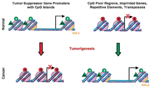
Figure 7: Differences in DNA methylation levels between normal and cancerous tissues. In normal tissue, lack of DNA methylation on promoter regions of tumor suppressor genes allows transcription to be initiated and expression to be turned on. In regions of repetitive elements, oncogenes, and transposons, promoter hypermethylation allows gene expression to be turned off. In cancer, a hypermethylated promoter for tumor suppressor genes is indicative of transcriptionally inactive genes. On the other hand, promoters for imprinted genes, repetitive elements, transposons, and oncogenes are hypomethylated, allowing gene expression. Figure taken from Sharma and colleagues [150].
34 3.2 Histone Post-Translational Modifications
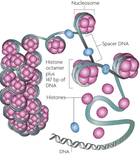
The ability for a cell’s nucleus to contain very long strands of DNA would not be accomplished without the presence of histone proteins [174, 175]. DNA is tightly wrapped around histones to form nucleosomes [147, 174, 175]. The nucleosome is formed by a histone octamer [147, 174]. Each octamer is made up of four histone proteins that form the nucleosome core: H2A, H2B, H3, and H4 [147]. Nucleosomes are connected to one another by linker DNA and are packed together to form chromatin [147, 174].
Figure 8: Nucleoprotein complex of DNA and histone forms chromatin.
DNA is wrapped around a histone octamer made up of two copies of each histone subunit forming a nucleosome. Each nucleosome is separated by spacer DNA [176].
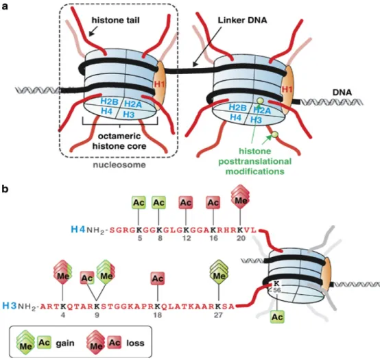
Each histone contains a C-terminus region, which forms the globular portion of each nucleosome, and an N-terminus region, forming a tail that can be covalently modified post-translationally by a variety of different enzymes. Depending on the post-translational modifications, chromatin can be either loosely or tightly organized, referred to as euchromatin and heterochromatin, respectively [147, 174, 177]. Whereas euchromatin favors gene transcription, heterochromatin is so tightly configured that gene transcription is almost non-existent [147, 174, 178]. The two most commonly studied histone modifications are acetylation and methylation [147, 150]. Other post-translational modifications that play a role in gene expression changes include phosphorylation, sumoylation, and ubiquitination [147, 150].
Fascinatingly, two models exist regarding the effects of histone acetylation on structural chromatin changes and regulation of gene expression. The first model describes acetylation of histone H3 and H4 subunits playing key roles in changing chromatin polarity [147]. The abundance of lysine residues on histone tails makes them inherently positive in polarity [147]. Hence, when lysine residues are not acetylated, they are tightly compacted with the negatively charged DNA strands to form heterochromatin [147]. On the other hand, when lysine residues are acetylated, the positive charges are neutralized, supporting the loose chromatin structure favoring gene transcription [147]. In the second model, however, acetylation of lysine residue 14 on histone 3 (H3K14ac) by P300 induces nucleosome eviction, mediated by the histone chaperone Nap1 [179]. Once the nucleosome is removed, transcription factor complexes are assembled and gene expression is turned on [179]. Luebben and colleagues demonstrated the importance of H3K14ac in mutation studies where they