Identification of blood pressure genes in the Dahi sait-sensitive hypertension model
par Julie Dutil
Département de Biologie Moléculaire Faculté des études supérieures
Thèse présentée à la Faculté des études supérieures en vue de l’obtention du grade de Philosophiae Doctor
en Biologie Moléculaire T Août 2005 Conaré
/
c-11 \ MAI , ©Julie Dutil, 2005de Montréal
Direction des bibliothèques
AVIS
L’auteur a autorisé l’Université de Montréal à reproduite et diffuser, en totalité ou en partie, par quelque moyen que ce soit et sur quelque support que ce soit, et exclusivement à des fins non lucratives d’enseignement et de recherche, des copies de ce mémoire ou de cette thèse.
L’auteur et les coauteurs le cas échéant conservent la propriété du droit d’auteur et des droits moraux qui protègent ce document. Ni la thèse ou le mémoire, ni des extraits substantiels de ce document, ne doivent être imprimés ou autrement reproduits sans l’autorisation de l’auteur.
Afin de se conformer à la Loi canadienne sur la protection des renseignements personnels, quelques formulaires secondaires, coordonnées ou signatures intégrées au texte ont pu être enlevés de ce document. Bien que cela ait pu affecter la pagination, il n’y a aucun contenu manquant.
NOTICE
The author of this thesis or dissertation has granted a nonexciusive license allowing Université de Montréal to reproduce and publish the document, in part or in whole, and in any format, solely for noncommercial educational and research purposes.
Ihe author and co-authors if applicable retain copyright ownership and moral
rights in this document. Neither the whole thesis or dissertation, nor substantial extracts from it, may be printed or otherwise reproduced without the author’s permission.
In complîance with the Canadian Privacy Act some supporting forms, contact information or signatures may have been removed from the document. While this may affect the document page count, it does flot represent any loss of content from the document.
Cette thèse intitulée:
Identification of blood pressure genes in the Dahi sait-sensitive hypertension model
présentée par Julie Dutil
A été évaluée par un jury composé des personnes suivantes:
Dr Richard Bertrand président-rapporteur Dr Alan Deng directeur de recherche Dr Christian Deschepper membre du jury Dr Joseph Dent examinateur externe Dr Pierre Moreau
Summary in English
Hypertension afflicts 21% of the Canadian population and is a leading cause of death as it is associated with an increased risk of cardiovascular diseases. Blood pressure (BP) regulation is a complex trait controlled by multiple genes and influenced by environment. Little is known about the genes responsible for BP regulation. Inbred rats genetically predisposed to hypertension have been developed to facilitate the study of the genetic basis to this disease. Preliminary linkage and congenic experiments have shown the presence of at least one BP quantitative trait loci (QTL) in a 80cM region of rat chromosome (chr) 2. The objective of the present thesis was to identify and characterize the rat chr 2 regions involved in the genetic control of hypertension. We are using congenic strains in which various segments from the Dahl sait-sensitive (DSS) rat chr 2 are replaced by the homologous segments coming from the Milan normotensive rat (MNS). This model has allowed to narrow down the rat chr 2 BP QTL to three non-overlapping regions each containing at least one distinct BP QTL: C2QTL1 (5.7cM), C2QTL2 (3.5cM) and C2QTL3 (1.5cM). These closely linked BP QTL showed epsitatic and additive interactions. We also identified a QTL for vascular smooth muscle celi (VSMC) number acting independently of BP. The gene coding for the angiotensin receptor lb Agtr1b) is located in the region containing the QTL for $MCN. A gtr]b is a candidate for the SMCN QTL because 1) two non-synonymous mutations were found in the coding region between DSS and MNS rats, and 2) contractile responses to Ang II are reduced in rats harbouring the MNS allele of the gene compared with DS$ rats. The cunent work provide new insights about the genetic determination of hypertension and of vascular remodelling disorders. In the future, these knowledge may be useful in the development of new therapeutic targets, genetic diagnostic tools and pharmacogenomics.
Keywords: genetics of hypertension, Dahi salt-sensitive rat, quantitative trait loci, aortic hyperpiasia, vasoreactivity.
Résumé en français
L’hypertension affecte 21% de la population canadienne et est un facteur de mortalité important car elle est associée à un risque accru de maladies cardiovasculaire. La régulation de la pression artérielle (PA) est un trait complexe impliquant plusieurs gènes et une composante environnementale. Les gènes responsables de la régulation de la PA sont peu connus. Les modèles de rats consanguins ont été développé pour faciliter l’étude des bases génétiques de l’hypertension. Des études préliminaires de liaison génétique et de lignées congéniques ont démontré la présence d’au moins un iocus pour trait quantitatif (QTL) pour la PA dans une région de 80 cM sur le chromosome (chr) 2 du rat. L’objectif de cette thèse était d’identifier, de définir et de caractériser les régions du chr 2 du rat impliquées dans la détermination génétique de l’hypertension. Pour atteindre cet objectif, nous utilisons des lignées congéniques dans lesquelles divers segments du chr 2 du rat Dahl sait-sensitive (DSS) ont été remplacé par la région homologue provenant du rat Milan normotendu (MNS). Ce modèle a permis de réduire la région d’intérêt du QTL du chr 2 du rat à 3 régions non-chevauchantes contenant chacune un QTL pour la PA distinct: C2QTL1 (5.7 cM), C2QTL2 (3.5 cM) and C2QTL3 (1.5 cM). Ces loci ont démontré des interactions additives et épistatiques. Nous avons aussi identifié un QTL pour le nombre de cellules musculaire lisses (SMCN) vasculaires agissant indépendamment de la PA. Le gène codant pour le récepteur lb de l’angiotensine (A gtrl b) est situé dans la région contenant le SMCN QTL. Agtrlb est un gène candidat pour le SMCN QTL puisque 2 mutations non-synonymes ont été identifiées dans la région codante entre les séquences du rat DSS et du MNS, et 2) la réponse contractile à l’angiotensine II est diminuée dans les rats portant les allèles MNS du gène comparativement aux rats DSS. Ce travail apporte de nouvelles précisions sur les déterminants génétiques de l’hypertension et des maladies de remodelage vasculaire. Ces connaissances pourraient être utiles au développement de nouvelles cibles thérapeutiques, d’outils de diagnostique génétique et à l’avancement de la pharmacogénomique.
Mots clé: génétique de l’hypertension, rat Dahl sait-sensitive, ioci pour trait quantitatif, hyperplasie aortique, vasoréactivité.
Summary in English Résumé en français Table of content List of tables List of figures List of abbreviations CHAPTER 1: INTRODUCTION
1.1 PHYSIOLOGY 0F BLOOD PRESSURE 1
1.1.1 Definition 1 1.1.2 Hemodynamics 2 1.1.3Regulation 2 1.1.3.1 2 3 3 3 4 4 4 4 5 5 5 7 7 8 8 8 9 Table of Contents Page I II III XIII XIII XIV Nervous system 1.1.3.1.1 Afferents inputs
1.1.3.1.2 Integration of afferent inputs 1.1.3.1.3 Efferent outputs
1.1.3.1.3.1 PSNS 1.1.3.1.3.2 SNS 1.1.3.2 Endocrine system
1.1.3.2.1 Renin angiotensin system (RAS)
1.1.3.2.1.1 Angiotensin II proteolytic cascade 1.1.3.2.1.2 Angiotensin receptors and signalling
1.1.3.2.1.2.1 ATY 1.1.3.2.1.2.2 AT2 1.1.3.2.1.3 Local RAS
1.1.3.2.1.4 Biological actions of Ang II 1.1.3.2.2 Natriuretic peptides
1.1.3.2.2.1 Receptors and signalling 1.1.3.2.2.2 Biological actions
1.1.3.2.2.2 Metabolism 9 1.1.3.3 Vasoactive substances 9 1.1.3.3.1 Endothelin 9 1.1.3.3.2 Nitric oxide 10 1.1.3.3.lKinins 12 1.1.3.3.2 Eicosanoids 12 1.2 HYPERTENSION 13
1.2.1 Definition and classifications 13
1.2.2 Sequelae 14
1.2.2.1 Oxidative stress and endothelial dysfunction 16
1.2.2.2 Atherosclerosis 16
1.2.2.3 Vascular remodeling 17
1.2.2.4 Cardiac remodeling 18
1.2.2.5 Coronary artery disease 19
1.2.2.6 Congestive heart failure 20
1.2.2.7 Stroke and cerebraÏ aneurysms 20
1.2.2.7 Renal damage 21
1.2.3 Epidemiology and burden 21
1.2.4 Treatment 22
1.3 GENETICS 0F HYPERTENSION 22
1.3.1 Monogenic hypertension 23
1.3.1.1 Glucocorticoid Remediable Aldosteronism 23 1.3.1.2 Apparent Mireralocorticoid Excess 24
1.3.1.3 Liddle’s syndrome 24
1.3.1.4 Pseudohypoaldosteronism type II 25
1.3.2 Essential Hypertension 26
1.3.2.1 Evidence fora genetic contribution 26
1.3.2.2 Rat models 28
1.3.2.2.2 The Dahi Sait-Sensitive rat 29 1.3.2.2.2.1 Development of the strain 29
1.3.2.2.2.2 Biood pressure 30
1.3.2.2.2.3 Hemodynamics 30
1.3.2.2.2.4 Kidney 31
1.3.2.2.2.5 Vasculature: endothelial dysfunction 31 1.3.2.2.2.6 Vasculature: Na+ICa2+ exchanger 32 1.3.2.2.2.7 Renin angiotensin system 32
1.3.2.3 Genetic tools 33
1.3.2.3.1 Genetic markers 33
1.3.2.3.2 Mapping of genetic markers 34 1.3.2.3.2.1 Linkage mapping 34 1.3.2.3.2.2 Radiation hybrid mapping 34 1.3.2.3.2.3 Physical mapping 35 1.3.2.3.3 Quantitative trait locus definition 36 1.3.2.3.4 Co-segregation analyses 36 1.3.2.3.4.1 Candidate gene approach 39 1.3.2.3.4.2 Total genome scan approach 39
1.3.2.3.4 Congenic strains 40
1.3.2.3.5 Congenic substrains 42
1.3.2.4 Candidate genes for essential hypertension 44 1.3.2.4.1 Renin angiotensin system 44 1.3.2.4.1.1 Angiotensinogen gene 44
1.3.2.4.1.2 Renin gene 46
1.3.2.4.1.3 Angiotensin I converting enzyme gene 47 1.3.2.4.1.4 Angiotensin II receptor gene 47
1.3.2.4.2 Others 48
1.3.2.4.2.1 SA gene 48
1.3.2.4.2.2 Nitric oxide synthase gene 49 1.3.2.4.2.3 Epithelial Na channel gene 49 1.3.2.4.2.4 WNK kinases genes 50
1.3.2.4.3 Limitations of linkage and association studies 51
1.4 RATIONALE ANI OBJECTIVE 52
CHAPTER 2: METHODS 53
2.1 GENETIC MAP 53
2.2 CONGENIC SUBSTRAINS 53
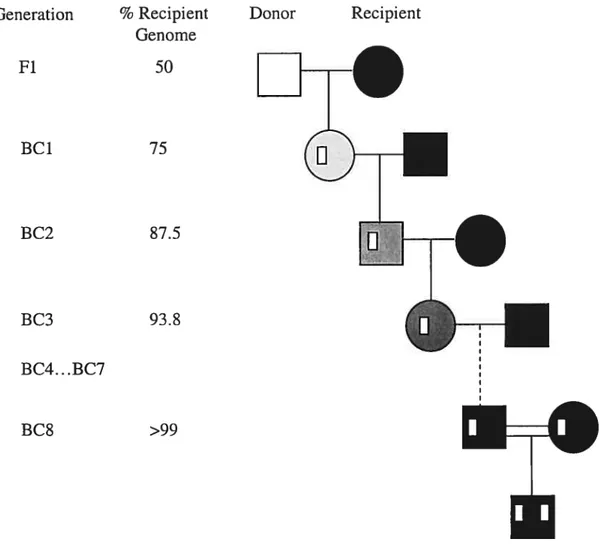
2.2.1 Nomenclature and genetic boundaries of congenic strains 54
2.3 PREPARATION 0F ANIMALS 55
2.3.1 Radiotelemetry 56
2.4 STATISTICAL ANALYSIS 58
CHAPTER 3: Mapping a Blood Pressure Quantitative Trait Locus to a 5.7 59 cM region in Dahi Sait Sensitive Rats
Mammalian Genonie (2001) 12: 362-365
3.1 ABSTRACT 60
3.2 INTRODUCTION 60
3.3METHODS 61
3.3.1 AnimaIs 61
3.3.2 Breeding scheme for generating substrains 62
3.3.3 Preparation of rats for BP measurements 62
3.3.4 BP measurements 63
3.3.6 DNA extraction and genotyping 63
3.3.7 Radiation hybrid mapping 64
3.4 RESULTS 64
3.4.1 Congenic strain monitoring 64
3.4.2 BP study design 64
3.4.3 BP measurements 64
3.4.4 Mapping of a BP QTL by analyzing BP effects associated with 65 chromosome segments
3.5 DISCUSSION 65
3.6 AKNOWLEDGEMENTS 67
3.7 REFERENCES 67
3.8 FIGURE LEGENDS AND FIGURES 69
CHAPTER 4: Further chromosomal mapping of a blood pressure 73 quantitative trait locus in Dahi rats on Chromosome 2 using
congenic strains Physiological Genomics (2001) 6: 3-9 4.1 ABSTRACT 74 4.2 INTRODUCTION 74 4.3METHODS 75 4.3.1 Animais 75
4.3.3 Preparation of rats for BP measurements 77
4.3.4 BP measurements 78
4.3.5 Statistical analysis 72
4.3.6 DNA extraction and genotyping 78
4.3.7 Radiation hybrid mapping 78
4.4 RESULTS 79
4.4.2 BP study design 79
4.4.3 BP measurements 79
4.4.4 Mapping of a BP QTL by analyzing BP effects associated with 79 chromosome segments
4.5 DISCUSSION 80
4.6 AKNOWLEDGEMENTS 83
4.7 REFERENCES 83
4.8 FIGURE LEGENDS AND FIGURES 85
CHAPTER 5: Multiple quantitative trait loci for blood pressure interacting 91 epistatically and additively on Dab! Rat Chromosome 2
Hypertension (2005) 45: 557-564
5.1 ABSTRACT 92
5.2 INTRODUCTION 93
5.3METHODS 93
5.3.2 Construction of congenic substrains 94
5.3.3 BP measurements 94
5.3.4 Statistical analysis 95
5.4RESULTS 95
5.4.1 A chromosome marker defining separate QTLs 95 5.4.2 Construction of new congenic substrains to fine map multiple BP 96
QTLs
5.4.3 Epistatic and additive QTL-QTL interactions 97 chromosome segments
5.5 DISCUSSION 97
5.5.1 Comprehensive congenic coverage divulging multiple BP QTL5 in 97 a closely linked region.
5.5.2 Epistatic and additive QTL interactions among C2QTL1, C2QTL2 99 and C2QTL3.
5.5.3 Perspectives 100
5.6 AKNOWLEDGEMENTS 100
5.7 REFERENCES 101
5.8 FIGURE LEGENDS AND FIGURES 106
CHAPTER 6: A quantitative trait locus for aortic smooth muscle ceil 116 number acting independently of blood pressure: implicating
the angiotensin receptor AT1B gene as a candidate
Physiotogicat Genomics (2005) 21: 362-369
6.2 INTRODUCTION 118
6.3METHODS 119
6.3.1 Animais 119
6.3.2 Marker development 119
6.3.3 BP studies and tissue extractions 120
6.3.4 Measurements of aortic cross sectionai areas 121
6.3.5 Determination of vascular SMCM 121
6.3.6 DNA content and apoptosis 122
6.3.7 Vasoreactivity studies 122
6.3.8 Statistical analysis 123
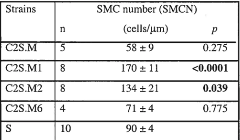
6.4 RESULTS 123
6.4.1 Localization of a QTL for vascuiar SMCN independent of BP QTLs 123 6.4.2 The angiotensin-receptor ATÏB (Agtrlb) gene as a candidate for 125
the SMCN QTL
6.5 DISCUSSION 126
6.5.1 A QTL for the vascuiar SMCN functions independently of QTL5 127 for hypertension
6.5.2Agtr]b as a candidate for a SMCN QTL 129
6.6 AKNOWLEDGEMENTS 130
6.7 REFERENCES 131
6.8 FIGURE LEGENI)S AND FIGURES 136
7.1 Effect of the chr. 2 congenic strains on heart rate 144
7.1.1 Circadian rhythms in HR and BP 144
7.1.2 Localization ofaHR QTL 145
7.2 QTL-QTL interactions 146
7.3 QTL for SMC number implicating the AT1B gene as a candidate 149 7.3.1 Model for the regulation of SMC number independently of BP 149 7.3.2 Agtr]b as a candidate gene for SMC number 150 7.3.3 Further implication of Agtr]b as a candidate gene for the SMC 151
number QTL
7.4 The next step: from a region to a gene 151
7.4.1 Identification of functional and positional candidates 152
7.4.2 Identification of sequence polymorphisms 152 7.4.3 Demonstrating the causative link between a variant and the BP 152
effect
7.5 Identification of causative genes for complex trait: is it possible? 153
7.6 Alternative strategies to identify genes for complex traits 154
7.6.1 DNA Microaffays 154
7.6.2 Importance of intermediate phenotypes 155
7.7 Clustering of functionally related QTL 156
7.7.1 Implications of gene clustering and interactions on QTL detection 157
7.8 The animal model: bacgrounf effect 158
7.8.1 Residual background 158
7.9 Relevance to other rat models of hypertension 159
7.10 Relevance human hypertension 161
7.11 Perspectives 161
7.11.1 Basic sciences 161
7.11.2 Clinical 162
7.8.2.1 Gene therapy 162
7.8.2.2 Genetic diagnosis and pharmacogenomics 163
7.9 Summary and conclusions 164
REFERENCES 165
List of Tables
Table I Properties of the different isoforms of NOS li
Table II Risk of cardiovascular or renal event in hypertensive subjects 14 Table III Nomenclature and genetic markers defining the congenic strains 55
presented in chapters 3-6
List of Figures
Figure 1 The sequelae of hypertension 15
Figure 2 The principle of linkage 38
Figure 3 Construction of a congenic strain. 41
Figure 4 Substitution mapping using congenic strains 43 Figure 5 Measure of the blood pressure by radiotelemetry 57 Figure 6 Construction of double congenic strains for rat chr.2 148
Lïst of abbrevïations
AA Arachidonic acid
ACE Angiotensin converting enzyme
Ach Acetylcholine
ACTH Adrenocorticotropic-hormone
AGT Angiotensinogen
AME Apparent mireralocorticoid excess
Ang I Angiotensin I
Ang II Angiotensin II
ANP Atrial natriuretic peptide
AT1 Ang II receptor 1
AT2 Ang II receptor 2
BCÏ Backcross generation
BNP Brain natriuretic peptide
BP Blood pressure
Ca2+ Calcium
cAMP Cyclic adenosine monophosphate cGMP Cyclic guanosine monophosphate CHD Coronary heart disease
CHF Congestive heart failure CHES Canadian heart health survey
chr. Chromosome
cM centiMorgans (cM)
CNP C-type natriuretic peptide CNS Central nervous system
C02 Carbon dioxide
CO Cardiac output
COX Cyclooxygenase
cR CentiRays
DBP Diastolic blood pressure
DG Diacylglycerol
DSR Dahl sait-resistant
DSS Dahi sait-sensitive
EET Epoxyeicosatrienoic acids
ENaC Amiloride-sensitive epitheliai sodium channel
eNOS Endotheliai NOS
ET-1 Endothelin 1
EST Expressed sequence tag
FAK Focal adhesion kinase
Fi First filial generation GFR Glomerular filtration rates
GH the New Zealand
GPK GPR protein kinase
GPR G protein-coupled receptor
GTP Guanosine triphosphate
HR Heart rate
iNOS Inducible NOS
1P3 Inositol triphosphate
JAKISTAT Janus Kinase ISignals transducers and activators transcription LDL Low density lipoproteins
Lew Lewis
LH Lyon hypertensive
LT Leukotrienes
LVH Left ventricular hypertrophy MAP Mean arterial pressure
MAPK Mitogen-activated protein kinases
MHS Milan hypertensive
IvllvIE Metalloendopeptidase
mmHg Milimeter of mercury
MNS Milan normotensive
NaC1 Sodium chloride
NCCT Na-Cl co-transporter
NCX SodiumlCalcium Na+/Ca2+ exchanger
Nedd4 neuronal precursor celis expressed, developmentally downregulated
NO Nitric oxide (NO)
NOS NO synthase (NOS)
NPR Natriuretic peptide receptor NTS Nucleus tractus solïtarius
02 Oxygen
PDE Phophodiesterase
PGs Prostaglandins
PHAII Pseuhypodoaldosteronism type II
PW2 4,5 biphosphate
PKA Protein kinase A
PKC Protein kinase C
PLA2 Phospholipase A2
PLC Phospholipase C
PLD Phospholipase D
PNS Peripheral nervous system
PP Pulse pressure
QTL Quantitative trait locus RAS Renin angiotensin system
RFLP Restriction fragment lenght polymorphism
RH Radiation hybrid
RIIIP Renal interstitial hydrostatic pressure ROMK Renal outer medullary potassium channel ROS Reactive oxygen species
S-GC Soluble guanylyl cyclase
SNP Single nucleotide p01 ymorphism
RVLN Rostral ventrol ateral nucleus SBP Systolic blood pressure
SHR Spontaneously hypertensive rat SHR-SP SHR stroke prone
SMC Smooth muscle celis
TPR Total peripheral resistance
TXs Thromboxanes
UTR Untranslated region
WNK With no lysine
WKY Wistar Kyoto
1 1BHSD 11 B-hydroxysteroid dehydrogenase 20-HETE 20-hydroxyeicosatetraenoic acid 5,6-EET 5,6-epoxyeicosatrienoic acid
INTRODUCTION
1.1 PHYSIOLOGY 0F BLOOD PRESSURE
The cardiovascular system is responsible for the distribution of blood to ail parts of the organism. It is composed of a pump, the heart and of a network of conduits, the blood vessels. The heart provides the force necessary for the blood to circulate through the arteries and veins. The blood is the vehicle ensuring the transport of several substances. A proper blood flow allows the distribution of heat, ensures that the nutriments and oxygen (02) reach the tissues and that carbon dioxide (C02) and waste are removed from the tissues. Blood also ensures the maintenance of a stable pH in the organism with the buffer substances it contains.
1.1.1 Definition
Blood pressure (BP) is defined as the force exerted by the blood per unit of vessel wall area. Circulation of the blood in the body follows a pulsatile pattem reaching its highest peak during ventncular contractions of the heart and its lowest level between cardiac contractions. The foi-ce necessary for the propulsion of the blood in the entire organism relies on the strength of heart beats and on the pressure gradient present in the cardiovascular system. In effect, there is a progressive lowering of the blood pressure as it travels from large arteries such as aorta to arterioles until it reaches heart atria through vena cava. Systolic blood pressure (SBP) corresponds to the blood pressure during ventricular contraction whereas diastolic blood pressure (DBP) is defined as the blood pressure between each ventricular contraction. Mean arterial pressure (MAP) is the average between diastolic and systolic blood pressures and reflects the pressure to which small vessels are continuously submitted. Pulse pressure (PP) is calculated by subtracting DAP to SAP and is an indication of the compliance of large arteries. BP is measured in millimetres of mercury (mmHg). 1
1.1.2 Hemodynamics
From a hemodynamic point cf view, MAP is function cf two major variables: j) cardiac output (CO), which is the quantity of blood ejected by the left ventricle during systole; and ii) total penpheral resistance (TPR) exerted by small arteries and arterioles. Thereby:
MAP=CO X TVR
Where CO is influenced by systolic precharge and heart rate, and total vascular resistance is determined by the rayon of small arteries and the physical properties of the blood which include viscosity and concentration of fibrinogen. 1
1.1.3 Regulation
A complex system of regulation is responsible blood pressure control te ensure constant blood flow to tissues and adjustment to meet specific demands cf the organism. Regulation is achieved thrcugh the conttcl of blood volume and cf resistance vessels. It involves the participation cf the endocrine system, of the central and peripheral nervous systems and cf vasoactive substances acting lccally.
1.1.3.1 Nervous system
The nervous system is a major player in the BP homeostasis as it is involved in short term and long-term regulation. The effects cf the nervous system on the cardiovascular system require the integration of afferent input from visceral organs and varicus brain regions by the central nervous system (CNS) and the transduction of the input in the appropriate cardiovascular adaptation via the parasympathetic and sympathetic divisions of the autonomic peripheral nervous system (PNS). The autonomïc nervous system is responsible for controlling vital functions including heart rate and BP. Its actions are mostly involuntary. The autonomic nervous system is dïvided into the parasympathetic nervous system (PSNS) and the sympathetic nervous system (SNS) on the basis cf structural and functional differences. Peripheral nervous system projects to target organs where it regulates cardiovascular function. The influence cf the two divisions is opposed. 2
1.1.3.1.1 Afferents inputs
The hypothalamus, the baroreceptors and the chemoreceptors ensure a constant monitoring of the cardiovascular system. The hypothalamus is the brain region responsible for the integration of environmental and behavioural input. It is the link between the endocrine and the neural systems to maintain homeostasis. Arterial baroreceptors are located mainly in the carotid sinus and in the wall of the aortic arch. They are ïnvolved in adaptation to rapid and important changes in blood pressure, like it is the case during postural changes. Cardiac baroreceptors are positioned in the walls of
atria and ventricles. Baroreceptors respond to an increase in pressure or stretching. Chemoreceptor are found proximity to baroreceptors in carotid sinus and aortic arch. They are specialized in detecting changes in 02, C02 and H levels. 2,3
1.1.3.1.2 Integration of afferent inputs
The nucleus tractus solitarius (NTS) of the medulla oblongata is the principal site of termination of afferent fibres arising from cardiovascular receptors, hypothalamus and cerebral cortex. Signal from different types of cardiovascular afferent fibres have different sites of termination in the NTS. The NTS integrates information from afferent fibres and adjust the cardiovascular system accordingly. 2,3 The NT$ controls the parasympathetic nerves of the penpheral nervous system directly and projects to the rostral ventrolateral nucleus (RVLN) in the medulla. The RVLN is responsible for controlling the output of the sympathetic preganglionic neurons. The signalling of the NTS to RVLN is inhibitory, which means that excitatory signalling to NTS are translated by an increase in inhibition of RVLN and sympathetic output.
1.1.3.1.3 Efferent outputs
Both division of the penpheral nervous system require two neurons to reach the target organs: a preganglionic neuron to conduct influx from the central nervous system to the ganglion and a postganglionic neuron which transmit the signal form the ganglion to the target organ. Preganglionic neurons of the PSNS and SNS signal by releasing the neurotransmitter acetylcholine (Ach) which binds to nicotinic receptors on postganglionic cells. Ail major organs are innervated by both, the P$NS and the SNS,
with the exception of the adrenal medulïa which is under the control of the SNS exc!usively. 2 The PSNS inhibitory tone predominates at rest whereas SNS stimulation is triggered in a stress or exercise situation.
1.1.3.1.3.1 PSNS
The parasympathetic system exerts its effect via the vagal postgang!ionic nerves using acetylcholine as the main neurotransmitter. Parasympathetic postganglionic neurons are very short since the ganglion is located at organ. These neurons are cholinergic as they release Ach which binds muscarinic receptors at target organs. Effects of PSNS signalling include a decreased rate and force of contraction of the heart and vesse! dilation.
1.1.3.1.3.2 SNS
The sympathetic neurons are !onger since they run from the gang!ion in the spinal cord to the target organ. They are adrenergic as they release the neurotransmitter norepinephrine which can bind to several subtypes of the adrenergic receptors. Sympathetic stimulation increases heart rate and cardiac output through f31 adrenergic receptors, provokes constriction of arteries and veins through OEl adrenergic receptors, and lead to dilation of vessels and renin secretion through f32 adrenergic receptors. AI! the effects of sympathetic activation contribute to the ‘fight or flight response aiming at preparing the body to respond to danger. More specifical!y, SNS actions allow to redistribute the blood away from skin and viscera to concentrate it to skeletal musc!es, brain and heart, it stimulates heart beat, raises BP and promotes the release of adrenaline in the bloodstream by the adrena! medu!Ia. This latter action of the SNS is necessary for ensuring that ail the body is reached by SNS effects. 5,6
1.1.3.2 Endocrine system
1.1.3.2.1 Renin angiotensin system (RAS)
The circulating RAS is a major system responsible for maintenance of body fluid vo!ume homeostasis and regulation of BP. It consists of a proteolytic cascade that
resuits in the production of a biologically active peptide, angiotensin II (Ang II), which exerts its effects via a signal transduction system.
1.1.3.2.1.1 Angiotensin II proteolytic cascade
Generation of Ang II begins with the molecule angiotensinogen (AGT), expressed constitutively from the liver and secreted in the circulation. Under normal conditions, AGT levels remain constant at a concentration near its Km for cleavage by renin .
AGT is cleaved by the aspartic protease renin to yield angiotensin I (Ang I). In the lddney, pre-prorenin (406aa) is first synthesized and is converted to prorenin by the removal of 23 amino acids as it enters the rough ER. Through a secretory pathway, prorenin is processed to renin by the cleavage of 43 amino acids. In response to low perfusion pressures, low sodium delivery to the distal tubules or
13
adrenergic stimulation, renin is secreted in the circulation. The opposite situation inhibits renin secretion from the kidneys. Inhibition of renin secretion by the product of the cascade itself, Ang II allows negative feedback regulation. 2 As the biologically inactive peptide Ang I reaches pulmonary circulation, it is converted to active Ang II by the Angiotensin converting enzyme (ACE). ACE is a zinc metallopeptidase that exists in 3 isoforms: somatic ACE, soluble ACE and testicular ACE. Somatic ACE is mainly responsible for the conversion of Ang I to Ang II by cleavage of a dipeptide from its C-terminal end. ACE lias a broad substrate specificity. In addition to cleaving Ang I, it can also inactivate bradykinin by removal of a C-terminal dipeptide.1.1.3.2.1.2 Angiotensin receptors and signalling
Ang II exerts its effect by binding to two receptors, the Ang II receptor 1 (AT1) and the Ang II receptor 2 (AT2).
1.1.3.2.1.2.1 AT1
Most known effect of Ang II are mediated through coupling with the AT1 receptor. The AT1 receptor is a member of the G-protein-coupled 7-transmembrane receptor family. The tertiary structure of the 4 extracellular loops is stabilized by disulfide bonds and contains 3 glycosylation sites. The cytoplasmic tau is rich in serine and threonine
residues, which are potential phosphorylation sites. Ail receptors exhibit a wide tissue distribution in aduit including spleen, liver, lddney, heart, vascular smooth muscle celis (SMC), blood vessels, adrenal gland, liver and brain. Molecular cloning studies have distinguished 2 receptor subtypes, AT1a and AT1b differing in their distribution and regulation. At the molecular level, AT1a and AT1b show differences in the carboxy domain of the protein and in the 3’ and 5’ untranslated region (UTR). The 2 receptor subtypes are present in the mouse and in the rat. In human, only one AT1 subtype is found. 8,9
Upon activation by Ang II, the AT1 receptor couples to G proteins of the Gi/Gq family which in mm activate a wide range of signalling cascades including the phospholipase C (PLC), phospholipase D (PLD), and phospholipase A2 (PLA2) pathways. In addition, Ang II stimulation of the Ail receptor has an inhibitory effect on adenylate cyclase. 10
12
Activation of PLC leads to the production of inositol triphosphate (1P3) and diacylglycerol (DG) from the hydrolysis phosphatidylinositol 4,5 biphosphate (PP2). In parallel, PLD generates choline and phosphatidic acid (PA) from phosphatidylcholine (PC). PA is subsequently converted to DG. W3 production is involved in the mobilization of intracellular calcium (Ca2) resulting in contraction of smooth muscle celis (SMC) and vasoconstriction. DG and intracellular Ca2 mobilization both contribute to the activation of protein kinase C (PKC) which mediates the proliferative effects of Ang II via the phosphorylation and activation of the mitogen-activated protein kinases (MAPK). PC is also hydroÏyzed by phospholipase A2 upon Ang II binding to AT1R. This cascade generates arachidonic acid (AA), a precursor for the production of eicosanoid vasoactive substances. 8,9,11
The consequence of the inhibition of adenylate cyclase is a decrease in cyclic AIVW
(cAMP) levels and as a resuit a reduction in protein kinase A (PKA) activity. In renal proximal tubules, the net effect is a reduction in Na’H exchange inhibition leading to an augmented sodium reabsorption. In the adrenal cortex, a decrease in adenylate
cyclase activity resuits in a reduction in inhibition of adrenocorticotropic-hormone (ACTH)-stimulated aldosterone secretion. ‘“
Effects of AT1R on stimulation of growth and proliferation involves additional signalling mechanisms including the Janus kinase signal transducers and activators transcription (JAKISTAT) system and ras/raf activation of MAPK. 12
1.1.3.2.1.2.2 AT2
The structure of the AT2 receptor resembles that of the G-protein-coupled 7-transmembrane receptor famiiy. Despite the similarities to the family, a G protein coupling AT2 to its signaliing pathways has yet to be identified. The AT2 receptor shares 34% sequence homology with the AT1 receptor. ° Contrarily to AT1 which is expressed mainly in aduit tissue, AT2 expression is stronger in foetuses and declines after birth. Weak AT2 expression remains in aduit endothelium, myocardium, brain, adrenal gland, ovaries and uterus
Description of signalling from AT2 receptor remains incomplete. Its effects are thought to balance the effects of ATY on cellular proliferation. In effect, AT2 is believed to have effects in cell differentiation and anti-proliferation. These effects are thought to be mediated by the activation of phosphotyrosine phosphatase which in tum inactivates mitogen-activated protein (MAP) kinase. 10 AT2 is also involved in vasodilatation and natriuresis via a local increase in nitric oxide and bradyldnin. 10
1.1.3.2.1.3 Local RAS
In addition to the well described endocrine RAS, evidence for the local generation of Ang II within tissues accumulates. In addition to their presence in the circulation, the components of the RAS are also found in several tissues including brain, kidney, adrenals, testis and arterial wall. 14 It is believed that the circulating RAS mediates the acute effects of Ang II on vasoconstriction, sait and water homeostasis and cardiac rhythm. In contrast, tissue RAS seems to be involved in the establishment of the long term effect on Ang II including celluiar proliferation and differentiation. 13
1.1.3.2.1.4 Biological actions of Ang II
In the vasculature, Ang II increases total peiipheral resistance by directly causing vasoconstriction and indirectly by facilitating SNS transmission. Ang II is a mitogenic factor for vascular SMCs and myocardium. In addition, Ang II interacts with other endocrine factors as it is one of the main stimulus for the synthesis of aldosterone by the adrenal cortex and the release ACTH and vasopressin by the pituitary. ACTH stimulates the adrenals to release aldosterone. Aldosterone acts on distal tubules where it stimulates sodiumlpotassium exchange leading to increased sodium reabsorption. Vasopressin triggers the insertion of water pores in the distal tubules and the collecting duct of the kidney, thereby promoting water reabsorption. Ang II also regulates sodium and fluid reabsorption from proximal tubules of the kidney by affecting localiy the constriction state of the renal vasculature. From its action on the CNS, Ang II induces thirst and sait appetite. Inhibition of renin secretion in the lddney provides a negative feedback loop mechanism for the effects of Ang
1.1.3.2.2 Natriuretic peptïdes
The natriuretic peptide famiiy inciudes the atriai natriuretic peptide (ANP), the brain natriuretic peptide (BNP) and the C-type natriuretic peptide (CNP). ANP is synthesized and released mainly by the atria and BNP is synthesized and reieased predominantiy by the ventricuiar myocytes. 16 These peptides exert their function as circulating hormones.
17
Even though ANP and BNP are constitutively released from the heart, mechanical stimuli such as atrial stretch or endocrine stimuli may affect their rate of synthesis and secretion 16 CNP is a paracrine factor produced by the cerebrai cortex and endotheiial ceils of the vesseis. 18
1.1.3.2.2.1 Receptors and signalling
Natriuretic peptides exert their function by binding to the natriuretic peptide receptors (NPR) A and B. NPR-A and NPR-B have a guanyiyl cyclase intracellular domain that catalyses the formation of cyciic guanosine monophosphate (cGMP) second messenger from guanosine triphosphate (GTP). Targets of cGMP include cGMP-dependent protein
kinases and cGMP-gated ion channels. 16 ANP and BNP have higher affinity for NPR A whereas CNP associates preferentially with NPR-B “r.
1.1.3.2.2.2 Biological actions
Natriuretic peptides have an important influence on the cardiovascular homeostasis. More specifically, ANP and BNP antagonize the RAS by inhibiting the synthesis and release of renin and aldosterone. They also increase natriuresis by constriction of efferent arterioles and dilation of afferent arterioles in the glomeruli which results in an increased glomerular filtration rate (GFR), they inhibit the effects of endothelin, vascular SMC proliferation, and SNS. In a general way, the effects of ANP and BNP oppose those of the RAS. ‘ In contrast to ANP and BNP, CNP lias only minimal natriuretic effects and doesn’t inhibit the RAS. It is an venous and arterial vasodilator which can inhibit endothelin and SMC proliferation. 17,18
1.1.3.2.2.2 Metabolism
Metabolism of natnuretic peptides include enzymatic degradation by neutral metalloendopeptidase (MME) and receptor-mediated clearance via NPR-C. In effect, NPR-C is similar to NPR-A and NPR-B, but lacks the guanylate cyclase activity due to a truncated intracellular domain. 16
1.1.3.3 Vasoactive substances
Regulation of blood flow is also achieved by locally acting vasoactive compounds that control the contractile state in a paracrine or autocrine fashion. Some of the vasoactïve substances have a constrictor action (endothelins, Ang II, eicosanoids) and some of them exert a dilator effect (nitric oxide, ldnins, eicosanoids). The balance of vasoconstrictors and vasodilators will determine the contraction state of the vessel. A detafled description of the effects of Ang II on the vasculature can be found in section 1.1.3.2.1.
1.1.3.3.1 Endothelin
The endothelium responds to mechanical stress and to humoral stimulation by producing endothelin 1 (ET-1). Humoral factors such as Ang II, vasopressin and catecholamines
can also trigger the release of ET-1 by endothelial ceils. The endothelial ceils first produce a precursor, preproendothelin, which is processed to a 21 amino acid bioiogically active peptide by a series of cleavage reactions occurring inside or outside the endothelial ceils. Upon release by endothelial ceils, ET-1 binds to ET1A and ET1B receptors at the surface of underlying SMC. Endothelin receptors are members of the 7 transmembrane domain family of receptors coupled to G proteins and signal through PLC, PLD, PLA2, and intracellular calcium modulation. Activation of the MAPK is involved in mediating the effect of ET-1 on cellular growth. 19
ET-1 induces vasoconstriction and stimulates SMC proliferation in the vasculature. The vasculature of lddneys is especially sensitive to the vasoconstrictor effects of ET-1, which resuits is a decrease in sodium excretion and water retention. ET1 can also indirectly induce vasodilatation through ET1B receptor via the release of NO. In the heart, ETY increases contractility and stimulates ANP production and release. It has growth promoting effect on myocytes and cardiac fibroblasts, mediates collagen production leading to fibrosis and impaired myocardial relaxation. 20
1.1.3.3.2 Nitricoxïde
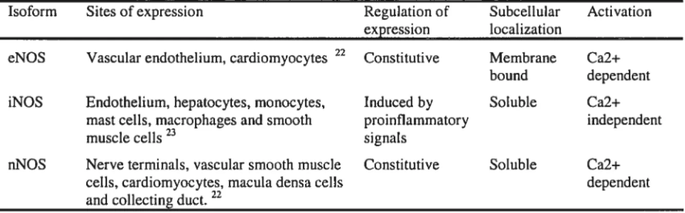
Nitric oxide is a small lipophilic molecule that exerts its function by diffusing to nearby cells. 21 is produced from the precursor L-arginine in a reaction catalyzed by nitnc oxide synthase (NOS). 22 NOS exists in 3 different isoforms that are differentially expressed throughout the cardiovascular system and the kidney. 22 The constitutive forms of NOS (eNOS and nNOS) are aiways expressed and activated upon binding of calcium and calmodulin. In contrast, iNOS is expressed upon stimulation by proinflammatory signais and act in a calcium-independent fashion. 23 In the cell, iNOS and nNOS isoforms are soluble whereas eNOS is membrane bound. 23 Table I summarizes the sites of expression, the regulation of expression, the subcellular localization and the requirements for the activation of the different isoforms of NOS.
Table I Properties of the different isoforms of NOS
Isoform Sites of expression Regulation of Subcellular Activation expression localization
eNOS Vascular endothelium, cardiomyocytes 22 Constitutive Membrane Ca2+ bound dependent iNOS Endothelium, hepatocytes, monocytes, Induced by Soluble Ca2+
mast ceils, macrophages and smooth proinflammatory independent
muscle cells23 signals
nNOS Nerve terminals, vascular smooth muscle Constitutive Soluble Ca2+ celis, cardiomyocytes, macula densa celis dependent and collecting duct.22
NO has a wide spectrum of biological actions throughout the body. In the cardiovascular system, NO is involved in the maintenance of vascular tone, in cardiac contractility, and in vascular remodeling. 22 By diffusing to nearby platelets in the lumen of the blood vessels, NO inhibits platelet aggregation and adhesion 21 In the lddney, NO contributes to water and sodium homeostasis, hemodynamics and renin secretion. 24 NO also acts as a neurotransmitter in the central and peripheral nervous systems, and participates in immune responses. 25
NO exerts its functions via cGMP-dependent and cGMP-independent pathways. 25 It acts as a paracrine signal and an intracellular messenger. 26 The short half life of NO limits its capacity to spread and restricts its actions to cells in close proximity. 21 Activation of eNOS and nNOS by transient intracellular calcium increases promotes the formation of NO at relatively low concentrations. 25 Most of the effects of eNOS and nNOS derived NO are mediated through binding to the soluble guanylyl cyclase (S GC).27 Binding of NO to S-CG leads to an increase in cyclic guanosine monophosphate (cGMP) levels. 27 Subsequently, cGMP activates downstream effectors including the activation of cGMP-gated ion channels, cGMP-dependent protein kinases (PKG), and cGMP- regulated phosphodïesterase (PDE). 27 In contrast, induction of iNOS in the context of a immune response leads to the production of high levels of NO over a long period of time 25 In this situation, NO exerts pro-apoptotic effects by releasing the cytochrome c from the mitochondria, by increasing p53 expression, and by regulating the expression of apoptosis associated proteins. 25
It is interesting to note that nNOS and eNOS are both involved in the regulation of the vascular tone. In knock out mice for the gene encoding nNOS, no changes in vascular tone was observed. 22 In contrast, in the knock out mice lacking the eNOS gene, the BP increase was accompanied by an increase in peripheral resistance. 22 From these data, it was proposed that eNOS might play a role in the regulation of vascular tone under basal conditions, whereas nNOS would be involved in adjusting the vessel contractility in response to specific modifications of its environment. 22 It was shown that nNOS activity is regulated at the transiational level in response to hypoxia leading to changes in vesse! contractility. 26
1.1.3.3.1 Kinins
Kinin peptides are formed from the precursor ldnininogen. The glycoprotein enzyme kallikrein is responsible for the formation of bradyldnin and kallidin in plasma and tissue, respectively. Kinins exert their effects through the kinin receptors Bi and B2. The B2 receptor, a G-protein coupled receptor mediates most of the known biological actions of ldnins. Kinins are converted to metabolically inactive peptides by the action of kininases. 28
Kinins are known to produce vasodilatation in vessels of the heart, lddney, intestines, skeletal muscles and liver. 2$ Effect of ldnins on the renal vasculature is believed to promote natriuresis and diuresis. 29 Other functions of ldnins include inflammatory process and stimulation of pain receptors. 2$
1.1.3.3.2 Eicosanoids
Eicosanoids are 20-carbon unsaturated fatty acids synthesized in response to a mechanical trauma, cytokines, growth factors or bradykinin. 30 Because eicosanoids are rapidly metabolized, they act locally on neighbouring cells. Biosynthesis of eicosanoids is initiated with the release of arachidonic acid (AA) from membrane phospholipids by PLA2 or from DG by PLC pathway. AA is further oxidized by 1) the cyclooxygenase (COX) pathway to produce prostaglandins (PGs) and thromboxanes (TX5), 2) the lipoxygenase pathway producing leukotnenes (LT) and 3) the P-450 epoxygenase
pathway resulting in the formation of epoxyeicosatrienoic acids (EETs). Eicosanoids exert their effects by binding G-protein linked receptors in an autocrine or paracrine fashion. 31
Eicosanoids are involved in a large array of biological functions including immunity, vascular and renal functions. Products of the COX pathway can lead either to vasodilatation of resistance vessels and diminution of SNS activity by a decrease in release of norepinephrine from sympathetic nerves (PGI2, PGE2) or to stimulation of VSM cells contraction and facilitation of sympathetic activity (TXA2, PGF2). 32 Through their effects on renai vasculature, proastaglandin and thromboxanes also affect salt and water excretion. 32 PGI2, PGE2 also increase renin release in the kidney and inhibit platelet aggregation. In contrast TXA2 is a potent platelet aggregator. 31 Leukotrienes are synthesized mostly by mast celis, eosinophils, neutrophils and macrophages. The effects of LTC4, LTD4, LTE4 leukotrienes include vasoconstriction and increased vascular permeability and LTB4 act as a chemo attractant Major cytochrome P450-derived eicosanoids include the vasodilator 5,6-epoxyeicosatrienoic acid (5,6-EET) and the vasoconstrictor 20-hydroxyeicosatetraenoic acid (20-HETE). In the preglomerular microvasculature, vasoconstriction by 20-HETE decreases GFR and consequently promotes sait and water reabsorption. 32
1.2 HYPERTENSION
1.2.1 Definition and classifications
A disruption in the homeostasis of the systems regulating blood pressure resuits in a condition known as hypertension. According to the recommendations established by the World Health Organization, blood pressure (SBP/DBP in mmHg) can be classified in the following categories: optimal (<120/80 mmHg), normal (<130/85 mmHg), normal high (130- 139185-89mmHg), hypertension grade 1 (140-159/90/99 mmHg), hypertension grade 2 (160-179/100-109 mmHg) and hypertension grade 3 ( 180/1 10 mmHg)
For 5% of the hypertensive patients, high blood pressure is explained by an underlying renal or endocrine abnormality. This type of hypertension for which the physiological cause is known is called secondary hypertension. The remaining 95% of hypertensive patients suffer from primary or essential hypertension. The pathological process leading to the development of this type of hypertension remains unknown.
1.2.2 Sequelae
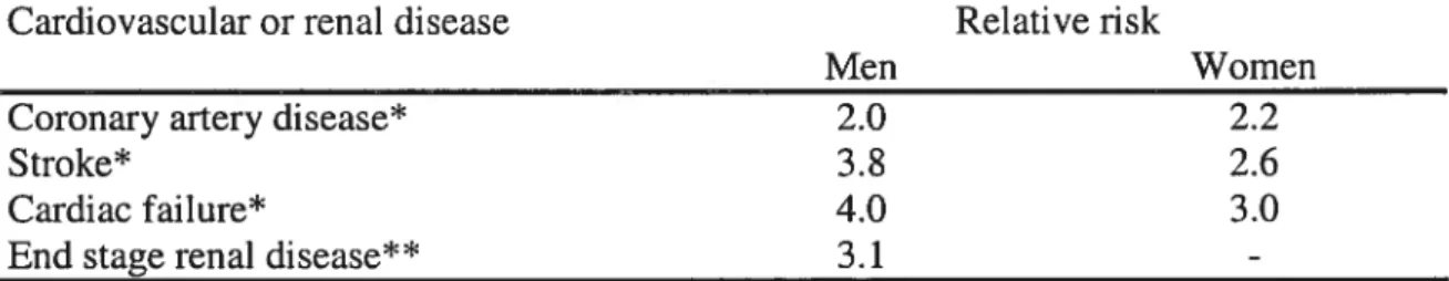
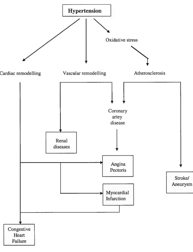
Hypertension is often referred to as the ‘suent killer’ because it is asymptomatic and painless until complications develop. However, with time, hypertension is associated with an increased risk of cardiovascular or renal complication (Table II). Oxidative stress and endothelial dysfunction, atherosclerosis and remodeling of the heart and vessels are at the basis of a sequelae to hypertension including coronary artery diseases, renal damage, angina pectoris, myocardial infarction, stroke and congestive heart failure (Figure 1).
Table II Risk of cardiovascular or renal event in hypertensive subjects Cardiovascular or renal disease Relative risk
Men Women
2.0 2.2
3.8 2.6
4.0 3.0
3.1
-Coronary artery disease* Stroke*
Cardiac failure*
End stage renal disease**
The relative risk refers to the ratio of incidence rate of cardiovascular events/renal disease among hypertensive subjects to the incidence rate cardiovascular events/renal disease among normotensives subjects. Adjusted for age. * Adapted from Kannel, W.B. ; **Adapted from Klag, M.J.
Hypertension
N
Oxidative stressN
Congestive Heart Failure Coronary artey diseasej
- Angina Pectoris Myocardial InfarctionCardiac remodelling Vascular remodelling Atherosclerosis
Stroke/ Aneurysm
1.2.2.1 Oxidative stress and endothelial dysfunction
Reactive oxygen species (ROS) are a family of molecules including free radicals (molecules that possess unpaired electrons) and other molecules that have an oxidizing effect. 38 They are part of normal cellular processes such as metabolic events, or growth factors and cytokines signaling. Under normal physiological conditions, scavenging mechanisms protect against the toxic effects of excess ROS. The rate of oxidant formation is balanced by the rate of their elimination. 40 Oxidative stress refers to the situation where there is an overproduction of ROS that cannot be buffered by scavenging mechanisms.
Several evidences are in favor of a role of oxidative stress in the development of hypertension. Correction of oxidative stress lowers BP whereas creation of oxidative stress raises BP in most animal models of hypertension. 41 Xanthine oxidase, NADHINADPH oxidase and uncoupled eNOS lias been extensively studied as sources of ROS in pathological situations. It is noteworthy that Ang II induced hypertension in rats is accompanied by an increase in expression and activity of NADHINADPH oxidase. 41 Mechanical stress on the vascular waII can also trigger increases in NADHINADPH oxidase. 38 As a resuit of increased oxidative stress, the endothelium dependent vascular relaxation is impaired, a condition known as endothelial dysfunction. 40 the consequence, at least in part, of a diminished bioavailability of NO. 42 The reduction in NO-mediated relaxation could be explained by a decreased synthesis, increased breakdown or interaction and inactivation of NO by other endothelium derived substances. u For example, ROS react with NO which resuits in the production of peroxinitrite, a cytotoxic oxidant that has poor relaxing properties on the vessels. 42
1.2.2.2 Atherosclerosis
Atherosclerosis is a chronic immune-inflammatory disease leading to the progressive accumulation of plaques wïthin the arterial wall. Several epidemiological studies have revealed that hypertension is associated with an increased frequency of complications due to atherosclerosis.
According to the response-to-injury hypothesis, elevated blood pressure would result in sustained endothelial damage which would be responsiNe for initiating the formation of a plaque. Regions of curvature and in close proximity to brandi sites in the large arteries are especially susceptible to plaque formation. 46 early stages of atherosclerosis, several elements are required for the plaque to form: 1) increased permeability of the endothelium facilitates the migration of oxidized low density lipoproteins (LDL) through the endothelium into the underlying extracellular matrix, 2) oxidized LDL stimulate blood monocytes to migrate towards the endothelium, 3) following their adhesion to the activated endothelium, the monocytes migrate across the endothelium to reach the intima. In the intima, the majority of the monocytes differentiates in macrophages, and starts intemalizing lipids to become foam cells. As the plaque progresses, the foam celis aggregate and a core of lipids and necrotic debris is formed. ‘
A fibrous cap composed of extracellular matrix components and recruited SMC cover the necrotic core. 46
Atherosclerotic plaques built up over the years and as they grow in size, they can interfere with blood flow partially or completely. In this advanced state of the disease, calcification and increased degradation of the extracellular matrix by matrix metalloproteinases predispose to plaque rupture. Once the plaque is released from the arterial wall, k produces a thrombus that can travel in the circulation and eventually block blood flow in an organ different from the site of plaque formation.
1.2.2.3 Vascular remodeling
Essential hypertension has been associated with an increase in peripheral resistance in animal models and human. 40 The resistance vessels responsible for this increased peripheral resistance are the small arteries and arterioles. ‘
The vascular resistance cf a blood vessel is function of its structural properties (media: lumen ratio and media thickness) and its active properties (force per cross sectional area). 48 The increased peripheral resistance observed in hypertension is explained by: 1) vascular remodeling
of the vessels, which affects their structural properties, and 2) modifications of the active properties of the vessel and hence, of the vascular tone.40
During the development of hypertension, inward hypertrophic remodeling and inward eutrophic remodeling have been observed in small arteries. Inward hypertrophy is characterized by a decrease in the lumen diameter accompanied by an increase in the cross sectional area of the vessel.48 In eutrophic remodeling the lumen diameter also decreases but there is no change in the cross sectional area. 48 Therefore, eutrophic and hypertrophic remodeling lead to a reduced lumen diameter. However, the increase in wall thickness is only present in hypertrophic remodeling. In contrast, eutrophic remodeling is a rearrangement of the same material around a smaller lumen. In hypertension, a process called rarefaction also contributes to the increase in vascular resistance. Rarefaction refers to a decrease in the number of parallel-connected vessels and is observed in arterioles. 50
Vascular remodeling of resistance vessels is the consequence of humoral factors, oxidative stress and mechanical factors. 40 It involves cell growth, cell death by apoptosis, celi migration and modifications of the extracellular matrix. 51 Vasoactive agents (Ang II, ET-1, catecholamine, vasopressin), growth factors and cytokines mediate vascular remodeling via activation of the MAP kinases. 40 The increase in wall stress, that results from high BP indLtces the stimulation of integrins, recruitment of the focal adhesion kinase (FAK), and remodeling processes in the vasculature through MAP kinase signaling. 48 Reactive oxygen species also contribute to vascular remodeling by stimulating growth of the vascular smooth muscle cells and accumulation of ECM. 40
1.2.2.4 Cardiac remodeling
In situations where the work load is increased, the heart responds by a compensatory increase in mass, or hypertrophy. Dunng exercise, a repeated but episodic mechanical stress is imposed to the heart. In this case, the increase in cardiac mass is explained by the homogenous remodeling of cardiomyocytes and non-cardiomycocytes. In contrast to exercise, hypertension exerts a constant mechanical and non-mechanical stress on the
heart. An increase in left ventricular hypertrophy (LVH) is accompanïed by proliferation of fibroblasts, accumulation of extracellular matrix (ECM) components and loss of cardiomyocytes by apoptosis or necrosis. Pathologie remodeling of the heart may also lead to dilated cardiomyopathy, a condition characterized by a decrease in wall thickness, increase in volume of the heart chambers and !oss of contractility. It is a consequence of increased blood volume in the heart and cardiomyocyte death. In hypertrophie cardiomyopathy, sarcomeres are added in parallel, leading to a thickening of the cardiomyocytes. In contrast, dilated cardiomyopathy presents a lengthening of the cardiomyocytes due to addition of new sarcomeres in series. 52
Mechanical and non-hemodynamic factors are the initiators of molecular events leading to a complex reorganization of the cardiac tissue. At the surface of the ce!! membranes, integrins act as mechanoreceptors that can detect changes in the work load of the heart.
When submitted to mechanica! force, the interaction between ECM components and integrins triggers intracel!ular signalling via the activation of the focal adhesion kinase (FAK). Among the mechanisms leading to the build-up of fibrosis in the myocardium, a disregulation of the MMP-1 was shown to be involved. MMP1 is the metalloproteinase responsible for degrading co!lagen type I, one of the predominant ECM component observed in fibrosis. Activation of G-protein coupled receptors by Ang II, ET-1, and activators of the adrenergic receptors, is also known to mediate the different manifestations of pathologica! hypertrophy. 52
1.2.2.5 Coronary artery disease
The coronary arteries are the vessels that distribute oxygenated b!ood to the heart. Myocardial ischemia refers to the !ack of oxygenation of the myocardium tissue resulting from a partial or complete obstruction of the coronary arteries. Hypertension can facilitate partial obstruction by an atherosclerotic plaque growing in the coronary arteries combined to an increased metabo!ic demand from thickening of the ventricular wall in LVH. The combination of plaque formation in the coronary arteries and increased metabo!ic demand due to LVH results in a b!ood flow to the heart that is flot sufficient to meet the organ requirements, a condition called coronary heart disease
(CHD). Chest pain resulting from partial blockage of the arteries in CI-ID is known as angina pectoris and is usually triggered by physical exertion or emotional stress. Altematively, the blood supply to heart can be interrupted completely by a thrombus originating from a plaque rupture. Complete privation of oxygen to heart tissue is known as myocardial infarction or heart attack. In this case, part of heart muscle will die from ischemia. The extend of the damage will depend on the proportion of myocardium that suffers from the lack of oxygen.
1.2.2.6 Congestive heart failure
Congestive heart failure (CHF) refers to a condition in which the heart cannot supply enough blood to meet the metabolic requirements of the body, due to a progressive weakening of the pump function of the organ. LVH or loss of myocytes due to past events CHD or MI are important risk factors for the development of CHF. 56 In addition to an increase in LVH, remodeling of the heart is accompanied fibrosis. One of the consequence of the accumulation of ECM is a decreased compliance of the left ventricle resulting in diastolic dysfunction. To maintain the cardiac output and
fuI
the left ventricle to normal levels, the heart compensates the diastolic dysfunction by increasing the left atrial pressure. Pulmonary oedema can occur if the elevated atrial pressure is transmitted to the pulmonary circulation. In advanced states of CHF, the condition progresses to a state of uncompensated CHF resulting in a lowenng of the CO that will eventually lead to pump failure. 581.2.2.7 Stroke and cerebral aneurysms
Stroke is the term used to define the vascular events leading to cerebral ischemia. Several studïes came to the conclusion that hypertension is the most important modifiable risk factor for stroke. In addition, active treatment of hypertension can reduce the occurrence of stroke by 25 to 45 %. Damage to the brain linked to hypertension can occur via several mechanisms. In large vessels, cerebral ischemia can resuit from partial or total blockage of a cerebral artery by an atherosclerotic plaque or from the interruption of the blood flow by a thrombus. Another type of stroke occurs from cerebral hemorrhage. The latter can be the result of an aneurysm, which us a blood
filled sac forming in the wal! of an artery due to increased pressure on a thrombus or from bursting of an artery. Because the sma!! diameter penetrating end arteries that supp!y cerebrai tissue arise directly from main arterial trunk, these are especia!ly vuinerable to the effect of high blood pressure.
1.2.2.7 Renal damage
As a resuit of hypertension, glycoproteins and collagen matrix form a homogeneous deposit on the wall of the renal vessels accompanied by atrophy of smooth muscle ceils and irregular thickening of the basement membrane. Preglomerular arterioles also undergo hypertrophic and hyperpiasic remodelling of the smooth muscle cells. At the molecular level, these modifications of the renal microvasculature are known to involve changes in cell cycle and activation of genes encoding proteins relating to growth and secretion of matrix. Deposition of fibrin and fibrinogen is also observed. A graduai obstruction of the biood flow to the giomeruli is associated with a decrease in function and subsequent atrophy due to the resulting ischemia. Altematively, the kidney can suffer direct damage from an increase in blood pressure entering the glomeruli due to the incapacity of preglomerular vesse! to adjust diameter in responses to changes in blood pressure. These mechanisms lead to glomeruiosclerosis and eventually renal failure.
In larger arteries, lesions attributable to atheromatous plaque obstruction and smooth muscle hyperplasia and fibrosis can lead to renai artery stenosis, which is a complete or partial narrowing of the artery. If occurring unilaterally, renal arterial stenosis will lead to secondary hypertension.
1.2.3 Epidemiology and burden
Statistics indicate that hypertension, obesity, physical inactivity, and tobacco smoking are among the modifiable risk factors of developing a cardiovascular disease. 80.2% of Canadian adults aged between 20 and 59 years have at least one of these risk factors. 60 High blood pressure alone affects 21.1% of the Canadian population according to the
The biennial publication of the Heart and Stroke Foundation of Canada ranks cardiovascular diseases as the leading cause of death in Canada, responsible for 74 626 deaths in 2002 only. 62 In addition to the high mortality associated with cardiovascular diseases, Health Canada classifies cardiovascular diseases as the most costly contributor to both direct and indirect health costs, accounting for 11.6 % of total illness costs in Canada. In 1998, the cost rising from cardiovascular disease was evaluated at 18.5 billion dollars, 6.8 billion in direct cost related to hospitalisation, prescription drug consumption and physician care expenditure and 11.7 billion dollars in indirect cost explained by disability and mortality. 6
1.2.4 Ireatment
When it cornes to treating hypertension, modification to the lifestyle and pharmacological treatments are often needed to reach therapeutic BP levels. Lifestyle modifications can reduce SBP by 4.6- 11.4 mmHg and the diastolic pressure by 2.5 - 7.5
mrnHg. 63 Changes recommended include weight loss, diminution of alcohol and salt consumption and regular exercise. 64 In cases where BP is higher than stage I hypertension, various classes of drugs can be used alone or in combination to reach target values of BP. The major classes of agents available to fight hypertension are the diuretics that act by depleting body sodium, the sympathoplegic agents that act on the SNS, the direct vasodilators that relax smooth muscle celis and the molecules that prevent the actions of the RAS such as the ACE inhibitors and the angiotensin receptor blockers. 65 Reduction of the blood pressure below 140/90 mmHg is achieved in only 13% of the patients suffering from hypertension 61 leaving room for improvement in awareness and efficiency of treatment.
1.3 GENETICS 0F HYPERTENSION
Primary hypertension can be sub-divided into monogenic and essential hypertension. Monogenic hypertension represents 5% of alI cases of primary hypertension. It is explained by a single gene defect inherited following a Mendelian transmission. The
remaining 95% of hypertension cases are classified as essential hypertension and are under a polygenic influence with an environmental component. The search for genes responsible for Mendelian forms of hypertension has been a successfui one, but the genes invoived in essentiai hypertension remain unknown.
1.3.1 Monogenic hypertension
Mendelian forms of hypertension include Liddie’s syndrome, glucocorticoid remediable aidosteronism (GRA), apparent mineraiocorticoid excess (AME), and pseudohypoaldosteronism type II (PHAII). Ail these forms of hypertension affect homeostasis of sait and water reabsorption. Even though Mendelian forms of hypertension are more rare and severe than essential hypertension, there was a tremendous effort to better understand their etiology in the hope that it wouid lead to dues about the pathophysiology of essential hypertension.
1.3.1.1 Glucocorticoid Remediable Aldosteronism
Normaily, cortisol is synthesized in the zona fasciculata of adrenai cortex whereas aldosterone is synthesized in the zona giomerulosa. Ïïf3hydroxylase encoded by the gene CYP11B1 is invoived in the synthesis of cortisoi and the aldosterone synthase encoded by the gene CYP]]B2 is part of the biosynthetic pathway leading to aldosterone production. CYPJJBJ and CYP]]B2 are both located on human chromosome (chr) 8. Both genes have 9 exons sharing 95% DNA simiiarity and $ introns simiiar to 90%. 66 The major difference between CYPJJBJ and CYPJ]B2 resides in the 5’flanking region. CYPJJBJ expression is regulated by adrenocorticotropic hormone (ACTH) and CYP]1B2 is reguiated by angiotensin II.
In GRA, an autosomai dominant form of hypertension, there is an increased and ectopic synthesis of aldosterone. 67 was shown that in families suffering from GRA, there is a chromosomai rearrangement resulting from unequal meiotic cross over between CYFJ1B1 and CYP]]B2 leading to an additional copy of a chimeric gene made of the S’flanking sequence and first few exons of CYPJJBJ and of the 3’exons of CYP]]B2.
68,69
control of ACTH. The rate of cortisol to aldosterone secretion is normally 1000:1. 70 Under the control of ACTH aldosterone is produced in excessive quantities therefore leading to sait retention, volume expansion and hypertension.
1.3.1.2 Apparent Mineralocorticoid Excess
Mineralocorticoid receptors have similar affinity for mineralocorticoid aldosterone and glucocorticoid cortisol. It was proposed that specificity of aldosterone its receptor in target tissues is achieved by tissue specific expression of 11 f3-hydroxysteroid dehydrogenase (11f3HSD) which converts cortisol to cortisone, an anaiog that has no affinity for the mineralocorticoid receptor. 71 Therefore, in the distal tubule, 1 1I3HSD acts as a protector of the mineralocortïcoid receptor by preventing high leveis of cortisoi to occupy the aidosterone receptor. 72
AME is an autosomal recessive form of hypertension. It is caused by stimulation of the mineralocorticoid receptor despite low levels of aldosterone. Mutations in the lddney isoform of 11f3HSD that affect enzyme activity or pre m-RNA spiicing have been identified in AME patients. A deficiency of 113HSD in the lddney impairs the conversion of cortisol to cortisone, thereby leaving the mineralocorticoid receptor unprotected from high levels of cortisol. 72 The activation of the aldosterone receptor by cortisol leads to hypertension through sodium and water retention.
1.3.1.3 Liddle’s syndrome
The amiloride-sensitive epithelial sodium channel (ENaC) is expressed in the distal portion of kidney tubules and is regulated by aldosterone. This channel is made up of three subunits: Œ,
E3,
and ‘y. Reabsorption of sodium through ENaC is an importantdeterminant of BP homeostasis.67
Liddle is characterized by early onset severe hypertension. Early linkage studies have shown association between f3ENaC locus on human chr. 16 and Liddle’s Syndrome. Further analysis of ENaC gene have revealed mutations in the
f3
subunit gene75,76 and in the ‘y subunit gene “. Ail mutations identified consist in a premature stop codon orframeshift mutation in the carboxy terminal domain of the protein resulting in the removal of the normal carboxy terminal. Subsequently, it was shown that truncation or frameshift of the carboxy terminal domain does flot affect the biophysical properties of the channel but causes an increase in the number of receptors present in the plasma membrane. 78 Nedd4 (neuronal precursor celi expressed developmentally downregulated) is a ubiquitin-protein ligase that binds to afryENaC PY (xPPxY) motif located in the COOH terminus of the channel. Upon ENaC/Nedd4 interaction, the channel is ubiquitinated and targeted for degradation by lysosomes. ‘ The loss of Nedd4 binding sites in Lidddle’s syndrome may resuit in impaired degradation of the channel and hence, increased channel activity would result in abnormal renal reabsorption of sodium and water, and explains hypertension.
1.3.1.4 Pseudohypoaldosteronïsm type II
Pseudohypoaldosteronism type II (PHAII), or Gordon’s syndrome, is an autosomal dominant form of hypertension. ° In PHAII, hypertension is accompanied by hyperkalemia, hyperchioremic metabolic acidosis, and normal to high levels of aldosterone. 72 Genomic wide linkage search of families affected with PHAII have revealed strong linkage on regions of chr. 1, 12 and 17. 72 Subsequently, identification of two of the genes responsible for PHA11 lead to the discovery of a novel family of serine/threonine kinases, the WNK.
The WNK (with no lysine kinases) are recognized by the lack of a key lysine residue in the catalytic domain. 82 In the epithelium of the distal nephron, WNK4 is necessary for the coordination of the transcellular and paracellular flux to achieve NaC1 and K+ homeostasis. This function is accomplished by: 1) inhibition of the NCCT (NaCÏ co transporter) by preventing its insertion into the membrane, 2) induction of clathrin dependent reduction of renal outer medullary potassium channel (ROMK) expression at celi surface, and 3) regulation of paracellular permeability to Cl- by phosphorylation of claudin proteins in the tightjuctions. 80 In the kidney, WNK1 expression is restricted to the cytoplasm of distal epithelial cells.83 Its function is to prevent WNK4-induced inhibition of NCCT. 80 The mechanism behind the inhibitory effect of WNK1 on
WNK4 is flot fully understood, but it may involve a direct interaction between the two kinases.80
Mutation in genes encoding WNK1 (on human chr.12) and WNK4 (on human chr.17) are responsible for PHAII. In the gene coding for WNK4, missense mutations clustered to a highly conserved sequence 20-25 amino acids downstream of the coiled cou domains of the enzyme have been identified. The mutated WNK4 protein looses the ability to retain NCCT in the cytoplasm leading to increased expression of NCCT on the celi surface and excessive Na+ reabsorption. In a gain of function fashion, the mutations in WNK4 gene increase intemalization of the ROMK channel and the permeability of tight junctions to Ci- leakage across the epithelium thereby explaining hyperkalemia associated with PHAII. ° The mutation in the WNKJ gene are large deletions in the first intron resulting in increased WNK1 expression without altering the protein structure. 83 The presence of higher leveis of WNK1 in the kidney increases the inhibitory effect of WKN1 on WNK4, which results in increased amount of NCCT at the celi surface and increased sodium reabsorption. 80
1.3.2 Essential Hypertension
1.3.2.1 Evidence for a genetic contribution
As described in the previous section, the search for a genetic basis to several forms of monogenic hypertension has been a successful one. However, Mendelian forms of hypertension represent merely 5% of ail cases, the remaining being polygenic with an environmental component. The challenge now resides in identifying genes involved in essential hypertension.
It has been known for a long time that hypertension aggregates in certain families. Familial clustering of high BP could be the consequence of genetic inheritance of blood pressure gene or genes, household environment or a combination of both. Familial aggregation studies have shown that clustering occurs early in life since siblings living apart do flot become dissimilar and spouses do flot become more similar as they share

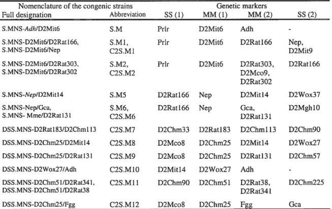


![Figure 2 Congenic Substrains — D2Rat303 — D2Chm33 — D2Ratl 83 — D2Chml22 — D2Ratl66 — D2Rat22l — D2Chml 13 — D2Chm9O — D2Chrn5 I — D2Chrn46 — jIjme — D2Rat3$ — D2Chrn225 — D2McoS — D2Chrn25 — D2Chrn24 — Gucythi — Gucylal Pgg — Gea —D2RatÎ31 — D2Chrn57 — D2Mgh 10CHROMOSOME 2Linkage MapcM-I—D2Mco]5ED2Mco131(3.4Prit4.7D2Mco23D2Mit66.6D2Ratl352.9Agtrib9.1D2Rat9610.3D2Rat3 035.7—D2RatI66D2Chrn9OAinie6.4D2Mco$D2Mitl IFgg2.9GeaD2RatI3I 4.6 D2Mih10 .11?) lai D2MItI4 2.6 D2Wox27 5.4 D2Mco4 9.0 D2N9] Gamk2d 6.7 27.7 D2N35 6.6 — Ad/i DSS C2S.M7 C2S.v1fl C2S.M12 C28.M9 C2S.M$ C2S.M10/f C2QTL1 C2QTL3Iri}C2Q1L2_.4tpla /D2MItI4—D2Wox27— IdÏi U MAP (mrnHe) 170±4 145±4 144±2 160±7 139±8 139±5 162±7 (nl 8) (n8) (n8) (n9) (n5) (n8) (n5)](https://thumb-eu.123doks.com/thumbv2/123doknet/2052702.5495/131.918.153.882.135.1121/figure-congenic-substrains-gucythi-gucylal-chromosome-linkage-agtrib.webp)