HAL Id: dumas-01075721
https://dumas.ccsd.cnrs.fr/dumas-01075721
Submitted on 20 Oct 2014
HAL is a multi-disciplinary open access archive for the deposit and dissemination of sci-entific research documents, whether they are pub-lished or not. The documents may come from teaching and research institutions in France or abroad, or from public or private research centers.
L’archive ouverte pluridisciplinaire HAL, est destinée au dépôt et à la diffusion de documents scientifiques de niveau recherche, publiés ou non, émanant des établissements d’enseignement et de recherche français ou étrangers, des laboratoires publics ou privés.
Treatment of refractory open-angle glaucoma using
ultrasonic circular cyclocoagulation : a prospective case
series
Caroline Dupuy
To cite this version:
Caroline Dupuy. Treatment of refractory open-angle glaucoma using ultrasonic circular cyclocoagu-lation : a prospective case series. Human health and pathology. 2014. �dumas-01075721�
AVERTISSEMENT
Ce document est le fruit d'un long travail approuvé par le
jury de soutenance et mis à disposition de l'ensemble de la
communauté universitaire élargie.
Il n’a pas été réévalué depuis la date de soutenance.
Il est soumis à la propriété intellectuelle de l'auteur. Ceci
implique une obligation de citation et de référencement
lors de l’utilisation de ce document.
D’autre part, toute contrefaçon, plagiat, reproduction illicite
encourt une poursuite pénale.
Contact au SICD1 de Grenoble :
[email protected]
LIENS
LIENS
Code de la Propriété Intellectuelle. articles L 122. 4
Code de la Propriété Intellectuelle. articles L 335.2- L 335.10
http://www.cfcopies.com/V2/leg/leg_droi.phpUNIVERSITE JOSEPH FOURIER FACULTE DE MEDECINE DE GRENOBLE
Année : 2014 N°
TREATMENT OF REFRACTORY OPEN-ANGLE GLAUCOMA USING ULTRASONIC CIRCULAR CYCLOCOAGULATION: A PROSPECTIVE CASE
SERIES
THESE
PRESENTEE POUR L’OBTENTION DU DOCTORAT EN MEDECINE
DIPLÔME D’ETAT
Caroline DUPUY
Née le 25 novembre 1985 A TOULON, France
THESE SOUTENUE PUBLIQUEMENT A LA FACULTE DE MEDECINE DE GRENOBLE
Le jeudi 16 octobre 2014
DEVANT LE JURY COMPOSE DE
Président du jury : Monsieur le Professeur Jean-Paul ROMANET Directeur de thèse : Monsieur le Professeur Florent APTEL
Membres : Monsieur le Professeur Christophe CHIQUET Monsieur le Docteur Fabrice ROMANO
UNIVERSITE JOSEPH FOURIER FACULTE DE MEDECINE DE GRENOBLE
Année : 2014 N°
TREATMENT OF REFRACTORY OPEN-ANGLE GLAUCOMA USING ULTRASONIC CIRCULAR CYCLOCOAGULATION: A PROSPECTIVE CASE
SERIES
THESE
PRESENTEE POUR L’OBTENTION DU DOCTORAT EN MEDECINE
DIPLÔME D’ETAT
Caroline DUPUY
Née le 25 novembre 1985 A TOULON, France
THESE SOUTENUE PUBLIQUEMENT A LA FACULTE DE MEDECINE DE GRENOBLE
Le jeudi 16 octobre 2014
DEVANT LE JURY COMPOSE DE
Président du jury : Monsieur le Professeur Jean-Paul ROMANET Directeur de thèse : Monsieur le Professeur Florent APTEL
Membres : Monsieur le Professeur Christophe CHIQUET Monsieur le Docteur Fabrice ROMANO
1
Professeurs des Universités- Praticiens Hospitaliers 2013-2014
Occupation Actuelle Discipline Universitaire
ALBALADEJO Pierre Depuis 01/09/2008 Anesthésiologie-réanimation ARVIEUX-BARTHELEMY Catherine Depuis de 01/09/2007 Chirurgie générale BACONNIER Pierre
Depuis 01/10/1993 Biostat, informatique médicale et technologies de communication
BAGUET Jean-Philippe
Depuis 01/09/2006 Cardiologie
BALOSSO Jacques
Depuis 01/09/2003 Radiothérapie
BARRET Luc
Depuis 01/10/1992 Médecine légale et droit de la santé
BAUDAIN Philippe
Depuis 01/05/1990 Radiologie et imagerie médicale
BEANI Jean-Claude
Depuis 01/10/1992 Dermato-vénérologie
BENHAMOU Pierre Yves
Depuis 01/09/2003 Endocrinologie, diabète et maladies métaboliques
BERGER François
Depuis 01/09/2001 Biologie cellulaire
BETTEGA Georges
Depuis 01/09/2013 Chirurgie maxillo-faciale et stomatologie
BONAZ Bruno
Depuis 01/09/2001 Gastro-entérologie, hépatologie, addictologie
BOSSON Jean-Luc
Depuis 01/01/2006 Biostat, informatique médicale et technologies de communication
BOUGEROL Thierry
Depuis 01/09/1998 Psychiatrie d'adultes
BOUILLET Laurence
Depuis 01/09/2012 Médecine interne
BRAMBILLA CHRISTIAN
Depuis 01/10/1989 Pneumologie
BRAMBILLA Elisabeth
Depuis 01/10/1993 Anatomie et cytologie pathologiques
BRICAULT Ivan
Depuis 01/09/2011 Radiologie et imagerie médicale
BRICHON Pierre-Yves
Depuis 01/10/1993 Chirurgie thoracique et cardio-vasculaire
CAHN Jean-Yves
Depuis 01/09/2004 Hématologie
CARPENTIER Françoise
Depuis 01/09/1997 Thérapeutique, médecine d'urgence
CARPENTIER Patrick
Depuis 01/10/1990 Chirurgie vasculaire, médecine vasculaire
CESBRON Jean-Yves
CHABARDES Stephan
Depuis 01/09/2010 Neurochirurgie
CHABRE Olivier
Depuis 01/09/2002 Endocrinologie, diabète et maladies métaboliques
CHAFFANJON Philippe
Depuis 01/09/2005 Anatomie
CHAVANON OlivierDepuis
01/09/2006 Chirurgie thoracique et cardio-vasculaire
CHIQUET Christophe
Depuis 01/09/2007 Ophtalmologie
CHIROSSEL Jean-Paul
Depuis 01/06/1990 Anatomie
CINQUIN Philippe
Depuis 01/10/1992 Biostat, informatique médicale et technologies de communication
COHEN Olivier
Depuis 01/09/2003 Biostat, informatique médicale et technologies de communication
COUTURIER Pascal
Depuis 01/09/2007 Gériatrie et biologie du veillissement
CRACOWSKI Jean-Luc
Depuis 01/09/2009 Pharmacologie fondamentale, pharmacologie clinique
DE GAUDEMARIS Régis
Depuis 01/07/1992 Médecine et santé au travail
DEBILLON Thierry
Depuis 01/09/2003 Pédiatrie
DEMATTEIS Maurice
Depuis 01/09/2010 Addictologie
DEMONGEOT Jacques
Depuis 01/10/1989 Biostat, informatique médicale et technologies de communication
DESCOTES Jean-Luc
Depuis 01/09/1997 Urologie
ESTEVE François
Depuis 01/09/2004 Biophysique et médecine nucléaire
FAGRET Daniel
Depuis 01/10/1992 Biophysique et médecine nucléaire
FAUCHERON Jean-Luc
Depuis 01/09/2001 Chirurgie générale
FERRETTI Gilbert
Depuis 01/09/2000 Radiologie et imagerie médicale
FEUERSTEIN Claude
Depuis 01/07/1992 Physiologie
FONTAINE Eric
Depuis 01/01/2006 Nutrition
FRANCOIS Patrice
Depuis 01/09/1998 Epidémiologie, économie de la santé et prévention
GARBAN Frédéric
Depui 01/09/2011 Hématologie, transfusion
GAUDIN Philippe
3
GAVAZZI Gaetan
Depuis 01/09/2011 Gériatrie et biologie du veillissement
GAY Emmanuel
Depuis 01/09/2004 Neurochirurgie
GODFRAIND Catherine
Depuis 01/09/2013 Anatomie et cytologie pathologiques
GRIFFET Jacques
Depuis 01/03/2010 Chirurgie infantile
HALIMI Serge
Depuis 01/10/1990 Nutrition
HENNEBICQ Sylviane
Depuis 01/09/2012 Biologie et médecine du développement et de la reproduction
HOFFMANN Pascale Depuis 01/09/2012 Gynécologie-obstétrique HOMMEL Marc Depuis 01/09/1995 Neurologie JOUK Pierre-Simon Depuis 01/09/1997 Génétique JUVIN Robert Depuis 01/10/1993 Rhumatologie KAHANE Philippe Depuis 01/09/2007 Physiologie KRACK Paul Depuis 01/09/2003 Neurologie KRAINIK Alexandre
Depuis 01/09/2009 Radiologie et imagerie médicale
LABARERE José
Depuis 01/09/2012 Epidémiologie, économie de la santé et prévention
LANTUEJOUL Sylvie
Depuis 01/09/2008 Anatomie et cytologie pathologiques
LECCIA Marie-Thérèse
Depuis 01/09/2002 Dermato-vénérologie
LEROUX Dominique
Depuis 01/09/1996 Génétique
LEROY Vincent
Depuis 01/09/2007 Gastro-entérologie, hépatologie, addictologie
LETOUBLON Christian
Depuis 01/05/1992 Chirurgie générale
LEVY Patrick
Depuis 01/09/1997 Physiologie
MACHECOURT Jacques
Depuis 01/10/1989 Cardiologie
MAGNE Jean-Luc
Depuis 01/07/1990 Chirurgie vasculaire
MAITRE Anne
Depuis 01/09/2007 Médecine et santé au travail
MAURIN Max
MERLOZ Philippe
Depuis 01/10/1991 Chirurgie orthopédique et traumatologie
MORAND Patrice
Depuis 01/09/2007 Bactériologie-virologie
MOREAU-GAUDRY Alexandre
Depuis 01/09/2013 Biostat, informatique médicale et technologies de communication
MORO Elena Depuis 01/09/2012 Neurologie MORO-SIBILOT Denis Depuis 01/09/2005 Pneumologie MOUSSEAU Mireille Depuis 01/09/1994 Cancérologie MOUTET François
Depuis 01/10/1990 Chirurgie plastique, reconstructrice & esthétique, brulologie
PALOMBI Olivier Depuis 01/09/2011 Anatomie PARK Sophie Depuis 01/09/2013 Hématologie PASSAGIA Jean-Guy Depuis 01/09/1994 Neurochirurgie PAYEN DE LA GARANDERIE Jean-François Depuis 01/09/1996 Anesthésiologie-réanimation PELLOUX Hervé
Depuis 01/09/2001 Parasitologie et mycologie
PEPIN Jean-Louis
Depuis 01/09/2004 Physiologie
PERENNOU Dominique
Depuis 01/04/2008 Médecine physique et de réadaptation
PERNOD Gilles
Depuis 01/09/2007 Médecine vasculaire
PIOLAT Christian
Depuis 01/09/2009 Chirurgie infantile
PISON Christophe Depuis 01/09/1994 Pneumologie PLANTAZ Dominique Depuis 01/09/2003 Pédiatrie POLACK Benoît Depuis 01/09/1998 Hématologie POLOSAN Mircea
Depuis 01/09/2013 Psychiatrie d'adultes
PONS Jean-Claude Depuis 01/09/1998 Gynécologie-obstétrique RAMBEAUD Jean-Jacques Depuis 01/07/1991 Urologie REYT Emile Depuis 01/10/1992 Oto-rhyno-laryngologie RIGHINI Christian Depuis 01/09/2010 Oto-rhyno-laryngologie
5
ROMANET J. Paul
Depuis 01/10/1991 Ophtalmologie
SARAGAGLIA Dominique
Depuis 01/07/1992 Chirurgie orthopédique et traumatologie
SCHMERBER Sébastien
Depuis 01/09/2005 Oto-rhyno-laryngologie
SCHWEBEL Carole
Depuis 01/09/2012 Réanimation, médecine d'urgence
SCOLAN Virginie
Depuis 01/09/2013 Médecine légale et droit de la santé
SERGENT Fabrice
Depui 01/09/2011 Gynécologie-obstétrique
SESSA Carmine
Depuis 01/09/2005 Chirurgie vasculaire
STAHL Jean-Paul
Depuis 01/10/1992 Maladies infectieuses, maladies tropicales
STANKE Françoise
Depuis 01/09/2011 Pharmacologie fondamentale
TAMISIER Renaud
Depuis 01/09/2013 Physiologie
TIMSIT Jean-François Réanimation
TONETTI Jérôme
01/09/2007 au 31/12/2010 Chirurgie orthopédique et traumatologie
TOUSSAINT Bertrand
Depuis 01/09/2008 Biochimie et biologie moléculaire
VANZETTO Gérald
Depuis 01/09/1999 Cardiologie
VUILLEZ Jean-Philippe
Depuis 01/09/1999 Biophysique et médecine nucléaire
WEIL Georges
Depui 01/09/2011 Epidémiologie, économie de la santé et prévention
ZAOUI Philippe
Depuis 01/09/2002 Néphrologie
ZARSKI Jean-Pierre
Maîtres de Conférences des Universités- Praticiens Hospitaliers 2013-2014
Occupation Actuelle Discipline universitaire
APTEL Florent
Depuis 01/09/2012 Ophtalmologie
BOISSET Sandrine
Depuis 01/09/2012 Bactériologie, virologie
BONNETERRE Vincent
Depuis 01/09/2011 Médecine et santé au travail
BOTTARI Serge
Depuis 01/10/1993 Biologie cellulaire
BOUTONNAT Jean
Depuis 01/09/2000 Cytologie et histologie
BOUZAT Pierre
Depuis 01/09/2012 Anesthésiologie-réanimation
BRENIER-PINCHART M.Pierre
Depuis 01/11/2001 Parasitologie et mycologie
BRIOT Raphaël
Depuis 01/09/2009 Thérapeutique, médecine d'urgence
CALLANAN-WILSON Mary
Depuis 01/09/2002 Hématologie, transfusion
DECAENS Thomas Depuis 01/09/2013 DERANSART Colin Depuis 01/09/2004 Physiologie DETANTE Olivier Depuis 01/09/2009 Neurologie DIETERICH Klaus Depuis 01/09/2012 Génétique DUMESTRE-PERARD Chantal Depuis 01/09/2004 Immunologie EYSSERIC Hélène
Depuis 01/10/2009 Médecine légale et droit de la santé
FAURE Julien
Depuis 01/09/2008 Biochimie et biologie moléculaire
GILLOIS Pierre
Depuis 01/09/2010 Biostat, informatique médicale et technologies de communication
GRAND Sylvie
Depuis 01/09/1995 Radiologie et imagerie édicale
GUZUN RitaDepuis 01/09/2012 Nutrition
LAPORTE François
Depuis 01/10/1991 Biochimie et biologie moléculaire
LARDY Bernard
Depuis 01/09/2007 Biochimie et biologie moléculaire
LARRAT Sylvie
7 LAUNOIS-ROLLINAT Sandrine Depuis 01/09/2001 Physiologie LONG Jean-Alexandre Depuis 01/09/1999 Urologie MAIGNAN Maxime
Depuis 01/09/2013 Médecine d'urgence
MALLARET Marie-ReineDepuis
01/08/1992 Epidémiologie, économie de la santé et prévention
MARLU Raphaël
Depuis 01/09/2013 Hématologie
MAUBON Danièle
Depuis 01/09/2010 Parasitologie et mycologie
MC LEER (FLORIN) Anne
Depuis 01/09/2011 Cytologie et histologie
MOUCHET Patrick
Depuis 01/10/1992 Physiologie
PACLET Marie-Hélène
Depuis 01/09/2007 Biochimie et biologie moléculaire
PAYSANT François
Depuis 01/02/2008 Médecine légale et droit de la santé
PELLETIER Laurent
Depuis 01/01/2006 Biologie cellulaire
RAY Pierre
Depuis 01/09/2003 Génétique
RIALLE Vincent
Depuis 01/09/2001 Biostat, informatique médicale et technologies de communication
ROUSTIT Matthieu
Depuis 01/08/1990 Pharmacologie clinique
ROUX-BUISSON Nathalie
Depuis 01/09/2012 Biochimie et génétique moléculaire
SATRE VéroniqueDepuis
01/09/2005 Génétique
SEIGNEURIN Arnaud
Depuis 01/09/2013 Epidémiologie, économie de la santé et prévention
STASIA Marie-Josée
REMERCIEMENTS
Aux membres de mon jury :
A Monsieur le Professeur Jean-Paul ROMANET, vous avez été notre Maître pendant notre spécialisation. Nous avons apprécié l’énergie que vous déployez au service de
l'ophtalmologie. Merci de m'avoir fait l'honneur d'accepter la présidence de ce jury.
A Monsieur le Professeur Florent APTEL, merci de m'avoir fait l'honneur de travailler sur ce sujet, dont tu es l'un des principaux instigateurs. C'est un privilège d'avoir pu travailler à tes côtés.
A Monsieur le Professeur Christophe CHIQUET, pour votre implication dans nos travaux de recherche et notre formation théorique.
A Monsieur Fabrice ROMANO, il semblait tout naturel de solliciter le concours de l'inventeur de ce dispositif novateur. Merci d'avoir accepté avec tant de gentillesse et simplicité de siéger à mon jury de thèse.
A tous ceux qui ont participé à ma formation :
En ophtalmologie : Diane BERNHEIM, Olivier SAVY, Matthieu TONINI, Hafide KHAYI, Karine PALOMBI, Elizabeth RENARD, Adel CHIBANI, Jean-Yves MILLET, Ruxandra Hera, Pierre PEGOURIE, Dominique SATGER, Viviane VINH, Brigitte GONZALVEZ, Sylvie BERTHEMY PELLET, Patrice MOYENIN, Pierre ALBINET
En chirurgie maxillo-faciale : Monsieur le Professeur Georges BETTEGA, Monsieur le Professeur Jacques LEBEAU, Madame Béatrice MORAND, Antoine GROSDIDIER, Cynthia HAMOU, Brice CARLOT
En neuroradiologie : Monsieur le Professeur Alexandre KRAINIK, Monsieur le Professeur Jean-François LEBAS, Madame Florence TAHON, Monsieur Kamel BOUBAGRA, Monsieur Jean-Ashok VASDEV, Monsieur Victor CUVINCIUC, Monsieur Omer EKER En bactériologie : Monsieur le Professeur Max MAURIN.
9
A mes co-internes :
Des plus anciens (Tiffany, Eva, Magali, Aurélie, Ralitsa, Antoine) qui m'ont transmis leur savoir dans la joie et la bonne humeur, aux plus jeunes (Marco, Georges, Perrine, Julie, Rachel, Adrianne, Olivier, Floriane, Perrine, Bruno, Julie, Kim, Oualid et François Xavier), sans oublier les marseillais (Joséphine et Frédérique), l'équipe EBO 2014 et la promo 2009 (Thierry, Benjamin, Nischal, Mathilde, Cécile), avec qui j'ai eu plaisir à partager ces 5 années.
Aux secrétariats de Grenoble et Chambéry, orchestrés magistralement par Madame
Catherine TARANTINI et Madame Bernadette RASSAT. Merci pour le temps passé à sortir et ressortir les dossiers pour nos travaux !
Aux équipes soignantes de Grenoble, chapeautées par Madame Catherine BOZON et
Madame Joëlle COET, et de Chambéry, pour leur gentillesse et leur disponibilité.
A l'équipe de microbiologie pour leur aide inestimable et leur disponibilité : Vivien
SUTERA, Jeanne Noëlle DEL BANO et Annie BLACHON.
A l'équipe d'Annecy, et particulièrement le Docteur TONINI et le Dr KHAYI, pour
m'accueillir parmi eux avec tant de bienveillance et gentillesse.
A mes amis, avec qui je partage la passion du sport, de la pagaie, du cinéma d'auteur et le
savon : Béatrice, Clotilde, Louis, Jean-François, Marion, Laurent, Gérald, Hélène, Julie, Amélie, Benoit, Aurélie, Claire, Marie, Irène, Christine, Thibaut, Morine, Sandrine et Claire.
A ma famille, pour son précieux et éternel soutien.
Treatment of Refractory Open-Angle Glaucoma using Ultrasonic
Circular Cyclocoagulation: a Prospective Case Series
11
ABSTRACT
Objective: To evaluate the efficacy and safety of the Ultrasonic Circular Cyclo Coagulation
(UC3) procedure in patients with refractory primary open-angle glaucoma.
Research design and methods: Prospective non-comparative interventional case series
performed in 2 French glaucoma centers. 28 eyes of 28 patients with primary open-angle glaucoma, intraocular pressure (IOP) > 21 mmHg, an average of 1.4 failed previous surgeries and an average of 3.8 hypotensive medications were insonified with a therapy probe comprising 6 piezoelectric transducers. The 6 transducers were activated and all patients were treated with a 6 seconds exposure time. Complete ophthalmic examinations were performed before the procedure, and at 1 day, 1 week, 1, 2, 3, 6 and 12 months after.
Main outcome measures: Primary outcomes were surgical success (defined as IOP reduction
from baseline ≥ 20% and IOP > 5mmHg) at the last follow-up visit, and vision-threatening complications. Secondary outcomes were mean IOP at each follow-up visit compared to baseline, medication use, complications, and re-interventions.
Results: IOP was significantly reduced (p<0.05), from a mean preoperative value of 29.0 ±
7.2 mmHg (n= 3.8 hypotensive medications) to a mean value of 21.6 ± 9.4 mmHg at last follow-up (n= 3.8 hypotensive medications and n= 1.29 procedures) (mean IOP reduction of 26%). Complete success (IOP reduction >20% without re-intervention and without hypotensive medications adjunction) was achieved in 50% of eyes at last follow-up (mean IOP reduction of 45% in these same eyes) and qualified success (IOP reduction >20% with possible re-interventions) was achieved in 68% of eyes at last follow-up. No major intra- or post-operative complications occurred.
Conclusions: UC3 procedure seems to be an effective and well-tolerated method to reduce intraocular pressure in patients with primary open-angle glaucoma.
INTRODUCTION
Many methods and energy sources for destroying the ciliary processes have been investigated, resulting in coagulation necrosis of the ciliary body following heating (laser, microwave) or freezing (cryotherapy) 1-8. All these methods have two major drawbacks which limit their clinical use. Firstly, they are non-selective of the organ to be treated, often resulting in damage to the adjacent structures and ocular inflammation. Laser energy is mainly absorbed by the pigmented tissues, and therefore can also damage the iris and the choroid. Cryotherapy and cyclodiathermy also result in a large area of treatment having unpredictable dimensions. Secondly, these methods have an unpredictable dose-effect relationship, which prevents accurate prediction of the treatment effect. Published studies report a 6% to 64.3% risk of visual acuity decrease, 0.5% to 37.5% risk of ocular phthisis, 12.4% to 27% risk of chronic inflammation, 2% to 6% risk of corneal dystrophy, 10% to 35% risk of cataract formation, and 12.9% to 80% risk of failure one year after the procedure 1-8.
To overcome the drawbacks of the current and past methods of cyclodestruction, and taking advantage of recent breakthroughs in the field of high-intensity focused ultrasound (HIFU) technology, a new device was recently developed, the aim being to achieve a selective and precise destruction of the ciliary body, and sparing the adjacent ocular structures 9-12. In the first human pilot study designed to evaluate the feasibility and safety of the method,
conducted in patients with refractory and very advanced secondary glaucoma (neovascular, congenital, uveitic, aphakic), twelve patients were enrolled and followed during at least one year 12. No complications occurred during the treatment. Intraocular pressure (IOP) was significantly reduced from a mean preoperative value of 37.9 ± 10.7 mmHg to a mean
13
at 1 day, 1 week, 1, 3 and 6 months respectively, and to a mean value of 24.7 ± 8.5 at the last follow-up visit. An IOP reduction of 33.9% was obtained at the last follow-up visit. No major intra- or post-operative complications occurred.
Subsequent to the first human pilot study, the device obtained CE approval to treat refractory glaucoma (at least one previous failed filtering surgery). The present study was designed and conducted to evaluate the efficacy and safety of ultrasonic circular cyclocoagulation in
patients with primary open-angle glaucoma and less advanced disease than those treated in the pilot study.
PATIENTS AND METHODS
This study was designed, conducted, and reported according to the World Glaucoma Association guidelines on the design and reporting of glaucoma surgical trials.13
HIFU device
The HIFU device has been previously described in detail.9-12 A coupling cone made of polymer is placed in direct contact with the eye, which allows good placement of the transducers in terms of centrationand distance. At the base of the coupling cone, a suction ring allows the application of a low-level vacuum and enables the cone to maintain contact with the eye. A 30-mm diameter, 15-mm high ring containing six active piezoelectric elements is inserted in the upper part of the coupling cone. The cavity created between the eye, the cone, and the probe (4 mL) is filled with room temperature saline solution (BSS,
Alcon Inc., Fort Worth, TX). Three device models with different ring diameters, equipped with the six transducers, are available. Depending on the diameter, the six elliptical cylinder-shaped impacts were centered on an 11 mm, 12 mm, or 13 mm diameter circle and spread over the circumference of the eye while avoiding the nasal-temporal meridian. In each patient, the ring model whose focal zones actually matched the ciliary body was determined by
ultrasound biomicroscopy (UBM) imaging of the anterior segment performed at baseline. The location of the focal zones was simulated using the UBM images, and the model that best targeted the ciliary body was chosen.11
Patients
We conducted this prospective pilot investigation in two university-affiliated glaucoma centers. The clinical trial protocol was submitted to an institutional review board (IRB
#5891). As all patients were treated and followed as done in routine clinical practice with this device, the institutional review board stated that ethical approval was not required. The study followed the tenets of the Declaration of Helsinki and was conducted in conformity with the standards of ISO 14155 (Clinical Investigation of Medical Devices for Human Subjects). All patients provided both verbal and written informed consent. Inclusion criteria were men or women aged 18 years or older; diagnosis of refractory primary open-angle glaucoma with at least one previous incisional glaucoma surgery or poor candidates to glaucoma filtering surgery; average baseline IOP of 21 mm Hg or more while on maximally tolerated medical treatment. Exclusion criteria were pregnancy; concomitant systemic medications that could affect IOP; diagnosis of normal tension glaucoma; history of refractive surgery, retinal detachment or ocular tumor; intraocular surgery or laser within the past month; and ocular infection in the past 2 weeks.
15
Procedures and Follow-up
Baseline evaluation included best-corrected visual acuity; slit lamp biomicroscopy with gonioscopy and mydriatic fundus examination, Goldmann applanation tonometry with three measurements, ultrasound pachymetry, visual field when applicable, and UBM. Visual field was performed using an automated diagnostic system (Humphrey Field Analyzer; 24–2 SITA-standard program; Carl Zeiss Meditec, Dublin, CA), and UBM with a 50-MHz probe (Aviso; Quantel Medical, Clermont-Ferrand, France). For UBM, radial and transverse scans were obtained at 0°, 45°, 90°, 135°, 180°, 225°, 270°, and 315° meridians.11
All HIFU procedures were performed by two authors (FA, JFR) under topical (n = 3), peribulbar (n = 14), or general (n = 11) anesthesia, depending on patient and physician preferences. The following parameters were used: aspiration (external), 150 mm Hg; operating frequency, 21 MHz; number of sectors activated, 6; acoustic power, 2.45 W; duration of each of the six shots, 6 seconds and time between each shot, 20 seconds. Postoperatively, patients were treated topically with tobramycin and dexamethasone (Tobradex; Alcon Inc., Fort Worth, TX), four times a day for 4 weeks. Preoperative hypotensive medications were maintained unchanged throughout the course of the study, without any washout period before the baseline IOP measurements.
Best-corrected visual acuity, slit lamp biomicroscopy with mydriatic fundus examination, and Goldmann applanation tonometry were performed postoperatively at 1 day, 1 week, 1 month, 3 months, 6 months, and 12 months. Ultrasound biomicroscopy was performed
postoperatively at 1 week, 1 month, and 3 months. All IOP measurements were taken at the same time of day as the preoperative IOPs.
End points and Statistical analysis
Primary outcomes were surgical success at the last follow-up visit, and vision-threatening complications. Complete success was defined as IOP reduction from baseline ≥ 20% and IOP > 5mmHg without re-intervention and without hypotensive medications adjunction. Qualified success was defined as IOP reduction from baseline ≥ 20% and IOP > 5mmHg with possible re-intervention. Vision-threatening complications were the number of standardized severe complications defined in the World Glaucoma Association guidelines on the design and reporting of glaucoma surgical trials.13 Secondary outcomes were mean IOP at each follow-up visit compared to baseline, medication use, complications, and re-interventions.
Student’s t-test was used to compare means and percentages, and X2 tests were used for the analysis of dichotomous variables. Statistical significance was set at P < 0.05. Statistical software (SPSS version 17.0; SPSS, Inc., Chicago, IL) was used for data analysis.
RESULTS
Patients characteristics
Patients characteristics are summarized in Table 1. Twenty-eight patients were enrolled, treated and followed at least up to the 6-month visit. Mean follow-up time was 9.3 ± 3.1 months [Range: 6 – 12 months].
Efficacy
Mean and relative intraocular pressure reductions from baseline of each follow-up are shown in Table 2.Complete surgical success was achieved in 14 of 28 (50%) patients at the last
17
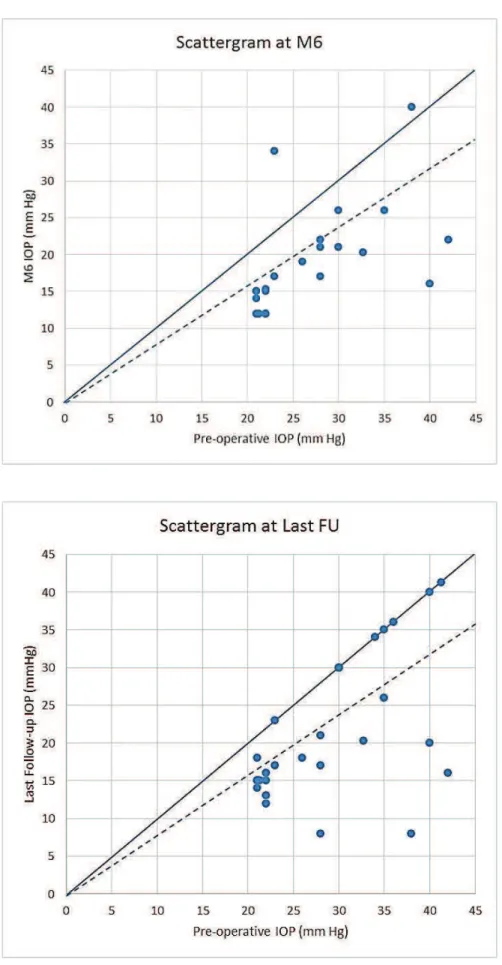
follow-up visit. Qualified surgical success was achieved in 19 of 28 (68%) patients at the last follow-up visit. Graphs showing IOP reduction in all patients and responders patients
(qualified success) over time are displayed in Figure 1.Scattergrams of postoperative versus preoperative IOP in all patients at 6 months and at the last follow-up are shown in Figure 2.
Safety
No complications occurred during any of the procedures. One patient presented a serious choroidal detachment which did not reach the posterior pole seven days after treatment, with complete resolution at two months. No other major intraoperative or postoperative
complications occurred. None of the patients encountered IOP spikes or major IOP increases in the early follow-up (IOP > baseline IOP + 10 mm Hg in the first 7 days). Clinical
examinations showed little or no signs of intraocular inflammation, particularly with no cases of hypopyon or anterior or posterior synechia.
Mean visual acuity remained statistically unchanged (best-corrected visual acuity logMAR 0.84 ± 1.09 before surgery and logMAR 1.09 ± 1.20 at last follow-up; P = 0.42). Visual acuity loss of 3 lines or more was observed in four patients (15.4%).
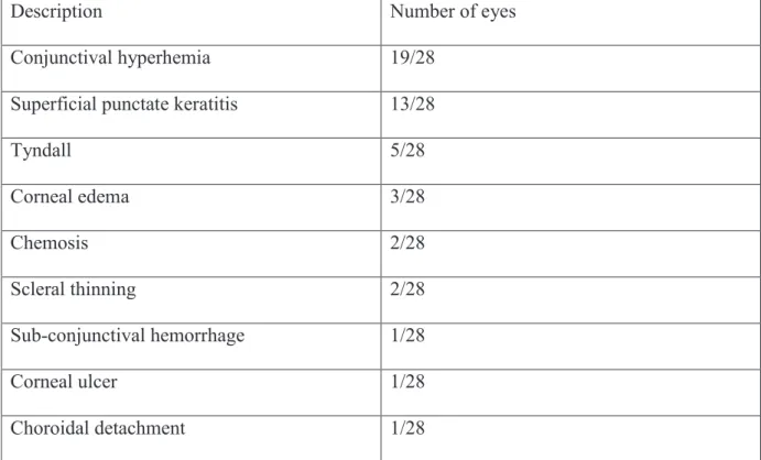
Superficial punctate keratitis occurred in 13 of 28 patients (46.4%) and central superficial corneal ulceration in 3 of 28 patients (10.7%). Other minor postoperative complications are listed in Table 3.
DISCUSSION
The initial pilot study conducted with the device for ultrasound circular cyclocoagulation was performed in patients with refractory primary or secondary glaucoma (at least one previous incisional glaucoma surgery) and a limited residual visual acuity and visual field
(best-corrected visual acuity less than 20/60; and visual field defect with a minimum of one location in the paracentral region exhibiting repeatable abnormality at the P < 0.5% level).11 Patients included had primary open-angle glaucoma, neovascular glaucoma, congenital glaucoma, primary angle-closure glaucoma and iridocorneal endothelial glaucoma. IOP was reduced from a mean preoperative value of 37.9 ± 10.7 mmHg to a mean postoperative value of 26.3 ± 5.1 mmHg at 6 months and 24.7 ± 8.5 mmHg at the last follow-up visit, respectively. An IOP reduction of 33.9% was obtained at the last follow-up visit. Surgical success (defined by an IOP reduction greater than or equal to 20% and an IOP more than 5 mmHg) was obtained in 10 of 12 patients (83.3%) at the last visit.
In the present case-series, twenty-eight patients with primary open-angle glaucoma and much less advanced disease were enrolled and followed during at least 6 months. Results between these two studies in terms of efficacy (mean IOP reduction and rate of responders) and safety were rather similar. IOP was reduced from a mean preoperative value of 29.0 ± 7.2 mmHg to a mean value of 21.6 ± 9.4 mmHg at last follow-up. Qualified success (IOP reduction >20% and IOP> 5 mmHg with possible re-treatment) was achieved in 68% of eyes, with an average IOP decrease of 45% in the same eyes. One reason explaining the slightly better results of the pilot study compared to the present study could be that one fourth of the patients of the pilot study had had diode laser cyclocoagulation procedures prior to having ultrasound
19
patients of the present study hade had diode laser cyclocoagulation before ultrasound
treatment. One other reason could be the small number of patients included in the pilot study (n=12), making the observed results more sensitive to IOP variability. The tolerability was also good in the present study, as reported in the pilot study, with no IOP spikes or major IOP increases in the early follow-up and, in the long term no cases of severe hypotony or phthisis, which are some of the most serious adverse effects of the currently available cyclodestructive methods.1-8 Clinical examinations showed little or no signs of intraocular inflammation and visual acuity remained statistically unchanged.
This study could help to define the optimal indications of this new method of treatment. The results, particularly the good intraoperative safety profile and postoperative tolerability, likely indicate that this new method of cyclocoagulation using ultrasound may be indicated in eyes refractory to one or several filtering surgeries, but with visual acuity and a visual field still relatively unaffected. In these eyes, where surgery may no longer be a valid option, physicians are sometimes prone to rule out the use of the current methods of cyclocoagulation, including diode laser, because of the possible side effects and the poor predictability of the IOP
decrease.
Approximately one-third of patients of the present study do not respond to the treatment, with usually no IOP reduction. Several reasons can be advanced to explain the complete absence of IOP reduction in some cases. One explanation may be that the amount of ciliary body tissue coagulated was not sufficient or was offset by an increased secretion of the ciliary body remaining intact. One other reason could be that the ciliary body targeted was not actually reached by the ultrasound beam during the procedure due to, possibly, an incorrect
through the apex of the cornea or partially oblique scans instead of perfectly radial scans, leading to an underestimated size of the ciliary ring and thus the required probe), or due to suboptimal intraoperative centering or moving of the device (which is positioned and maintained manually by the operator), or because of excessive pressure exerted on the
coupling cone on the eye during the procedure with consequent deformation of the sclera and the ciliary body below.
Some patients having an IOP response insufficient to reach the target IOP were retreated in the present study. The six transducers were activated and the probe was rotated with the intention of targeting different areas of the ciliary body. The diameter of the probe was generally unchanged for the second treatment (n = 6/8 eyes retreated without diameter change, 1/8 increased, 1/8 reduced). IOP decrease was frequently significant after the
retreatment (n = 5/8 eyes retreated were complete successes). This finding could corroborate the hypothesis that the amount of ciliary body tissue treated during the first procedure was not sufficient and/or that the position of the device was not optimal.
A second large multicenter study evaluating the long-term efficacy and safety of this procedure on a larger number of patients having less advanced open-angle glaucoma is
currently in progress (clinicaltrial.gov registration # NCT01338467). 14 As the tolerability and safety profile of this new method of cyclocoagulation is good, it also seems logical to
compare it to filtering surgery as a second line treatment, after medical treatment. At the time of writing, a third clinical trial is ongoing in patients with primary open-angle glaucoma uncontrolled with medical treatment and naïve of any glaucoma filtering surgery
21
trials should also be conducted to directly compare the efficacy and safety of HIFU cyclocoagulation with that of trabeculectomy or diode laser.
CONCLUSIONS
In summary, the present case-series study shows that ultrasonic coagulation of the ciliary body using high-intensity focused ultrasound delivered by miniaturized high-frequency transducers seems to be an effective method of reducing IOP in patients with open-angle glaucoma refractory to filtering surgeries but still having a good residual visual acuity and visual field . The procedure seems to be safer than other previous available cyclodestructive procedures.
THESE SOUTENUE PAR : DUPUY Caroline
TITRE : Treatment of Refractory Open-Angle Glaucoma using Ultrasonic Circular
Cyclocoagulation: a Prospective Case Series
CONCLUSION
La cyclocoagulation par ultrasons focalisés de haute intensité semble être une méthode efficace et bien tolérée pour réduire la pression intraoculaire chez les patients présentant un glaucome primaire à angle ouvert réfractaire aux chirurgies filtrantes.
Cette méthode permet une réduction de la pression intraoculaire moyenne de 45% dans 68% des cas.
Une étude prospective est actuellement en cours, étudiant l'efficacité et la tolérance de cette méthode chez les patients présentant un glaucome primaire à angle ouvert non contrôlé par les traitements médicaux et naïfs de toute chirurgie filtrante.
23
REFERENCES
1. Hamard P, Gayraud JM, Kopel J, et al. Treatment of refractory glaucomas by transscleral cyclophotocoagulation using semiconductor diode laser. Analysis of 50 patients followed up over 19 months. J Fr Ophthalmol 1997;20:125–133.
2. Al-Ghamdi S, al-Obeidan S, Tomey K F, al-Jadaan I. Transscleral neodymium:YAG laser cyclophotocoagulation for end-stage glaucoma, refractory glaucoma, and painful blind eyes. Ophthalmic Surg 1993;24:526–529.
3. De Roetth A Jr. Cryosurgery for the treatment of glaucoma. Trans Am Ophthalmol Soc 1965;63:189–204.
4. Maus M, Katz LJ. Choroidal detachment, flat anterior chamber, and hypotony as
complications of neodymium: YAG laser cyclophotocoagulation. Ophthalmology 1990;97:69 –72.
5. Uram M. Ophthalmic laser microendoscope ciliary process ablation in the management of neovascular glaucoma. Ophthalmology 1992;99:1823–1828.
6. Kosoko O, Gaasterland DE, Pollack IP, Enger CL. Long-term outcome of initial ciliary ablation with contact diode laser transscleral cyclophotocoagulation for severe glaucoma: the Diode Laser Ciliary Ablation Study Group. Ophthalmology 1996;103:1294–302.
7. Sabri K, Vernon SA. Scleral perforation following trans-scleral cyclodiode. Br J Ophthalmol 1999;83:502–503.
8. Vernon SA, Koppens JM, Menon GJ, Negi AK. Diode laser cycloablation in adult
glaucoma: long-term results of a standard protocol and review of current literature. Clin Exp Ophthalmol 2006;34: 411–420.
9. Aptel F, Charrel T, Palazzi X, et al. Histologic effects of a new device for high-intensity focused ultrasound cyclocoagulation. Invest Ophthalmol Vis Sci 2010;51:5092-8.
10. Charrel T, Aptel F, Birer A, et al. Development of a miniaturized HIFU device for glaucoma treatment with conformal coagulation of the ciliary bodies. Ultrasound Med Biol 2011; 37:742-54.
11. Aptel F, Charrel T, Lafon C, et al. Miniaturized high-intensity focused ultrasound device in patients with glaucoma: a clinical pilot study. Invest Ophthalmol Vis Sci 2011; 52:8747-53.
12. Aptel F, Lafon C. Therapeutic applications of ultrasound in ophthalmology. Int J Hyperthermia 2012; 28:405-18.
13. Shaarawy T, Sherwood MB, Grehn F. Guidelines on Design and Reporting of Glaucoma Surgical Trials. Amsterdam: Kugler Publications; 2009.
14. Aptel F, Denis P, Rouland JF, et al. Ultrasonic Circular Cyclo Coagulation in patients with Primary Open-Angle Glaucoma: preliminary results of a Multicenter Clinical Trial. Presented at ARVO 2013.
25
FIGURE CAPTIONS
Figure 1. Graphs showing IOP reduction (mean ± standard deviation) and the mean number
of procedures performed in all patients (up) and responder patients (qualified success) (down) over time.
Figure 2. Scattergrams showing the IOP before ultrasonic circular cyclocoagulation versus at
6 months (top) and at last follow-up (bottom) for all patients. All points below the diagonal line represent eyes in which there was a decrease in IOP. All points below the dashed line represent eyes in which IOP reduction was reduced by at least 20% from the preoperative value.
Figure 3. UBM examinations taken at month 3 showing two ultrasound- induced lesions in
the superior (A) and inferior (B) circumference of one patient with complete success. Note that the areas of treatment are located at the junction between the sclera and the base of the ciliary body.
TABLES
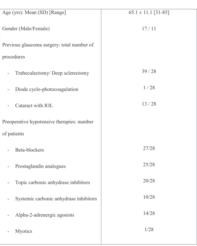
Table 1. Demographic characteristics.
Age (yrs): Mean (SD) [Range]
Gender (Male/Female)
Previous glaucoma surgery: total number of procedures
- Trabeculectomy/ Deep sclerectomy - Diode cyclo-photocoagulation - Cataract with IOL
Preoperative hypotensive therapies: number of patients
- Beta-blockers
- Prostaglandin analogues
- Topic carbonic anhydrase inhibitors - Systemic carbonic anhydrase inhibitors - Alpha-2-adrenergic agonists - Myotics 65.1 ± 11.1 [31-85] 17 / 11 39 / 28 1 / 28 13 / 28 27/28 25/28 20/28 10/28 14/28 1/28
27
Number of Preoperative hypotensive therapies: Mean (SD) [Range]
Follow-up (months): Mean (SD) [Range]
3.8 ± 1.6 [0-7]
9.3 ± 3.1 [6-12]*
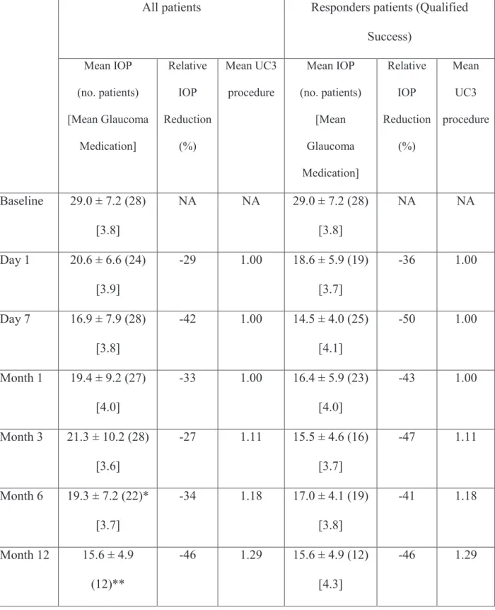
Table 2. IOP at baseline and during follow-up with number of procedures and hypotensive
medications.
All patients Responders patients (Qualified Success) Mean IOP (no. patients) [Mean Glaucoma Medication] Relative IOP Reduction (%) Mean UC3 procedure Mean IOP (no. patients) [Mean Glaucoma Medication] Relative IOP Reduction (%) Mean UC3 procedure Baseline 29.0 ± 7.2 (28) [3.8] NA NA 29.0 ± 7.2 (28) [3.8] NA NA Day 1 20.6 ± 6.6 (24) [3.9] -29 1.00 18.6 ± 5.9 (19) [3.7] -36 1.00 Day 7 16.9 ± 7.9 (28) [3.8] -42 1.00 14.5 ± 4.0 (25) [4.1] -50 1.00 Month 1 19.4 ± 9.2 (27) [4.0] -33 1.00 16.4 ± 5.9 (23) [4.0] -43 1.00 Month 3 21.3 ± 10.2 (28) [3.6] -27 1.11 15.5 ± 4.6 (16) [3.7] -47 1.11 Month 6 19.3 ± 7.2 (22)* [3.7] -34 1.18 17.0 ± 4.1 (19) [3.8] -41 1.18 Month 12 15.6 ± 4.9 (12)** -46 1.29 15.6 ± 4.9 (12) [4.3] -46 1.29
29 [4.4] Last FU [x] 21.6 ± 9.4 (28) [3.8] -26 1.29 16.0 ± 4.3 (19) [3.8] -45 1.29 NA = not applicable
*6 patients withdrew from the study because of the glaucoma surgery (5 patients) and Diode Laser (1 patient) performed less than 6 months after the UC3 procedure
*9 patients withdrew from the study because of the glaucoma surgery (7 patients) and Diode Laser (2 patients) performed less than 12 months after the UC3 procedure, 1 patient died, 6 patients not already controlled at 12 months.
Table 3. Details of postoperative complications.
Description Number of eyes
Conjunctival hyperhemia 19/28 Superficial punctate keratitis 13/28
Tyndall 5/28 Corneal edema 3/28 Chemosis 2/28 Scleral thinning 2/28 Sub-conjunctival hemorrhage 1/28 Corneal ulcer 1/28 Choroidal detachment 1/28
31
33
SERMENT D’HIPPOCRATE
En présence des Maî tres de cette Facult é, de mes chers condiscipl es et devant l’effigi e d’HIPPOCRATE,
Je promets et j e j ure d’être fidèl e aux lois de l ’honneur et de la probit é dans l’exerci ce de l a Médecine.
Je donnerai mes soi ns gratuit ement à l’indigent et n’exigerai j amais un salaire au dessus de mon travai l. Je ne participerai à aucun partage clandestin d’honorai res.
Admis dans l’inti mit é des maisons, mes yeux n’y verront pas ce qui s’y passe ; ma l angue tai ra l es secrets qui me seront confiés et mon état ne servira pas à corrompre l es mœurs, ni à f avoriser le cri me.
Je ne permettrai pas que des considérations de rel igion, de nat ion, de race, de part i ou de classe social e vi ennent s’i nterposer entre mon devoir et mon pati ent.
Je garderai l e respect absolu de la vie humaine.
Même sous la menace, je n’admettrai pas de fai re usage de mes connaissances médi cales cont re les lois de l ’h umanit é.
Respect ueux et reconnaissant envers mes Maîtres, j e rendrai à leurs enfants l’instruction que j ’ai reçue de l eurs pères.
Que l es hommes m’accordent l eur estime si j e suis fi dèle à mes promesses. Que j e sois couvert d’opprobre et méprisé de mes con frères si j’y manque.