DOI: 10.5897/AJAR2016.11421 Article Number: 7FA266D61487 ISSN 1991-637X
Copyright ©2016
Author(s) retain the copyright of this article http://www.academicjournals.org/AJAR
African Journal of Agricultural
Research
Full Length Research Paper
Prevalence of viruses infecting plantain (Musa sp., AAB
genome) in the major growing regions in Cote d’Ivoire
Kouakou Théodore Kouadio
1*, Caroline De Clerck
2, Thérèse Atcham Agneroh
1, Ludivine
Lassois
3, Olivier Parisi
2, Sébastien Massart
2, Philippe Lepoivre
2and M. Haïssam Jijakli
21
Laboratoire de Phytopathologie et de Biologie végétale, Département Agriculture et Ressources Animales, Institut National Polytechnique Félix Houphouët Boigny, BP 1313 Yamoussoukro, Côte d‟Ivoire.
2
Laboratoire de Phytopathologie Intégrée et Urbaine, Gembloux Agro-Bio Tech, Université de Liège, Passage des Déportés 2, B-5030 Gembloux, Belgium.
3
BIOSystem Engineering (BIOSE) Department, Gestion Des Ressources Forestières, Gembloux Agro Bio Tech, University of Liege, Passage des Déportés 2, B-5030 Gembloux, Belgium.
Received 12 July, 2016; Accepted 15 September, 2016
This study was undertaken to determine the prevalence of viruses, viz Banana streak Obino l’ewai virus (BSOLV), Cucumber mosaic virus (CMV), Banana mild mosaic virus (BanMMV) and Banana bract mosaic virus (BBrMV) causing serious diseases on plantain in Cote d'Ivoire. From April 2010 to July 2010 and June 2011 to August 2011, 103 farmers' plots located in 13 important plantain productions regions were screened. In all, 424 samples having symptoms of yellow or moderate chlorotic streaks were analyzed by reverse transcriptase-PCR and by immunocapture-PCR. Viruses identified were BSOLV, BanMMV and CMV respectively in proportions of 78%, 63% and 5.4% of the samples analyzed. Mixed infections of these three viruses were found in the 13 regions while CMV was present only in 3 regions. None of the samples collected were infected by BBrMV. Infected suckers used by farmers to establish their banana field could be the cause of these viral infections. The results showed that 9% of symptomatic samples were not associated with the presence of one or the other of the viruses studied. Further study is required to identify reported viruses in banana and plantain across the world.
Key words: Plantain, farming modes, viruses, reverse transcription polymerase chain reaction (RT-PCR), immunocapture polymerase chain reaction (IC-PCR), Cote d‟Ivoire.
INTRODUCTION
Plantain is a staple food for millions of people in many countries of West and Central Africa and an important source of income for producers (Lescot and Ganry, 2010). According to FAO (2012), plantain is the third food crop produced in Cote d‟Ivoire after yam and cassava.
Indeed, originally grown around homes, plantain has, for decades, greatly expanded because of its association with food and/or industrial crops (Traore et al., 2009). The different cultivars of bananas, including plantains, whatever the areas and production methods, are affected
*Corresponding author. E-mail: tkouadiothed@gmail.com.
Author(s) agree that this article remain permanently open access under the terms of the Creative Commons Attribution License 4.0 International License
by a wide range of pests and diseases (Ploetz, 2004). Viral diseases play a destructive role reducing the levels of production and the quality of products in both industrial and village plantations and limiting the exchanges of germplasm for improvement programs (Geering, 2007). The main viruses natura lly infecting banana are Banana bunchy top virus (BBTV, Babuvirus, Nanoviridae), Cucumber mosaic virus (CMV, Cucumovirus, Bromoviridae), several species of Banana streak virus (BSVs, Badnavirus, Caulimoviridae), Banana bract mosaic virus (BBrMV, Potyviridae) and Banana mild mosaic virus (BanMMV, Flexiviridae). The symptoms caused by BBTV on banana, consisting of a marginal chlorosis and bunching of leaves at the top of the pseudostem, forming a rosette with a “bunchy top” appearance are very characteristic (Su et al., 2003). On the other hand, for the other viruses the visual diagnosis is not enough because the symptoms are not specific (Iskra-Caruana et al., 2008).
In addition, during mixed infections between BanMMV and BSVs or CMV, often encountered in plantation, symptoms of BanMMV are masked by those caused by other viruses (Lockhart, 2002; Reichel et al., 2003). Banana bunchy top virus, BSVs and BanMMV are associated with the genus Musa (Kumar et al., 2014) while CMV has an extremely wide host range with over 1,200 plant species representing 100 plant families (Jacquemond, 2012). Banana bract mosaic virus known for a long time to have been associated with the genus Musa has been reported for the first time on cardamom plants (Elettaria cardamomum Maton), an herbaceous monocotyledonous rhizome of the Zingiberaceae family, in India (Siljo et al., 2012). Among the 5 viruses, only 4 (BBTV, BSVs, BanMMV, CMV) have been reported in Africa (Pietersen and Thomas, 2005), BBrMV being limited in some Asian countries has been detected in Colombia and Ecuador in Latin America (Alarcon et al., 2006, cited by Quito-Avila et al., 2013 ; Quito-Avila et al., 2013). The spread of these viruses takes place, according to each species, either by insect vectors from primary foci of infection, or through the use of propagation materials already infected or by agricultural tools for CMV (Pietersen and Thomas, 2005).
However, an original mode of transmission, the vertical transmission from integrated functional viral sequences of BSVs (endogenous BSVs) in the genome of bananas of the species M. balbisiana (B genome) was also highlighted by several authors including Geering et al. (2005) and Cote et al. (2010). This phenomenon called viral activation leads to the generation of infectious virions responsible of streak symptoms on banana and may be due to abiotic and biotic stress action (in vitro culture, interspecific crosses) (Dallot et al., 2001; Lheureux et al., 2003; Cote et al., 2010). Banana streak virus is therefore considered the most atypical virus among the major five viruses infecting banana. Indeed, this disease known as streak mosaic may be the result of
a complex of species of BSVs. Eleven distinct species of free BSVs are known such as Banana streak Obino l‟ewai virus (BSOLV), Banana streak gold finger virus (BSGFV), Banana streak mysore virus (BSMYV), Banana streak imove virus (BSIMV) and Banana streak cavendish virus (BSCAV) (James et al., 2011; Stainton et al., 2015). As for BanMMV, very few studies are available on this virus and namely on its spreads; for now the only known mode of transmission is the plant material (Teycheney et al., 2005). On the other hand, BanMMV seems to have no impact on the host plant in terms of morphology and yield when it is in simple infection but it is important in co-infection (Caruana, 2003).
Cucumber mosaic virus and BSV affect bananas (Cavendish, AAA genome) in the fields of industrial exploitation for more than three decades in Cote d‟Ivoire (Lassoudière, 1979; Aka et al., 2009). The works carried out on the economic impacts of BSV showed that yield losses could reach 70% after a growth cycle on the cultivar Poyo (Cavendish, AAA genome) in the location of Azaguié (Lassoudière, 1979). On plantain (AAB genome), the only study by Kouadio et al. (2013) reported for the first time the presence of BanMMV. However, to date no study has been conducted to determine the prevalence and geographical distribution of the three viruses already identified as well as those known worldwide in the production areas on plantains (AAB genome) in the country. A thorough knowledge of the viruses infecting plantains based on farming mode is therefore necessary for the development of control methods. Such measures include the strengthening of quarantine and an appropriate management of plant genetic resources. The aim of this study is to determine the prevalence of the major viruses infecting plantains in the main producing regions in Cote d'Ivoire.
MATERIALS AND METHODS
Surveys and collection of samples
Surveys were conducted from April 2010 to July 2010 and June 2011 to August 2011. Hundred and three farmers‟ plots located in 13 regions (South, Southeast, East, center and center-west) of Cote d‟Ivoire were covered (Figure 1). The choice of collection areas was based on their importance for the production of plantains (Koffi, 2007). Generally, 4 to 10 samples were collected by plot depending on the size and distribution of the disease.
The sample selection was based on the presence of viral symptoms (Figure 2) and in total 424 plantain samples were collected in farmers‟ plots. During the surveys an inventory of plants grown in association with plantain was also achieved by taking a census of plants grown in plots visited. In addition, information on the plantain cultivar, the health status of planting material as well as its origin was asked to the producer. We also recorded the location of plantain plants showing symptoms of virus diseases from the sampling plot. On the collection sites, the samples were packed in plastic bags with a unique code and stored on ice during their transportation to the laboratory where the samples were preserved by drying over calcium chloride (CaCl2) and kept at 4°C for
Figure 1. Map of Cote d‟Ivoire showing the location of regions 1 to 13 where field grown banana plantain crops were surveyed for mosaic virus diseases in 2010 and 2011: 1: Abengourou; 2: Aboisso; 3: Agboville; 4: Bongouanou, 5: Bouaflé; 6: Daloa; 7: Gagnoa; 8: Issia; 9: Oumé; 10: Sinfra; 11: Toumodi; 12: Yamoussoukro; 13: Zuénoula
A
B
Figure 2. Yellow and/or chlorotic streak symptoms on 2 plantain samples collected in Cote
d‟Ivoire: A : sample Y26 collected in Yamoussoukro ; B : Sample GB9 collected in Agboville.
Molecular detection of CMV, BanMMV and BBrMV by reverse transcriptase-polymerase chain reaction (RT-PCR)
Molecular detections by RT-PCR were performed to detect the
presence of CMV, BanMMV and BBrMV in the plantain samples collected. The primers used come from the genes encoding the coat proteins of these viruses (Table 1). The samples were put into a grinding bag in the ratio of 0.5 g of fresh leaves for 2 ml (control)
Table 1. Primer sequences and target templates.
Primer name Primer sequence (5'-3') Target virus Reference
BanCP1 GGATCCCGGGTTTTTTTTTTTTTTTTTV BanMMV Teycheney et al., 2005
BanCP2 TATGCNGCNTTYGAYTTCTTRGAYG BanMMV Teycheney et al., 2005
BractN2 ACATGGAGTATGATGGATAAGG BBrMV Iskra-Caruana et al., 2008
BractNR GTGTGCYTCTCTAGCCCT GTT BBrMV Iskra-Caruana et al., 2008
CMV3' TTTTAGCCGTAAGCTGGATGGACAACCC CMV Sharman et al., 2000
CMV5' TATGATAAGAAGCTTGTTTCGCGCA CMV Sharman et al., 2000
BSV cl1 ATGGCCTTAATAGTCTTTCGTGAT BSOLV Dallot et al. 2001
BSV cl2 GGTGGCGCTGAG GATGTG BSOLV Dallot et al. 2001
or 50 mg of dried leaves for 1 ml of extraction buffer at pH 7.2 (137 mM NaCl; 8 mM Na2HPO4; 1.5 mM KH2PO4; 2.7 mM KCl; 80 mM
Na2SO3; 3 mM NaN3, 0.05% Tween 20), the dried leaves being
previously soaked for 15 min in the extraction buffer. Grinding using an electric grinder (Power Plus X022) was then performed. The juice clarified by decantation (maintained on ice) was collected in Eppendorf tubes before being diluted 100 times with sterile distilled water for the RT-PCR reaction. The reaction was performed in a volume of 25 μl (20 μl of reaction mixture + 5 μl of the sample) by using the Titan RT-PCR kit (Rock, Mannheim, Germany). The reaction mixture consisted of 5 μl of Buffer (5× Concentrate) Titan RT-PCR, 0.5 μl of dNTP, 0.5 μl of each primer at 25 μM, 1.25 μl of DTT (100mM), 0.5 μl of enzyme mix Titan, and 11.75 μl of sterile distilled water. All amplification reactions included negative and positive controls from banana plants grown in the greenhouse as well as blanks made of sterile distilled water. The amplification was performed in a thermocycler (My CyclerTM, Biorad, USA) according to the following program: 50°C for 30 min for the reverse transcription, 94°C for 5 min for the first denaturation followed by 40 cycles (94°C for 30 s for the second and the following denaturations; 54°C for 1 min for annealing; 72°C for 2 min for elongation) and final elongation at 72°C for 10 min. The last step was the revelation of amplification products on agarose gel 1% [1 g agarose for 100 ml of Tris Acetate EDTA (TAE) 1 x buffer] containing 10 μl of ethidium bromide in buffer TAE 1×. The electrophoretic migration took place under a constant current of 120 mA for 45 min. The gel was then visualized under ultra violet light allowing the observation of amplified bands. It is important to clarify that there was no simultaneous detection; the collected samples have been separated for indexing each of the three viruses BanMMV, BBrMV and CMV.
Detection of BSOLV by immunocapture (IC)-PCR
The detection of BSOLV was performed by IC-PCR using a specific polyclonal antiserum (SEDIAG, France) and the BSV cl1 and BSV cl2 primers targeting the gene encoding the viral capsid protein and detecting the species BSOLV (Table 1). The difficulty to detect exogenous forms of BSV species makes the IC-PCR a commonly used technique for detection of that virus (Agindotan et al., 2006; Geering et al., 2011). The IC-PCR was performed following the protocol described by Dallot et al. (2001).
RESULTS
Analysis of cropping systems of plantain
Following the surveys carried out in 2010-2011 in 103
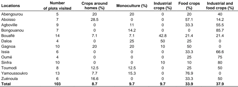
plots located in the 13 regions, there are three major farming systems of plantain: crops around homes, monoculture and the associated cropping with 3 types. The types of this associated cropping are the association with other food crops, the association with industrial crops and the association with other food and industrial crops (Table 2). The associated cropping system of plantain is the most common with a proportion of 81.5% of all plots visited. Only 8.7% and 9.7% of the farmers respectively practice crops around their homes and monoculture (without association with other crops). These farmers are located in the regions of Abengourou, Aboisso, Bouafle, Gagnoa, Toumodi, Yamoussoukro and Zuénoula. The industrial crop associated with the plantain is cocoa followed by the rubber tree. Among the food crops we have cocoyam (Xanthosoma sagitifolium) and corn that have been observed in the majority of plots visited in the 13 regions. Other food crops include pepper, eggplant and yam. Regarding the type of planting material, farmers use local cultivars of "French" and "False-Horn" plantain. Of the 424 samples collected, 82 samples were taken on the cultivar "Agnrin", French plantain with 98% (74/82) coming from 3 regions (Abengourou, Bongouanou and Aboisso). The 342 samples of the cultivar "afoto" False-Horn Plantain come from plots located in the 10 other regions. In addition, in all plots visited, the material of new plantations consists of suckers from old plantations without prior phytosanitary indexing. During surveys, the banana plants were at different ages including bananas less than a year and bananas more than one crop planting cycle.
Overall prevalence of viruses in the 13 regions visited
The results of the identification of the viruses in the 424 samples collected in 66 plots plantains with symptomatic plants over the 103 visited are summarized in Table 3. The specific primers used for the molecular detection of these viruses generated DNA fragments about 500bp, 500bp, 350bp and 300 bp respectively for CMV, BSOLV, BanMMV and BBrMV (Figure 3). Expected sizes of the
Table 2. Percentage of presence by region visited of the different cultivation systems including plantain in farmers‟ fields in Cote
d‟Ivoire between 2010 and 2011
Locations Number of plots visited Crops around homes (%) Monoculture (%) Industrial crops (%) Food crops (%) Industrial and food crops (%) Abengourou 5 20 20 0 20 40 Aboisso 7 28.5 0 0 57.1 14.2 Agboville 9 0 11 0 33.3 55.5 Bongouanou 7 0 14.2 0 0 85.7 Bouaflé 14 7.1 7.1 42.8 21.4 21.4 Daloa 4 0 25 50 25 0 Gagnoa 10 20 20 10 50 0 Issia 6 0 0 0 33.3 66.6 Oumé 4 0 0 0 25 75 Sinfra 10 0 0 10 10 80 Toumodi 8 12.5 12.5 0 25 50 Yamoussoukro 13 7.7 15.3 0 76.9 0 Zuénoula 6 16.6 0 0 33.3 50 Total 103 8.7 9.7 9.7 33.9 37.9
amplicons were observed in all positive controls. On the other hand, no amplification was observed for negative controls (healthy plants) and the blanks. In all, 91% (386/424) of the samples collected were found to be infected with at least one of the four viruses in this study (Table 3). The viruses identified were BSOLV, BanMMV and CMV respectively in the proportions of 78%, 63% and 5.4% of the samples analyzed. None of the collected survey samples was positive for BBrMV. BSOLV and BanMMV dominated either single or mixed infections. Single infections accounted for 39% of the samples tested (that is 167 over 424) of which 115 samples infected with BSOLV (27%) and 52 samples infected with BanMMV (12%). As for mixed infections BSOLV + BanMMV, they were around 46% of the samples analyzed. A double infection BSOLV + CMV was observed in one of the locations (Bongouanou) whereas no mixed infection BanMMV + CMV was identified. A triple infection BSOLV + BanMMV + CMV was observed in 4.7% of the samples collected. BSOLV and BanMMV were detected in the 13 regions visited while CMV was positively indexed in the samples of 3 regions [Bouaflé (14/89), Gagnoa (3/38) and Bongouanou (6/45)]. In all, the percentages of symptomatic samples infected with at least one of the four viruses studied are above 79% in all areas of collection(Table 3) including a value of 100% in the locations of Abengourou and Daloa. Although BSOLV and BanMMV were present in all the locations visited, a difference of prevalence was nevertheless observed in the samples collected (Figure 4). Thus, BSOLV was the most present virus in 9 of the 13 locations visited, with high proportions in the locations of Zuénoula (93%), Bongouanou (93%), Abengourou (86%), Issia (84%) and Bouaflé (84%) while BanMMV was dominant in the locations of Sinfra (76.7%), Gagnoa (76%) and Oumé
(69%). The single BSOLV infections were higher in the locations of Abengourou (52%), Issia (50%), Agboville (45%) Aboisso (45%) and Yamoussoukro (38%).The percentages of the most important and prevailing single infections due to BanMMV in our samples were 28% and 26% respectively in Gagnoa and Oumé. The mixed infections BSOLV + BanMMV ranged from 21% (Aboisso) to 80% (Toumodi).
Prevalence of virus based on plantain cultivars and cropping systems
From the two cultivars of plantain subgroup observed in the plots visited, the proportion of infections due to BSOLV were 78% (64/82) and 79% (270/342) respectively for the types of cultivars French and False-Horn in the samples analyzed. Similarly, BanMMV was present in 58% (48/82) and 64% (220/342) of the cultivars French and False-Horns plantains analyzed. CMV was detected in 4% (3/82) of the samples of the cultivar type French and 6% (20/342) of the samples of cultivar type False-Horns. These results show that there are few differences in the proportions of viral infections among the plantain cultivars.
The analysis of the frequency of viral infections in the samples tested, based on cropping systems including plantain reveals a high frequency of BSOLV followed by BanMMV (Figure 5). In at least 56% of cases, the observation of the chlorotic streaks symptoms was explained by the presence of BSOLV. We can note that this frequency of BSOLV was higher (81%) in the associations of plantain with other food and industrial crops although it was present in other cropping systems. This trend is similar for BanMMV where the frequency
M 1 2 3 4 5 6 7 8 9 10 11 12 M MM M (3a) 500 pb M 1 2 3 4 5 6 7 8 9 10 11 12 M (3b) 500 pb (3c) M 1 2 3 4 5 6 7 8 9 10 11 13 M 10 13 M 500 pb
Figure 3. (a) Molecular detection of BSOLV by IC-PCR in samples of plantains collected in
Cote d‟Ivoire, M: Marker of molecular weight of 100 bp (Ladder DNA), 1-2: Healthy plant in the greenhouse; 3-10: Amplicons (500 bp) of 8 samples of banana; 11: Mix + sterile distilled water; 12: Positive control BSOLV; (b) (1%) agarose gel electrophoresis of the amplification products obtained by reverse-transcriptase (RT) -PCR for the detection of CMV: M: Marker of molecular weight 100 bp (DNA Ladder), 1-9: Amplicons of 500 bp in 9 symptomatic banana samples; 10: Mix + sterile distilled water, 11 healthy plant in the greenhouse, 12: Positive control CMV; (c) (1%) agarose gelelectrophoresis of amplification products obtained by RT-PCR for detection of BanMMV: M: Marker of molecular weight 100 bp (DNA Ladder); wells 1-8: Samples of plantains collected; wells 9-10: Healthy plant and blank; 11 and 13 positive controls for BanMMV
was 70% in the associations of plantain with food and industrial crops. The presence of CMV remains relatively low but was higher in crop around homes (22%) and crop
associated with food crops including peppers, eggplant. CMV was not detected on plantain associated with cocoa, coffee and rubber.
Table 3. Occurrence of Banana streak Obino l‟ewai virus (BSOLV), Banana mild mosaic virus (BanMMV), Cucumber osaic virus (CMV) and
Banana bract mosaic virus (BBrMV) on plantain leaf samples with virus-like symptoms collected from farmers‟ fields surveyed during 2010-2011 in Cote d‟Ivoire.
Location Fieldsa No. Analyzed BSOLV CMV BanMMV BBrMV Positive total
Abengourou 5/5 23 20 0 11 0 23 (100)b Aboisso 6/7 29 19 0 10 0 23 (79) Agboville 5/9 20 14 0 7 0 16 (80) Bongouanou 5/7 45 42 6 34 0 44 (98) Bouafle 8/14 89 75 14 69 0 85 (95) Daloa 1/4 10 8 0 7 0 10 (100) Gagnoa 9/10 38 24 3 29 0 35 (92) Issia 5/6 26 22 0 10 0 23 (88) Oume 4/4 26 16 0 18 0 23 (88) Sinfra 4/10 30 22 0 23 0 28 (93) Toumodi 1/8 5 4 0 4 0 4 (80) Yamoussoukro 9/13 52 39 0 22 0 42 (81) Zuenoula 4/6 31 29 0 24 0 30 (97) Total 66/103 424 334 (78) 23 (5.4) 268 (68) 0 386 (91)
aFields with symptomatic plants/fields surveyed; bIn parentheses are percentages calculated over the total number of virus-infected plants. No, Number of
samples.
Frequ
encies
of
viruses
Figure 4. Frequencies of viruses identified in locations visited during field surveys in main plantain
growing areas in Cote d‟Ivoire.
DISCUSSION
The results of the molecular tests carried out on symptomatic samples of plantain collected in 2010-2011 in Cote d‟Ivoire have shown that these plants are infected by three viruses over the four tested. In addition, in the 13
regions visited, the viruses identified could infect local cultivars of plantains namely French and False-Horn plantains. Viruses were mentioned on plantains by Kouadio et al. (2013) in Cote d‟Ivoire, but this study provides some missing information to date on the prevalence of the viruses in the main production areas in
Freq uen c ies of vir uses
Figure 5. Frequencies of viruses identified according to plantain cropping systems.
relation to cropping systems including plantain. The results showed a predominance of BSOLV and BanMMV in the 13 regions visited and in different cropping systems including plantain. In Africa BSOLV was reported on plantain in several countries such as Benin, Nigeria, Ghana (Kumar et al., 2014). According to Caruana (2003), BanMMV has a worldwide distribution. This virus has indeed been detected in samples coming from Africa, America, Asia and Australia (Caruana, 2003). The high prevalence of BSOLV followed by BanMMV in the main production areas of plantain in Cote d‟Ivoire suggests a viral transmission due to contaminated planting material through the transfers of plant material between producers.
In fact, the surveys have shown that farmers use suckers that have not been indexed and certified coming from old plantations. In addition, the symptomatic plants showed a random distribution in the plots of plantain visited. BSOLV can be transmitted through infected suckers (Kengamal et al., 2008; Kumar et al, 2014), although the infection by the activation of the viral sequences integrated in the genome of the species M.balbisiana can also take place under some conditions of stress such as in vitro culture (Cote et al., 2010) or a longer drought (Hauser, 2010). Plantains (AAB genome) grown in Cote d‟Ivoire are not derived from in vitro culture but rather from suckers as mentioned in our surveys and already shown by Traoré et al. (2009). Similarly, the collection of samples was conducted during the rainy seasons in the areas subjected to a similar type of
climate and located in the same agro-ecological zone, the Guinean zone. The major difference corresponded to the rainfalls between locations in the Centre (Yamoussoukro and Toumodi) and other locations with a higher amount of rainfall. Lassoudière (1979) had already shown a high prevalence of BSV on bananas (Cavendish, AAA group) in Cote d‟Ivoire. The hypothesis of the transmission by infected suckers is more likely especially as the mealybug vectors are less active in the long-distance spread of BSV (Kengamal et al., 2008; Kumar et al., 2014).
However, no diagnosis via molecular markers to discriminate episomal infections from those from an activation of integrated sequences has been carried out (Kumar et al., 2014). For BanMMV, the only known mode of transmission of this virus is the infected plant material (Teycheney et al., 2005). In this study conducted in Cote d'Ivoire, in general, there were few unique infections but it was most often mixed infections of BSOLV and BanMMV. The observation of symptoms of pronounced chlorotic streaks on plantain in the collected samples could be due in part to the mixed infection between these two viruses. BanMMV could amplify the severity of symptoms caused by BSVs when it is in co-infection (Lockhart, 2002; Caruana, 2003).
However, the incidence of the viruses identified in this study could not be carried out. While waiting for the study of the impact of these viruses, these results highlight the need to develop indexing and certification tools to produce healthy planting material. Moreover, given the
introduction of new improved hybrid varieties of banana (AAB and AAAB genomes) in Cote d'Ivoire (Kouakou et al., 2012), a study on the assessment and the risk management of the activation of viral sequences of different pathogenic species of BSVs integrated into the genome of M. balbisiana in these interspecific hybrids should be performed. In this context of increasing plantain cropping, it would be advisable to study this diversity of species of BSVs in Cote d‟Ivoire since this study took into account only one viral species. Indeed, the specific primers used for the detection of BSVs in the samples tested reveal the species BSOLV (Dallot et al., 2001).
The results also showed that CMV is present in the samples of plantains collected but with a relatively low percentage of infection (5%). The occurrence of CMV on plantain was also reported in Ghana and Nigeria (Kumar et al., 2014). In the present study, the virus was identified only in 3 regions (Gagnoa, Bongouanou, Bouaflé) and in cropping systems of plantain around homes and crops associated with food crops. Cucumber mosaic virus is also transmitted through vegetative propagation and by aphid vectors from a wide range of host plants (Jacquemond, 2012). It is the only banana virus known to date besides banana with over 1,200 host plants (Jacquemond, 2012). In Cote d‟Ivoire, CMV has been mentioned on several crops including banana, pepper, eggplant, yam (Aka et al., 2009; Séka et al., 2009; Sorho et al., 2014).
However, on plantain we observed a lower proportion of infected samples. The association of plantain with industrial crops including cocoa and rubber could explain the low viral presence due to CMV. To date, no study has reported a virus infection of these crops (cocoa, rubber) with CMV. In the location of Bouaflé (center-west) where came 60% (14/23) of the samples tested positive for CMV, it was noted that cultural association in the plantain plots was dominated by pepper and around the plots with eggplants. Similarly, Estelitta et al. (1996) revealed a high occurrence of CMV in banana plantations in association with the Solanaceae like pepper in India. This could also explain the prevalence of this virus in crops around the homes (22%). Indeed, in this cropping system, some plantains are planted in the immediate vicinity of homes on garbage dumping sites. From these refuses from the kitchens, there could be the development of other plants including food crops, potential hosts of CMV that will be in the banana environment. And as among the aphid vectors Aphis gossipii is present in Cote d'Ivoire (Lassoudière, 2012), the transmission will be done, followed by the spread from the infected suckers.
Banana bract mosaic virus was not detected in all samples tested in this work. This is the first study that confirms the absence of the virus in Cote d‟Ivoire. This suggests a strengthening of quarantine measures for the exchanges of plant material since the insect vectors including Aphis gossipii and Pentalonia nigronervosa are
present in Cote d'Ivoire (Lassoudière, 2012).
Overall, 9% of the symptomatic samples collected were negatively indexed to four viruses tested. This might be due to less virus titer in the collected samples. The symptoms of chlorotic streaks could be due to other unidentified viruses or other species of BSV namely BSGFV and BSMYV (Kumar et al., 2014) not targeted in this study. Banana bunchy top virus is one of the most damaging viruses on banana (Musa sp.) (Kumar et al., 2011) and it is expanding in Africa including Benin and Nigeria in West Africa (Kumar et al., 2011; Lokossou et al., 2012; Adegbola et al., 2013). However, unlike the other 4 banana viruses causing symptoms including mosaics that we indexed, Banana bunchy top disease has very characteristic symptoms on banana. During surveys and sample collections no plantain plant showed such symptoms. Even if we did not observe these symptoms, one should be attentive to the extension of BBTV in Africa since the aphid vector (Pentalonia nigronervosa) is already present (Lassoudière, 2012).
Conflict of Interests
The authors have not declared any conflict of interests.
ACKNOWLEDGEMENT
The authors wish to thank all the plantain producers in Cote d'Ivoire who have accepted surveys in their fields as well as the University of Liege (Belgium) via its Faculty Gembloux Agro-Bio Tech for the funding of this work.
REFERENCES
Adegbola RO, Ayodeji O, Awosusi OO, Atiri GI, Lava P (2013). First report of banana bunchy top virus in banana and plantain (Musa spp.) in Nigeria. Plant Dis. 97(2):290.
Agindotan B, Winter S, Lesemann D, Uwaifo A, Mignouna J, Hugues J, Thottapilly G (2006). Diversity of banana streak-inducing viruses in Nigeria and Ghana: twice as many sources detected by immunoelectron microscopy (IEM) than by TAS-ELISA or IC-PCR. Afr. J. Biotechnol. 5(12):1194-1203.
Aka AK, Kouassi NK, Agneroh TA, Amancho NA, Sangaré A (2009). Distribution et incidence de la mosaïque du concombre (CMV) dans les bananeraies industrielles au Sud-est de la Côte d‟Ivoire. Sci. Nature 6:171-183.
Caruana ML (2003). Analyse du Risque Phytosanitaire (ARP). Banana mild mosaic virus (BanMMV). BAN-v4, CIRAD Montpellier, France, 24 p.
Cote FX, Galzi S, Folliot M, Lamagnere Y, Teycheney PY, Iskra-Caruana ML (2010). Micropropagation by tissue culture triggers differential expression of endogenous Banana streak virus (eBSV) in the B genome of natural and synthetic interspecific banana plantain. Mol. Plant Pathol. 11:137-144.
Dallot S, Acuña P, Rivera C, Ramírez P, Côte F, Lockhart BE, Caruana ML (2001). Evidence that the proliferation stage of micropropagation procedure is determinant in the expression of banana streak virus integrated into the genome of the FHIA 21 hybrid (Musa AAAB). Arch.
Virol. 146:2179-2190.
Estelitta S, Radhakrishnan TC, Paul TS (1996). La chlorose infectieuse du bananier au Kerala, en Inde. InfoMusa 5(2):25-26.
FAOstat (2012). Production-Crops, Food and Agriculture Organization of the United Nation. www.faostat.fao.org
Geering ADW (2007). Viral pathogens of banana: outstanding questions and options for control. In: ISHS/ProMusa Symposium: Recent advances in banana crop protection for sustainable production and improved livehoods. Greenway Woods Resort, White River, South Africa pp. 10-1411.
Geering ADW, Parry JN, Thomas JE (2011). Complete genome sequence of a novel badnavirus, banana streak IM virus. Arch. Virol. 156:733-737.
Geering ADW, Pooggin MM, Olszewski NE, Lockhart BEL, Thomas JE (2005). Characterisation of Banana streak Mysore virus and evidence that its DNA is integated in the B genome of cultivated Musa. Arch. Virol. 150:787-796.
Hauser S (2010). Growth and yield response of the plantain (Musa spp.) hybrid „FHIA 21‟ to shading and rooting by Inga edulis on a southern Cameroonian ultisol. Proceedings of international conference on banana and plantain in Africa. Act. Hortic. 879:487-494.
Iskra-Caruana ML, Galzi S, Laboureau N (2008). A reliable IC one-step RT-PCR method for the detection of BBrMV to ensure safe exchange of Musa germplasm. J. Virol. Methods. 153:223-231.
Jacquemond M (2012). Cucumber mosaic virus. Adv. Virus Res. 84:439-504.
James A, Geijskes RJ, Dale JL, Harding RM (2011). Molecular characterisation of six badnavirus species associated with leaf streak disease of banana in East Africa. Ann. Appl. Biol. 158(3):346-353. Kengamal MY, Ranebennur H, Patil FS, Kulkarni UG (2008). Survey on
incidence of banana streak virus and its expression behavior. Res. Crops 9:172-177.
Koffi KS (2007). Rôle des ressources génétiques dans l‟essor du secteur bananier plantain en Côte d‟Ivoire. In: Vadouche R., Atta-Krah K., Achigan-daka GE, Eyog-Matig (eds.) Plant genetic resources and food security in West and Central Africa. Regional Conference, Ibadan, Nigeria. P 179.
Kouadio KT, Agneroh TA, De Clerck C, Lepoivre P, Jijakli MH (2013). First report of Banana mild mosaic virus infecting Plantain in Ivory Coast. Plant Dis. 97(5):693.
Kouakou KAK, Coulibaly S, Atchibri LOA, Kouamé G, Méité A (2012). Evaluation nutritionnelle comparative des fruits de trois hybrides de bananiers (CRBP39, FHIA17, FHIA21) avec ceux de la variété Orishele. Tropicultura 30(1):49-54.
Kumar PL, Hanna R, Alabi OJ, Soko MM, Oben TT, Vangu GHP, Naidu RA (2011). Banana bunchy top virus in sub-Saharan African: Investigations on virus distribution and diversity. Virus Res. 159:171-182.
Kumar PL, Selvarajan R, Iskra-Caruana ML, Chabannes M, Hanna R (2014). Biology, etiology and control of virus diseases of banana and plantain. Adv. Virus Res. 91:229-269.
Lassoudière A (1979). Mise en évidence des répercussions économiques de la mosaïque en tirets du bananier en Côte d‟Ivoire. Possibilités de lutte par eradications. Fruits 34:3-34.
Lassoudière A (2012). Le bananier. Un siècle d‟innovations techniques. Editions Quae, France. 352 p.
Lescot T, Ganry J (2010). Plantain (Musa spp.) cultivation in Africa: a brief summary of developments over the previous two decades. Proceedings IC on Banana & Plantain in Africa. Eds T. Dubois et al. Acta Hortic. 879:445-456.
Lheureux F, Carreel F, Jenny C, Lockhart BEL, Iskra-Caruana ML (2003). Identification of genetic markers linked to banana streak disease expression in inter-specific Musa hybrids. Theor. App. Genet. 106:594-598.
Lockhart BEL (2002). Management of viral diseases of banana, Acorbat. Memorias XV reunion, 217-222.
Lokossou B, Gnanvossou D, Ayodeji O, Akplogan F, Safioré A, Migan DZ, Pefoura AM, Hanna R, Kumar PL (2012). Occurrence of banana bunchy top virus in banana and Plantain (Musa sp.) in Benin. New Dis. Reports 25:13.
Pietersen G, Thomas JE (2005). Overview of Musa virus diseases. Plant virology in sub-Saharan Africa, IITA, NIGERIA. pp. 50-60. Ploetz RC (2004). Les maladies et les ravageurs: leur importance et
leur gestion. InfoMusa 13(2):11-16.
Quito-Avila DF, Ibarra MA, Alvarez RA, Ratti MF, Espinoza L, Cevallos-Cevallos JM, Peralta EL (2013). First report of Banana bract mosaic virus in „Cavendish‟ banana in Ecuador. Plant Dis. 97:1003.
Reichel H, Belalcazar S, Munera G, Arevalo E, Narvaez J (2003). First report of Banana mild mosaic virus isolated from Plantains (Musa AAB) in Colombia. Plant Dis. 87:1150.
Séka K, Diallo AH, Kouassi NK, Aké S (2009). Incidence du Yam mosaic virus (YMV) et du Cucumber mosaic virus (CMV) sur des variétés de Dioscorea spp. Cultivées
dans les régions de Bouaké et de Toumodi en Côte d‟Ivoire. Int. J. Biol. Chem. Sci. 3(4):694-703.
Sharman M, Thomas JE, Dietzgen RG (2000). Development of a multiplex immunocapture PCR with colourimetric detection for viruses of banana. J. Virol. Methods 89:75-88.
Siljo A, Bhat AI, Biju CN, Venugopal MN (2012). Occurrence of banana bract mosaic virus on cardamom. Phytoparasitica 40:77-85.
Sorho F, Dembele D, Chérif M, Bolou Bi A, Kassi F, N‟Guessan AC, Koné D (2014). Identification and distribution of some viral diseases of solanaceous in Cote d‟Ivoire. Int. J. Sci. 3(9):55-62.
Su HJ, Tsao LY, Wu ML, Hung TH (2003). Biological and molecular categorisation of strains of banana bunchy top virus. J. Phytopathol. 151:290-296.
Stainton D, Halafihi M, Collings DA, Varsani A (2015). Genome sequence of Banana streak MY virus from the Pacific Ocean Island of Tonga. Gen. Announc 3(3):e00543-15.
Teycheney PY, Laboureau N, Iskra-Caruana ML, Candresse T (2005). High genetic variability and evidence for plant-to-plant transfer of banana mild mosaic virus. J. General Virol. 86:3179-3187.
Traoré S, Kobenan K, Kouassi KS, Gnonhouri G (2009). Plantain cultivation systems and pests management by smallholder producers in Côte d‟Ivoire. J. Appl. Biosci. 19:1094-1101.