Direction des bibliothèques
AVIS
Ce document a été numérisé par la Division de la gestion des documents et des archives de l’Université de Montréal.
L’auteur a autorisé l’Université de Montréal à reproduire et diffuser, en totalité ou en partie, par quelque moyen que ce soit et sur quelque support que ce soit, et exclusivement à des fins non lucratives d’enseignement et de recherche, des copies de ce mémoire ou de cette thèse.
L’auteur et les coauteurs le cas échéant conservent la propriété du droit d’auteur et des droits moraux qui protègent ce document. Ni la thèse ou le mémoire, ni des extraits substantiels de ce document, ne doivent être imprimés ou autrement reproduits sans l’autorisation de l’auteur.
Afin de se conformer à la Loi canadienne sur la protection des renseignements personnels, quelques formulaires secondaires, coordonnées ou signatures intégrées au texte ont pu être enlevés de ce document. Bien que cela ait pu affecter la pagination, il n’y a aucun contenu manquant.
NOTICE
This document was digitized by the Records Management & Archives Division of Université de Montréal.
The author of this thesis or dissertation has granted a nonexclusive license allowing Université de Montréal to reproduce and publish the document, in part or in whole, and in any format, solely for noncommercial educational and research purposes.
The author and co-authors if applicable retain copyright ownership and moral rights in this document. Neither the whole thesis or dissertation, nor substantial extracts from it, may be printed or otherwise reproduced without the author’s permission.
In compliance with the Canadian Privacy Act some supporting forms, contact information or signatures may have been removed from the document. While this may affect the document page count, it does not represent any loss of content from the document.
Contribution de l'aire motrice supplémentaire et du cervelet dans divers stades d'apprentissage moteur
par
Maria-Cristina V;lsilescu
Département de Psychologie Faculté des Arts et des Sciences
Thèse présentée à la Faculté des études supérieures en vue de l'obtention du grade de Philosophie Doctor (Ph.D.)
en psychologie - recherche et intervention option neuropsychologie clinique
décembre, 2007
©Vasilescu,2007
Université de Montréal Faculté des études supérieures
Cette thèse intitulée:
Contribution de l'aire motrice supplémentaire et du cervelet dans divers stades d'apprentissage moteur
présentée par: Maria-Cristina Vasilescu
a été évaluée par un jury composé des personnes suivantes:
YVES JOANETTE, PH.D. Président-rapporteur· MARYSE LASSONDE, PH. D. Directeur de recherche JULIEN DOYON, PH. D. Co-directeur de recherche JULIE CARRIER, PH. D. Examinateur interne PHILIP JACKSON, PH. D. Examinateur externe ALAN SMITH, PH. D. Représentant du doyen de la fes
RÉSUMÉ
L'apprentissage moteur permet l'acquisition d'actes moteurs qui n'étaient pas
présents à la naissance. Les données d'études évaluant les substrats neuronaux de
l'apprentissage moteur suggèrent que des régions cérébrales responsables du contrôle moteur, telles que l'aire motrice supplémentaire (AMS) et le cervelet, seraient également impliqués dans l'apprentissage moteur. Afin de mettre en évidence les rôles potentiels de . ces aires dans l'apprentissage moteur, le type de tâche ainsi que la phase d'apprentissage moteur seraient des variables à considérer. L'objectif principal de .cette thèse était donc de découvrir le rôle essentiel de l' AMS et du cervelet dans différentes phases d'un apprentissage moteur séquentiel et d'adaptation.
Les deux études présentées ont été construites avec un design expérimental similaire. Elles évaluaient respectivement l'apprentissage d'une séquence motrice par l'entremise de la tâche de temps de réaction sérielle ainsi que l'apprentissage d'une tâche
d'adaptation motrice cinématique. La stimulation transcrânienne magnétique répétée
(STMr) a été utilisée afin de créer des 'lésions virtuelles' chez des participants sains lors de l'apprentissage d'une des deux tâches. Les participants ont été entraînés intensivement pendant une session de pratique et ont été testés le jour suivant. Les stimulations de STMr
ont été appliquées soit au niveau de l'AMS soit au niveau du cervelet et ce au début et à la
fin de la première j oumée d'apprentissage intensif. La performance des groupes ayant eu une stimulation magnétique a été comparée au groupe contrôle (sans stimulation).
Les résultats de l'étude portant sur l'apprentissage moteur séquentiel ont montré que
l'AMS et le cervelet sont nécessaires lors de la phase initiale d'apprentissage, mais non après entraînement intensif ou le jour suivant au retest. Les résultats de l'étude portant sur l'adaptation motrice ont quant à eux montré que le cervelet jouait un rôle essentiel dans l'acquisition et le maintien de ce type d'apprentissage alors que l' AMS ne semble pas contribuer à ce type d'apprentissage.
Généralement, les résultats de ces deux études appuient des modèles d'apprentissage moteur provenant de la littérature d'imagerie fonctionnelle et soutiennent
une dissociation fonctionnelle de ces deux types d'apprentissage moteur. En outre, des résultats novateurs ont été révélé dans les deux études. L'ensemble de ces découvertes mettent en évidences la portée de l'utilisation de la STMr chez les sujets sains afin d'étudier les fonctions cognitives de l' AMS et du cervelet.
Mots-clés: aire motrice supplémentaire, cervelet, apprentissage d'une séquence motrice, apprentissage d'une adaptation motrice, stimulation transcrânienne magnétique.
ABSTRACT
Motor learning enables improvement in performance of motor acts that are not hard wired in the brain from birth. Increasing evidence on the neural substrates involved in motor leaming suggests that motor execution areas such as the supplementary motor area
(SMA) and cerebellum contribute to motor leaming as weIl. However, it remains
controversial to which learning phase and to what type of task these two brain regions bring a more substantial contribution to. The main objective of this thesis was to shed light on the critical role of the SMA and cerebellum to different acquisition phases of a motor sequence and an adaptation task.
The two studies presented herein had similar designs. One involved acquisition of a motor sequence through the seriaI reaction time task (SRTT) and the other investigated motor adaptation learning of a kinematic task. Repetitive transcranial magnetic stimulation (rTMS) was employed to create 'virtuallesions' in healthy participants while they acquired either tasks. Participants trained intensively during a first session and were re-tested the following day. Different groups of participants underwent rTMS on either the SMA or the cerebellum at the beginning and at the end of task acquisition. The performance of the stimulated groups was compared to that of a control group without magnetic stimulation.
The results of the motor sequence learning study showed that both the SMA and cerebellum are necessary in the beginning of sequence acquisition, but not after intensive training or at re-test the following day. The results of the motor adaptation experiment, revealed a critical contribution of the cerebellum in acquisition and storage of adapted movements. The SMA was not shown to contribute to this type of leaming.
In general, the results of the two present studies support motor leaming models from the imaging literature and bring further evidence for a functional dissociation of these two motor tasks. New findings were uncovered by employing the TMS technique in both
motor sequence and adaptation experiments. These highlight the value of using
interference techniques in healthy participants to study cognitive functions in the SMA and the cerebellum.
Keywords: supplementary motor area, cerebellum, motor sequence leaming, motor adaptation leaming, transcranial magnetic stimulation.
TABLE OF CONTENTS
IDENTIFICATION DU JURy ... ... ii
, , RESUME ... iii
ABSTRACT ... v
TABLE OF CONTENTS ...•... vii
LIST OF FIGURES ... ix
ABBREVIA TION LIST ... xi
REMERCIEMENTS ... xii
PREAMBLE ... xiii
CHAPTER 1: INTRODUCTION ... 1
1. Multiple memory types ... 2
1.1 Declarative memory ... 3
1.2 Non-dec1arative memory ... 3
2. Another view on memory taxonomy ... .4
3. Motor skillleaming ... " ... 5
3.1. Definition of motor ski11leaming ... 5
3.2 Motor leaming stages ... 5
3.3 Types ofmotor tasks ... 6
3.4 Putative neural correlates ofmotor ski11leaming ... 8
4. Models of neural correlates ofmotor ski11leaming ... .10
4.1 Doyon & Ungerleider's proposaI of motor skill acquisition ... 10
4.2 Hikosaka's model ofmotor sequence acquisition ... .13
5. The cerebellum ... 16
5.1 Brief functional anatomy and connectivity ... 16
5.2 Motor control in the neocerebellum ... 17
5.3 Motor leaming in the cerebellum ... 17
5.4 Animal models to study motor leaming in the cerebellum ... 18
5.6 Motor adaptation studies ... 21
6. Supplementary motor area (SMA) ... 23
6.1 Brief functional anatomy and connectivity ... .23
6.2 Motor control in the SMA ... '" ... '" ... '" .... .24
6.3 Motor sequence studies ... 25
6.4 Motor adaptation studies ... ; ... 27
7. Critiques ofhuman imaging and 1esion studies ... 28
8. Advantages of TMS over imaging techniques and naturallesions ... 29
8.1 Presentation of the TMS technique ... .29
9. Motor leaming studies involving rTMS of the SMA and Cerebellum ... 30
SYNTHESIS, GOALS AND HYPOTHESES ... 32
CHAPTER II : EXPERIMENTAL STUDIES ... ' ... 34
Article 1 - Contribution of the Cerebellum to Procedural Learning of Motor Adaptation Examined Through Transcranial Magnetic Stimulation ... 35
Article 2 - The Implication of the Cerebellum and the Supplementary Motor Area in Procedural Learning of a Motor Sequence Task: a TMS Investigation ... 85
CHAPTER III: GENERAL DISCUSSION ... 121
1. Review of goals, methods and main results ... 122
2. Integration of findings with hypotheses from literature ... 123
3. Mechanisms of action of the rTMS technique ... 125
4. Cellular mechanisms underlying motor leaming in the SMA and the cerebellum ... 126
5. Strengths and limitations of the presented studies ... 126
6. Future research ... 129
LIST OF FIGURES
CHAPTERI: INTRODUCTION
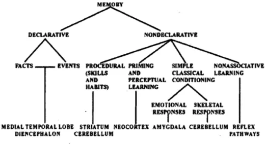
Figure 1. Taxonomy oflong-term memory systems, adapted from Squire (2004) ... 2 Figure 2. Cortical and subcortical input to the motor areas, adapted from Ghez (1991) .... .10
Figure 3. Model of mot or leaming, reproduced with permission from Doyon &
8enali (2005) ... 12 Figure 4. Model of motor leaming, reproduced with permission from Hikosaka
et al. (1999) .... ~ ... 15
CHAPTERII : EXPERIMENTAL STUDIES
Article 1 : Contribution oUhe Cerebel/um to Procedural Learning ofMotor Adaptation Examined Through Transcranial Magnetic Stimulation
Figure 1. SMA and the cerebellum stimulation sites ... 72 Figure 2. The eight-target tracking task ... 73 Figure 3. Timeline of the motor adaptation task acquisition ... 74 Figure 4. Percentage of correct responses (i.e. number of trials dufing which participants
reached the target within the time limit). Illustration of the means and standard errors of the mean for each block of trials ... 75 Figure 5. Total distance to reach the target during leaming. Illustration of me ans and
standard errors of the mean ofindividual blocks ... 76 Figure 6. Illustration oftypical travelled distance after intensive within-day training in
Article 2 : The Implication orthe Cerebellum and the Supolementary Motor Area in Procedural Learning ora Motor Sequence Task: a TMS Investigation
Figure 1. Coronal and transversal views of the SMA and the cerebellurn stimulation
x
sites ... 109 Figure 2. Seriai Reaction Time Task ... 110 Figure 3. Timeline of the motor sequence task acquisition ... 111 Figure 4. Mean RT and standard-errors of the mean for a1l45blocks during sequence
SMA
Pre-SMA
MlSRT
SRTT
MRI fMRIPET
TMS
rTMS
VOR ECLTP
LTD
ABBREVIATION LIST
Supplementary Motor Area Pre- Supplementary Motor Area Primary Motor Area
SeriaI Reaction Time SeriaI Rection Time Task Magnetic Resonance Imaging
Functional Magnetic Resonance Imaging Positron Emission Tomography
Transcranial Magnetic Stimulation
Repetitive Transcranial Magnetic Stimulation Vestibulo-ocular Reflex
Eyeblink Conditioning Long-T erm Potentiation Long-Term Depression
REMERCIEMENTS
Le doctorat fut pour moi une opportunité de découverte professionnelle et personnelle. Ma réussite n'aurait pas été possible sans l'appuis de ma famille et de mes amis, de mes superviseurs et des membres du laboratoire.
Je remercie mes parents Mioara Militaru et Marin-Cristian Vasilescu et ma soeur Rose-Marie pour leur soutien durant toutes ces années d'études. Je remercie également ma tante Nela, mon oncle Miki, mon cousin Stefan et ma cousine Raluca qui ont contribué au travers des années à forger celle que je suis devenue aujourd 'hui.
Je remercie mes chers amis qui ont chacun à leur façon illuminé mon existence. Un grand merci à Mihai, Guillaume, Latifa, Gaspard, Mihaela, Alexandra, Beatriz, Sandrine, Guylaine, Anne.
Un grand merci à mes superviseurs de recherche Maryse Lassonde et Julien Doyon. Sans leurs commentaires et encouragements, ce travail n'aurait pas été possible! Je remercie également ma superviseure de neuropsychologie clinique, Thérèse Botez-Marquard pour ses vifs encouragements et sa confiance constante en moi.
Finalement, je remercie mes collègues de laboratoires pour leur agréable présence dans ma VIe: Martin, Louis, Christine, Anne, Maude, Luke, Olivier, Miriam, Raby, Amélie, Marie-Claude, Valérie ainsi que Stéphane Dénis et Maria Sachez.
PREAMBLE
What would be life without motor memory? Dressing in the moming, using the utensils for eating breakfast, walking down stairs out of one's house and driving the car to work would be a forever challenge. Indeed, the main goal. of motor leaming is to improve perfonnance of those purposeful motor acts that are not hard wired in the brain from birth.
The neural substrates underlying motor leaming are unde~ CUITent investigation. A
hypothesis in the literature is that brain areas regulating motor control such as the supplementary motor area and the cerebellum are also responsible for motor leaming. Motor leaming theories from the literature suggest that important variables detennining the contribution of these areas to motor learning are the type of motor task and the level of expertise the participants perfonn the task at. The aim of this thesis is to study the critical role of the supplementary motor area and cerebellum in different stages of a motor sequence and a motor adaptation paradigm.
In the first chapter, mot or leaming will be introduced in the context of multiple memory systems, followed by a presentation of its behavioural characteristics, including its stages and the main paradigms employed to investigate it. The two main neural circuits responsible for motor control and the putative neural structures responsible for motor leaming will presented. The roles of the SMA and cer.ebellum in motor leaming will be discussed in the context of two recent leaming models. Additional imaging and clinical literature conceming the implication of the SMA and cerebellum in motor execution and motor leaming will be briefly reviewed. Repetitive TMS will be introduced as a new complementary technique to imaging and clinical research and the few studies that employed rTMS on the SMA or cerebellum to explore motor leaming will be presented.
In the second chapter, two rTMS experimental studies will be presented, one involving motor sequence and the other, motor adaptation acquisition. These studies had
similar designs. Repetitive TMS was employed to create 'virtual lesions' in healthy
participants while they acquired either task. Training was intensive during a first session, followed by a re-test the second day. Different groups of participants underwent rTMS on either the SMA or the cerebellum at the beginning and at the end of intensive task training. The performance of the stimulated groups was compared to that of a control group without magnetic stimulation.
In the third chapter, a review of the findings from the two studies will be presented, followed by an integration of the results with CUITent motor leaming models and a discussion conceming the effects of rTMS in the neural substrates it affects. Then, the two experiments will be examined in the context of their strengths and limitations. Finally, several lines of research will be suggested.
CHAPTERI: INTRODUCTION
1. Multiple memory types
The idea of multiple memory systems in the brain has received a lot of scientific attention since a report of a patient with important resection of the medial temporal lobes presented normal acquisition of a motor skill in the absence of any conscious memory of
having practiced the task before (Scoville & Milner, 1957; Milner, 2005). A memory
system is 'a particulàr neural network that mediates a specifie form of mnemonic
processes' (Brewer, Gabrieli, Preston, Vaidya, & Rosen, 2007). A well-accepted
classification separates declarative and non-declarative memory systems (Squire, 1992;
Squire & Knowlton, 1995; Squire, 2004) (see Figure 1 for a classification of the different
memory types and the brain structures thought to subserve them).
DECLARATIVE
A
FACI'S IVE1'ffS NONDECLARATIVE SIMPLE CLASSlCAL LIARNING PfRCltPTUAL CONDITIONING .LIARNING ~ lMOTIONAL SKEUTAL RESryNSESRESjNnS
MEDIAL TEMPOR4L LOBE STRIATVM !I1EOCORTlX AMYCDALA CEREBELLUM REFLEX DIENCEPHALON CBREBELLUM PATHWAYS
1.1 Declarative memory
Declarative memory consists of remembering fact or events. It involves consCÎous
retrieval of a list of items (words, pictures, etc.) previously encoded in a single-trial fashion
(Squire et al., 1996). It has been further distinguished as: semantic (Le. memory for events
or facts, which can be either true or false, such as remembering the capital of France) or episodic (Le. memory tied to the context in which it was acquired, the time, the place and
the sense that 'it happened to me', such as recalling one's lunch from yesterday). The vast
majority of memory research has been devoted to the study of dec1arative memory. Based on evidence from human and animai research, it was uncovered that the neural substrates subserving declarative memory are dependent on structures of, and connected to the medial
temporal lobe: the hippocampus, the parahippocampal gyrus, the rhinal cortex, the
amygdaIa, the diencephalon (the dorso-median and ventro-anterior nucleus of the
thalamus) and ventromedian regions of the pre frontal cortex (Squire, 1992; Squire, 2004; Petri & Mishkin, 1994; Schacter, 1987; Mishkin & Appenzeller, 1987).
1.2 Non-declarative memory
Cohen & Squire (1980) introduced the terrn 'nondec1arative memory' to refer to
those memories that are encoded and retrieved implicitly and non-intentionally, corresponding to a facilitation effect on performance due to prior experience (Schacter,
Chiu, & Ochsner, 1993; Doyon, 1997). The major component of non-declarative memory
is skills or procedural memory (Robertson & Cohen, 2006). Skill learning is the graduai
domain. An accepted notion in the literature is that the striatum andcerebellum are
critically involved in learning of skills (Salmon & Butters, 1995; Doyon, 1997; Doyon &
Ungerleider, 2002). However, compared to declarative memory, the neural substrates of skills or procedural memory are less well-established. For instance, procedural memory is believed to be independent of medial temporal lobe structures. Yet, this idea has been recently challenged since the medial temporal lobe has been found to support procedural
learning (Keele, Ivry, Mayr, Hazeltine, & Heuer, 2003). This finding points to a different
conception of memory systems.
2. Another view on memory taxonomy
Another popular taxonomy distinguishes two grand memory categories based on the characteristic of awareness. In this view, one memory class is termed explicit because it involves conscious and intentional recalling of an item; while the other, implicit, because it involves facilitation of performance as a result of previous experience with a material and
may not be associated with conscious recoUection of having studied that material (Graf &
Schacter, 1985; Schacter et al., 1993; Schacter, 19·87). Equivalence between declarative memory and explicit memory and between nondeclarative and implicit memory is generaUy accepted in the literature (Gabrieli et al., 2003). However, if we consider a memory as 'aU the information encoded during a task' (Robertson & Cohen, 2006), a procedural task may have implicit and explicit components. Several imaging studies suggest that learning a skill involves both implicit and explicit aspects and that these aspects may be reflected in
3. Motor skilllearning 3.1. Definition of Motor skilllearning
Motor skill leaming is defined as an improvement in time and space in the precision of movements as a result of repeated practice and interactions with the environment (Willingham, 1998). Motor skills often include a visual component, hence they are also called visuo-motor skills. In the laboratory, they are typically measured by a decrease in reaction time, number of errors, a decrease in the number of trials needed to reach a
. l
criterion or by a change in the synergy (coordination of muscles) and kinematics of movements.
3.2 Motor learning stages
Motor leaming develops gradually and constantly, taking place over several training sessions (Kami, 1996). Kami et al. (1998) have proposed a model with two leaming stages, one requiring practice (fast and slow 1eaming phases) and another that does not require further training (consolidation phase). At the very beginning of the acquisition process, there is a 'fast leaming' phase with considerable within-session improvement in
both speed and accuracy. The participants' performance is significantly and rapid1y
. enhanced. Within six hours post-training follows the consolidation phase. This is defined as an improvement in performance as a consequence of a latent period without practice
following the fast leaming phase (Kami & Sagi, 1993; Kami et al., 1998) or as resistance to
Ghilardi, 2005). Then, following repeated practice, there is the 'slow' learning stage' corresponding to further improved performance. When the participants receive further training beyond the slow phase, they reach the automatic phase, during which motor skills require minimal conscious effort, they are resistant to interference by other tasks and to the passage of time (Doyon et al., 2002).
3.3 Types of motor tasks
Many daily activities involve acquiring motor skills: driving a car, knitting, typing, riding a bicycle, etc. In order to study them in the laboratory, a variety of experimental tasks have been developed. Depending on their different cognitive demands, these tasks have been divided in two broad categories: motor sequence and motor adaptation tasks
(Sanes, Dimitrov, & Hallett, 1990; Hallett, 1996; Doyon, Penhune & Ungerleider, 2003).
Motor sequence tasks are those that combine sequences of movements into a more precise and effective motor plan such as learning to type or play the piano (Sanes & Dogues, 2000). This grand class of motor skills has been by far the most studied in the literature. In the laboratory, the most usèd paradigm is the seriaI reaction time task (SRTT)introduced by Nissen and Bullemer (1987). On each trial of the task, a cue is presented on the center of the screen at one of four spatially distinct locations. The participant has an equal nùmber of corresponding response keys and is instructed to press the correct keys as quickly as possible on each trial while making as few errors as possible. eues appear in a repeating sequence. Sequence learning is measured by a decrease in reaction time to the repeating stimulus sequence or by the difference in response time between sequenced items compared
to those administered in random. Several other sequence tasks have been employed in the
literature: those that incorporate timing information (Penhune & Doyon, 2002; Penh une &
Doyon, 2005; Sakai, Ramnani, & Passingham, 2002), visuo-spatial information (Hikosaka
et al., 1996; Sakai et al., 1998; Sakai et al., 1999) or solely motor information such as thumb-to-finger opposition (Kami et al., 1995; Kami et al., 1998). Several other paradigms,
including maze tracing (van Mier, Perlmutter, & Petersen, 2004) and rotor pursuit tasks
(Grafton, Woods, & Tyszka, 1994; Hatakenaka, Miyai, Mihara, Sakoda, & Kubota, 2007)
have been employed to explore both the behavioral and the neural aspects of sequence acquisition (for reviews, see Rhodes et al., 2004; Ashe et al., 2006).
Motor adaptation tasks are those that compensate for environmental changes such as leaming how to move the wheel in the opposite direction when driving a car backwards. Motor adaptations have been further divided into dynamic and kinematic paradigms. Kinematic adaptations are those that convert between coordinate systems (as between the position of the driving wheel and the position of the car on the street) and dynamic adaptations are those that relate 'motor commands to the motion of the system' (Wolpert,
Ghahramani, & Flanagan, 2001) (adjusting the forces applied to the driving wheel and the
resulting car movement, taking account the inertia of the wheel and the friction between the
wheels and the pavement). A typic~l laboratory task to study dynamic adaptation is
adapting to a unusual force-fiels in reaching movements (Thoroughman & Shadmehr, 2000;
Shadmehr & Moussavi, 2000; Smith, Ghazizadeh, & Shadmehr, 2006). A kinematic
adaptation laboratory task is one that converts movements based on a visual transformation, such as when the relationship between a mouse movement and the cursor on the screen is
altered (Contreras-Vidal & Kerick, 2004; Della-Maggiore & McIntosh, 2005; Graydon,
Friston, Thomas, Brooks, & Menon, 2005; Krakauer, Ghez, & Ghilardi, 2005) or when the
relationship between the direction of gaze and arm movement is changed through the use of
wedge prisms on the eyes (Norris, Greger, Martin, & Thach, 2001; Goedert & Willingham,
2002; Richter et al., 2002).
3.4 Putative neural correlates of motor skilllearning
Imaging studies have revealed many brain regions associated to motor and
visuo-motor leaming: the prefrontal cortex (Hazeltine, Grafton, & Ivry, 1997; Honda et al., 1998;
Eliassen, Souza, & Sanes, 2001; Sakai et al., 2002; Willingham et àl., 2002), the primary
motor area (Kami et al., 1995; Kami et al., 1998; Grafton, Salidis, & Willingham, 2001;
Penhune et al., 2005), the premotor area (Wu, Kansaku, & Hallett, 2004; Matsumura et al.,
2004; OIson et al., 2006), the supplementary motor cortex (Gordon, Lee, Flament, Ugurbil,
& Ebner, 1998; Honda et al., 1998; Doyon et al., 2002; van Mier et al., 2004; Heun et al., 2004), the parietal cortex (Sakai et al., 1998; Willingham et al., 2002; OIson et al., 2006;
Landau & D'esposito, 2006) and subcortical regions such as the cerebellum (Flament,
Ellerman, Kim, Ugurbil, & Ebner, 1996; Doyon et al., 2002; Imamizu et al., 2000;
Imamizu, Kuroda, Miyauchi, Yoshioka, & Kawato, 2003; Koeneke, Lutz, Wustenberg, &
Jancke, 2004; Penhune & Doyon, 2005) and the basal ganglia (Poldrack, Prabhakaran,
Seger, & Gabrieli, 1999; Bischoff-Grethe, Goedert, Willingham, & Grafton, 2004; Seidler
et al., 2005; Seidler, Noll, & Chintalapati, 2006). Several imaging studies have scanned
activation of brain areas correlated to the level of expertise (Grafton et al., 1994; Petersen,
van Mier, Fiez, & Raichle, 1998; Sakai et al., 1998; Jueptner et al., 1997; Krebs et al.,
1998; Nezafat, Shadmehr, & Holcomb, 2001; Hikosaka, Miyashita, Miyachi, Sakai, & Lu,
1998; Doyon et al., 2002; Floyer-Lea & Matthews, 2004; Floyer-Lea & Matthews, 2005;
Poldrack et al., 2005; Landau et al., 2006). These authors argue for distinct brain structures contributing to different motor leaming phases.
From a neuropsychological perspective, Willingham (1998) proposed that motor leaming develops directly of motor control processes, as these bec orne increasingly tuned to a particular task, thus functioning more efficiently. Although Willingham does not attribute a specific role to the primary motor area (Ml) in his control-based leaming theory, the primary motor area is being increasingly involved in motor leaming. Converging evidence from neurophysiological animal and human studies have uncovered Ml
activity-dependent plasticity associated with motor skill leaming (for reviews, see Sanes &
Donoghue, 2000; Ungerleider, Doyon, & Kami, 2002). Thus, these data point to Ml 's
implication in early acquisition, storage and consolidation of simple motor skills.
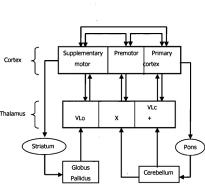
Ml together with the premotor and the supplementary motor area of the frontal cortex have cortical connections between themselves as well as subcortical inputs. Anatomical studies have revealed that these interconnected brain regions form two distinct cortico-subcortical circuits responsible for motor control: the striato-thalamo-cortical and the
Supplementary Premotor Primary motor ortex VLo Globus Pallidus
x
VLc +Figure 2. Cortical and subcortical input to the motor areas. Adapted from Ghez (1991). Cortical areas: SMA, supplementary motor area; PM, premotor cortex; Ml, primary motor
cortex. Thalamic nuclei: Vlo, ventrolateral nucleus, oral division; X, area X; VLc,
ventrolateral nucleus, caudal division; VPLo, ventroposterior nucleus, oral division.
As such, the SMA and cerebellum, each part of the striatal and
cortico-cerebellar circuits, respectively, might also play a role in motor leaming. In the following,
two leaming models will bepresented. The first one sheds light on the cerebellum's
contribution to motor sequence and adaptation leaming (Doyon & Benali, 2005) and the
second one highlights the implication of the SMA to motor sequence acquisition (Hikosaka,
Nakamura, Sakai, & Nakahara, 2002).
4. Models of Neural Correlates of Motor Skill Learning
4.1 Doyon & Ungerleider's proposaI of motor skill acquisition
In a model of motor skill leaming based on the imaging literature, Doyon and
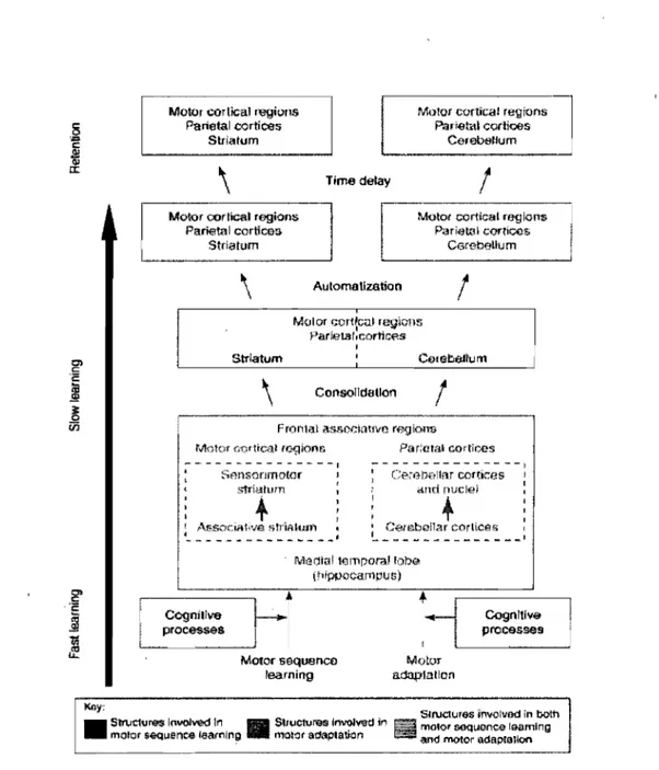
Benali, 2005) proposed that the two cortico-subcortical circuits involvedin motor control, participate in motor learning depending not only on the leaming phase, but also on the type of task (motor sequence learning versus motor adaptation). The authors' hypothesis is that the early phase of motor sequence and motor adaptation acquisition is supported by both the cortico-cerebellar and the cortico-striatal networks, inc1uding other areas such as the prefrontal, parietal and limbic cortices. However, starting from the consolidation, to the slow and more to the automatic stage, the two neural circuits are thought to specialize: the striatum and cortical associated regions are believed to subserve motor sequence learning but not motor adaptation and the cerebellum and cortical associated regions are thought to support motor adaptation, but not motor sequence (Figure 3).
Motoc cortical ragions Parietal cor1ices
Slriatum
Motor ctlr!îcal rtl'gions Parietal cortiœs
CerebeUum
\
Time delay1
Motor cortical regionsParietal corlîœs Stria tom
\
Automatlzation1
\
Consolldallon1
Frontal assor.kmvo regions
Motor cortical rc.gions
.... -, - - - --- ---.-- -
-.
: Sm;sonmotor striatmn'"
! ,.\~Soci.n1ive &tt'tf'th.:lf1 « .f _ _ _ _ _ "" ... _ _ _ _ _ _ _ .... ' Pariatal CQrtioos Careooltar cortk:es ami ouclei+
: C~(ebéna( cortîces----_
...._-- --_
... ' Medial temparallobe (hippocarnpus) Cognitive processes Motor sequence learning Motor adaptation Cognitive proœs!IesK6~: , Structures ïnvolvad in both
• Sb\Jcture<$ Involved ln mOIOt sequence learnlng • Slructure<$ InvoIved", _ motot adaptation lm and l'I'IClor adapuIIlon mOlor OOQuenca leaming
Figure 3. Model ofmotor learning. Reproduced with permission from Doyon & Benali
Doyon and Ungerleider's model makes testable predictions conceming the contribution of the cerebellum depending on the type oftask. The cerebellum is believed to contribute only to the early, but not to the late phase of motor sequence acquisition, while, it is thought to bring an important contribution to the whole time-course of motor adaptation leaming (early phase, consolidation and automatic perfonnance of an acquired ski Il). This model does not make particular predictions with regards to the contribution of SMA in motor leaming. Instead, the motor regions are said to participate to both tasks and to aIl phases of leaming. Yet, the SMA has important connections to the striatum, structure that is believed to play a role in the early phase of motor adaptation, but to be involved throughout the time-course of motor sequence acquisition. In the following, we will present a framework of motor sequence acquisition in which the SMA is thought to play a central role.
4.2 Hikosaka's mode) ofmotor sequence acquisition
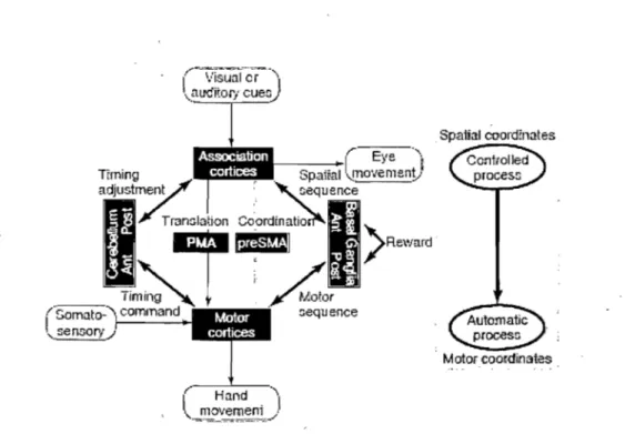
i-likosaka and colleagues (Hikosaka et al., 1999; Nakahara, Doya, & Hikosaka,
2001; Hikosaka, Nakamura, Sakai, & Nakahara, 2002); have proposed a model of the
underlying neural substrates subserving acquisition of motor sequences based on known anatomical connections of different cortico-subcortical loops and integrating data from neurophysiological experiments in animaIs (recording and les ions in monkeys) and humans. They proposed that motor sequences are acquired separately by two cortico-subcortical networks working in parallel, each with its own coordinates: the spatial mechanism fonned by fronto-parietal areas connected to associative regions of the
cerebellum and the basal ganglia; the motor mechanism formed by motor cortices (including Ml and SMA) connected to motor regions of the cerebellum and basal ganglia
(see Figure 4). A conyersion is thought to take place between spatial and motor
coordinates. Spatial sequences are believed to be effector unspecific, processed explicitly, acquired rapidly and requiring high attentional demands; while motor sequences are effector-specific, processed implicitly, acquired slowly and requiring low attentional
demands. Motor skill learning may be initiated by either spatial or motor mechanisms. It
is thought that optimization of performance is achieved by the cerebellum that processes sensori-motor or timing errors and by the basal ganglia processing reward or likelihood values. In a case where a sequence like trial-and-error begins by being explicit and then becomes implicit, the spatial mechanism would initiate leaming and guide the motor mechanisms. An implicit sequence like the SRT task, would be initiated by the motor mechanism which would guide the spatial mechanism. Because retention of a motor skill in the long-term is supported by two parallel systems, when the motor sequence mechanism is damaged, learning can be supported by the spatial system working simultaneously.
Spatial ooord:nates
j " _ , ,_~ 1 • ( Eyc 4"""
•• I!l.t-:<:G;'
~paina ~.-~I "" ""110' ' 'e n~lQ ~y nt ,:::equence
" Motor coordil:lates
Figure 4. Modei of motor Iearning, reproduced with permission from Hikosaka et al. (1999).
In this framework, the SMA as part of the motor network wouid be important for improvement of speed in early implicit sequence Ieaming and aiso for the Iong-term retention of such skill when the performance is implicit, rapid and with Iow attentionai demands. As for the cerebellum, its posterior part wouid be criticai in early sequence acquisition to optimize performance of such parameters às timing. The authors believe that the anterior cerebellum (including the dentate nucleus) subserves Iater sequence Iearning.
This modei does not make predictions for the other important class of motor skills, adaptations. In the following, the Iiterature will be reviewed in the perspective of the role of the cerebellum and the SMA in motor execution and in motor Iearning of different tasks (with emphasis on motor sequence and adaptation studies).
5. Tbe cerebellum
5.1 Brief functional anatomy and connectivity
The cerebellum is functionally divided in three main parts: the vestibulocerebellum, the spinocerebellum and the neocerebellum. It is now well established that each of these functional regions have a specific role in fine motor control and in coordinated movements
(Timmann. & Daum, 2007; Timmann & Diener, 2007). The vestibulocerebellum is
implicated in eye movement and body equilibrium, the spinocerebellum controls muscle tone and adjusts ongoing movements and the neocerebellum is involved in initiation,
planning and timing of movement (Ghez, 1991). We will further focus on the
neocerebellum since lesions in this area, not only produce slight motor control deficits, but also leaming deficits (Thack, 1996).
The neocerebellum is the largest cerebellar structure comprising the cerebellar
hemispheres and the dentate nucleus. It receives information through the ponto-cerebellar
fibers from ail sensory modalities (pre-processed information from second-order neurons), the motor, cognitive and associative cortices (Thach, 1996). The neocerebellum is thus implicated in motor behaviour of the limbs through the ponto-cerebellar-thalamo-cortical loop. The motor cortex sends information to the neocerebellum via the cortico-ponto-cerebellar pathway and the dentate nucleus projects back to the motor cortex via the
5.2 Motor control in the neocerebellum
The fact that the cerebellum regulates movement control (Houk, Buckingham, &
Barto, 1996) is illustrated by lesions of the hemispheres. These produce motor
coordination deficits without a motor deficit as such, ipsilateral to the lesion and manifest themselves as disturbance in limb movements, intentional tremor (when the dentate nucleus is involved), decomposition of movement or dysynergy, dysmetria and dysdiadochokinesis (Timmann et al., 2007). Dysynergy is characterized by a trouble in the amplitude, direction and force of a multijoint movement. Dysmetria is an inability to control the range of movements while dysdiadochokinesis is an incapacity to perform rapid, alternating movements.
5.3 Motor learning in the cerebellum
The role of the cerebellum in motor learning was first modeled by Albus and Marr based on its unique cellular architecture (Albus, 1971; Marr, 1969). The gist of their proposaI is that the cerebellum is important for response-context linkage through a mechanism of detection and correction of errors (Thach, 1997; Mauk, 1997; Boyden,
Katoh, & Raymond, 2004). This is based on the fact that the sole output of the cerebellum
are Purkinje cells which project to the deep nuclei and that the Purkinje cells are influenced by two different types of input. The first source of afferent input is constituted by a system of mossy fibers-granule cellcontacting only one Purkinje cell. This system represents the 'context', carrying sensory and other ongoing activity of the nervous system. The second source of afferent input is the climbing fiber that has a strong synapse to one Purkinje cell.
The climbing fibers have the role of the 'error' signal, firing when a Ilew movement is to be learned. When climbing fibers fire, they instruct the Purkinje cells to strengthen their contact with granule cells. In this manner, the correct movement is tied to the context, such that with repetitions, the context automatically evokes the correct movement.
5.4 Animal models to study motor learning in the cerebellum
Mechanisms of motor leaming in the cerebellum have been extensively studied in animaIs with two main paradigms: the vestibulo-ocular reflex (VOR) and classical
conditioning of the eyeblink (EC) (Christian & Thompson, 2003; du Lac, Raymond,
Sejnowski, & Lisberger, 1995). In the VOR, when moving the head, the eyes move
reflexihly in the opposite direction to stabilize the image on the retina and prevent blurred vision. The VOR can adapt when altering conditions in vision (e.g. wearing prisms) pro duce errors. In the EC, upon repeated association of atone before an air puff delivered onto the eyes, the presentation of the tone al one evokes an eye blink. Lesion and recording studies in this research domain have found evidence for the Marr-Albus theory. The mossy fibers convey the context (the tone or head movements), climbing fibers convey the error signal (the air puff Q.r image motion) and the cerebellar and vestibular nuclei . carry expression of the conditioned eyeblink and VOR adaptation, respectively (for reviews see,
Mauk, 1997; Lee & Thompson, 2006). Conclusions from this domain of research have
revealed that the relative contribution of the cerebella's cortex versus its nuclei to motor learning might depend on the type and amount of training (Mauk, 1997). Although still a matter of present investigation (Shutoh et al., 2006), it seems that short-term acquisition of
the VOR and EC depends on the cerebellar cortex and that long-tenn storage involve the cerebellar and vestibular nuc1ei and that these are mediated by different plasticity
mechanisms (for reviews, see Lee & Thompson, 2006; Boyden, Katoh & Raymond, 2004).
5.5 Motor sequence studies
Human functional imaging studies have shown activations in the cerebellum during early learning of several sequence tasks such as the SRT (Eliassen et al., 2001; Doyon et
al., 2002), sequential finger-to-thumb opposition (Friston, Frith, Passingham, Liddle, &
Frackowiak, 1992), sequence by trial-and-error (Toni, Krams, Turner, & Passingham, 1998;
Jenkins, Brooks, Nixon, Frackowiak, & Passingham, 1994), pursuit rotor task (Grafton,
Woods & Tyszka, 1994) or rhythmic sequence (Sakai, Ramnani & Passingham, 2002;
Penhune & Doyon, 2005). However, following practice, commensurate with better
perfonnance, either in early or later phases of sequence acquisition, several researchers
have noted a decrease in the cerebellar cortex activity (Doyon, Owen, Petri des, Sziklas, &
Evans, 1996; Doyon et al., 2002; Friston, Frith, Passingham, Liddle, & Frackowiak, 1992;
Grafton, Woods & Tyszka, 1994; Jueptner et al., 1997; Grafton, Salidis & Willingham,
2001; vanMier, Perlmutter & Petersen, 2004; Floyer-Lea & Matthews, 2005). The
observation that the cerebellar cortex is active during early sequence acquisition but that it decreases with leaming is attributed, in many imaging studies, to the role of the cerebellar cortex in errors. Indeed, in a recent imaging studies on rhythmic sequence learning
Penhune & Doyon (2005) have found correlations between activation in the cerebellar
during sequence leaming (Grafton et al., 1992; Grafton et al., 1995; Rauch et al.; 1995) or related activity in the cerebellum to expression of performance, but not leaming (Seidler et al.,2002).
Clinical studies with patients having cerebellar lesions have uncovered important deficits in early acquisition of the SRTT. Pascual-Leone et al. (1993) have reported severe impairment in both implicit and explicit leaming of the SRTT, while Molinari et al. (1997) have shown an important impairment when the task was implicit, but not when it was
explicitly taught prior to the experimentation. Gomez-Beldarrain, Garcia-Monco, Rubio, &
Pascual-Leone (1998) revealed that cerebellar patientshad deficits in acquiring the SRTT with the hand ipsilateral to the les ion, but not with the contralateral hand. In another study, patients with cerebellar lesions were unable to leam either a spatial or a temporal sequence
in the SRTT (Shin & Ivry, 2003). Interestingly, in a single experiment, cerebellar patients
were impaired in late, but not in the early phase of SRTT acquisition (Doyon et al., 1997). Inconsistencies between clinical studies are possibly due to the heterogeneity of cerebellar lesions: atrophies, vascular lesions, tumors or degenerative diseases. Yet, another view is that the severe impairment in motor sequence leaming of cerebellar patients is simply due
to performance deficits (Hallett, 1996; Timmann & Diener, 1996). In agreement with this
idea, Frings, Boenisch, Gerwig, Diener, & Timmann (2004) have revealed that cerebellar
patients were not impaired in acquiring and detecting various auditory and sensory sequences with low motor demands.
Imaging and lesion studies have not yet reached a consensus regarding the role of
implication of the cerebeIlum, more particularly its hemispheres, in the early fast phase of a motor sequence acquisition, but not consistently in later phases, when the task is weIl-leamed.
5.6 Motor adaptation studies
A few imaging studies employing fMRI and PET have addressed the specific role of the cerebellum in leaming of a motor adaptation task. High activation in the cerebellar hemispheres has been found during early adaptation leaming of kinematic (Imamizu et al., 2000; Flament et al., 1996) and dynamic tasks (Nezafat et al., 2001). In these studies, the early activation of the lateral cerebellum was related to visuo-spatial errors on the task. The finding regarding the role of the cerebellum in the detection and correction errors is weIl
documented in the literature (Doyon & Ungerleider, 2002; Mier, 2000; Diedrichsen,
Hashambhoy, Rane, & Shadmehr, 2005). However, areas of the cerebellum were found to
be active in late phases of adaptation learning (after consolidation), when errors were minimal: an are a close to theposterior superior fissure (Imamizu et al., 2000), the dentate nucleus (Flament et al., 1996; Floyer-Lea et al., 2004) and the right cerebellar cortex (Krebs et al., 1998). These findings hint at the possibility that the cerebellum might not only be involved in the processing of errors and thus have a subsidiary role in early adaptation learning, but that this structure also constitutes a memory storage site of adaptation skills. Indeed, Imamizu and colleagues (Imamizu et al., 2000; Imamizu et al., 2003) have observed activity in the cerebellar hemispheres throughout adaptation leaming and have attributed this activity to acquisition of 'internaI models' by this structure.
Functional imaging studies show correlations of brain activity and behaviour. Thus, even if the cerebellum might be important for learning, it might not be essential for it to occur. Natural lesions to the human cerebellum were found to impair learning of several
motor adaptation tasks: visuomotor adaptation to prisms (Martin, Keating, Goodkin, &
Bastian, 1996; Martin, 2003; Weiner, Hallett, & Funkenstein, 1983), mirror drawing
(Laforce & Doyon, 2001), adaptation of anticipatory muscle activity during catching (Lang
& Bastian, 1999) and adaptation to a new force field (Maschke, Gomez, Ebner, & Konczak,
2004; Smith & Shadmehr, 2005). In the later two studies, cerebellar patients were impaired
on several dependent variables when acquiring a dynamic task, results that were interpreted as a difficulty in establishing and updating internaI models of limb dynamics. In the study
by Smith & Shadmehr (2005), cerebellar patients (with cerebellar degeneration of several
etiologies) showed deficits in the use error from a previous trial to change motor commands to the next despite intact ability to correct errors during the trial itself. Maske et al. (2004) have noted a negative correlation between the severity of progressive cerebellar ataxia and
extent of learning. Their patients showed little learning-related aftereffects, no
generalization of learned movements to targets outside the learned space and di mini shed retention 3hrs later.
In sum, the cerebellar cortex seems to be involved in the fast leaming phase and in later phases of motor adaptation learning.
6. Supplcmcntary motor arca (SMA) 6.1 Bricf functional anatomy and connectivity
The SMA may be anatomically divided in the pre-SMA, anterior to the coronal plane through the anterior commissure, and the SMA-proper, posterior to the plane
(Rizzolatti, Luppino, & Matelli, 1998). Work on monkeys has revealed that only the
SMA-proper has anatomical connections to Ml and to the spinal cord, while the pre-SMA has
abundant connections to the prefrontal cortex (Luppino, Matelli, Camarda, & Rizzolatti,
1993; Wang, Shima, Sawamura, & Tanji, 2001). In humans, it was shown that the
pre-SMA and pre-SMA send projections to different parts of the striatum, pre-SMA-proper having a similar connectivity to that of Ml (Lehericy et al., 2004). Using imaging in the monkey,
and Picard & Strick (2003) observed that motor-re1ated activity in SMA-proper was
coupled to that of Ml. Based on their different connectivity and function, Picard & Strick
(2001) argued that the pre-SMA is resembles a prefrontal region and the SMA, a motor region. From now on, we will focus on the SMA-proper simply referred to as SMA.
Conncctivity betwccn SMA and ccrebellum
The SMA and several other cortical regions project to the contralateral cerebellar hemisphere through the pontine nuc1ei (Ghez, 1991). The SMA although a major target of basal ganglia output through the thalamus, also receives minor dentate input from the
cerebellum (Akkal, Dum & Strick, 2007; Sakai, Inase & Tanji, 2002; Sakai, Inse & Tanji,
6.2 Motor control in the SMA
The motor representation of the SMA is contralateral (Fried et al., 1991) when the task requires movements on one side, but SMA neurons have shown activity during
movement selection of the two limbs (Hoshi & Tanji, 2004). Electrophysiological
recording and imaging in humans suggest that the SMA is somatotopically organized (Fried
et al., 1991; Fontaine, Capelle, & Duffau, 2002). Lesions in the human SMA speak of its
role in initiating and planning of motor activity. Ressectioning the SMA unilaterally in humans produces the SMA syndrome characterized by severe and reversible motor and speech deficits post-operation first described by (Laplane, Talairach, Meininger, Bancaud,
& Orgogozo, 1977). Motor deficits immediately after SMA removal consist of global reduction of spontaneous movements contralaterally (Krainik et al., 2001; Zentner,
Hufnagel, Pechstein, Wolf, & Schramm, 1996) of variable intensity and related to the
extent of SMA removal (Russell & Kelly, 2003). In addition, there have been reports of
~, heminegle~t and apraxia in the contralaterallimb (Bannur & Rajshekhar, 2000). Lastly, an almost complete recovery follows within days to weeks with residual reduction in
controlateral motor activity (Bannur & Rajshekhar, 2000; Duffau et al., 2003). In the
long-term, the motor function is essentially completely recovered, the only permanent sequela reported are disturbance of altemating movements (Zentner et al., 1996). Laplane et al. (1977) have also suggested an implication of the SMA in sequential movements. Recording studie's in monkeys have indeed shown an implication of the SMA in control of
6.3 Motor sequence studies
SMA activation in well-Ieamed movement sequences was noted in several PET and fMRI studies. Jueptner et al. (1997) have observed significant activity in the SMA when comparing overleamed to newly acquired trial-and-error sequences. Doyon et al. (2002) have shown an increase of SMA activity in late compared to early acquisition of the SRT task. Gordon et al. (1998) have specifically revealed strong SMA activity in well-Ieamed movement sequences, but not in simple repetitive key presses. Poldrack et al. (2005) have implicated the SMA in well-Ieamed sequences by showing a decrease in SMA activity when comparing well-Ieamed SRTT trials to random ones. Recently, a functional ne ar-infrared spectroscopy study has $hown an increase in SMA activity after within-session leaming of a rotor pursuit task, but not in beginning of task acquisition (Hatakenaka et al., 2007). However, activation of the SMA in late sequence leaming has not been consistently
observed in imaging studies. In a fMRI study, Toni et al. (1998) have revealed a
modulation of SMA activity in the early phase of within-day intensive sequence acquisition. Compared to the baseline, SMA was minimally active in the first third of the experiment, significantly increased in the second third and decreased at the end of the experiment. Several fMRI studies have also shown SMA activation in early leaming of
different versions of the SRT task (Daselaar, Rombouts, Veltman, Raaijmakers, & Jonker,
2003; Heun et al, 2004; Landau & D'Esposito, 2007), while other imaging studies have·
correlated activity in the SMA during early sequence acquisition to improvement in
performance. Van Mier, Perlmutter & Petersen, (2004) have uncovered early
maze. Floyer-Lea & Matthews (2004) noted decreases in SMA activation associated to early improvement in performance on a tracking task. The inconsistent activation in the SMA regarding several sequence leaming stages might stem from the fact that imaging
studies report relative levels of activations between tasks. It is posible that a significant
performance improvement during motor leaming corresponds to an increasing involvement of the SMA. Indeed, in imaging studies, the SMA might be found more active wh en comparison between relative levels of activations involve time points in leaming corresponding to performance improvement.
Clinical studies regarding the role of SMA in motor leaming are scarce because this area is involved in motor execution. Lesion research suggest that the SMA is critical in early and late sequence leaming. In a controlled a case study of a patient with a left SMA lesion, Ackerman et al. (1996) have reported impaired acquisition of a motor sequence through the SRTT at the beginning of leaming. Botez (1992) has reported anecdotal cases of loss of automatic complex voluntary movements in patients with SMA lesions: a secretary who while being able to type the right letters had considerably lost her typing rapidity; a person could not play the piano anymore, but was able to play each note separately. In the monkey, chemical inactivation of the SMA bilaterally produced errors on a sequence of movements performed from memory, even when animaIs were able to associate a visual cue with the different movement to make or when animaIs with the same
lesion performed adequately other types of reaching movements (Shima & Tanji, 1998).
In sum, several studies involve the SMA in the late stage of sequence acquisition, while others suggests an implication of this structure in early sequence leaming as weIl.
6.4 Motor adaptation studies
The SMA has been mostly implicated in sequence learning and much less in motor adaptation. Several functional imaging studies have not registered activation in the SMA
during early or late learning of adapted movements (Krebs et al., 1998; Shadmehr &
Holcomb, 1997). Using fMRI, Seidler, Noll & Chintaiapati (2006) have noted activations in several cortical and subcortical regions associated with early learning of a kinematic adaptation task. In this study, SMA activity was associated to sensori-motor processes and thus considered subsidiary. In another recent imaging study (Imamizu, Higuchi, Toda, & Kawato, 2007), learning of a kinematic task was associated with development of an internai model in the cerebellum, while activation of the SMA was explained as a result of cerebellar output to this region.
Animal studies have not been conclusive either. Padoa-Schioppa, Li, & Bizzi 2004) havè recorded the neuronal activity in the SMA as monkeys adapted to an external perturbing force field. Their results indicate that the SMA participates in movement dynamics (the forces exerted by the muscles that cause the movement) during motor preparation (before massive activity is registered in Ml) and execution. However, results concerning Iearning of adapted movements were less conclusive since SMA cells
underwent plastic changes in control sessions as weIl. Paz, Natan, Boraud, Bergman, &
Vaadia (2005) have used recording techniques in monkeys while they adapted to new kinematic movements. They noticed piastic-related changes in the SMA during early adaptation to kinematic movements, while Iater learning of the same task involved plastic changes in Ml.
In sum, if any, the SMA seems to have a marginal role in learning of adapted movements.
7. Critiques of human imaging and les ion studies
Human imaging techniques such as PET and fMRI have the advantage of registering who le brain activity during task execution. However, the activated brain regions are merely associated or 'correlated' with behaviour. Not only sorne of the activated regions may be superfluous (faise positives) to the studied behaviour, but other areas that do not show activation couid in principle constitute a crucial node in the computing of the studied cognitive task (false negatives). lndeed, no matter how well-controlled, imaging studies do not provide information about brain-behaviour causal relationships. Neuropsychologicai studies complement imaging research by providing causal links between brain and behaviour. However, clinical research has several disadvantages related to lesion's focality, localization and compensation by other brain areas. Indeed many natural Iesions are not necessarily small enough or placed in the same area for ail patients, thus restricting spatial resolution and reliability of group studies. Moniover, the lesion is determined by the area of irrigation of the cerebrovascular system and not by modules of brain that neuroscientists are interested in. In addition, depending on the passage of time from the appearance of the lesion to the testing time, the patient's behaviour might reflect the ability of the rest of the brain to compensate (Kolb & Whishaw, 1998). More particularly, patients with cerebellar and SMA lesions have associated motor
execution troubles that are difficult to disentangle from motor learning deficits (Gentilucci et al., 2000; Gentilucci et al., 2000; Timmann et al., 2007).
8. Advantages of TMS over imaging techniques and c1inical studies Transcranial magnetic stimulation (TMS) allows non-invasive and temporary
disruption of a brain region in healthy and diseased individuals. It is a unique method to
establish true functional significance of imaging studies by providing causal information
about the activated brain area and the· related behaviour. Compared to c1assical
neuropsychological studies, it has the added advantage of well-controlled temporary 'virtuallesions' in healthy participants.
8.1 Presentation of the TMS technique
The TMS technique enables the induction of currents of physiological amplitude in the cortical surface of the brain. Intense pulses of current are passed through a coil that is placed above the subject's scalp. This current generates a time-varying magnetic field (1.5 to 3 Tesla) that penetrates undeflected into the subject's brai n, inducing a secondary current of inverse orientation and parallel to the current from the coil (Hallett, 2000). The brain-induced current corresponds to simultaneous neuronal firing in the stiinulated region and
propagation of neuronal activity (Ilmoniemi, Ruohonen, & Karhu, 1999). In one mode of
TMS stimulation, a single pulse or a short train of pulses induce a brief 'virtual lesion' by temporarily disrupting neural processing. In the repetitive mode, longer trains of rTMS modify cortical excitability of the stimulated area for several minutes after the end of the
stimulation period. Low frequency rTMS (SI Hz) was shown to depress transiently cortical excitability and cause an effect lasting for a period of half the time of the stimulation period (Chen et al., 1997) white high frequency TMS (> 1 Hz to 50 Hz) was shown to increase
cortical excitability (Pascual-Leone, Valls-Sole, Wassermann, & Hallett, 1994).
9. Motor learning studies involving rTMS of the SMA and Cerebellum
Many recent rTMS studies have employed rTMS on Ml to explore learning of different motor tasks (Muellbacher et al., 2002; Kobayashi, Hutchinson, Theoret, Schlaug,
& Pascual-Leone, 2004; Robertson, Press, & Pascual-Leone, 2005; Carey, Fregni, &
Pascual-Leone, 2006; Cothros, Kohler, Dickie, Mirsattari, & Gribble, 2006; Shemmell,
Riek, Tresilian, & Carson, 2007). Fewer such studies have induced behavioral effects via
rTMS over the SMA or cerebellum to investigate motor control or motor learning. Gerloff, Corwell, Chen, Hallett, & Cohen et al. (1997) have applied high frequency rTMS over the SMA during performance of overleamed finger sequences and produced accuracy errors in the complex sequence only', The authors concluded to a critical role of the SMA in planning of complex overleamed movement sequences. Muri, Rivaud, Vermersch, Leger,
& Pierrot-Deseilligny (1995) employed single pulse TMS and impaired acquisition of memory-guided saccades, Early sequence acquisition of the SRT task was not impaired by
5Hz rTMS over the SMA (Pascual-Leone, Wassermann, Grafman, & Hallett, 1996), while
moderate practice levels of short sequences were hindered by 1 Hz rTMS over the SMA (Verwey, Lammens, & van Honk, 2002). Two studies have shown motor behavioral effects by applying 1 Hz rTMS on the cerebellum: stimulation of the medial cerebellum
increased variability in self-paced finger tapping (Theoret, Haque, & Pascual-Leone, 2001) and stimulation of the lateral cerebellum increased movement times on a pegboard task
(Miall & Christensen, 2004). In a single experiment, 1Hz rTMS over the lateral cerebellum
hindered early acquisition of the SRT task (Torriero, Oliveri, Koch, Caltagirone, &
Petrosini,2004). None ofthese motor sequence learning studies employing rTMS over the SMA or cerebellum have allowed intensive training to attest the time-course of the TMS effect. To our knowledge, no study has employed rTMS over the SMA or cerebellum to investigate motor adaptation.
SYNTHE SIS, GOALS AND HYPOTHESES
Imaging and clinical studies suggest that the SMA and cerebellum play a role in motor learning, besides their established role in motor control. Still a matter of debate in the literature concems the contribution of the SMA and cerebéllum to different motor learning stages and the type of tasks to which they bring a more substantial contribution to. The majority of imaging and lesion evidence reviewed thus far suggest that the cerebellum plays a particular role throughout the acquisition of adapted movements (early phase, consolidation and storage) (Doyon & Benali, 2005), while it seems to be critical only in the early phase of sequence acquisition (Molinari et al., 1997; Shin and Ivry, 2003). An important body of research points to a specifie role of the SMA in learning of sequential procedures. Yet, the stage of learning to which the SMA is more important remains
controversial. The Hikosaka's model of sequential procedures (1999) specifically
hypothesizes an implication of the SMA in the early fast phase of sequence learning with an emphasis on performance improvement. In the same time, several imaging studies point to a role of the SMA in late stages of motor sequence acquisition (Doyon et al., 2002; Wu, . Kansku & Hallett, 2004; Poldrack et al., 2005; Megumi et al., 2007). Although few studies addressed the issue of the role of the SMA in motor adaptation learning, these suggest a subsidiary role of the SMA in this type of learning.
The main goal of the present thesis was to complement imaging and lesion research via the use of rTMS to shed light on the critical role of the SMA and cerebellum in several stages of acquisition of a motor sequence and an adaptation task. Temporary 'virtual lesions' were created via 1Hz rTMS on either the SMA or cerebellum at the beginning and