Amyloid Burden and White Matter Hyperintensities Mediate Age-Related Cognitive Differences
Pénélope S. Dupont, PhD candidate a, b, Christian Bocti, MD c, d, Maude Joannette, PhD candidate a,
b, Marie Maxime Lavallée, PhD candidate a, b, Jim Nikelski, PhD e, Guillaume T. Vallet, PhD g,
Howard Chertkow, MD e, f, Sven Joubert, PhD a, b
aDépartement de psychologie, Université de Montréal, bCentre de recherche de l’Institut universitaire
de gériatrie de Montréal (CRIUGM), cUniversité de Sherbrooke, Service de neurologie, Départment of médecine, dResearch Center on Aging and Memory Clinic, CIUSS Estrie-CHUS, eLady Davis
Institute for Medical Research, McGill University, fDepartment of Neurology and Neurosurgery, McGill University, gUniversité Clermont Auvergne, Laboratoire de Psychologie Sociale et Cognitive
(CNRS, UMR6024), France Corresponding author: Pénélope Sévigny Dupont
Corresponding author’s address: 4545 Chemin Queen Mary, Montreal, QC, Canada H3W 1W6 Corresponding author’s phone and fax: 1 514-340-3540, ext. 4779
ABSTRACT
This study examined the additive vs synergistic contribution of beta-amyloid (Aβ) and white matter hyperintensities (WMH) across seven cognitive domains in 104 cognitively normal older adults. It also measured the extent to which age-related differences in cognition are driven by measurable brain pathology. All participants underwent neuropsychological assessment along with MRI and PiB-PET imaging for Aβ quantification. WMH severity was quantified using the ARWMC scale. Stepwise regressions, moderation and mediation modelling were performed. Our findings show that Aβ deposition single-handedly predicts poorer episodic memory performance, and that Aβ and WMH contribute additively to poorer performance in working memory and language while carrying synergistic associations with executive functions and attention. Through mediation modelling, we demonstrated that the influence of age over episodic memory, working memory, executive functions and language is fully mediated by brain pathology. This study permits to conclude that, in healthy older adults; 1) Aβ burden and WMH have synergistic associations with some cognitive domains and; 2) age-related differences in most cognitive domains are driven by brain pathology associated with dementia.
Keywords: Cognitive aging; Alzheimer’s disease; Neuroimaging
Abbreviations: Aβ = beta-amyloid; AD = Alzheimer’s disease; MCI = mild cognitive impairment;
PiB = Pittsburgh Compound B; SUVR = standard uptake value ratio; WMH = white matter hyperintensities
Introduction
Pathological changes are common in the aging brain, even in older adults who remain fully functional. For example, it has been consistently shown that significant cerebral beta-amyloid deposition (Aβ) – one of the pathological hallmarks of Alzheimer’s disease (AD) along with neurofibrillary tangles – is present in about a third of cognitively normal older adults (Jack et al., 2017; Jansen et al., 2015). Longitudinal studies suggest that healthy older adults harbouring high Aβ burden are about fourfold more likely to convert to mild cognitive impairment (MCI) and that MCI individuals are also more likely to convert to AD in the following years compared to their same-aged peers with low Aβ (Chen et al., 2014; Doraiswamy et al., 2012; Villemagne et al., 2011).
The amyloid cascade hypothesis, which posits that Aβ deposition is the initiating event in a sequence of neuropathological changes leading to dementia, has remained for a long time the dominant
explanation for AD (Hardy and Selkoe, 2002; Jack et al., 2013). More recently, though, the impact of other pathologies coexisting with Aβ burden - especially of presumed vascular origin - is gaining increasing recognition in the pathogenesis of AD (Gorelick et al., 2011; Iadecola, 2013). For
instance, in a comprehensive data-driven model of the spatiotemporal ordering of preclinical AD progression, vascular dysregulation was identified as the first pathological event, followed by Aβ deposition, metabolic dysfunction, functional impairment and cortical atrophy (Iturria-Medina et al., 2016).
White matter hyperintensities (WMH), which reflect vascular brain pathology (i.e., small vessel disease), have been linked to an increased risk of developing dementia, whether in combination with Aβ or independently (Mortamais et al., 2014). WMH and Aβ have received considerable attention as
potential biomarkers of preclinical dementia because: 1) they occur in a significant proportion of healthy older adults (Burns et al., 2005; de Leeuw et al., 2001); and 2) they are key drivers of age-related cognitive decline in healthy older adults (Vemuri et al., 2015). Although the deleterious influence of WMH on cognition in healthy older adults is viewed as largely global, the bulk of research points towards a more direct link with executive components of cognition and processing speed (Hedden et al., 2012; Moon et al., 2017; Murray et al., 2010; Papp et al., 2014). In contrast, high Aβ burden in cognitively normal older adults has been associated with decline primarily in episodic memory, although some studies have shown decline in semantic memory, working
memory, executive functions and visuospatial functions as well (Baker et al., 2017; Duke Han et al., 2017; Hedden et al., 2013).
The specific nature of age-associated changes across specific cognitive domains remains to be better understood, however, since many studies have used relatively rudimentary neuropsychological testing. Finer discrimination between multiple cognitive domains in the context of preclinical dementia is valuable in order to distinguish subtle changes in cognition most suggestive of underlying pathology from those which are more likely benign in nature. In the same way, the question as to whether the effects of Aβ burden and WMH on cognition are independent or interactive also remains open. Some authors have advanced that Aβ and WMH yield an additive effect on cognition in healthy older adults (Lo et al., 2012; Vemuri et al., 2015), while others have suggested a synergistic interaction, whereby cognitive deterioration associated with coexisting pathologies is greater than the sum of their distinctive effects (Iadecola, 2010; Zekry et al., 2002). A more comprehensive view of the interrelationships between AD and cerebrovascular disease
early identification of at-risk individuals. So far, clinical trials targeting Aβ have shown very limited success in altering the clinical course of AD, which may be attributable to treatment being
administered tardily in the disease course, when significant neurodegeneration has already occurred (Gauthier et al., 2016; Mangialasche et al., 2010; Musiek and Holtzman, 2015). Thus, timely intervention (at the presymptomatic stage) holds a better promise of achieving therapeutic efficacy. For that purpose, the first step is to hone our ability to reliably recognize the earliest signs of pathological aging.
The first objective of this study was to investigate the independent contribution of Aβ and WMH across seven cognitive domains, and to examine whether there was an interaction between these pathological biomarkers on cognition. Another main objective was to assess the extent to which age-related differences in cognitive function may be driven by Aβ deposition and WMH using partial least squares path (mediation) modelling. We sought to answer these questions in a cross-sectional study carried out in a cohort of cognitively normal older adults. Aβ deposition was modelled on PET imaging with 11C-Pittsburgh Compound B ligand (PiB), and WMH were quantified with a
semi-quantitative visual rating scale.
Methods
Participants
In this cross-sectional study, we recruited 104 participants aged 65 years and older. The majority of participants in our study was recruited from the CRIUGM pool of healthy older participants, but a portion of our sample was also recruited through advertisements (73 and 27%, respectively), because we wanted to recruit participants across a broader range of educational achievement (i.e.,
overrepresentation of educated participants in the CRIUGM pool). All participants underwent a detailed neuropsychological assessment, an MRI scan and a PiB-PET scan. Volunteers with
untreated diabetes or heart disease, and other health conditions known to adversely impact cognition including untreated mental health disorders were excluded. A history of moderate to severe
traumatic brain injury, substance abuse or neurological disorders, as well as general anaesthesia in the last six months, were also criteria for exclusion. Participants expressed no significant cognitive complaint and were judged to be free of dementia or MCI at the time of their participation, using the following criteria. Participants had to perform within 2 SD of the mean on a self-reported memory questionnaire (van der Linden et al., 1989). Two specific domains of the questionnaire were used for screening, “Conversations” and “Movies and books”, which have been shown to be particularly sensitive to objective memory decline (Clement et al., 2008). We further screened for general cognitive decline using the Montreal Cognitive Assessment (MoCA), on which participants had to score ≥23. This cut-off has been deemed optimal in a recent meta-analysis (Carson et al., 2018). Additionally, performance of participants had to be > -1.5 SD when compared to the mean of age-matched controls on at least one of two learning tasks used for screening, the Logical Memory subtest of the Wechsler Memory Scale (WMS-III; delayed recall) and the Delayed Matching-to-Sample - 48 items (DMS-48; 2 min. delay). In an effort to screen for depressive symptoms,
participants had to score < 11/30 on the Geriatric Depression Scale (GDS). This study protocol was reviewed and approved by the CRIUGM and the Montreal Neurological Institute and Hospital (MNIH) Research Ethics Boards. All participants provided written informed consent prior to their participation in the study.
Participantsunderwent detailed neuropsychological testing covering the following cognitive domains: episodic memory, working memory, executive functions, language, attention, processing speed and visuospatial abilities. Composite scores were calculated for each of these cognitive domains. In order to do so, we computed correlation matrices including all performance indices of subtests related to a specific cognitive domain based on their theoretical framework. For each matrix (or cognitive domain), all performance scores significantly intercorrelated at r ≥ 0.30 were selected, thereby ensuring internal validity of our composite measures (this cut-off corresponds to a medium effect size according to accepted standards) (Cohen, 1988). These raw scores were then converted into standardized z-scores (based on our sample’s mean and SD). Cognitive measures for which a higher score indicates a poorer performance (such as z-scores reflecting errors or execution times) were reversed scored. The z-scores were then averaged together to produce composite cognitive scores, which were themselves converted into z-scores to facilitate data interpretation. When only one test score was available in a participant for a specific cognitive domain, a composite score was not calculated. This resulted respectively in 3 and 4 missing values for executive functions and attention. Outliers were defined as z ± 3.29 (P < 0.001); and then two extreme scores (z < -3.29) were replaced with the minimum threshold value (-3.29). The distribution normality of the composite cognitive scores was verified upon visual inspection of frequency distributions and normal probability plots. Detailed information on the neuropsychological battery and the list of performance indices (by cognitive domain) are shown in Supplemental Table S1. Matrices showing correlation coefficients between composite cognitive scores and between performance scores included in the composites are provided in Supplemental Tables S2. and S3.
Brain Imaging
Aβ quantification in individuals without dementia (Mintun et al., 2006). PET images of Aβ were acquired on a Siemens/CTI ECAT HR+ scanner in 3D imaging mode at the McConnell Brain Imaging Centre. Each scanning session yielded a collection of 7 dynamic fames comprised of 63 axial slices, using a 128x128 matrix (voxel dimensions: x,y = 2.059 mm, z = 2.425 mm). In order to minimize in-scanner time, scanning was started 50 minutes following bolus injection, during which time 7 frames (6x300 sec, 1x600 sec) were collected. Global standard uptake value ratios (SUVR) were computed by creating a region-weighted average of PiB ratio values, which, in turn, were derived by normalizing cortical tissue radioactivity concentration to cerebellar grey matter counts. Cerebellar grey matter was used as a reference region because it is largely spared from Aβ
deposition (Klunk et al., 2004). These SUVR units were regarded as a continuous measure of Aβ burden in this study. PiB PET and MRI imaging each took place within an average of 85.12 days (SD = 73.26) and 5.62 days (SD = 34.11) of the last neuropsychological testing date.
MRI scans were obtained on a 3T Siemens Trio MRI at the Unité de Neuroimagerie Fonctionnelle. High resolution anatomical images were acquired using an optimized MPRAGE protocol
(TR = 2.3 s, TE = 2.94 ms, TI = 900 ms, flip angle = 9, FOV = 256×240, voxel
1mm×1mm×1.2mm). This MPRAGE protocol allowed co-registration of PiB PET to each subject's MRI image. In addition to this sequence, an Axial Proton Density (PD)/T2 weighted protocol (TR 3000 ms, TE 12ms and 99 ms, FOV 240 mm, matrix 228 x 256, 48 slices, slice thickness 3mm) and a FLAIR protocol (TR 9000ms, TE 105 ms, T1 2500 ms, FOV 220 mm, matrix 256 x 256, slice thickness 4mm) was used for quantification of WMH in participants.
Severity of WMH was assessed by a certified neurologist in our team (C.B.) using the ARWMC scale, a semi-quantitative visual rating tool yielding a 4-point scale (0 = no lesion; 1 = focal lesion(s); 2 = confluent lesions; 3 = diffuse lesions) over five cerebral regions (frontal,
parieto-occipital, temporal, infratentorial and basal ganglia) for the right and left hemispheres separately (Wahlund et al., 2001). A global brain measure of WMH was calculated by summing scores on all five regions bilaterally and was treated as a continuous variable in our analyses.
Statistical Analysis
Pearson correlations were calculated between age and all studied variables. We used partial
correlations adjusted for age in a first attempt to explore associations between Aβ burden, WMH and cognitive domains. Independent samples t-tests were also conducted to assess possible sex
differences in biomarkers, cognitive abilities and number of years of education. Independence between Aβ and WMH was verified and confirmed prior to carrying out the main analyses.
We then employed a stepwise multiple regression approach to estimate the additive and distinctive contribution of different potential predictors on cognition. A stepwise regression analysis was conducted for each cognitive domain by entering age, sex, Aβ and WMH as independent variables and cognitive composite score as the outcome. The same regression analyses were then repeated including number of years of education as an independent variable. The stepwise procedure allowed for the selection of predictor variables in a step-by-step iterative manner, adding variables in order of their additive contribution to the model while simultaneously removing variables no longer reaching significance levels, until no variable could be further added or removed. Statistical criteria for the inclusion and removal of predictor variables in the final model were set to an F probability (P-value) ≤ 0.050 and ≥ 0.051, respectively. Potential multicollinearity issues were tested using the tolerance (1-R2) and the Variance Inflation Factor (VIF) diagnostic indexes. Normality of the residuals was
In order to verify the presence of an interaction between Aβ and WMH pathologies on cognitive abilities, we used multiple regression models with an added interaction term. A significant interaction could be of two types: 1) one (more plausible) synergistic, whereby Aβ and WMH’s combined effects on cognition are greater than the sum of their individual effects; 2) and the other antagonistic, where both biomarkers cancel out each other’s effects, either partially or completely. To answer that question, simple linear moderation models were computed based on the following equation: Ŷ = i1 + b1X + b2M + b3XM, where X represents WMH and M represents Aβ. The
function of such a model is to determine whether the effect of a one-unit change in X on Ŷ is dependent upon M (or conditional). Otherwise stated, the moderation analysis establishes whether WMH’s effect on cognitive performance varies across different values of Aβ (or vice-versa). In our analyses, Aβ was selected as the moderator variable (M) and WMH as the independent variable (X), but this choice was arbitrary as interchanging the position of X and M would not affect the
interaction term. Age, sex and education were entered as covariates in the moderated regressions. An interaction was deemed significant at P < 0.05. The moderated regression models were built using Hayes’ PROCESS macro for SPSS (model 1) (Hayes, 2013).
As one of our main objectives was to test whether age’s influence on cognition was driven by Aβ deposition and WMH, we constructed parallel multiple mediation models using ordinary least squares path analysis with the PROCESS macro (model 4). In these structural equation models, Aβ burden and WMH were entered as potential mediators of the relationship between age (independent variable) and each composite cognitive score (outcome). In order to control for potential spurious or epiphenomenal associations, sex and education were included in the path models as covariates, thus statistically removing their influence on the mediation paths. Bias-corrected bootstrap confidence intervals for the indirect effect (based on 10,000 bootstrap samples) were used to assess the
significance of mediation effects. A significant mediation effect is indicated when the bootstrapped confidence interval does not contain zero with 95% confidence. Full mediation occurs when path c’ (direct effect) is non-significant whereas path a (independent variable’s effect on mediator), path b (mediator’s effect on outcome) and path c (total effect) are significant at P < 0.050. The indirect mediation effect of age on cognitive performance through the mediator variable(s) is obtained from the product of paths a and b (a x b). The direct effect of age on cognition is calculated by subtracting the indirect effect from the total effect [c’ = c - (a x b)]. All statistical analyses in this study were conducted with IBM SPSS Statistics (version 24.0; Armonk, NY). False discovery rate (FDR)-corrected P-values accounting for multiple testing across the 7 cognitive domains for the standardized regression coefficients (β), moderation interaction terms and indirect (mediation) effects are displayed in Supplemental Table S4.
Results
Characteristics of participants included in the analyses are displayed in Table 1. Unsurprisingly, age was associated with greater Aβ deposition (r = 0.388, P < 0.001) and WMH severity (r = 0.399,
P < 0.001), and with poorer cognitive performance across all cognitive domains: episodic memory (r
= -0.223, P = 0.023), working memory (r = -0.286, P = 0.003), executive functions (r = -0.360,
P < 0.001), language (r = -0.282, P = 0.004), attention (r = -0.399, P < 0.001), processing speed (r =
-0.538, P < 0.001) and visuospatial abilities (r = -0.320, P = 0.001). There was no significant correlation between age and education (P = 0.759). Importantly, Aβ burden and WMH were not correlated with one another (P = 0.342) when controlling for age; therefore, they were interpreted as independent variables in our statistical analyses. Controlling for age, education correlated marginally and negatively with Aβ (r = -0.186, P = 0.059) but not with WMH (P = 0.732).
scores, Aβ burden was inversely associated with episodic memory (r = -0.359, P < 0.001), working memory (r = -0.248, P = 0.012), attention (r = -0.296, P = 0.003) and marginally with executive functions (r = -0.182, P = 0.070), while WMH were inversely associated with working memory (r = -0.284, P = 0.004), executive functions (r = -0.272, P = 0.006), language (r = -0.326, P = 0.001) and attention (r = -0.206, P = 0.041). Independent samples t-tests revealed a male advantage in working memory (t(102) = -2.146, P = 0.034) and visuospatial abilities (t(102) = -3.585, P < 0.001). Men were also slightly more educated than women in our sample; t(102) = -2.099, P = 0.042. There were no significant sex differences in age (P = 0.770), Aβ burden (P = 0.673) or WMH severity (P = 0.377).
Testing the independent and additive contribution of Aβ burden and WMH on cognition Age, sex, Aβ and WMH were entered as potential predictors of cognitive composite scores in stepwise regression analyses. As a trend was observed for an inverse relationship between Aβ and education, the same regressions were repeated in separate analyses this time including education, so as not to overlook Aβ associations due to the shared variance with education (see Table 2 for
detailed stepwise regressions results with and without education). In the first set of analyses (without education), Aβ burden was the only predictor retained in the stepwise model for episodic memory. Working memory was best predicted by the combination of WMH, Aβ and sex. Similarly, both biomarkers – but not age and sex – made an additive contribution in the stepwise regression models for executive functions, attention and language. Age did not contribute in predicting episodic memory, working memory, executive functions, attention and language in the stepwise models. In contrast, processing speed was best predicted by age alone, while visuospatial abilities were best predicted by sex and age.
When education was included in the stepwise regression analysis, Aβ burden was no longer a significant predictor of executive functions and language. Rather, executive functions were best predicted by WMH, education and age, while language was best predicted by WMH and education. Age, Aβ, WMH and education (that is, all independent variables except for sex) were found to be significant predictors of attention when considering education as a potential predictor. Education was also a significant predictor of visuospatial abilities after sex and age in the stepwise model. However, education made no distinctive contribution to episodic memory, working memory and processing speed.
Consistent with the correlational analyses, Aβ burden, WMH and age were negative predictors of cognitive performance when selected in the stepwise models. Inversely, education was a positive predictor of cognition when significant. Among collinearity statistics, all tolerance values were well above 0.100 (min = 0.648) and all VIF values were well below 5.00 (max = 1.543), indicating that multicollinearity was not a concern for the regressions. The residuals of the regressions were normally distributed.
Testing the synergistic interaction between Aβ burden and WMH on cognition
To test the possibility of synergy (or antagonism) between Aβ burden and WMH on cognition, moderated regressions were run for each composite cognitive score, controlling for age, sex and education. The moderation analysis revealed a significant interaction term between biomarkers on executive functions (F(1,94) = 14.334, P < 0.001) and attention (F(1,93) = 11.556 , P = 0.001). More specifically, a one-unit SUVR increase in Aβ pathology potentiated WMH’s deleterious influence on executive functions and attention by 0.390 SD and 0.298 SD, respectively. The
for three levels of Aβ burden and WMH (-1, 0 and 1 SD using sample’s mean) are portrayed in Fig. 1. Besides, there was a marginally significant interaction effect between Aβ burden and WMH for language (F(1,97) = 2.77, P = 0.0995) and processing speed (F(1,97) = 2, P = 0.097). WMH’s negative effects on cognition increased by 0.169 SD for language and 0.167 SD for processing speed consequently to a one unit increase in Aβ SUVR. The moderation analyses detected no interaction between biomarkers for episodic memory (P = 0.576), working memory (P = 0.582) and visuospatial abilities (P = 0.432). Detailed results of the moderation analyses are provided in Table 3.
Taken together, results from the stepwise and moderated regressions indicate that Aβ and WMH yield superadditive (synergistic) effects on executive functions and attention, whereas their shared effects on working memory and language are additive in nature.
Mediation of age-related cognitive differences by Aβ burden and WMH
Mediation analyses were conducted to assess whether Aβ burden and WMH act as potential mediators of the relationship between age and cognition. Path models with a significant mediation effect are presented in Fig. 2. The path analysis revealed that Aβ deposition fully mediates the relationship between age and episodic memory (see Fig. 2A). The bootstrap confidence interval for the indirect effect (a1 x b1 = -0.023) was entirely under zero (CI1: -0.051, -0.008), indicating that the
indirect effect of age on episodic memory performance through Aβ burden was significant. Both Aβ and WMH pathologies mediated the effect of age on working memory and attention (see Fig. 2B and 2D). The confidence intervals for the indirect mediation effects of age on working memory through Aβ [(a1 x b1 = -0.017) and WMH (a2 x b2 = -0.020) did not contain zero [(CI1: -0.038, -0.005), (CI2:
-0.035, -0.010)] and were thus significant. Similarly, the mediation effects on attention were
-0.005)]. However, while Aβ burden and WMH together fully mediated the influence of age on working memory, the mediation effect was only partial for attention. The indirect mediation effect of age through WMH was significant for executive functions [(a2 x b2 = -0.020); (CI2: -0.04, -0.005)]
and for language [(a2 x b2 = -0.023), (CI2: -0.041, -0.010)]; complete mediation by WMH was
observed in both cognitive domains (see Fig. 2C and 2E). In contrast, there was no significant mediation effect by either biomarker for processing speed or visuospatial abilities.
Discussion
One of the main findings of this study, as demonstrated through mediation modelling, is that age-dependent differences in episodic memory, working memory, executive functions and language were explained either fully or partially – as is the case for attention – by the presence of brain pathologies associated with AD (Aβ burden) and cerebrovascular disease (WMH). Changes in other domains such as processing speed and visuospatial skills, however, were explained only by age and/or sex. These results are important because they suggest that the extent of age-dependent changes in specific domains in healthy older individuals is related to the extent of underlying brain pathology. Another significant finding was that Aβ and WMH were found to contribute additively to decreased
performance in working memory and language, while they were found to contribute synergistically to decreased performance in executive functions and attention. Evidence for the existence of a synergistic interaction between both brain pathologies on functions dependent upon frontal-subcortical circuitry contributes in attaining a clearer understanding of the specific role of brain pathology in cognitive aging.
It must be pointed out that episodic memory stood out as the only cognitive domain to be explained solely by Aβ deposition, and that Aβ single-handedly predicted episodic memory performance in our
study. This distinctive contribution of Aβ burden to episodic memory is in agreement with a meta-analysis showing that AD pathology has closer and more robust associations with episodic memory decline than any other cognitive domains in healthy older adults (Hedden et al., 2013). It is
noteworthy that Aβ-dependent memory decline in healthy older adults, as in MCI participants, has been considered to be a signature of preclinical dementia (Pike et al., 2007; Sliwinski et al., 2003). It should also be noted, however, that episodic memory – as a hallmark of AD - has been more
systematically investigated than other cognition domains, and this may introduce a bias. Hence, associations between Aβ and other cognitive skills may have been overlooked in initial studies. In fact, a recent meta-analysis has linked positive Aβ status in cognitively intact older adults to poorer performance not only in episodic memory, but also in global cognitive functioning, language, visuospatial abilities, processing speed, working memory, attention and executive functions (Duke Han et al., 2017). In the current study, beyond its role in episodic memory performance, Aβ burden also mediated age-related differences in working memory and attention, along with WMH.
Moreover, Aβ burden and WMH were negatively and synergistically associated with executive functions and attention, which rely to a significant extent on frontal lobe function (Tsuchida and Fellows, 2013). Although mixed findings have been reported previously, some researchers have suggested that specific components of executive functioning (especially switching and inhibition) (Doherty et al., 2015; Kantarci et al., 2012; Mielke et al., 2016) and working memory (Lim et al., 2012) are vulnerable to Aβ deposition. Our findings are consistent with this view, especially considering that our composite measure of executive functions was composed primarily of tasks assessing switching and inhibition.
The role of WMH in driving age-related differences in working memory, executive functions and attention is also in accordance with previous work relating cerebrovascular pathology and cognition
in normal older adults (Hedden et al., 2012; Moon et al., 2017; Murray et al., 2010; Papp et al., 2014). These particular cognitive functions are thought to be vulnerable to WMH because they depend on prefrontal and subcortical loops, which are more vulnerable to disruption by widespread microvascular lesions (Prins and Scheltens, 2015). Vascular cognitive impairment may also occur through cortical atrophy (Kloppenborg et al., 2012; Rizvi et al., 2018) or other less studied
pathophysiological mechanisms, such as damage to the cholinergic neuronal system (Bocti et al., 2005; Richter et al., 2017; Roman and Kalaria, 2006).
More surprisingly, adverse effects of age on language and semantic-related tasks were fully
mediated by WMH severity. The current literature reviewing the impact of WMH on verbal abilities in normal aging remains scarce, as most studies have focused primarily on executive functions and processing speed. Lexical and semantic difficulties in AD and even mild cognitive impairment are well documented (Benoit et al., 2017; Joubert et al., 2010; Joubert et al., 2008; Langlois et al., 2016) and have been associated with structural and functional alteration within regions of the semantic network (Barbeau et al., 2012; Joubert et al., 2010; Pineault et al., 2018). There is some evidence for the role of white matter alterations in developmental language disorders such as dyslexia (Zhao et al., 2016). The language-semantic network is complex and involves a wide range of interconnected brain regions, and it is possible that widespread axonal damage may compromise the connectivity of this network. A recent study with a cohort of healthy older participants showed that executive control required to access representations of stored lexical-semantic knowledge relied on the integrity of the arcuate fasciculus, a major language-related white matter pathway (Hoffman et al., 2017). Our results do not allow distinguishing between storage vs access difficulties in language-semantics, but the notion that the integrity of white matter pathways is necessary for executive components of language and efficient communication between distributed cortical regions forming
the language-semantic network is an interesting avenue and deserves future investigation.
In agreement with previous studies in cognitively normal older adults and in individuals with MCI, there was no significant correlation between these two biomarkers when controlling for age, supporting the prevailing notion that Aβ and vascular pathologies reflect distinct pathological pathways of independent aetiology (Haight et al., 2013; Hedden et al., 2012; Vemuri et al., 2015; Villeneuve and Jagust, 2015). Further, Aβ and cerebrovascular pathologies also appear to occur independently in preclinical older adults (Knopman et al., 2013). In individuals with AD, however, those with high Aβ deposition also tend to harbour greater WMH volume; hence, it has been hypothesized that WMH may impart a “second hit” to already existent AD pathology, thus precipitating the clinical expression of the disease (Provenzano et al., 2013). Consistent with this view, there is some evidence suggesting that AD may induce WMH through degenerative, non-vascular processes. Although the pathophysiological mechanisms of interaction between Aβ and cerebrovascular burden remain elusive, several tentative explanations have been put forward. For instance, accumulation of tau protein in the cortex secondary to Aβ deposition may promote WMH development (and axonal loss) directly via neuronal death and more indirectly by disrupting fast anterograde axonal transport mechanism (McAleese et al., 2015). WMH, in turn, are thought to potentiate the deleterious impact of AD pathology and accelerate neurodegeneration through various processes including oxidative stress, inflammation and impaired cerebral perfusion (Iadecola, 2010). An alternative hypothesis is that vascular brain damage disrupts lymphatic drainage of vessels located in the periventricular tissue and prevent proper clearance of waste material such as Aβ peptides (Ramirez et al., 2016; Weller et al., 2009).
It could be reasonably contended that these mechanisms of interaction between AD and vascular pathways may not yet be detectable in asymptomatic individuals because they emerge later in the course of the disease. Challenging this proposition, however, recent work has provided evidence that Aβ deposition and total WMH volume in healthy older adults have synergistic associations with grey matter neurometabolic alterations typically associated with AD, especially in the posterior cingulate and precuneus (Schreiner et al., 2018). Therefore, AD and cerebrovascular disease pathophysiology may yield mutually potentiating effects which, albeit subtle, are detectable even in the absence of objective cognitive impairment (i.e. in the hypothetical preclinical stage of dementia). Pertaining to cognitive effects of Aβ deposition and WMH in normal older adults, our results advocate the existence of both additive and synergistic associations, depending on the cognitive domains. It is worth noting that the presence of synergy in several cognitive domains lends support to the notion that Aβ and WMH interact at a physiological level through one or several of the aforementioned mechanisms. Thus, despite reflecting initially independent disease pathways, Aβ and WMH pathologies appear to exert interactive effects on cognition in healthy older adults. This finding strongly suggests that AD and cerebrovascular disease pathways converge at some point in time in preclinical individuals, and that their interactive effects and co-occurrence are likely to increase with the disease progression.
In contrast with the results reported above, Aβ burden and WMH did not significantly predict performance in processing speed and visuospatial abilities when age was accounted for in the regression models. Therefore, other factors or pathogenic mechanisms may be responsible for age-related differences in processing speed and visuospatial function. The lack of an association between biomarkers and processing speed, while not entirely unexpected regarding Aβ burden, stands against
a body of work suggesting that processing speed is particularly vulnerable to early white matter changes and cerebral small vessel disease (Gunning-Dixon and Raz, 2000; Prins et al., 2005; van den Heuvel et al., 2006; Wright et al., 2008). It could be that other surrogates of cerebrovascular damage, such as vascular risk factors or brain infarcts, would be more readily responsible for slowed
information processing with age than WMH (Benjamin et al., 2014; Knopman et al., 2001;
Saczynski et al., 2009; Viswanathan et al., 2015). Alternatively, the failure to link higher WMH with reduced processing speed could also be attributable to relatively low WMH severity in our sample. Nonetheless, our findings are fairly in line with those of a recent meta-analysis in which progression of WMH was associated with decline in global cognitive functioning with markedly greater effects for attention and executive functions as opposed to memory and processing speed (Kloppenborg et al., 2014).
On the whole, our results appear quite robust when compared to analogous cross-sectional studies of similar sample size on cognitively normal older adults. One likely explanation is that our carefully selected neuropsychological assessment included the most sensitive and relevant tests specific to each cognitive domain. For instance, we avoided neuropsychological tests with ceiling effects in healthy older adults (e.g. we chose the RAVTL instead of the CVLT for assessing verbal memory). Indeed, many studies have relied on a global cognitive composite score or a single measure of memory, and few studies have sought to assess the distinctive contribution of Aβ and
cerebrovascular pathologies across a wide range of cognitive domains. Another strength to the current study is that in addition to controlling for education in our analyses, we also recruited participants with different levels of education, thus avoiding the common pitfall of recruiting only highly educated participants. Education is often a source of bias because; 1) highly educated adults are more likely to volunteer in cognitive aging studies and; 2) higher education, which is a proxy of
cognitive reserve, may mask associations between brain pathologies and cognition (Stern, 2012). The use of continuous measures of Aβ burden and WMH is more statistically powerful and a better reflection of reality than dichotomous measures based on a threshold, which can be viewed as more arbitrary (Farrell et al., 2017).
A few limitations to this study should also be noted. First of all, the cross-sectional nature of the current results limits the scope of interpretation of our findings, as well as our sample size which is relatively modest when compared with some other cohorts. However, we compensated by recruiting participants from a wider spectrum of educational achievement and by using more sensitive
neuropsychological tests. Visual rating of WMH severity may prove to be less sensitive to early white matter changes in healthy individuals than automated volumetric assessment of WMH because of potential floor or ceiling effects and does not as readily lend itself to regional characterization.
Conclusions
Altogether, our results demonstrate that age-related changes in multiple components of cognitive functioning are driven by specific neuropathological biomarkers of dementia in healthy older adults. Furthermore, we argue that pathological aging is a multifaceted and insidious process occurring over a continuum of severity rather than a discrete entity. In particular, the present study demonstrates that Aβ and cerebrovascular pathologies can yield synergistic effects on cognition, especially for functions that rely on frontal-subcortical circuits (i.e., executive functions and attention). Although many have speculated on the existence of superadditive cognitive effects of Aβ and WMH in healthy older adults, this notion had never, to our knowledge, received tangible empirical support before this study. Most research effort in that direction has failed to find a significant interaction between Aβ and WMH on cognition, thus pointing towards additive effects (Gordon et al., 2015; Lo et al., 2012;
Vemuri et al., 2015). While it is difficult to pinpoint why our findings differ from those of previous work in this regard, this could be partly imputable to important discrepancies in cognitive
assessment, such as reliance on a single composite measure or rating scale in some studies. In another study on this matter, a trend was noticed for worse cognition in healthy older adults harboring significant levels of both AD and cerebrovascular pathologies as compared to other groups, although there was no significant interaction effect (Marchant et al., 2012). Most
importantly, our primary finding that brain pathology mediates age-related differences in cognitive performance of older adults has significant implications for future research on cognitive aging and may serve as a leverage for the development of interventions to mitigate cognitive decline associated with aging in individuals without objective cognitive deficits. In light of the current study findings, what truly remains a challenge is to define cognitive aging in the absence of significant brain pathology associated with AD and cerebrovascular disease.
Acknowledgments
This work was supported by a grant from the Canadian Institutes of Health Research (MOP123376) and the Institute of Aging (IA0120269). SJ was supported by a Chercheur boursier senior award from the Fonds de recherche du Québec - Santé (FRQ-S). PSD was supported by a doctoral award from the FRQ-S.
Author Contributions
P.S.D., S.J., C.B., and H.C. were responsible for conception and design of the study. P.S.D., S.J., M.J., M.M.L. and G.T.V. contributed to acquisition and statistical analysis of data, and J.N., H.C., and C.B. were responsible for MRI/PET preprocessing and interpretation. P.S.D. and S.J.
contributed to drafting the manuscript and P.S.D. was responsible for drafting the figures. All authors reviewed the manuscript and approved the final version of the manuscript.
Potential Conflicts of Interest
References
Baker, J.E., Lim, Y.Y., Pietrzak, R.H., Hassenstab, J., Snyder, P.J., Masters, C.L., Maruff, P., 2017. Cognitive impairment and decline in cognitively normal older adults with high amyloid-beta: A meta-analysis. Alzheimers Dement (Amst) 6, 108-121.
Barbeau, E.J., Didic, M., Joubert, S., Guedj, E., Koric, L., Felician, O., Ranjeva, J.P., Cozzone, P., Ceccaldi, M., 2012. Extent and neural basis of semantic memory impairment in mild cognitive impairment. J Alzheimers Dis 28(4), 823-837.
Benjamin, P., Lawrence, A.J., Lambert, C., Patel, B., Chung, A.W., MacKinnon, A.D., Morris, R.G., Barrick, T.R., Markus, H.S., 2014. Strategic lacunes and their relationship to cognitive impairment in cerebral small vessel disease. Neuroimage Clin 4, 828-837.
Benoit, S., Rouleau, I., Langlois, R., Dostie, V., Kergoat, M.J., Joubert, S., 2017. The impact of time and repeated exposure on famous person knowledge in amnestic mild cognitive impairment and Alzheimer's disease. Neuropsychology 31(7), 697-707.
Bocti, C., Swartz, R.H., Gao, F.Q., Sahlas, D.J., Behl, P., Black, S.E., 2005. A new visual rating scale to assess strategic white matter hyperintensities within cholinergic pathways in dementia. Stroke 36(10), 2126-2131.
Burns, J.M., Church, J.A., Johnson, D.K., Xiong, C., Marcus, D., Fotenos, A.F., Snyder, A.Z., Morris, J.C., Buckner, R.L., 2005. White matter lesions are prevalent but differentially related with cognition in aging and early Alzheimer disease. Arch Neurol 62(12), 1870-1876.
Carson, N., Leach, L., Murphy, K.J., 2018. A re-examination of Montreal Cognitive Assessment (MoCA) cutoff scores. Int J Geriatr Psychiatry 33(2), 379-388.
Chen, X., Li, M., Wang, S., Zhu, H., Xiong, Y., Liu, X., 2014. Pittsburgh compound B retention and progression of cognitive status--a meta-analysis. Eur J Neurol 21(8), 1060-1067.
Clement, F., Belleville, S., Gauthier, S., 2008. Cognitive complaint in mild cognitive impairment and Alzheimer's disease. J Int Neuropsychol Soc 14(2), 222-232.
Cohen, J., 1988. Statistical power analysis for the behavioral sciences, 2nd ed. L. Erlbaum Associates, Hillsdale, N.J.
de Leeuw, F.E., de Groot, J.C., Achten, E., Oudkerk, M., Ramos, L.M., Heijboer, R., Hofman, A., Jolles, J., van Gijn, J., Breteler, M.M., 2001. Prevalence of cerebral white matter lesions in elderly people: a population based magnetic resonance imaging study. The Rotterdam Scan Study. J Neurol Neurosurg Psychiatry 70(1), 9-14.
Doherty, B.M., Schultz, S.A., Oh, J.M., Koscik, R.L., Dowling, N.M., Barnhart, T.E., Murali, D., Gallagher, C.L., Carlsson, C.M., Bendlin, B.B., LaRue, A., Hermann, B.P., Rowley, H.A., Asthana, S., Sager, M.A., Christian, B.T., Johnson, S.C., Okonkwo, O.C., 2015. Amyloid burden, cortical thickness, and cognitive function in the Wisconsin Registry for Alzheimer's Prevention. Alzheimers Dement (Amst) 1(2), 160-169.
Doraiswamy, P.M., Sperling, R.A., Coleman, R.E., Johnson, K.A., Reiman, E.M., Davis, M.D., Grundman, M., Sabbagh, M.N., Sadowsky, C.H., Fleisher, A.S., Carpenter, A., Clark, C.M., Joshi, A.D., Mintun, M.A., Skovronsky, D.M., Pontecorvo, M.J., Group, A.A.S., 2012. Amyloid-beta assessed by florbetapir F 18 PET and 18-month cognitive decline: a multicenter study. Neurology 79(16), 1636-1644.
Duke Han, S., Nguyen, C.P., Stricker, N.H., Nation, D.A., 2017. Detectable Neuropsychological Differences in Early Preclinical Alzheimer's Disease: A Meta-Analysis. Neuropsychol Rev 27(4), 305-325.
Farrell, M.E., Kennedy, K.M., Rodrigue, K.M., Wig, G., Bischof, G.N., Rieck, J.R., Chen, X., Festini, S.B., Devous, M.D., Sr., Park, D.C., 2017. Association of Longitudinal Cognitive Decline With Amyloid Burden in Middle-aged and Older Adults: Evidence for a Dose-Response
Relationship. JAMA Neurol 74(7), 830-838.
Gauthier, S., Albert, M., Fox, N., Goedert, M., Kivipelto, M., Mestre-Ferrandiz, J., Middleton, L.T., 2016. Why has therapy development for dementia failed in the last two decades? Alzheimers Dement 12(1), 60-64.
Gordon, B.A., Najmi, S., Hsu, P., Roe, C.M., Morris, J.C., Benzinger, T.L., 2015. The effects of white matter hyperintensities and amyloid deposition on Alzheimer dementia. Neuroimage Clin 8, 246-252.
Gorelick, P.B., Scuteri, A., Black, S.E., Decarli, C., Greenberg, S.M., Iadecola, C., Launer, L.J., Laurent, S., Lopez, O.L., Nyenhuis, D., Petersen, R.C., Schneider, J.A., Tzourio, C., Arnett, D.K., Bennett, D.A., Chui, H.C., Higashida, R.T., Lindquist, R., Nilsson, P.M., Roman, G.C., Sellke, F.W., Seshadri, S., American Heart Association Stroke Council, C.o.E., Prevention, C.o.C.N.C.o.C.R., Intervention, Council on Cardiovascular, S., Anesthesia, 2011. Vascular contributions to cognitive impairment and dementia: a statement for healthcare professionals from the american heart association/american stroke association. Stroke 42(9), 2672-2713.
Gunning-Dixon, F.M., Raz, N., 2000. The cognitive correlates of white matter abnormalities in normal aging: a quantitative review. Neuropsychology 14(2), 224-232.
Haight, T.J., Landau, S.M., Carmichael, O., Schwarz, C., DeCarli, C., Jagust, W.J., Alzheimer's Disease Neuroimaging, I., 2013. Dissociable effects of Alzheimer disease and white matter hyperintensities on brain metabolism. JAMA Neurol 70(8), 1039-1045.
Hardy, J., Selkoe, D.J., 2002. The amyloid hypothesis of Alzheimer's disease: progress and problems on the road to therapeutics. Science 297(5580), 353-356.
Hayes, A.F., 2013. Introduction to mediation, moderation, and conditional process analysis : a regression-based approach. The Guilford Press, New York.
Hedden, T., Mormino, E.C., Amariglio, R.E., Younger, A.P., Schultz, A.P., Becker, J.A., Buckner, R.L., Johnson, K.A., Sperling, R.A., Rentz, D.M., 2012. Cognitive profile of amyloid burden and white matter hyperintensities in cognitively normal older adults. J Neurosci 32(46), 16233-16242. Hedden, T., Oh, H., Younger, A.P., Patel, T.A., 2013. Meta-analysis of amyloid-cognition relations in cognitively normal older adults. Neurology 80(14), 1341-1348.
Hoffman, P., Cox, S.R., Dykiert, D., Munoz Maniega, S., Valdes Hernandez, M.C., Bastin, M.E., Wardlaw, J.M., Deary, I.J., 2017. Brain grey and white matter predictors of verbal ability traits in older age: The Lothian Birth Cohort 1936. Neuroimage 156, 394-402.
Iadecola, C., 2010. The overlap between neurodegenerative and vascular factors in the pathogenesis of dementia. Acta Neuropathol 120(3), 287-296.
Iadecola, C., 2013. The pathobiology of vascular dementia. Neuron 80(4), 844-866.
Iturria-Medina, Y., Sotero, R.C., Toussaint, P.J., Mateos-Perez, J.M., Evans, A.C., Alzheimer's Disease Neuroimaging, I., 2016. Early role of vascular dysregulation on late-onset Alzheimer's disease based on multifactorial data-driven analysis. Nat Commun 7, 11934.
Jack, C.R., Jr., Knopman, D.S., Jagust, W.J., Petersen, R.C., Weiner, M.W., Aisen, P.S., Shaw, L.M., Vemuri, P., Wiste, H.J., Weigand, S.D., Lesnick, T.G., Pankratz, V.S., Donohue, M.C., Trojanowski, J.Q., 2013. Tracking pathophysiological processes in Alzheimer's disease: an updated hypothetical model of dynamic biomarkers. Lancet Neurol 12(2), 207-216.
Jack, C.R., Jr., Wiste, H.J., Weigand, S.D., Therneau, T.M., Knopman, D.S., Lowe, V., Vemuri, P., Mielke, M.M., Roberts, R.O., Machulda, M.M., Senjem, M.L., Gunter, J.L., Rocca, W.A., Petersen,
R.C., 2017. Age-specific and sex-specific prevalence of cerebral beta-amyloidosis, tauopathy, and neurodegeneration in cognitively unimpaired individuals aged 50-95 years: a cross-sectional study. Lancet Neurol 16(6), 435-444.
Jansen, W.J., Ossenkoppele, R., Knol, D.L., Tijms, B.M., Scheltens, P., Verhey, F.R., Visser, P.J., Amyloid Biomarker Study, G., Aalten, P., Aarsland, D., Alcolea, D., Alexander, M., Almdahl, I.S., Arnold, S.E., Baldeiras, I., Barthel, H., van Berckel, B.N., Bibeau, K., Blennow, K., Brooks, D.J., van Buchem, M.A., Camus, V., Cavedo, E., Chen, K., Chetelat, G., Cohen, A.D., Drzezga, A., Engelborghs, S., Fagan, A.M., Fladby, T., Fleisher, A.S., van der Flier, W.M., Ford, L., Forster, S., Fortea, J., Foskett, N., Frederiksen, K.S., Freund-Levi, Y., Frisoni, G.B., Froelich, L., Gabryelewicz, T., Gill, K.D., Gkatzima, O., Gomez-Tortosa, E., Gordon, M.F., Grimmer, T., Hampel, H., Hausner, L., Hellwig, S., Herukka, S.K., Hildebrandt, H., Ishihara, L., Ivanoiu, A., Jagust, W.J., Johannsen, P., Kandimalla, R., Kapaki, E., Klimkowicz-Mrowiec, A., Klunk, W.E., Kohler, S., Koglin, N.,
Kornhuber, J., Kramberger, M.G., Van Laere, K., Landau, S.M., Lee, D.Y., de Leon, M., Lisetti, V., Lleo, A., Madsen, K., Maier, W., Marcusson, J., Mattsson, N., de Mendonca, A., Meulenbroek, O., Meyer, P.T., Mintun, M.A., Mok, V., Molinuevo, J.L., Mollergard, H.M., Morris, J.C., Mroczko, B., Van der Mussele, S., Na, D.L., Newberg, A., Nordberg, A., Nordlund, A., Novak, G.P., Paraskevas, G.P., Parnetti, L., Perera, G., Peters, O., Popp, J., Prabhakar, S., Rabinovici, G.D., Ramakers, I.H., Rami, L., Resende de Oliveira, C., Rinne, J.O., Rodrigue, K.M., Rodriguez-Rodriguez, E., Roe, C.M., Rot, U., Rowe, C.C., Ruther, E., Sabri, O., Sanchez-Juan, P., Santana, I., Sarazin, M., Schroder, J., Schutte, C., Seo, S.W., Soetewey, F., Soininen, H., Spiru, L., Struyfs, H., Teunissen, C.E., Tsolaki, M., Vandenberghe, R., Verbeek, M.M., Villemagne, V.L., Vos, S.J., van Waalwijk van Doorn, L.J., Waldemar, G., Wallin, A., Wallin, A.K., Wiltfang, J., Wolk, D.A., Zboch, M., Zetterberg, H., 2015. Prevalence of cerebral amyloid pathology in persons without dementia: a meta-analysis. JAMA 313(19), 1924-1938.
Joubert, S., Brambati, S.M., Ansado, J., Barbeau, E.J., Felician, O., Didic, M., Lacombe, J., Goldstein, R., Chayer, C., Kergoat, M.J., 2010. The cognitive and neural expression of semantic memory impairment in mild cognitive impairment and early Alzheimer's disease. Neuropsychologia 48(4), 978-988.
Joubert, S., Felician, O., Barbeau, E.J., Didic, M., Poncet, M., Ceccaldi, M., 2008. Patterns of semantic memory impairment in Mild Cognitive Impairment. Behav Neurol 19(1-2), 35-40.
Kantarci, K., Lowe, V., Przybelski, S.A., Weigand, S.D., Senjem, M.L., Ivnik, R.J., Preboske, G.M., Roberts, R., Geda, Y.E., Boeve, B.F., Knopman, D.S., Petersen, R.C., Jack, C.R., Jr., 2012. APOE modifies the association between Abeta load and cognition in cognitively normal older adults. Neurology 78(4), 232-240.
Kloppenborg, R.P., Nederkoorn, P.J., Geerlings, M.I., van den Berg, E., 2014. Presence and progression of white matter hyperintensities and cognition: a meta-analysis. Neurology 82(23), 2127-2138.
Kloppenborg, R.P., Nederkoorn, P.J., Grool, A.M., Vincken, K.L., Mali, W.P., Vermeulen, M., van der Graaf, Y., Geerlings, M.I., Group, S.S., 2012. Cerebral small-vessel disease and progression of brain atrophy: the SMART-MR study. Neurology 79(20), 2029-2036.
Klunk, W.E., Engler, H., Nordberg, A., Wang, Y., Blomqvist, G., Holt, D.P., Bergstrom, M.,
Savitcheva, I., Huang, G.F., Estrada, S., Ausen, B., Debnath, M.L., Barletta, J., Price, J.C., Sandell, J., Lopresti, B.J., Wall, A., Koivisto, P., Antoni, G., Mathis, C.A., Langstrom, B., 2004. Imaging brain amyloid in Alzheimer's disease with Pittsburgh Compound-B. Ann Neurol 55(3), 306-319.
Knopman, D., Boland, L.L., Mosley, T., Howard, G., Liao, D., Szklo, M., McGovern, P., Folsom, A.R., Atherosclerosis Risk in Communities Study, I., 2001. Cardiovascular risk factors and cognitive decline in middle-aged adults. Neurology 56(1), 42-48.
Knopman, D.S., Jack, C.R., Jr., Wiste, H.J., Weigand, S.D., Vemuri, P., Lowe, V.J., Kantarci, K., Gunter, J.L., Senjem, M.L., Mielke, M.M., Roberts, R.O., Boeve, B.F., Petersen, R.C., 2013. Brain injury biomarkers are not dependent on beta-amyloid in normal elderly. Ann Neurol 73(4), 472-480. Langlois, R., Joubert, S., Benoit, S., Dostie, V., Rouleau, I., 2016. Memory for Public Events in Mild Cognitive Impairment and Alzheimer's Disease: The Importance of Rehearsal. J Alzheimers Dis 50(4), 1023-1033.
Lim, Y.Y., Ellis, K.A., Pietrzak, R.H., Ames, D., Darby, D., Harrington, K., Martins, R.N., Masters, C.L., Rowe, C., Savage, G., Szoeke, C., Villemagne, V.L., Maruff, P., Group, A.R., 2012. Stronger effect of amyloid load than APOE genotype on cognitive decline in healthy older adults. Neurology 79(16), 1645-1652.
Lo, R.Y., Jagust, W.J., Alzheimer's Disease Neuroimaging, I., 2012. Vascular burden and Alzheimer disease pathologic progression. Neurology 79(13), 1349-1355.
Mangialasche, F., Solomon, A., Winblad, B., Mecocci, P., Kivipelto, M., 2010. Alzheimer's disease: clinical trials and drug development. Lancet Neurol 9(7), 702-716.
Marchant, N.L., Reed, B.R., DeCarli, C.S., Madison, C.M., Weiner, M.W., Chui, H.C., Jagust, W.J., 2012. Cerebrovascular disease, beta-amyloid, and cognition in aging. Neurobiol Aging 33(5), 1006 e1025-1036.
McAleese, K.E., Firbank, M., Dey, M., Colloby, S.J., Walker, L., Johnson, M., Beverley, J.R., Taylor, J.P., Thomas, A.J., O'Brien, J.T., Attems, J., 2015. Cortical tau load is associated with white matter hyperintensities. Acta Neuropathol Commun 3, 60.
Mielke, M.M., Machulda, M.M., Hagen, C.E., Christianson, T.J., Roberts, R.O., Knopman, D.S., Vemuri, P., Lowe, V.J., Kremers, W.K., Jack, C.R., Jr., Petersen, R.C., 2016. Influence of amyloid and APOE on cognitive performance in a late middle-aged cohort. Alzheimers Dement 12(3), 281-291.
Mintun, M.A., Larossa, G.N., Sheline, Y.I., Dence, C.S., Lee, S.Y., Mach, R.H., Klunk, W.E., Mathis, C.A., DeKosky, S.T., Morris, J.C., 2006. [11C]PIB in a nondemented population: potential antecedent marker of Alzheimer disease. Neurology 67(3), 446-452.
Moon, S.Y., de Souto Barreto, P., Chupin, M., Mangin, J.F., Bouyahia, A., Fillon, L., Andrieu, S., Vellas, B., group, M.D., 2017. Associations between white matter hyperintensities and cognitive decline over three years in non-dementia older adults with memory complaints. J Neurol Sci 379, 266-270.
Mortamais, M., Artero, S., Ritchie, K., 2014. White matter hyperintensities as early and independent predictors of Alzheimer's disease risk. J Alzheimers Dis 42 Suppl 4, S393-400.
Murray, M.E., Senjem, M.L., Petersen, R.C., Hollman, J.H., Preboske, G.M., Weigand, S.D., Knopman, D.S., Ferman, T.J., Dickson, D.W., Jack, C.R., Jr., 2010. Functional impact of white matter hyperintensities in cognitively normal elderly subjects. Arch Neurol 67(11), 1379-1385. Musiek, E.S., Holtzman, D.M., 2015. Three dimensions of the amyloid hypothesis: time, space and 'wingmen'. Nat Neurosci 18(6), 800-806.
Papp, K.V., Kaplan, R.F., Springate, B., Moscufo, N., Wakefield, D.B., Guttmann, C.R., Wolfson, L., 2014. Processing speed in normal aging: effects of white matter hyperintensities and hippocampal volume loss. Neuropsychol Dev Cogn B Aging Neuropsychol Cogn 21(2), 197-213.
Pike, K.E., Savage, G., Villemagne, V.L., Ng, S., Moss, S.A., Maruff, P., Mathis, C.A., Klunk, W.E., Masters, C.L., Rowe, C.C., 2007. Beta-amyloid imaging and memory in non-demented individuals: evidence for preclinical Alzheimer's disease. Brain 130(Pt 11), 2837-2844.
Pineault, J., Jolicoeur, P., Grimault, S., Bermudez, P., Brambati, S.M., Lacombe, J., Villalpando, J.M., Kergoat, M.J., Joubert, S., 2018. Functional changes in the cortical semantic network in amnestic mild cognitive impairment. Neuropsychology 32(4), 417-435.
Prins, N.D., Scheltens, P., 2015. White matter hyperintensities, cognitive impairment and dementia: an update. Nat Rev Neurol 11(3), 157-165.
Prins, N.D., van Dijk, E.J., den Heijer, T., Vermeer, S.E., Jolles, J., Koudstaal, P.J., Hofman, A., Breteler, M.M., 2005. Cerebral small-vessel disease and decline in information processing speed, executive function and memory. Brain 128(Pt 9), 2034-2041.
Provenzano, F.A., Muraskin, J., Tosto, G., Narkhede, A., Wasserman, B.T., Griffith, E.Y., Guzman, V.A., Meier, I.B., Zimmerman, M.E., Brickman, A.M., Alzheimer's Disease Neuroimaging, I., 2013. White matter hyperintensities and cerebral amyloidosis: necessary and sufficient for clinical
expression of Alzheimer disease? JAMA Neurol 70(4), 455-461.
Ramirez, J., Berezuk, C., McNeely, A.A., Gao, F., McLaurin, J., Black, S.E., 2016. Imaging the Perivascular Space as a Potential Biomarker of Neurovascular and Neurodegenerative Diseases. Cell Mol Neurobiol 36(2), 289-299.
Richter, N., Michel, A., Onur, O.A., Kracht, L., Dietlein, M., Tittgemeyer, M., Neumaier, B., Fink, G.R., Kukolja, J., 2017. White matter lesions and the cholinergic deficit in aging and mild cognitive impairment. Neurobiol Aging 53, 27-35.
Rizvi, B., Narkhede, A., Last, B.S., Budge, M., Tosto, G., Manly, J.J., Schupf, N., Mayeux, R., Brickman, A.M., 2018. The effect of white matter hyperintensities on cognition is mediated by cortical atrophy. Neurobiol Aging 64, 25-32.
Roman, G.C., Kalaria, R.N., 2006. Vascular determinants of cholinergic deficits in Alzheimer disease and vascular dementia. Neurobiol Aging 27(12), 1769-1785.
Saczynski, J.S., Sigurdsson, S., Jonsdottir, M.K., Eiriksdottir, G., Jonsson, P.V., Garcia, M.E., Kjartansson, O., Lopez, O., van Buchem, M.A., Gudnason, V., Launer, L.J., 2009. Cerebral infarcts and cognitive performance: importance of location and number of infarcts. Stroke 40(3), 677-682. Schreiner, S.J., Kirchner, T., Narkhede, A., Wyss, M., Van Bergen, J.M.G., Steininger, S.C., Gietl, A., Leh, S.E., Treyer, V., Buck, A., Pruessmann, K.P., Nitsch, R.M., Hock, C., Henning, A., Brickman, A.M., Unschuld, P.G., 2018. Brain amyloid burden and cerebrovascular disease are synergistically associated with neurometabolism in cognitively unimpaired older adults. Neurobiol Aging 63, 152-161.
Sliwinski, M.J., Hofer, S.M., Hall, C., Buschke, H., Lipton, R.B., 2003. Modeling memory decline in older adults: the importance of preclinical dementia, attrition, and chronological age. Psychol Aging 18(4), 658-671.
Stern, Y., 2012. Cognitive reserve in ageing and Alzheimer's disease. Lancet Neurol 11(11), 1006-1012.
Tsuchida, A., Fellows, L.K., 2013. Are core component processes of executive function dissociable within the frontal lobes? Evidence from humans with focal prefrontal damage. Cortex 49(7), 1790-1800.
van den Heuvel, D.M., ten Dam, V.H., de Craen, A.J., Admiraal-Behloul, F., Olofsen, H., Bollen, E.L., Jolles, J., Murray, H.M., Blauw, G.J., Westendorp, R.G., van Buchem, M.A., 2006. Increase in periventricular white matter hyperintensities parallels decline in mental processing speed in a non-demented elderly population. J Neurol Neurosurg Psychiatry 77(2), 149-153.
van der Linden, M., Wijns, C., von Frenkell, R., Coyette, G., Seron, X., 1989. Un questionnaire d’auto-évaluation de la mémoire (QAM). Bruxelles : Editest.
Vemuri, P., Lesnick, T.G., Przybelski, S.A., Knopman, D.S., Preboske, G.M., Kantarci, K., Raman, M.R., Machulda, M.M., Mielke, M.M., Lowe, V.J., Senjem, M.L., Gunter, J.L., Rocca, W.A., Roberts, R.O., Petersen, R.C., Jack, C.R., Jr., 2015. Vascular and amyloid pathologies are independent predictors of cognitive decline in normal elderly. Brain 138(Pt 3), 761-771.
Villemagne, V.L., Pike, K.E., Chetelat, G., Ellis, K.A., Mulligan, R.S., Bourgeat, P., Ackermann, U., Jones, G., Szoeke, C., Salvado, O., Martins, R., O'Keefe, G., Mathis, C.A., Klunk, W.E., Ames, D., Masters, C.L., Rowe, C.C., 2011. Longitudinal assessment of Abeta and cognition in aging and Alzheimer disease. Ann Neurol 69(1), 181-192.
Villeneuve, S., Jagust, W.J., 2015. Imaging Vascular Disease and Amyloid in the Aging Brain: Implications for Treatment. J Prev Alzheimers Dis 2(1), 64-70.
Viswanathan, A., Macklin, E.A., Betensky, R., Hyman, B., Smith, E.E., Blacker, D., 2015. The Influence of Vascular Risk Factors and Stroke on Cognition in Late Life: Analysis of the NACC Cohort. Alzheimer Dis Assoc Disord 29(4), 287-293.
Wahlund, L.O., Barkhof, F., Fazekas, F., Bronge, L., Augustin, M., Sjogren, M., Wallin, A., Ader, H., Leys, D., Pantoni, L., Pasquier, F., Erkinjuntti, T., Scheltens, P., European Task Force on
Age-Related White Matter, C., 2001. A new rating scale for age-related white matter changes applicable to MRI and CT. Stroke 32(6), 1318-1322.
Weller, R.O., Djuanda, E., Yow, H.Y., Carare, R.O., 2009. Lymphatic drainage of the brain and the pathophysiology of neurological disease. Acta Neuropathol 117(1), 1-14.
Wright, C.B., Festa, J.R., Paik, M.C., Schmiedigen, A., Brown, T.R., Yoshita, M., DeCarli, C., Sacco, R., Stern, Y., 2008. White matter hyperintensities and subclinical infarction: associations with psychomotor speed and cognitive flexibility. Stroke 39(3), 800-805.
Zekry, D., Duyckaerts, C., Moulias, R., Belmin, J., Geoffre, C., Herrmann, F., Hauw, J.J., 2002. Degenerative and vascular lesions of the brain have synergistic effects in dementia of the elderly. Acta Neuropathol 103(5), 481-487.
Zhao, J., Thiebaut de Schotten, M., Altarelli, I., Dubois, J., Ramus, F., 2016. Altered hemispheric lateralization of white matter pathways in developmental dyslexia: Evidence from spherical deconvolution tractography. Cortex 76, 51-62.
TABLES
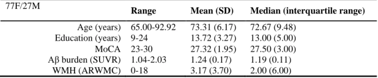
TABLE 1. Characteristics of Participants
77F/27M
Range Mean (SD) Median (interquartile range)
Age (years) 65.00-92.92 73.31 (6.17) 72.67 (9.48)
Education (years) 9-24 13.72 (3.27) 13.00 (5.00)
MoCA 23-30 27.32 (1.95) 27.50 (3.00)
Aβ burden (SUVR) 1.04-2.03 1.24 (0.17) 1.19 (0.11)
TABLE 2. Stepwise regressions models by cognitive domain Significant Predictors Std. β (p-value) F (p-value) R2 Episodic Memory Aβ burden -0.409 (<0.001) 20.513 (<0.001) 0.167 Working Memory WMH -0.323 (<0.001) 11.870 (<0.001) 0.132 Aβ burden -0.316 (0.001) 0.224 Sex 0.196 (0.025) 0.263 Executive Functions a (without education) WMH -0.339 (<0.001) 13.565 (<0.001) 0.143 Aβ burden -0.275 (0.003) 0.217 b (with education) WMH -0.287 (0.003) 14.515 (<0.001) 0.143 Education 0.341 (<0.001) 0.257 Age -0.253 (0.007) 0.310 Attention
a (without education) Aβ burden -0.381 (<0.001) 16.458 (<0.001) 0.158
WMH -0.309 (0.001) 0.253
b (with education) Age -0.209 (0.039) 11.002 (<0.001) 0.159
Aβ burden -0.276 (0.004) 0.233 WMH -0.244 (0.010) 0.280 Education 0.195 (0.026) 0.317 Language a (without education) WMH -0.385 (<0.001) 12.425 (<0.001) 0.160 Aβ burden -0.195 (0.032) 0.197 b (with education) WMH -0.413 (<0.001) 16.797 (<0.001) 0.160 Education 0.300 (0.001) 0.250 Processing Speed Age -0.538 (<0.001) 41.578 (<0.001) 0.290 Visuospatial Abilities
a (without education) Sex 0.325 (<0.001) 13.304 (<0.001) 0.112
Age -0.311 (0.001) 0.209
b (with education) Sex 0.272 (0.003) 11.834 (<0.001) 0.112
Age -0.320 (<0.001) 0.209
Education 0.238 (0.008) 0.262
Outcomes (cognitive domains) are identified in the first column of the table. Predictors included in the final model are listed in order of their additive contribution in the stepwise regression model. Results of the stepwise regressions are shown without education (a) and with education (b)
separately when inclusion of education as a potential predictor influenced the final model. R2*100
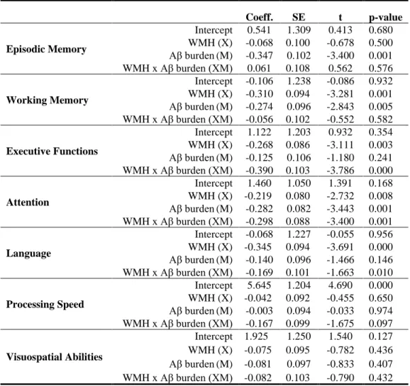
TABLE 3. Moderation models by cognitive domain
WMH, Aβ burden and composite cognitive scores are expressed in standardized z-scores (derived from the sample’s mean and SD). For simplicity, covariates (age, sex and education) are not shown in this table but were accounted for in all models. The interaction term for the moderation analysis (WMH x Aβ burden) is shown in the fourth row for each model.
Coeff. SE t p-value Episodic Memory Intercept 0.541 1.309 0.413 0.680 WMH (X) -0.068 0.100 -0.678 0.500 Aβ burden (M) -0.347 0.102 -3.400 0.001 WMH x Aβ burden (XM) 0.061 0.108 0.562 0.576 Working Memory Intercept -0.106 1.238 -0.086 0.932 WMH (X) -0.310 0.094 -3.281 0.001 Aβ burden (M) -0.274 0.096 -2.843 0.005 WMH x Aβ burden (XM) -0.056 0.102 -0.552 0.582 Executive Functions Intercept 1.122 1.203 0.932 0.354 WMH (X) -0.268 0.086 -3.111 0.003 Aβ burden (M) -0.125 0.106 -1.180 0.241 WMH x Aβ burden (XM) -0.390 0.103 -3.786 0.000 Attention Intercept 1.460 1.050 1.391 0.168 WMH (X) -0.219 0.080 -2.732 0.008 Aβ burden (M) -0.282 0.082 -3.443 0.001 WMH x Aβ burden (XM) -0.298 0.088 -3.400 0.001 Language Intercept -0.068 1.227 -0.055 0.956 WMH (X) -0.345 0.094 -3.691 0.000 Aβ burden (M) -0.140 0.096 -1.466 0.146 WMH x Aβ burden (XM) -0.169 0.101 -1.663 0.010 Processing Speed Intercept 5.645 1.204 4.690 0.000 WMH (X) -0.042 0.092 -0.455 0.650 Aβ burden (M) -0.003 0.094 -0.033 0.974 WMH x Aβ burden (XM) -0.167 0.099 -1.675 0.097 Visuospatial Abilities Intercept 1.925 1.250 1.540 0.127 WMH (X) -0.075 0.095 -0.782 0.436 Aβ burden (M) -0.081 0.097 -0.833 0.407 WMH x Aβ burden (XM) -0.082 0.103 -0.790 0.432
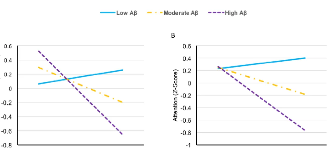
Figure 1. Cognitive performance at low (-1 SD), moderate (0 SD) and high (1 SD) levels of Aβ burden and WMH (adjusted for age, sex and education)
The regression slopes show WMH associations with executive functions (A) and attention (B) at three levels of Aβ burden and WMH (-1.00 SD, 0.00 SD and 1.00 SD based on our sample’s statistics). Differences of slopes illustrate the synergistic interaction between WMH and Aβ over these cognitive domains. The moderated regression analyses were corrected for age, sex and education.
Figure 2. Path models with a significant mediation effect of the biomarkers
Aβ, WMH and cognitive composite scores are expressed in z-scores (SD units). Age and education are expressed in years. As such, path a is expressed in SD increment of Aβ (a1) and WMH (a2)
associated with an increment of one year of age, while path b is expressed in SD decrement of cognitive performance associated with an increment of one SD in Aβ (b1) and WMH (b2). Path c
(total effect) is expressed in SD decrement in performance associated with an increment of one year of age regardless of Aβ and WMH. Significant mediators are highlighted by grey shading.
Significant paths are designated by a solid line and non-significant paths by dashed lines. In all the pictured cognitive domains except for attention, the direct effect of age on cognition (c’) was non-significant, indicating complete mediation. The mediation effect is only partial for attention. Sex and education were included as covariates in all mediation models. The reported mediations paths (i.e., a, b and c’) are adjusted for these covariates. Significant paths between covariates and other variables derived from the mediation analysis are indicated by light lines while non-significant paths are not shown on the figure for simplicity.