Non-invasive quantitative assessment
of scoliosis spinal surgery outcome
Lama Seoud*
a, Farida Cheriet
a,b, Hubert Labelle
b, Stefan Parent
ba
Computer engineering department, École Polytechnique de Montréal, C.P. 6079 Succ. Centre-ville,
Montréal, QC, H3C3A7 Canada;
bSainte Justine Hospital Reasearch Center, 3175 Chemin de la
Côte-Sainte-Catherine, Montréal, QC, H3T 1C5, Canada
ABSTRACT
Improving the appearance of the trunk is an important goal of scoliosis surgical treatment, mainly in patients' eyes. Unfortunately, existing methods for assessing postoperative trunk appearance are rather subjective as they rely on a qualitative evaluation of the trunk shape. In this paper, an objective method is proposed to quantify the changes in trunk shape after surgery. Using a non-invasive optical system, the whole trunk surface is acquired and reconstructed in 3D. Trunk shape is described by two functional measurements spanning the trunk length: the lateral deviation and the axial rotation. To measure the pre and postoperative differences, a correction rate is computed for both measurements. On a cohort of 36 scoliosis patients with the same spinal curve type who underwent the same surgical approach, surgery achieved a very good correction of the lateral trunk deviation (median correction of 76%) and a poor to moderate correction of the back axial rotation (median correction of 19%). These results demonstrate that after surgery, patients are still confronted with residual trunk deformity, mainly a persisting hump on the back. That can be explained by the fact that current scoliosis assessment and treatment planning are based solely on radiographic measures of the spinal deformity and do not take trunk deformity into consideration. It is believed that with our novel quantitative trunk shape descriptor, clinicians and surgeons can now objectively assess trunk deformity and postoperative shape and propose new treatment strategies that could better address patients' concern about their appearance.
Keywords: scoliosis, surgery, trunk surface topography, shape analysis.
1. INTRODUCTION
Scoliosis is a complex three-dimensional deformity of the spine and the ribcage that leads to visible deformations at the trunk surface1. The lateral shift of the trunk, as well as the hump on the back, represent the first signs of scoliosis. These
external manifestations constitute patients’ major concern and the reason for which they seek treatment2.
Current scoliosis clinical assessment is mainly based on frontal and lateral radiographs of the entire spine. These images allow clinicians to identify the type of the spinal curvature and its severity by means of the Cobb angle3, according to
which the treatment strategy is decided. For severe scoliosis (Cobb angle above 40°), a surgical treatment is usually considered and classifications based on the spinal curve type4, 5 help in determining the appropriate surgical approach.
The correction of the spine and achieving frontal and sagittal trunk balance are surgeons’ top priorities. While improving the appearance of the trunk is an important goal of scoliosis surgical treatment in patients' eyes.
Some clinical tools can be used non-invasively to document trunk asymmetries and the rib hump. The plumb line test1
consists in dropping a plumb line from C7 vertebral prominence and measuring its deviation from the intergluteal cleft. The scoliometer6 is an inclinometer that is placed on the back of the patient that is bending forward, perpendicularly to
the spine and at the apex of the curvature. It measures the angle of rotation of the back and quantifies the rib hump. Because of their portability and their non-invasiveness, these tools are most frequently used in a scoliosis screening context. However, several studies7-9 have pointed out a limited reliability associated to the plumb line test and the
scoliometer. The absence of reliable tools to assess external trunk deformities has led the clinicians in taking these deformations less into consideration in scoliosis management and treatment planning, while they are at the heart of patients’ concern. This often leaves patients confronted with residual trunk deformities.
The difference in trunk’s appearance before and after surgery is an important factor upon which depends patients’ satisfaction to treatment10. An objective and quantitative method to measure trunk deformities could be valuable for
I. MIN, A A I C7/71 X BS 10 20 30 40 R (degrees) C7/T1 / T12/L1 ..-._ ... J 60 60 i Recently, Se acquisitions. the trunk in t splines allow measurement The objectiv changes betw quantitatively 2.1 Trunk s At Sainte-Jus using a non-i reconstructs and merged t During the a process lasts also highly a Prior to the a the left and r markers can time, each tr anatomical la 2.2 Trunk s To quantify horizontal tru MPSIS and g Figu then cross-se functions of t
oud et al.11 pro
The trunk sha the axial plane ws taking into ts have proven e of this study ween preopera y the outcome surface acquis stine Hospital invasive optica in 3D one side to provide a su acquisition, the between 4 to 5 accurate13 with acquisition, ma right anterior be identified runk reconstruc andmarks: the shape measure visible trunk unk cross-sect going up to the ure 1- Computa ectional measu trunk levels (o oposed a new a ape is represen e and its deviat o consideration n to be reliable
is to use this n ative and posto
of scoliosis sp sition Research Cent al system made e of the trunk. urface of the co e patient stands 5 seconds12. T a precision of arkers are place
superior iliac on the texture ction is transpo left and right a
ements
deformities, ions are autom e VP. The cross
ational step for urements com n the right). approach to qua nted as a set of tions in the co n the deformit to changes in p novel quantitat operative trun inal surgery in
2. MET
ter (SJHRC), in e of four digitiz Using a calibr omplete trunk. T s in upright po The resulting m about 1.1±0.9 ed on the surfa spines and the ed mesh. In or osed into a pa anterior superio we used the matically extrac s-sections are e r trunk shape m mputation (in t antify scoliosis f three function oronal and sagities not only posture betwee tive descriptor k acquisitions n terms of trunk
THODOLO
n Montréal, the zers (Inspeck/C ration procedu The texture of osition with th mesh is dense; i mm overall th ace over 4 anat e midpoint of t der to compar atient-specific r or iliac spines a methodology cted along the equally spaced measurements, the middle) an s trunk deform nal measureme ittal planes. Th at the apex, b en same day ac of trunk deform s. This investig k appearance.OGY
e trunk surface Creaform®, Lé ure, the 4 reconthe surface is a he arms slightly it is composed he trunk surface tomical landm the posterior s re trunk surfac reference fram and the (MPSI presented in e trunk height
along the vert
starting with c nd functional
mities from non ents correspon he functional r but all along cquisitions. mities to comp gative study w e is acquired an évis, QC). Each nstructed portio also mapped o y abducted by d of approximat e.
marks: the verte superior iliac s ces acquired at me, based on th S). our previous (Figure 1, on tical axis. cross-sections e representation -invasive trunk nding to the ro representation u
the trunk heig pare retrospecti will help docu
nd reconstructe h digitizer acqu ons are then re onto the mesh.
the sides. The tely 60.000 no ebral prominen spines (MPSIS t different mom he 3D coordina paper11. Brie
the left), starti
extraction (on n of BSR and k surface tation of using B-ght. The ively the umenting ed in 3D uires and egistered e overall odes. It is nce (VP), S). These ments in ates of 3 efly, 300 ing from the left), d XG as
For each cross-section, two measurements are computed (Figure 1, in the middle). The first one quantifies the lateral deviation of the section in the coronal plane; it corresponds to the X-coordinate of the section’s center point (noted XG). The second measurement quantifies the section’s axial rotation; it is computed in the axial plane as the angle between the dual tangent to the back portion of the section and the coronal plane (noted BSR for back surface rotation).
The multilevel raw measurements are then converted to a functional form14 using a set of 10 cubic B-spline basis
functions (Figure 1, on the right). This allows for dimensionality reduction as well as measurements smoothing. BSR and XG indices are thus represented as continuous and smooth functions of trunk height (noted t): ( ) and ( ).
2.3 Assessment of the surgical outcome
In order to evaluate the changes in trunk shape after scoliosis surgery, we computed the relative correction rate (CR), independently for both measurements:
(%) = 100 × ( ) − ( ) ( )
(%) = 100 × ( ) − ( ) ( )
A positive value of the correction rate indicates an improvement in the appearance of the trunk, while a negative value indicates a worsening in trunk deformity.
3. VALIDATION METHOD
For this study, we used retrospective data of adolescent patients with scoliosis who were surgically treated at Sainte Justine’s Hospital in Montreal. We only considered data of patients with the same type (Lenke1A) of scoliosis spinal curvature according to Lenke’s radiographic classification4. Moreover, we only included data of patients who had a
trunk surface acquisition at most two months before surgery and an acquisition at least 6 months after surgery.
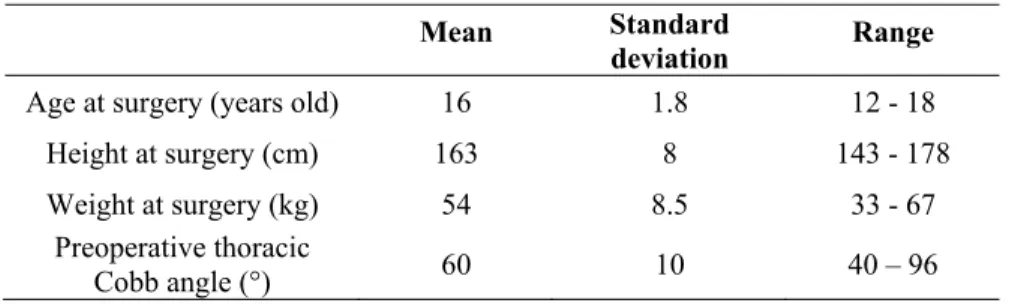
A total of 36 patients, of which 31 females and 5 males, met our inclusion criteria. Table 1 summarizes the information about the cohort.
Table 1 - Information about the cohort
Mean Standard
deviation Range
Age at surgery (years old) 16 1.8 12 - 18 Height at surgery (cm) 163 8 143 - 178 Weight at surgery (kg) 54 8.5 33 - 67
Preoperative thoracic
Cobb angle (°) 60 10 40 – 96
All patients in our cohort had the same posterior surgical approach; except one patient (P19) who had both anterior and posterior approaches. Surgeries were conducted by three different orthopedic surgeons.
4. RESULTS
Figure 2 illustrates the correction rates for BSR and XG among the cohort. It clearly shows that, in most cases, surgery achieves a very good correction of the lateral deviation of the trunk (median value of 76%) and a moderate correction of the back surface axial rotation (median value of 19%). Table 2 summarizes the descriptive statistics.
e
ccu
100 80 60 40 20 o -20 -a0 460 -ioo wn XG rl N M CL CL C C3 v1 iD I. co ai L C a a D_ aI
I'll
UM
O 'i N M a t, Cl. a a a a á 1 tD N CO Ql O 1 rl rl rl ci Ni. a a a a a
-"Hil
N N N N NIII
IF'
, oc Co
-N -N . - cL nt[
Figu Tab Figure 3 illu angles of 74° angle was the cases and su corrections o from 24° to BSR decreas Moreover, ev back of both ure 2 - Correcti ble 2- Descripti N= M St M ustrates the cas° and 68° resp e same with 18 uccessful in cl of the back surf 11°. While, fo ses from 16° to
ven though the patients after s ion rates (CR) ive statistics of =36 Mean tandard deviati Median
ses of two fem ectively). They 8° Cobb angle linicians eyes, face rotation an or patient P17, 14°, which is e correction rat surgery.
for back surfa f the correction on male adolescen y both underw after surgery. E , the changes nd the coronal , the correction insignificant. e of the Cobb ce rotation (BS n rates (CRBSR a CRBSR (%) 19 29 19 nts with scolio went the same s Even though th on the trunk trunk deviation n of the back s angle is above SR) and lateral and CRXG). CR osis (P16 and surgery. Postop he correction o surface are ra n are both sign surface rotatio e 70% in both c l shift (XG) for RXG (%) 49 58 76 P17) having s peratively, the of the spinal cu ather different nificant. The m on is almost in cases, a rib hum
r all the 36 pati
similar severit reduction of t rve was simila t. For patient maximal BSR d nexistent. The m mp still remain ients. ty (Cobb the Cobb ar in both P16 the decreases maximal ns on the
ç
1H
Back surface rotai
-20 -10 0 10 Degrees
Back surface rota
1 -20 -10 0 10 Degrees
\
T12/1_1 L5/S1 20 30 -50 - Preop - Postop on C7/11 112/L1 L5/51 -20 30 -50Coronal trunk devi
1
0 Millimeters
Coronal trunk devi
X
1
0 Millimeters alion Figu depth map of on the right s surgery for b The results o results on the spine. Trunk and reliable scoliosis on a In our previo reliability wi this new des trunk appeara Several stud questionnaire photographs Quantitative most deform deformity. T could be fla ure 3 – Clinica f the back of bo side illustrate th both patients. of this investiga e trunk shape. I k deformities ar measurement a second level ous publicatio ith respect to c criptor is used ance.dies have tried es administere of the patients approaches ha med level along
he rib hump se attened along
al examples (P1 oth patients be he functional m
ative study dem In fact, the cho re not taken in
s for assessin in clinical eval n11, we propo
changes in pat d for the first ti d to assess the ed to patients before and aft ave been propo the trunk heig everity for exa the trunk he
16 on the top ro fore and after s measurements (
5. D
monstrate how oice of the surg nto consideratio ng and quantif
luation and tre sed a novel de tient’s position ime to evaluate e cosmetic ou
and their par ter surgery16. H
osed in the liter ght. Neverthele ample might be eight resulting ow and P17 on surgery. The co (BSR and XG)
DISCUSSION
w a similar corr gical strategy is on for treatmen fying trunk de atment while it escriptor for s ning between t e objectively a utcome of scol rents15, or qua However, these rature17, 18. The ess, comparing e improved, su g in a more n the bottom ro olor map is the ) as functions oN
rection of the s s based solely nt planning. It eformities has t is at the heart scoliosis trunk two consecutiv and quantitativ liosis spinal s alitative surve e assessments a ey rely on trun g only maximu uggesting a cor extensive defow). The first tw e same in the 4 of trunk height
spinal deformit on the radiogr is believed tha
put the exter t of patients’ co deformity and ve acquisitions vely the surgica
surgery using eys for surgeo are highly subj nk surface mea um values over rrection of the formity. By c wo columns sh 4 images. The g t, before and af ty can lead to raphic evaluatio at the lack of o rnal manifesta oncerns. d we demonst s. In the presen al outcome in either self-ass ons to comple ective. asurements tak rlooks the exte deformity; ho considering th how the graphs fter different on of the objective ations of trated its nt study, terms of sessment ete using en at the nt of the owever it e whole
measurement profile, our functional measurements together with our correction rate’s definition overcome this limitation.
6. CONCLUSION
The results of this investigation demonstrate clearly that after surgery, patients are still confronted with residual trunk deformity, mainly a persisting hump on the back. That can be explained by the fact that current scoliosis assessment and treatment planning are based solely on radiographic measures of the spinal deformity. It is believed that with our novel quantitative trunk shape descriptor, clinicians and surgeons can now objectively assess trunk deformity and treatment outcome and think of possible new treatment strategies that could better address patients' concern about their appearance.
ACKOWLEDGEMENTS
The authors would like to thanks Philippe Debanné and Manivone Savann for their valuable technical help.
REFERENCES
[1] M. H. Pope, I. A. Stokes, and M. Moreland, "The biomechanics of scoliosis," Crit Rev Biomed Eng, vol. 11, pp. 157-88, 1984.
[2] M. Tones, N. Moss, and D. W. Polly, Jr., "A review of quality of life and psychosocial issues in scoliosis," Spine, vol. 31, pp. 3027-38, Dec 15 2006.
[3] J. R. Cobb, "Outline for the study of scoliosis," Am Acad Orthop Surg Instruct Lect, vol. 5, pp. 261-275, 1948.
[4] L. G. Lenke, R. R. Betz, J. Harms, K. H. Bridwell, D. H. Clements, T. G. Lowe, and K. Blanke, "Adolescent idiopathic scoliosis: a new classification to determine extent of spinal arthrodesis," J Bone Joint Surg Am, vol. 83-A, pp. 1169-81, Aug 2001.
[5] H. A. King, J. H. Moe, D. S. Bradford, and R. B. Winter, "The selection of fusion levels in thoracic idiopathic scoliosis," J Bone Joint Surg [Am], vol. 65, pp. 1302-13, 1983.
[6] W. P. Bunnell, "An objective criterion for scoliosis screening," J Bone Joint Surg Am, vol. 66, pp. 1381-7, Dec 1984.
[7] L. E. Amendt, K. L. Ause-Ellias, J. L. Eybers, C. T. Wadsworth, D. H. Nielsen, and S. L. Weinstein, "Validity and reliability testing of the Scoliometer," Phys Ther, vol. 70, pp. 108-17, Feb 1990.
[8] G. A. Murrell, R. W. Coonrad, C. T. Moorman, 3rd, and R. D. Fitch, "An assessment of the reliability of the Scoliometer," Spine, vol. 18, pp. 709-12, May 1993.
[9] P. Cote, B. G. Kreitz, J. D. Cassidy, A. K. Dzus, and J. Martel, "A study of the diagnostic accuracy and reliability of the Scoliometer and Adam's forward bend test," Spine, vol. 23, pp. 796-802; discussion 803, Apr 1 1998.
[10] M. Hawes, "Impact of spine surgery on signs and symptoms of spinal deformity," Pediatr Rehabil, vol. 9, pp. 318-39, Oct-Dec 2006.
[11] L. Seoud, J. Dansereau, H. Labelle, and F. Cheriet, "Multilevel analysis of trunk surface measurements for non-invasive assessment of scoliosis deformities," Spine, vol. 37, pp. E1045-E1053, August 2012.
[12] V. Pazos, F. Cheriet, J. Danserau, J. Ronsky, R. F. Zernicke, and H. Labelle, "Reliability of trunk shape measurements based on 3-D surface reconstructions," Eur Spine J, vol. 16, pp. 1882-91, Nov 2007.
[13] V. Pazos, F. Cheriet, L. Song, H. Labelle, and J. Dansereau, "Accuracy assessment of human trunk surface 3D reconstructions from an optical digitising system," Med Biol Eng Comput, vol. 43, pp. 11-5, Jan 2005.
[15] T. R. Haher, A. Merola, R. I. Zipnick, J. Gorup, D. Mannor, and J. Orchowski, "Meta-analysis of surgical outcome in adolescent idiopathic scoliosis. A 35-year English literature review of 11,000 patients," Spine, vol. 20, pp. 1575-84, Jul 15 1995.
[16] R. Buchanan, J. G. Birch, A. A. Morton, and R. H. Browne, "Do you see what I see? Looking at scoliosis surgical outcomes through orthopedists' eyes," Spine, vol. 28, pp. 2700-4; discussion 2705, Dec 15 2003.
[17] M. Asher, S. M. Lai, D. Burton, and B. Manna, "Maintenance of trunk deformity correction following posterior instrumentation and arthrodesis for idiopathic scoliosis," Spine, vol. 29, pp. 1782-8, Aug 15 2004.
[18] T. N. Theologis, R. J. Jefferson, A. H. Simpson, A. R. Turner-Smith, and J. C. Fairbank, "Quantifying the cosmetic defect of adolescent idiopathic scoliosis," Spine, vol. 18, pp. 909-12, Jun 1 1993.