Stimulation of Matrix Metalloproteinase-9 Expression in Human
Fibrosarcoma Cells by Synthetic Matrix Metalloproteinase Inhibitors
Erik Maquoi,* Carine Munaut,* Alain Colige,† Charles Lambert,† Francis Frankenne,* Agnès Noël,* Frank Grams,‡ Hans-Willi Krell,§ and Jean-Michel Foidart*
* Laboratoire de Biologie des Tumeurs et du Développement, † Laboratoire de Biologie des Tissus Conjonctifs, Université de Liège, Belgium; ‡Roche, Basel, Switzerland; and §Roche, Penzberg, Germany
Abstract
Enhanced expression and activation of matrix met-alloproteinase-2 (MMP-2) and MMP-9 have been associated with tumor progression, invasion, and metastasis. The use of synthetic MMP inhibitors to block the proteolytic activity of these enzymes recently emerged as a potential therapeutic tool to treat cancer. In this study, we report that GI129471, a synthetic broad-spectrum MMP inhibitor, efficiently reduced the in vitro invasiveness of HT1080 cells through type IV collagen, a major component of basement membranes. This reduced invasion was paralleled by a complete inhibition of pro-MMP-2 activation; however, GI129471 strongly increased the amount of secreted pro-MMP-9, which could be subsequently activated through a plasminogen-dependent mechanism. Quantitative RT-PCR and northern blot analysis revealed that GI129471 specifically increased the MMP-9 mRNA steady-state level. Moreover, transient transfection of HT1080 cells with β-galactosidase reporter vectors containing different lengths of the 5'-flanking region of the MMP-9 gene revealed an upregulation of the transcriptional activity of the corresponding promoter. Well-known modulators of MMP-9 expression such as 11-1β and TNF-α were not involved in this upregulation. These findings emphasize the complexity of the regulation of MMP expression and the requirement for a detailed characterization of the potential adverse side effects associated with the use of broad-Spectrum MMPIs.
Key Words: tumor; invasion; proteolysis; MMP inhibitor; plasminogen; transcriptional regulation.
INTRODUCTION
Tumor invasion and metastasis represent a complex process that depends on the cyclic repeat of three fundamental steps: (1) adhesion to the extracellular matrix (ECM) components, (2) local degradation of the ECM, and (3) migration through the degraded components [1]. Proteolysis of ECMs, and especially basement membranes, is considered a key event during this process [1, 2]. Several classes of proteinases, including aspartic-, cysteine-, and serine proteinases as well as matrix metalloproteinases (MMPs) have been implicated in tumor invasion [3]. The MMPs, a family of zinc-dependent endopeptidases, appear as essential contributors to tumor progression [4]. Due to their unusual ability to cleave and degrade several ECM components, including type IV collagen, fibronectin, and laminin as well as denatured collagens, MMP-2 and MMP-9 are thought to play critical roles during tumor invasion and metastasis [5-10].
The expression of MMP-9 is subjected to transcriptional regulation by different factors including cytokines, growth factors, ECM components, and other agents such as 12-O-tetradecanoyl-phorbol-13-acetate (TPA) [11-13]. In contrast, MMP-2 is constitutively expressed in many cell types and its transcription is not readily induced by these factors. Most MMPs are secreted as latent zymogens (pro-MMPs) requiring proteolytic processing in the N-terminal region in order to become catalytically active [14]. This proteolytic maturation is essentially mediated by serine proteinases (including plasmin and trypsin) or some activated MMPs. Unlike most MMPs, pro-MMP-2 appears resistant to these "classical" activators [15, 16] and a membrane-type MMP (MT1-MMP) has been shown to mediate pro-MMP-2 activation [16-18].
In addition to these transcriptional and posttranscriptional regulations, MMP activities are controlled by specific inhibitors known as tissue inhibitors of metalloproteinases (TIMPs). TIMPs inhibit active MMPs by forming with the enzyme tight 1:1 stoichiometric non covalent complexes [19]. Consequently, the net degradative potential of MMPs reflects the proportion of active enzymes not bound to TIMPs.
There is now a considerable body of evidence indicating that in many forms of human malignancies the regulation of MMP activities is abnormal. Several MMPs are expressed in cancers at higher levels than those found in normal tissues or benign tumors. Moreover, excessive ECM degradation observed during tumor invasion and metastasis frequently correlates with an imbalance of MMPs over TIMPs [20]. Hence, restoration of this balance by adding exogenous inhibitors was contemplated for anti-cancer therapy. Indeed, both
physiological (TIMPs) and nonphysiological MMP inhibitors (MMPIs) efficiently inhibited tumor invasion and metastasis in several in vitro and in vivo models [21-24]. However, due to their relatively large size and their short half-life in vivo, TIMPs were not considered suitable for therapeutic applications. Nonphysiological MMPIs essentially include low molecular weight synthetic inhibitors which inhibit the catalytic activity through zinc-binding chemical groups including hydroxamates, carboxylates, aminocarboxylates, sulphydryls, and phosphorus acid derivatives [23]. Among these MMPIs, hydroxamic acid based compounds were demonstrated as potent inhibitors of both tumor growth and invasion [21-23]. Several synthetic MMPIs are currently in phase II/III clinical trials in a number of solid tumors [20, 24].
This work was primarily undertaken to investigate the influence of one synthetic MMPI on the in vitro invasiveness of tumor cells. A stimulation of MMP-9 expression has been observed with some inhibitors and was documented. These results show that the use of broad-spectrum synthetic MMPIs for therapeutic purposes should be considered with caution.
MATERIALS AND METHODS
Cell culture and reagents.
HT1080 (human fibrosarcoma), CKEJ (human HPV-33 transformed cervix keratinocyte), C.IV.3, and S.I.5 (vector control and MTl-MMP-transformed A2058 human melanoma, respectively [25]), MCF7 and BT549 (two human mammary carcinomas), and human primary skin fibroblasts were maintained in Dulbecco's modified Eagle's medium (DMEM) supplemented with 10% (v/v) heat-inactivated fetal bovine serum,
penicillin-streptomycin (100 IU/ml-100 µg/ml), 2 mM glutamine, and 10 mM Hepes buffer at 37°C in a humid atmosphere (5% CO2-95% air). All culture reagents were purchased from Gibco-Life Technologies (Merelbeke, Belgium).
GI129471 ([4-(N-hydroxyamino)-2R-isobutyl-3S-(phenylthiomethyl)-succinyl]-L-phenylalanine-N-methylamide) [26, 27], BB-94 [26], and AG3340 [28] are broad-spectrum hydroxamic acid based synthetic MMPIs (all these inhibitors were kindly provided by Roche Diagnostics and synthesized according to the PCT Patent Applications WO 90/05719 [GI129471 and BB-94] and WO 95/04735 [AG3340]). Recombinant human TIMP-1 (rTIMP-1) was a gift of Prof. G. Murphy (School of Biological Sciences, University of East Anglia, Norwich, UK). Recombinant human TIMP-2 (rTIMP-2) was produced from transformed CHO cells conditioned medium as previously described [29]. Human plasminogen was purified by affinity chromatography on lysine-Sepharose [30]. In-terleukine-1β (0.05-5 ng/ml) and tumor necrosis factor-α (0.011-11 ng/ml) were purchased from Genzyme (Cambridge, MA). MAB936, a mouse anti-human MMP-9 monoclonal antibody was from R&D Systems. 4-Aminophenylmercuric acetate (APMA) was obtained from Sigma (St. Louis, MO).
Preparation of conditioned media and cell extracts.
Cell suspensions were prepared in serum-free medium and plated in 96-well plates (200 µl/well; Falcon, Becton-Dickinson, Meylan, France) as described [31]. Cells were treated with either GIl29471 (0.01-1 µM) dissolved in dimethylsulfoxyde (DMSO; 0.1%), TPA (10 ng/ml; Sigma, Bornem, Belgium), rTIMP-1 (0.01-1 µM), or rTIMP-2 (0.01-1 µM). Controls consisted of either culture medium supplemented with DMSO alone (0.1%) in the case of GI129471, or unsupplemented medium in the case of TPA and TIMPs treatments. The resultant culture supernatants were collected after different incubation times and stored at-20°C. Cell monolayers were washed once with 100 µl serum-free medium and either extracted by incubation in 50 µl of Ripa buffer (50 mM Tris-HCl, pH 7.4, 150 mM NaCl, 1% Nonidet P40, 1% Triton X-100, 1% Na-deoxycholate, 0.1% SDS; supplemented with Complete Mini protease inhibitor cocktail [Roche]) for 2 h at 4°C, or stored at -80°C before RNA extraction.
Gelatin zymography.
Analysis of gelatinolytic activities in conditioned media and cell extracts was performed by gelatin zymography as described [31]. The relative gelatinolytic activity corresponding to MMP-9 was evaluated by scanning densitometry of the gels using a GS-700 Imaging Densitometer (Bio-Rad, Hercules, CA) equipped with
cells to MMP-9 produced by control cultures. A value of 1 was arbitrarily given to the control conditions.
Isolation of RNA and Northern hybridization analysis.
Total RNA was extracted from the cell monolayers by RNAzol B treatment (Biogenesis, Bournemouth, UK). An aliquot of RNA (15 µg/lane) was electrophoresed on 1% agarose gels containing 10% formamide and transferred to nylon membranes (Hybond-N, Amersham, Gent, Belgium). Blots were probed with cDNAs labeled with 32P
using random priming synthesis (Roche Molecular Biochemicals, Mannheim, Germany). The cDNAs used as probes included human MMP-1, MMP-2, MT1-MMP, TIMP-1, and TIMP-2. The membranes were rehybridized with an oligonucleotide probe of human 28S rRNA (Clontech Laboratories, Palo Alto, CA), which served as a control. The amounts of the different transcripts were quantified by densitometric analysis of autoradiographs of the Northern blots. All results were corrected for RNA loading by densitometric data obtained for the 28S rRNA signals.
Quantitative RT-PCR for MMP-9 transcripts.
The MMP-9 mRNA and 28S rRNA steady-state levels were measured in 10-ng aliquots of total RNA by quantitative RT-PCR using a synthetic RNA (pCTR01) as external standard [32]. pCTR01 was introduced in each sample to monitor the variations in efficiency of MMP-9 and 28S RT-PCR reactions (5.103 and 4.106 copies/10 ng total RNA, respectively). RT-PCR was performed using the GeneAmp Thermostable RNA PCR Kit (Applied Biosystems, Foster City, CA) and two pairs of oligonucleotides (Eurogentec, Seraing, Belgium)
32P-labeled by T4 polynucleotide kinase (Roche Molecular Biochemicals):
GCGGAGATTGGGAAC-CAGCTGTA-3' and GACGCGCCTGTGTACACCCACA-3' for MMP-9 and
5'-GTTCACCCACTAATAGGGAACGTGA-3' and 5'-GATTCTGACTTAGAGGCGTTCAGT-3' for 28S. Reverse transcription was performed at 70°C for 15 min followed by 2 min incubation at 95°C for denaturation of RNA-DNA heteroduplexes. Amplification started by 15 s at 94°C, 20 s at 68°C, and 10 s at 72°C (29 cycles for MMP-9 and 15 cycles for 28S) and terminated by 2 min at 72°C. RT-PCR products were separated on acrylamide gels (10%) which were dried and auto-radiographed. The expected size for MMP-9 is 266 bp for the pCTR01 and 208 bp for the endogenous mRNA, and for 28S, 269 bp for the pCTR01 and 212 bp for the endogenous rRNA. Signals were quantified by scanning densitometry of the autoradiographies. The number of copies of endogenous MMP-9 mRNA and 28S rRNA were calculated by dividing the intensity of the signal for the external standard (pCTR01) by the intensity of the endogenous signal. All results were corrected for differences in RNA concentrations by dividing the number of copies of MMP-9 mRNA by the number of copies of 28S rRNA.
Quantification of cytokines and cytokine receptors by multiplex cDNA amplification.
Total RNA (1 µg) from HT1080 cells treated or not with GI129471 (1 µM) was reverse transcribed using the RETROscript first-strand synthesis kit (Ambion Inc., Austin, TX) and random primers as described by the manufacturer. Expression of GM-CSF, GM-CSFR, IL-1β, TNF-α as well as TNF-RI and TNF-RII mRNAs in HT 1080 cells treated or not with GI129471 (1 µM) was quantified using a CytoXpress-multiplex cDNA amplification kit (BioSource Europe, Nivelles, Belgium). The PCR thermocycle profile included an initial denaturating step at 96°C for 1 min, 2 cycles including a denaturating step at 96°C for 1 min, and an annealing step at 55°C for 4 min. The cycle program was continued for 33 cycles with the denaturating step at 94°C for 1 min and the annealing at 55°C for 2.5 min. The final elongation was at 72°C for 10 min. PCR products were analyzed on 2% agarose gel stained with ethidium bromide.
MMP-9 transcriptional analysis.
Two β-galactosidase vectors (pNA645 and pNA7.8) containing different lengths of the 5'flanking region of murine MMP-9 gene were constructed by ligating, respectively, 646 bp (-645 to +1 bp) and 7.8 kb (-7745 to +1 bp) of the MMP-9 promoter region [33] to the cloning site of the pNASSβ reporter vector (Clontech
Laboratories, Palo Alto, CA). Control vector (pβgalCT, Clontech Laboratories) consisted of the SV40 early promoter inserted upstream of the β-galactosidase gene and the SV40 enhancer inserted downstream. HT1080 cells in 6-well plates (2 × 10⁵ cells/well) were transiently cotransfected with the β-galactosidase reporter
constructs and the luciferase vector (pSV21uc, kindly provided by Dr. M. Grooteclaes, Université de Liège, Liège, Belgium; used to monitor transfection efficiency) by using the Lipo-fectamine reagent (GIBCO-Life Technologies). To achieve optimal transfection efficiency, 1 µg of DNA (0.8 µg of the β-galactosidase construct and 0.2 µg of the luciferase construct) and 10 µg of lipo-fectamine were used as well. Cells were washed with serum-free medium 18 h after the start of the transfection experiment. They were then incubated for 24 or 48 h in serum-free medium containing either GI129471 (1 µM) dissolved in DMSO (0.1%) or DMSO (0.1%) alone.
β-Galactosidase activity was measured in cell extracts by the chemiluminescent β-Gal Reporter Gene Assay
(Roche Molecular Bio-chemicals) and was represented as relative β-galactosidase activity after normalization by the luciferase activity. Luciferase activity was measured as previously described [34]. Triplicate cultures were used in each experiment, and three independent experiments were conducted.
Measurement of cellular metabolic activity.
The potential influence of the different MMPIs on cellular metabolism and/or proliferation was evaluated as previously described [31] using the WST-1 assay (Roche Molecular Biochemicals) according to the manufacturer's protocol.
Cytokine immunoassays.
Interleukin-1β (IL-1β) and tumor necrosis factor-α (TNF-α) levels in HT1080 conditioned media were quantified by ELISA (Medgenix, Belgium and Roche Molecular Biochemicals, respectively) according to manufacturer's instructions. The sensitivity limit of the assays were 2 and 12 pg/ml, respectively.
In vitro invasion assay.
The potential influence of GI129471 on HT1080 cell invasiveness was assayed using polycarbonate filters (8-µm pore size) of Transwell cell culture chamber inserts (Costar) as described [31]. Briefly, uncoated or type IV collagen-coated (22 µg/ filter) filters were used, respectively, for chemotaxis and chemoinvasion assays. Lower wells of the chambers contained medium supplemented with 20% fetal calf serum and 1% BSA (fraction V, ICN Biomedicals, Doornveld, Belgium) as chemoattractant. The upper wells were seeded with 6 x 10⁴ cells prepared
in serum-free medium. GI129471 (1 µM) or DMSO alone (0.1%) were added to both lower and upper wells of the chambers. Chambers were incubated for 48 h (in order to maintain a chemotactic gradient, media from both lower and upper wells were renewed after 24 h) and 4 h (for chemoinvasion and chemotaxis assays,
respectively), in a humid atmosphere at 37°C. After incubation, filters were removed from the chambers, fixed in 4% paraformaldehyde for 15 min, permeabilized for 10 min in methanol (-20°C), and stained with giemsa (4%). The upper surface of the filters was scraped with a cotton swab and cells having reached the lower surface of the filters were visually counted using a light microscope (Vanox AH3, Olympus) at 400-fold magnification. At least 20 random fields were counted per filter. Each experiment was performed in triplicate and repeated at least twice.
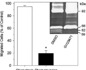
FIG. 1: Inhibition of HT1080 invasiveness by GI129471. HT1080 chemoinvasion (black bar) and chemotaxis (open bar) were evaluated in Transwell cell culture inserts coated or not with type IV collagen, respectively. HT 1080 cells were seeded in the presence of 1 µM GI129471 or vehicle alone (DMSO 0.1%: control) and the number of migrated cells was determined as described under "Materials and Methods." Results were expressed as percentages (mean ± SEM) of the migration observed in control condition. *, significant difference (Mann Whitney U-test, P = 0.000005; n = 12). Insert: in order to monitor the potential influence of GI129471 on MMPs secretion and activation during invasion, conditioned media from the invasion experiments were collected and analyzed by gelatin zymography.
Statistical analysis.
Results are expressed as the mean ± SEM of at least triplicate determinations. Statistical comparisons are based on the Mann-Whitney U test and were performed with the NCSS software. Differences were considered as statistically significant for P < 0.05.
RESULTS
Anti-invasive Activity of GI129471 on HT1080 Cells
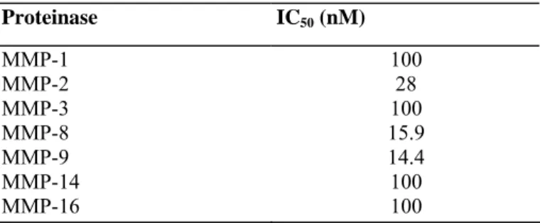
We first examined, by using Transwell chambers, the potential influence of an hydroxamic acid based broad-spectrum synthetic MMPI on HT1080 cell invasion through type IV collagen, a major component of basement membranes. In the presence of GI129471 (1 µM), HT1080 cells invasiveness was inhibited by about 80%, as compared to vehicle-treated cells (P = 0.000005, n = 12), whereas the ability of the cells to freely migrate through uncoated filters (measured by the chemotaxis assay) was not affected (Fig. 1). Similar anti-invasive properties have previously been reported with other hydroxamic acid based MMPIs [35-37] and TIMPs [38-40], as well as with anti-gelatinases anti- bodies [39, 41]. Because a reduced invasiveness could result from a cytotoxic effect of the inhibitor, cellular viability and metabolism were measured by the WST-1 assay after 48 h of treatment. The result of this assay completely ruled out this possibility (data not shown), indicating that the GI129471-dependent inhibition of HT1080 invasion was most probably mediated through a reduced proteolytic activity. Indeed, GI129471 has a broad spectrum of effects for different MMPs, with IC50 values ranging from 14
to 100 nM (Table 1). Gelatin zymography of conditioned media from the chemoinvasion assays revealed that, in control conditions, HT1080 secreted essentially two gelatinolytic enzymes of 66 and 92 kDa corresponding to the inactive pro-forms of MMP-2 and MMP-9, respectively. In addition, two activated forms of MMP-2 (62 and 59-kDa species) and a higher molecular weight gelatinase of approximately 120 kDa were also detected (Fig. 1, insert and Fig. 2A), the latter corresponding most likely to a complex between TIMP-1 and the 92-kDa pro-MMP-9 [42-44]. This high molecular weight band was recognized in Western blotting by MAB936, a
monoclonal anti-human MMP-9 antibody (data not shown). Upon treatment with GI129471, the activated forms of MMP-2 were undetectable, reflecting the inhibition of MT1-MMP activity (as already demonstrated with BB-94 [45]). Unexpectedly, the levels of both 92-kDa pro-MMP-9 and 120-kDa complex were strongly increased. However, oversecreted MMP-9 remained essentially inactive, as revealed by the presence of a very faint gelatinolytic band corresponding to the activated 84-kDa species.
TABLE 1: Pharmacologic Activities of GI129471
Proteinase IC50 (nM) MMP-1 100 MMP-2 28 MMP-3 100 MMP-8 15.9 MMP-9 14.4 MMP-14 100 MMP-16 100
MMP-9 Secretion by HT1080 Is Stimulated by GI129471
To further assess the stimulatory effect of GI129471 on MMP-9 secretion, HT1080 cells plated on plastic were treated for 6, 24, and 48 h with either GI129471 (0-1 µM) or TPA (10 ng/ml). Both conditioned media and cell extracts were analyzed by gelatin zymography (Figs. 2A and B). After a 6-h incubation period, no significant differences were seen between treated or untreated cells: they essentially produced pro-MMP-2 and pro-MMP-9 (data not shown). Later on, TPA treatment clearly stimulated the secretion of the 92-kDa pro-MMP-9. This stimulation was maximal (about 10-fold) after 24 h (data not shown) and then decreased to a fivefold stimulation at 48 h (Fig. 2A). TPA also induced the production of a 45-kDa protein with a weak gelatinolytic activity corresponding to MMP-1 [46], and increased the amount of 62 and 59-kDa active MMP-2 forms, accounting for the well-documented stimulatory effect of TPA on the MT1-MMP-dependent maturation process of pro-MMP-2 [25, 46, 47]. Similarly to TPA, treatment of the cells with GI129471 dose dependently stimulated the secretion of
pro-MMP-9 with a maximal 6.6-fold stimulation after 48 h and 1 µM GI129471 (Fig. 2 A, lanes 3-6). In
contrast, pro-MMP-2 processing was dose dependently inhibited as revealed by the progressive disappearance of both 62 and 59-kDa MMP-2 species. Analysis of cellular extracts of untreated or DMSO-treated cells revealed the presence of abundant cell-associated MMP-2 forms and only trace amount of MMP-9 (Fig. 2B). As observed in conditioned media, treatment with either TPA or GI129471 resulted in increased levels of cell-associated 92-kDa pro-MMP-9. Furthermore, an additional cell-associated gelatinolytic species of 85-92-kDa, which was recognized in Western blotting by a monoclonal anti-human MMP-9 antibody (data not shown), was also induced by both treatments (Fig. 2B). Incubation of cellular extracts of GI129471-treated HT1080 cells with an organomercurial (4-aminophenylmercuric acetate) at 37°C for 3 h failed to activate the different gelatinolytic species present in this lysate, while it activated the proforms detected in the supernatant (data not shown). The failure to activate the 85-kDa form in the lysate could probably be ascribed to the presence of inhibitors. This MMP-9 species might thus correspond to either the unglycosylated precursor of pro-MMP-9 previously identified in TPA-treated cells [48] or an active membrane-bound MMP-9 [44, 49].
FIG. 2: Modulation of MMP-9 and MMP-2 secretion and activation by HT1080 cells following treatment with TPA, GI129471, rTIMP-1, and rTIMP-2. Conditioned media (A and C) and cell lysates (B) obtained from cells cultured for 48 h with either (A and B) TPA (10 ng/ml), DMSO (D), or increasing concentrations of GI129471 (0.01-1 µM), or (C) rTIMP-1 or rTIMP-2 (0.01-1 µM) were analyzed by gelatin zymography. The relative gelatinolytic activity corresponding to MMP-9 was evaluated by scanning densitometry. Results were expressed as ratios of MMP-9 produced by TPA, rTIMP-1, rTIMP-2, or GI129471-treated cells to MMP-9 produced by control cultures (untreated or DMSO-treated cells, respectively). A value of 1 was arbitrarily given to the control conditions. Relative stimulation (fold) of MMP-9 secretion is shown.
The influence of two other broad-spectrum hydroxamic acid based MMPIs, Batimastat (BB-94: 0.01-1 µM) and AG3340 (0.01-1 µM) on MMP-9 secretion was also investigated. While both BB-94 and AG3340, at
concentrations ranging from 0.1-1 µM, completely inhibited pro-MMP-2 activation, only BB-94 increased MMP-9 secretion (data not shown), suggesting that the stimulation of MMP-9 secretion is not a general characteristic of this group of inhibitors. It should be noted that the concentrations of inhibitors tested in this study are compatible with those observed in vivo after intraperitoneal administration of BB-94 (plasma concentrations of 0.2-0.4 µM were detectable 28 days after a single administration [21]).
In order to determine if the stimulation of MMP-9 secretion observed with some of these synthetic inhibitors could also be induced by natural and more specific MMPIs, HT1080 cells were treated with 0.01-1 µM of rTIMP-1 or rTIMP-2 (Fig. 2C). In agreement with previously published data [16, 45, 50], rTIMP-1 slightly reduced the formation of the active 59-kDa MMP-2 but failed to prevent the conversion of the 66-kDa pro-MMP-2 into the 62-kDa intermediate species (Fig. 2C, lanes 1-4). In contrast, this pro-pro-MMP-2 maturation process was completely abolished by 2 (Fig. 2C, lanes 1 and 5-7). However, neither 1 nor rTIMP-2 were able to significantly modulate MMP-9 secretion, suggesting that this stimulatory process was not directly mediated by the inhibition of an active MMP.
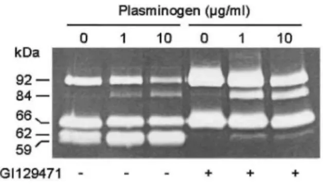
FIG. 3: Activation of secreted pro-MMP-9 by exogenously added plasminogen. HT1080 cultures treated with GI129471 (1 µM, +) or vehicle alone (-) were supplemented with increasing concentrations of plasminogen: 0, 1, and 10 µg/ml. Culture media were collected after 48 h and analyzed by gelatin zymography.
GI129471 Does Not Prevent the Plasminogen-Dependent pro-MMP-9 Processing by HT1080 Cells
In our model, most of the MMP-9 secreted by HT1080 cells was in an inactive proform. However, MMP-9 could be activated in vivo by other proteinases such as serine proteinases. In this respect, it has been previously
demonstrated that, in HT1080 cells, the conversion of plasminogen to plasmin through cell surface-bound urokinase-type plasminogen activator was able to activate endogenously secreted MMP-9 [29]. Here, we report that the addition of plasminogen to HT1080 cultures induced the conversion of 92-kDa pro-MMP-9 to activated 85-kDa form in a concentration-dependent manner (Fig. 3). This process was not affected by the treatment with GI129471. However, it should be noted that the presence of increasing concentrations of plasminogen
dramatically impaired the capacity of GI129471 to inhibit the chemoinvasion of HT1080 cells through type IV collagen-coated filters (data not shown).
MMP-9 Secretion Is Not Ubiquitously Increased by GI129471
In order to define whether the GI129471-increased MMP-9 secretion observed in HT1080 cells also occurred in other cell types, different human cells, including CKEJ (HPV-33 transformed cervix keratinocyte), S.I.5 (MT1-MMP-transfected A2058 melanoma), MCF7 and BT549 (two mammary carcinomas), and primary skin fibroblasts were treated with either TPA (10 ng/ml), GI129471 (1 µM), or vehicle (DMSO 0.1%). Analysis by gelatin zymography of 48-h conditioned media revealed that, despite a very heterogeneous basal level of MMP-9 secretion, TPA induced or increased this secretion in all cell types tested. In contrast, GI129471-mediated MMP-9 oversecretion was limited to HT 1080 and melanoma S.I.5 cells (Table 2).
TABLE 2: Modulation of MMP-9 Secretion by TPA and GI129471 in Different Human Cell Lines
Cell lines Vehicle TPA GI129471 Normal cells: Primary skin
fibroblasts 0 + + 0
Tumoral cells: HT1080
(fibrosarcoma) + + + + + +
CKEJ (cervix keratinocyte) + + + +
C.IV.3 (melanoma) 0 (+)a (+)a
S.I.5 (melanoma) (+)a + + +
MCF7 (breast carcinoma) 0 + + + 0
BT549 (breast carcinoma) 0 + + + (+)a
Note. Cells were cultured in the presence or absence (vehicle) of TPA (10 ng/ml) or GI129471 (1 µM). Conditioned media were collected after 48 h and analyzed by gelatin zymography. MMP-9 activity was evaluated visually. 0, no detectable level; + to + + + , minor to major level detected. * Extremely low level of MMP-9 detected.
GI129471 Increases MMP-9 mRNA Steady-State Level in HT 1080 Cells
The MMP-9 mRNA level in HT1080 cells treated for 6, 24, and 48 h with either TPA (10 ng/ml) or GI129471 (0-1 µM) was measured by quantitative RT-PCR. Quantification of the RT-PCR products revealed that both treatments increased the MMP-9 mRNA level (Fig. 4). Upon treatment with TPA, a rapid fivefold increase was already detected after 6 h (Fig. 4A, lanes 1-2). The stimulatory effect of GI129471 on MMP-9 mRNA level was dose dependent and maximal at the concentration of 1 µM (Fig. 4, lanes 3-6). When compared to TPA, the GI129471-mediated MMP-9 mRNA increase was delayed: the stimulation was maximal (three to four fold) after 24 h and then declined during the next 24 h (Fig. 4B).
Northern blot analysis was also used to compare the effects of TPA and GI129471 on the expression of other MMPs and TIMPs. Unlike TPA, GI129471 did not induce nor increase MMP-1, MT1-MMP, or TIMP-1 mRNA levels (data not shown). Our data are consistent with earlier reports demonstrating the upregulating activity of TPA on MMP-1, MMP-3, MMP-9, MT1-MMP, and TIMP-1 expression in different cell types [46, 47, 51].
GI129471 Stimulates the Transcriptional Activity of the MMP-9 Promoter in HT1080 Cells
To assess whether the GI129471-induced increase of MMP-9 mRNA level was a consequence of a transcriptional upregulation of the corresponding gene, we transiently transfected HT1080 cells with either pNA645 or pNA7.8 reporter vectors (Fig. 5). These constructs were made up of the 5'-flanking region of murine MMP-9 gene (-645 to +1 bp or -7745 to +1 bp, respectively) linked to the β-galactosidase reporter gene. pNA645 contained the cis-elements (two AP-1 as well as one Sp-1 and one NF-κB binding sites) essential for basal as well as TPA- and TNF-α-induced promoter activity [12]. In addition to these essential sequences, pNA7.8 also contained an additional AP-1 site as well as other yet unidentified upstream sequences.
Preliminary experiments have demonstrated the stimulatory effect of TPA on the transcriptional activity of these two constructs (data not shown). Transfected cells were treated for 24 and 48 h with GI129471 (1 µM) or vehicle alone, and the β-galactosidase activity was measured in cell extracts (Fig. 5). In the presence of GI129471,
β-galactosidase activity was slightly increased after 24 h with both vectors (1.4-fold and 2.8-fold for pNA645
and pNA7.8, respectively). After 48 h, the stimulatory effect of GI129471 was even more obvious with
β-galactosidase activities stimulated by about fourfold in both transfectants, thus confirming the delayed
stimulation already observed at both mRNA and protein levels. Analysis by gelatin zymography of the
corresponding conditioned media confirmed the increased MMP-9 secretion following GI129471 treatment (data not shown). These data clearly demonstrated that the GI129471-stimulated MMP-9 synthesis was, at least in part, mediated through an enhanced transcription of the MMP-9 gene.
FIG. 4: TPA and GIl29471 stimulated MMP-9 expression in HT1080 cells. HT1080 cells were treated or not (medium, Med.) for up to 48 h with TPA (10 ng/ml), DMSO (0.1%, D.), or increasing concentrations of GI129471 (0.01-1 µM). (A) MMP-9 mRNA levels were quantified by quantitative RT-PCR using a pCTR01 as external control RNA template. Arrows, external control; arrow heads, MMP-9 transcript. (B) Quantification of MMP-9 transcripts by densitometric analysis of autoradiographies of the gels. All results were corrected by densitometric data obtained for the 28S rRNA. A value of 1 was arbitrarily attributed to the relative level of MMP-9 mRNA measured in control conditions (medium and DMSO).
FIG. 5: Modulation of the transcriptional activity of MMP-9 promoter by GI129471. Left-side panel represents a map of the β-galactosidase reporter vectors with different lengths of the 5'-flanking region of murine MMP-9 gene (pNA645: -645 to +1 bp or pNA7.8: - 7745 to +1 bp). The arrow indicates the position of the transcription initiation site and the position of some consensus sequences including the TATA box; AP-1, NF-κB, and Sp-1 binding sites are also noted. Unsequenced regions are noted by a "?". These constructs or a control vector (pβgalCT) were transiently transfected into HT1080 cells as described under "Methods." Right-side panels: β-galactosidase activity was measured in cell extracts after 24 and 48 h of treatment with either GI129471 (1 µM: solid bars) or vehicle (DMSO: open bars). The calculated promoter activity of both pNA645 and pNA7.8 constructs was expressed as percentage ± SD of the activity of pβgalCT.
The GI129471 -Induced MMP-9 Overexpression in HT1080 Cells Is Not Mediated by IL-1β or TNF-α
The delayed stimulation of MMP-9 secretion triggered by GI129471 suggests that this inhibitor does not directly modulate the transcriptional activity of MMP-9 promoter but rather acts through an indirect mechanism. In this respect, several molecules, including cytokines such as IL-1β, TNF-α, epidermal growth factor, granulocyte macrophage-colony stimulating factor (GM-CSF), transforming growth factor-β, TNF-β, interferon-γ, IL-4, and IL-6 [52-54], as well as ECM components such as fibronectin, collagens, laminin, and tenascin [13, 55-57] have been reported as potent modulators of MMP-9 expression in different cell types. This raised the possibility that the effect of GI129471 on MMP-9 secretion might be mediated through modulations of the activities of these ECM components, cytokines, and/or their respective receptors. In order to detect a potential GI129471-dependent modulation of the expression profile of some of these cytokines and/or their receptors in HT1080 cells, we used a multiplex cDNA amplification assay to monitor the mRNA levels of GM-CSF, its receptor (GM-CSFR), IL-1β, IL-6, and its receptor (IL-6R) as well as TNF-α and its two receptors (type I and type II receptors, TNF-RI and TNF-RII, respectively). Our data reveal that the levels of TNF-α, CSF, and GM-CSFR mRNAs are extremely low and are not affected by the presence of GI129471 (Fig. 6). In accordance with this mRNA analysis, no immunoreactive TNF-α is detected in the corresponding conditioned media by ELISA measurement (data not shown). Altogether, these data suggest that neither TNF-α nor GM-CSF are likely to direct mediators of MMP-9 overexpression. In contrast, the mRNAs corresponding to IL-1β, IL-6, and its receptor as well as the two TNF-Rs are clearly detected but their relative abundance is not modulated upon treatment with GI129471 (Fig. 6), excluding a potential influence of this inhibitor on the steady-state mRNA levels of these genes. Despite this absence of transcriptional modulation of IL-1β, GI129471 induces a twofold increase of secreted IL-1β concentration as detected by ELISA (Fig. 7). A similar stimulation of IL-1β secretion by synthetic MMPIs has been previously documented by others [27, 58, 59] and results most probably from the inhibition of an MMP-mediated IL-1β degradation [60]. In order to assess whether this elevated IL-1β
concentration could mediate MMP-9 overexpression, HT1080 cells, treated or not with GI129471 (1 µM), were supplemented with increasing concentrations of recombinant human IL-1β (0-5 ng/ml) and 48-h conditioned media were analyzed by gelatin zymography. In accordance with previously published data [47, 61] our results do not reveal any modulation of MMP-9 secretion upon IL-1β treatment (data not shown), thus ruling out a potential involvement of this cytokine in this process. Further analysis will be required to better define the mechanism(s) that account for this transcriptional upregulation.
FIG. 6: Influence of GI129471 on the expression of GM-CSF, GM-CSFR, IL-lβ, IL-6, IL-6R, TNF-α, TNF-RI, and TNF-RII. HT1080 cells were treated with 1 µM GI129471 (lanes 1 and 3) or vehicle (lanes 2 and 4) for 24 h (lanes 1 and 2) and 48 h (lanes 3 and 4) and the different mRNA levels were measured by a multiplex cDNA amplification assay. Lane 5, control DNA.
FIG. 7: GI129471 increases IL-10 concentration in HT1080 cells. HT1080 cells were incubated in the presence of GI129471 (1 µM; closed circles) or vehicle (open circles). Conditioned media were harvested after 24 and 48 h and IL-1β levels determined by ELIS A.
DISCUSSION
Our findings clearly demonstrate that GI129471, a synthetic peptidomimetic broad-spectrum MMPI, could, in addition to its classical inhibitory activities, specifically upregulate both transcription and synthesis of pro-MMP-9. The activation of MMP-9 has been demonstrated to be essentially mediated by plasmin [29, 49]. In this study, we used HT1080 cells, which synthesize urokinase-type plasminogen activator (uPA) and its cell surface receptor (uPAR) but not plasminogen [29, 49]. As a consequence of the absence of plasminogen, HT1080 cells were unable to efficiently activate pro-MMP-9 under basal conditions (unsupplemented serum-free culture medium). During the chemoinvasion assay, HT1080 cells degrade and migrate through a layer of type IV collagen. Activated MMP-2 and -9 are well-known mediators of this type IV collagen degradation. However, we have previously demonstrated that, in the absence of exogenous plasminogen, the chemoinvasion of HT1080 cells relies essentially on activated MMP-2. Indeed, chemoinvasion was strongly reduced when the HT1080 cells were treated with a furin inhibitor, which decreased MMP-2 maturation but altered neither MMP-9 secretion, nor activation [31]. In contrast, when the cells were treated with increasing concentrations of plasminogen, cell surface-generated plasmin efficiently converted the 92-kDa pro-MMP-9 to a 84-kDa active species, resulting in a dose-dependent stimulation of chemoinvasion. These different observations are in good agreement with earlier studies [49] demonstrating that (1) type IV collagen degradation by HT1080 cells was MMP dependent (plasmin failed to directly degrade type IV collagen), and (2) that the addition of plasminogen greatly enhanced this degradation as a consequence of an increased activation of pro-MMP-9.
When the chemoinvasion assay was performed in the presence of increasing concentrations of GI129471, a dose-dependent stimulation of pro-MMP-9 secretion was observed. However, invasion was not promoted because (1) pro-MMP-9 processing was not enhanced (plasminogen was absent), and (2) the proteolytic activities of both the mature MMP-2 and the low level of mature MMP-9 were blocked by the MMPI, thus precluding type IV collagen degradation. In contrast, when both GI129471 and plasminogen were present during the invasion assay, the overexpressed pro-MMP-9 was efficiently activated and the inhibitory effect of GI129471 on chemoinvasion was reduced, most probably as a consequence of an altered balance between the concentrations of active MMPs and molecules of inhibitor. The increased MMP-9 expression is not a systematic consequence of the treatment with MMPIs because other synthetic (such as AG3340) as well as natural (such as TIMP-1 and TIMP-2) inhibitors are unable to elicit a similar response. It is worth noting that a similar stimulation of MMP-9 expression has also been observed in vivo in mice treated with galardin and BB-94, two other broad-spectrum hydroxamic acid based MMPIs [62, 63]. These divergent abilities for these different inhibitors to trigger MMP-9 expression might arise from either their distinct natures (i.e., peptidomimetic, such as GI129471, BB-94, and galardin, vs nonpeptidomimetic molecules such as AG3340) and/or their selectivity. Moreover, the MMPI-mediated upregulation of MMP-9 clearly differs from the well-characterized induction observed in the presence of TPA in that: (1) it requires a longer incubation period and (2) it is cell type specific, suggesting the
involvement of distinct mediators and/or signaling pathways. Among the numerous factors known to regulate MMP-9 expression, we selected IL-1β and TNF-α for further investigation because their bioactivities have previously been reported to be modulated by MMPs or MMP-like proteinases [60, 64]. However, our data do not reveal any implication for these two cytokines in MMP-9 upregulation.
Treatment with broad-spectrum MMPIs such as GI129471 can deeply alter the cellular environment. Indeed, a growing body of evidence suggests that the unspecific inhibition of MMPs and MMP-like proteinases (such as the adamalysins) might, among other things, lead to the accumulation of ECM components, the prevention of the exposure of cryptic protein domains, and the reduced shedding of several cell surface protein ectodomains such
as membrane-anchored growth factors, growth factor receptors, cell adhesion molecules, and ectoenzymes as well as proteins of unknown functions [4, 65, 66]. Because the expression of most MMPs is tightly controlled through complex signaling cascades initiated at the plasma membrane by an array of receptors (including integrins as well as cytokines and growth factor receptors) which sense the cellular environment, MMPI-induced alterations of this pericellular environment might therefore promote the accumulation of inducer(s) and/or prevent the generation of inhibitors that will affect downstream signaling cascades, ultimately resulting in modifications of the initial gene expression profile. Such a mechanism is illustrated by the type I collagen-induced activation of tyrosine kinases of the discoidin domain receptor which ultimately trigger the synthesis of MMP-1. However, the MMP-1 -mediated cleavage of collagen abolishes the receptor activation, creating a specific negative-feedback regulatory loop [67, 68]. In the presence of broad-spectrum MMPIs, the impaired collagen degradation might prevent this negative regulation, resulting in an uncontrolled stimulation of MMP-1 expression.
Our results should be paralleled with the recently halted clinical development of some synthetic MMPIs due to the occurrence of severe side effects, including inflammatory polyarthritis, in cancer patients treated with such molecules [24, 69]. In this context, it is worth noting that elevated MMP-9 levels were consistently reported in arthritic patients [70]. Moreover, MMP-9 was suggested to contribute to the progression of the disease [71], supporting the concept that the higher MMP-9 expression observed in the presence of some synthetic MMP inhibitors could play a causal role in the appearance of these clinical side-effects. Furthermore, recent data revealed that the treatment of tumor-bearing mice with BB-94 (a MMPI which upregulates MMP-9 expression in our in vitro model) inhibited the growth of inoculated primary tumors. However, it also promoted a liver-specific overexpression of MMP-9 as well as the development of liver metastasis [63, 72]. These observations should be related to previous reports demonstrating the pivotal role of MMP-9 in the formation of distant metastasis. Indeed, Kim and coworkers [9] reported that a rate-limiting step for metastasis was the breaching of the vascular wall (which contains large amounts of type IV collagen). Interestingly, they elegantly proved that a cooperation between uPA/uPAR system (which mediates the conversion of plasminogen into plasmin) and MMP-9 was required to complete this step. In addition, Hua and Muschel [73] showed that while the inhibition of MMP-9 in tumor cells did not affect their tumorigenicity, a strong reduction of the number of distant metastasis was observed. Altogether, these different reports support the concept that in vivo (i.e., in situations where endogenous plasminogen is present) MMP-9 is required for the formation of metastasis. It is worth noting that the elevated MMP-9 expression induced by some MMPIs was not restricted to tumor cells. Indeed, higher MMP-9 levels were also detected in livers from BB-94-treated mice that were not inoculated with tumor cells [63] as well as in cultured mammary epithelial cells [74]. Collectively, these observations reveal that an enhanced production of MMP-9 by tumor cells and/or the surrounding stromal cells might promote a metastatic dissemination. Altogether these different data emphasize the complexity of the regulation of MMP expression and the requirement for a detailed characterization of the potential adverse side effects associated with the in vivo administration of such broad-spectrum MMPIs.
Acknowledgments
We thank J.P. Cherami-Bien and L. Volders for their technical assistance and Prof. G. Murphy (School of Biological Sciences, University of East Anglia, Norwich, UK) for providing rTIMP-1. This work was supported by grants from the Communauté Française de Belgique (Actions de Recherche Concertées 93/98-171 and 95/00-191), the CGER-Assurances 1996/1999, the Association contre le Cancer, the Association Sportive contre le Cancer, the Loterie Nationale (9.4573.96F), the Fonds de la Recherche Scientifique Médicale (FRSM)
(3.4573.95 et 3.4595.98), the Fonds d'Investissement de Recherche Scientifique 1997-CHU Liège, Belgium, the Centre Anticancéreux près de l'Université de Liège, and the Fondation Léon Frédéricq, University of Liège, Liège (all in Belgium) and the General RE-Luxembourg, the Commission of European Communities (Concerted European Action Biotech BIO4-CT96-0464), and the Industry (Boehringer Mannheim GmbH, Germany). REFERENCES
1. Liotta, L. A., Steeg, P., and Stetler-Stevenson, W. G. (1991). Cancer metastasis and angiogenesis: An imbalance of positive and negative regulation. Cell 64, 327-336.
2. Mignatti, P., and Rifkin, D. B. (1993). Biology and biochemistry of proteinases in tumor invasion. Physiol. Rev. 73, 161-195. 3. Duffy, M. J. (1992). The role of proteolytic enzymes in cancer invasion and metastasis. Clin. Exp. Metastasis 10, 145-155.
4. McCawley, L. J., and Matrisian, L. M. (2000). Matrix metallo-proteinases: Multifunctional contributors to tumor progression. Mol. Med. Today 6, 149-156.
5. Brown, P. D., Bloxidge, R. E., Stuart, N. S. A., Gatter, K. C, and Carmichael, J. (1993). Association between expression of activated 72-kilodalton gelatinase and tumor spread in non-small-cell lung carcinoma. J. Natl. Cancer Inst. 85, 574-578.
6. Stetler-Stevenson, W. G., Liotta, L. A., and Kleiner, D. E. (1993). Extracellular matrix 6: Role of matrix metalloproteinases in tumor invasion and metastasis. FASEB J. 7, 1434-1441.
7. Zucker, S., Lysik, R. M., Zarrabi, M. H., and Moll, U. (1993). M(r) 92,000 type IV collagenase is increased in plasma of patients with colon cancer and breast cancer. Cancer Res. 53, 140-146.
8. Yu, A. E., Hewitt, R. E., Kleiner, D. E., and Stetler-Stevenson, W. G. (1996). Molecular regulation of cellular invasion-role of gelatinase A and TIMP-2. Biochem. Cell. Biol. 74, 823-831.
9. Kim, J., Yu, W., Kovalski, K., and Ossowski, L. (1998). Requirement for specific proteases in cancer cell intravasation as revealed by a novel semiquantitative PCR-based assay. Cell 94, 353-362.
10. Itoh, T., Tanioka, M., Yoshida, H., Yoshioka, T., Nishimoto, H., and Itohara, S. (1998). Reduced angiogenesis and tumor progression in gelatinase A-deficient mice. Cancer Res. 58, 1048-1051.
11. Okada, Y., Tsuchiya, H., Shimizu, H., Tomiya, K., Nakanishi, I., Sato, H., Seiki, M., Yamashita, K., and Hayakawa, T. (1990). Induction and stimulation of 92-kDa gelatinase/type IV collagenase production in osteosarcoma and fibrosarcoma cell lines by tumor necrosis factor alpha. Biochem. Biophys. Res. Commun. 171, 610-617.
12. Sato, H., and Seiki, M. (1993). Regulation mechanism of 92-kDa type IV collagenase gene expression which is associated with invasiveness of tumor cells. Oncogene 8, 395-405.
13. Khan, K. M. F., and Falcone, D. J. (1997). Role of laminin in matrix induction of macrophage urokinase-type plasminogen activator and 92-kDa MMP expression. J. Biol. Chem. 272, 8270-8275.
14. Nagase, H., and Woessner, J. F. (1999). Matrix metalloprotein-ases. J. Biol. Chem. 274, 21491-21494.
15. Okada, Y., Morodomi, T., Enghild, J. J., Suzuki, K., Yasui, A., Nakanishi, I., Salvesen, G., and Nagase, H. (1990). Matrix
metalloproteinase 2 from human rheumatoid synovial fibroblasts. Purification and activation of the precursor and enzymatic properties. Eur. J. Biochem. 194, 721-730.
16. Strongin, A. Y., Marmer, B. L., Grant, G. A., and Goldberg, G. I. (1993). Plasma membrane-dependent activation of the 72-kDa type IV collagenase is prevented by complex formation with TIMP-2. J. Biol. Chem. 268, 14033-14039.
17. Sato, H., Takino, T., Okada, Y., Cao, J., Shinagawa, A., Yamamoto, E., and Seiki, M. (1994). A matrix metalloproteinase expressed on the surface of invasive tumour cells. Nature 370, 61-65.
18. Holmbeck, K. et al. (1999). MT1-MMP-deficient mice develop dwarfism, osteopenia, arthritis, and connective tissue disease due to inadequate collagen turnover. Cell 99, 81-92.
19. Brew, K., Dinakarpandian, D., and Nagase, H. (2000). Tissue inhibitors of metalloproteinases: Evolution, structure and function. Biochem. Biophys. Acta 1477, 267-283.
20. Brown, P. D. (1999). Clinical studies with matrix metalloproteinase inhibitors. A.P.M.I.S. 107, 174-180.
21. Brown, P. D., and Giavazzi, R. (1995). Matrix metalloproteinase inhibition: A review of anti-tumor activity. Ann. Oncol. 6, 967-974. 22. Talbot, D. C., and Brown, P. D. (1996). Experimental and clinical studies on the use of matrix metalloproteinase inhibitors for the treatment of cancer. Eur. J. Cancer 14, 2528-2533.
23. Wojtowicz-Praga, S. M., Dickson, R. B., and Hawkins, M. J. (1997). Matrix metalloproteinase inhibitors. Invest. New Drugs 15, 61-75. 24. Hidalgo, M., and Eckhardt, S. G. (2001). Development of matrix metalloproteinase inhibitors in cancer therapy. J. Natl. Cancer Inst. 93, 178-193.
25. Maquoi, E., Frankenne, F., Baramova, E., Munaut, C., Sounni, N. E., Remade, A., Noël, A., Murphy, G., and Foidart, J.-M. (2000). Membrane type 1 matrix metalloproteinase-associated degradation of tissue inhibitor of tissue inhibitor of metalloproteinase 2 in human tumor cell lines. J. Biol. Chem. 275, 11368-11378.
27. McGeehan, G. M. Becherer, J. D., Bast, R. C, Boyer, C. M., Champion, B., Connolly, K. M., Conway, J. G., Furdon, P., Karp, S., Kidao, S., McElroy, A. B., Nichols, J., Pryzwansky, K. M., Schoenen, F., Sekut, L., Truesdale, A., Verghese, M., Warner, J., and Ways, J. P. (1994). Regulation of tumour necrosis factor-α processing by a metalloproteinase inhibitor. Nature 370, 558-561.
28. Castelhano, A. L., Home, S., Shengyan, L., Castelhano, L. A., Krantz, A., Liak, J. T., Yuang, Z., and Liak, T. J. (1995). PCT Patent Application, WO 95/04735.
29. Baramova, E. N., Bajou, K., Remacle, A., L'Hoir, C, Krell, H. W., Weidle, U. H., Noël A., and Foidart, J. M. (1997). Involvement of PA/plasmin system in the processing of pro-MMP-9 and in the second step of pro-MMP-2 activation. FEBS Lett. 405, 157-162.
30. Chibber, B. A. K., Deutsch, D. G., and Mertz, E. T. (1974). Affinity chromatography of plasminogen. Methods Enzymol. 34, 424-432. 31. Maquoi, E., Noël, A., Frankenne, F., Angliker, H., Murphy, G., and Foidart, J. M. (1998). Inhibition of matrix metalloproteinase 2 maturation and HT1080 invasiveness by a synthetic furin inhibitor. FEBS Lett. 424, 262-266.
32. Lambert, C. A., Colige, A. C, Munaut, C, Lapière, C. M., and Nusgens, B. V. (2001). Distinct pathways in the over-expression of matrix metalloproteinases in human fibroblasts by relaxation of mechanical tension. Matrix Biol. 20, 397-408.
33. Munaut, C, Salonurmi, T., Kontusaari, S., Reponen, P., Morita, T., Foidart, J. M., and Tryggvason, K. (1999). Murine matrix metalloproteinase 9 gene: 5' upstream region contains cis-act-ing elements for expression in osteoclasts and migrating kera-tinocytes in transgenic mice. J. Biol. Chem. 274, 5588-5596.
34. de Wet, W. J. (1987). R-loop analysis of procollagen messenger RNA for the assessement of human collagen mutations. Methods Enzymol. 145, 235-252.
35. Reich, R., Thompson, E. W., Iwamoto, Y., Martin, G. R., Deason, J. R., Fuller, G. C, and Miskin, R. (1988). Effects of inhibitors of plasminogen activator, serine proteinases, and collagenase IV on the invasion of basement membranes by metastatic cells. Cancer Res. 48, 3307-3312.
36. Thompson, E. W., Nakamura, S., Shima, T. B., Melchiori, A., Martin, G. R., Salahuddin, S. Z., Gallo, R. C, and Albini, A. (1991). Supernatants of acquired immunodeficiency syndrome-related kaposi's sarcoma cells induce endothelial cell chemo-taxis and invasiveness. Cancer Res. 51, 2670-2676.
37. Taraboletti, C, Carofalo, A., Belotti, D., Drudis, T., Borsotti, P., Scanziani, E., Brown, P. D., and Giavazzi, R. (1995). Inhibition of angiogenesis and murine hemangioma growth by batimastat, a synthetic inhibitor of matrix metalloproteinases. J. Natl. Cancer Inst. 87, 293-298.
38. Khokha, R., Waterhouse, P., Yagel, S., Lala, P., Overall, C. M., Norton, G., and Denhardt, D. T. (1989). Antisense RNA-induced reduction in murine TIMP levels confers oncogenicity on Swiss 3T3 cells. Science 243, 947-950.
39. Albini, A., Melchiori, A., Santi, L., Liotta, L. A., Brown, P. D., and Stetler-Stevenson, W. G. (1991). Tumor cell invasion inhibited by TIMP-2. J. Natl. Cancer Inst. 83, 775-779.
40. DeClerck, Y. A., Yean, T.D., Chan, D., Shimada, H., and Langley, K. E. (1991). Inhibition of tumor invasion of smooth muscle cell layers by recombinant human metalloproteinase inhibitor. Cancer Res. 51, 2151-2157.
41. Stearns, T. M. E., Wang, M., and Stearns, M. (1995). IL-10 blocks collagen IV invasion by "invasion stimulating factor" activated PC-3 ML cells: Upregulation of TIMP-1 expression. Oncol. Res. 7, 157-16PC-3.
42. Hibbs, M. S., Hasty, K. A., Seyer, J. M., Kang, A. H., and Mainardi, C. L. (1985). Biochemical and immunological characterization of the secreted forms of human neutrophil gelatinase. J. Biol. Chem. 260, 2493-2500.
43. Moll, U. M., Youngleib, G. L., Rosinski, K. B., and Quigley, J. P. (1990). Tumor promoter-stimulated Mr 92,000 gelatinase secreted by normal and malignant human cells: Isolation and characterization of the enzyme from HT1080 tumor cells. Cancer Res. 50, 6162-6170. 44. Ginestra, A., Monea, S., Seghezzi, G., Dolo, V., Nagase, H., Mignatti, P., and Vittorelli, L. (1997). Urokinase plasminogen activator and gelatinases are associated with membrane vesicles shed by human HT1080 fibrosarcoma cells. J. Biol. Chem. 272, 17216-17222. 45. Kinoshita, T., Sato, H., Takino, T., Itoh, M., Akizawa, T., and Seiki, M. (1996). Processing of a precursor of 72-kilodalton type IV collagenase/gelatinase A by a recombinant membrane-type 1 matrix metalloproteinase. Cancer Res. 56, 2535-2538.
46. Lim, Y.T., Sugiura, Y., Laug, W. E., Sun, B., Garcia, A., and DeClerck, Y. A. (1996). Independent regulation of matrix met-alloproteinases and plasminogen activators in human fibrosarcoma cells. J. Cell. Physiol. 167, 333-340.
47. Lohi, J., Lehti, K., Westermarck, J., Kähäri, V.M., and Keski-Oja, J. (1996). Regulation of membrane-type matrix metalloproteinase-1 expression by growth factors and phorbol 12-myris-tate 13-acetate. Eur. J. Biochem. 239, 239-247.
48. Toth, M., Gervasi, D. C, and Fridman, R. (1997). Phorbol ester-induced cell surface association of matrix metalloproteinase-9 in human MCF10A breast epithelial cells. Cancer Res. 57, 3159-3167.
49. Mazzieri, R., Masiero, L., Zanetta, L., Monea, S., Onisto, M., Garbisa, S., and Mignatti, P. (1997). Control of type IV collage-nase activity by components of the urokinase-plasmin system: A regulatory mechanism with cell-bound reactants. EMBO J. 16, 2319-2332. 50. Atkinson, S. J., Crabbe, T., Cowell, S., Ward, R. V., Butler, M. J., Sato, H., Seiki, M., Reynolds, J. J., and Murphy, G. (1995). Intermolecular autolytic cleavage can contribute to the activation of progelatinase A by cell membranes. J. Biol. Chem. 270, 30479-30485. 51. Mackay, A. R., Ballin, M., Pelina, M. D., Farina, A. R., Nason, A. M., Hartzler, J. L., and Thorgeirsson, U. P. (1992). Effect of phorbol ester and cytokines on matrix metalloproteinase and tissue inhibitor of metalloproteinase expression in tumor and normal cell lines. Invasion Metast. 12, 168-184.
52. Ries, C, and Petrides, P. E. (1995). Cytokine regulation of matrix metalloproteinase activity and its regulatory dysfunction in disease. Biol. Chem. 376, 345-355.
53. Meisser, A., Chardonnens, D., Campana, A., and Bischof, P. (1999). Effects of tumour necrosis factor-alpha, interleukin-1 alpha, macrophage colony stimulating factor and transforming growth factor beta on trophoblastic matrix metalloproteinases. Mol. Hum. Reprod. 5, 252-260.
54. Pei, X. H., Nakanishi, Y., Takayama, K., Bai, F., and Hara, N. (1999). Granulocyte, granulocyte-macrophage, and macrophage colony-stimulating factors can stimulate the invasive capacity of human lung cancer cells. Br. J. Cancer 79, 40-46.
55. Werb, Z., Tremble, P. M., Behrendtsen, O., Crowley, E., and Damsky, C. H. (1989). Signal transduction through the fi-bronectin receptor induces collagenase and stromelysin gene expression. J. Cell Biol. 109, 877-889.
56. Sarret, Y., Woodley, D. T., Goldberg, G. S., Kronberger, A., and Wynn, K. C. (1992). Constitutive synthesis of a 92-kDa kerati-nocyte-derived type IV collagenase is enhanced by type I collagen and decreased by type IV collagen matrices. J. Invest. Dermat. 99, 836-841.
57. Corcoran, M. L., Kibbey, M. C, Kleinman, H. K., and Wahl, L. M. (1995). Laminin SIKVAV peptide induction of monocyte/ macrophage prostaglandin E2 and matrix metalloproteinases. J. Biol. Chem. 270, 10365-10368.
58. Williams, L. M., Gibbons, D. L., Gearing, A., Maini, R. N., Feldmann, M., and Brennan, F. M. (1996). Paradoxical effects of a synthetic metalloproteinase inhibitor that blocks both p55 and p75 TNF receptor shedding and TNFα processing in RA synovial membrane cell cultures. J. Clin. Invest. 97, 2833-2841.
59. Gallea-Robache, S., Morand, V., Millet, S., Bruneau, J. M., Bhatnagar, N., Chouaib, S., and Roman-Roman, S. (1997). A
metalloproteinase inhibitor blocks the shedding of soluble cytokine receptors and processing of transmembrane cytokine precursors in human monocytic cells. Cytokine 9, 340-346.
60. Ito, A., Mukaiyama, A., Itoh, Y., Nagase, H., Thogersen, I. B., Enghild, J. J., Sasaguri, Y., and Mori, Y. (1996). Degradation of interleukin 1/3 by matrix metalloproteinases. J. Biol. Chem. 271, 14657-14660.
61. Lauricella-Lefebvre, M. A., Castronovo, V., Sato, H., Seiki, H., French, D. L., and Merville, M. P. (1993). Stimulation of the 92-kD type IV collagenase promoter and enzyme expression in human melanoma cells. Inv. Metast. 13, 289-300.
62. Lund, L. R., Romer, J., Bugge, T. H., Nielsen, B. S., Frandsen, T. L., Degen, J. L., Stephens, R. W., and Dano, K. (1999). Functional overlap between two classes of matrix-degrading proteases in wound healing. EMBO J. 18, 4645-4656.
63. Kriiger, A., Soeltl, R., Sopov, I., Kopitz, C, Arlt, M., Magdolen, V., Harbeck, N., Gänsbacher, B., and Schmitt, M. (2001). Hy-droxamate-type matrix metalloproteinase inhibitor batimastat promotes liver metastasis. Cancer Res. 61, 1272-1275.
64. Gearing, A. J. H. et al. (1994). Processing of tumour necrosis factor-α precursor by metalloproteinases. Nature 370, 555-557. 65. Hooper, N. M., Karran, E. H., and Turner, A. J. (1997). Membrane protein secretases. Biochem. J. 321, 265-279.
66. Werb, Z. (1997). ECM and cell surface proteolysis: Regulating cellular ecology. Cell 91, 439-442.
67. Shrivastava, A. et al. (1997). An orphan receptor tyrosine kinase family whose members serve as nonintegrin collagen receptors. Mol. Cell 1, 25-34.
68. Vogel, W., Gish, G. D., Alves, F., and Pawson, T. (1997). The discoidin domain receptor tyrosine kinases are activated by collagen. Mol. Cell 1, 13-23.
69. Drummond, A. H. et al. (1999). Preclinical and clinical studies of MMP inhibitors in cancer. Ann. N. Y. Acad. Sci. 878, 228-235. 70. Gruber, B. L., Sorbi, D., French, D. L., Marchese, M. J., Nuovo, G. J., Kew, R. R., and Arbeit, L. A. (1996). Markedly elevated serum MMP-9 (gelatinase B) levels in rheumatoid arthritis: A potentially useful laboratory marker. Clin. Immunol. Immuno-pathol. 78, 161-171.
71. Koolwijk, P., Miltenburg, A. M., van Erck, M. G., Oudshoorn, M., Niedbala, M. J., Breedveld, F. C, and van Hinsbergh, V. W. (1995). Activated gelatinase B (MMP-9) and urokinase-type plasminogen activator in synovial fluids of patients with arthritis. Correlation with clinical and experimental variables of inflammation. J. Rheumatol. 22, 385-393.
72. Delia Porta, P., Soeltl, R., Krell, H. W., Collins, K., O'Donoghue, M., Schmitt, M., and Kruger, A. (1999). Combined treatment with serine protease inhibitor aprotinin and matrix metalloproteinase inhibitor Batimastat (BB-94) does not prevent invasion of human esophageal and ovarian carcinoma cells in vivo. Anticancer Res. 19, 3809-3816.
73. Hua, J., and Muschel, R. J. (1996). Inhibition of matrix metal-loproteinase 9 expression by a ribozyme blocks metastasis in a rat sarcoma model system. Cancer Res. 56, 5279-5284.
74. Lee P. P., Hwang J. J., Mead L., and Ip M. M. (2001). Functional role of matrix metalloproteinases (MMPs) in mammary epithelial cell development. J. Cell. Physiol. 188, 75-88.