1
THE ROLE OF SLEEP IN THE CONSOLIDATION OF
REWARDED AND RELATIONAL MEMORY
Giulia Gaggioni Master in Neuroscience
Faculty of Science University of Geneva
September 2012
Supervisors: Prof. Sophie Schwartz and Dr. Kinga Igloi Jury:
Prof. Sophie Schwartz Dr. Christina Schmidt Dr. med Stephen Perrig
2
Acknowledgements
I would like to thank three wonder-women that I had the pleasure to meet during my Master in Geneva.
To Professor Sophie Schwartz, thank you for appointing me with a place in your lab, for sharing precious knowledge, and for supervising me always.
To Dr. Kinga Igloi, thank you for teaching, guiding, helping, and supporting me during this Master’s program. I have passed lovely moments with you in the lab, which I will remember forever.
To Dr. Virginie Sterpenich, thank you for introducing me to sleep research and teaching me how to score all the naps.
I would like to thank all the labnicers: I really appreciated the nice and cooperative atmosphere of the lab. In particular, thank you Christophe, Bruno, Sebastian, Niyati, Christoph, Hamdi, the classmates Maria and Laurent, and my Fabio for always helping me.
I would like also to thank Dr. Christina Schmidt and Dr. med Stephen Perrig for accepting to be the examiners.
Ringrazio di cuore i miei genitori, vi sono riconoscente.
Un grazie speciale va alla mia famiglia, ai miei animali e in particolare ai miei splendidi mici.
E ringrazio quelle persone che, quando avevo bisogno perché non era facile, c’erano.
3
Table of contents
1 INTRODUCTION ... 5
1.1 MEMORY AND THE PROCESSES OF MEMORY FORMATION ... 5
1.1.1 MEMORY SYSTEM ... 5
1.1.2 PROCESSES OF MEMORY FORMATION ... 6
1.2 SLEEP AND MEMORY ... 7
1.2.1 SLEEP STAGE AND MEMORY CONSOLIDATION ... 7
1.2.2 MODEL FOR MEMORY TRANSFER FROM HIPPOCAMPUS TO NEOCORTEX DURING SLEEP ... 9
1.2.3 SLEEP AND RELATIONAL MEMORY ... 10
1.3 REWARD SYSTEM ... 11
1.3.1 THE MESOLIMBIC DOPAMINERGIC SYSTEM ... 11
1.3.2 MESOLIMBIC-DOPAMINERGIC SYSTEM AND MEMORY CONSOLIDATION ... 11
1.3.3 MESOLIMBIC-DOPAMINERGIC SYSTEM AND MEMORY CONSOLIDATION DURING SLEEP ... 13
1.3.4 THE REWARD ACTIVATION MODEL (RAM) FOR SLEEP ... 14
1.4 HYPOTHESES ... 15
2 METHODS ... 16
2.1 PARTICIPANTS... 16
2.2 PROTOCOL ... 17
2.3 ANALYSIS OF BEHAVIORAL DATA ... 19
2.4 EEG DATA ACQUISITION AND ANALYSIS ... 20
2.5 fMRI DATA ACQUISITION AND ANALYSIS ... 21
3 PRELIMINARY RESULTS ... 22
3.1 MEMORY ACCURACY AND SLEEP ... 22
3.1.1 BEHAVIORAL RESULTS ... 22
3.1.2 EEG MEASURES: SLEEP SPINDLES ... 24
3.2 CONFIDENCE JUDGMENT AND SLEEP ... 26
3.2.1 BEHAVIORAL RESULTS ... 26
3.2.2 EEG MEASURES: STAGE 2 ... 27
3.3 FUNCTIONAL MRI RESULTS ... 28
4 DISCUSSION ... 28
4
Abbreviations list: DA: dopamine HP: hippocampus
LTP: long-term potentiation
ML-DA: mesolimbic-dopaminergic system MTL: medial temporal lobe
NAcc: nucleus accumbens
NREM sleep: non-rapid-eye movement sleep PFC: prefrontal cortex
PPT: pedunculopontine tegmental nucleus RAM: Reward Activation Model
REM sleep: rapid-eye movement sleep SO: slow oscillation
SWS: slow-wave sleep VS: ventral striatum
5
1 INTRODUCTION
Memory is one of the most fascinating features of humans. Oscar Wilde in The Importance of Being Earnest wrote: “Memory, my dear Cecily, is the diary that we all carry about with us.” Recent data suggest that sleep plays a crucial role in the consolidation of newly acquired memories. Yet the criteria, through which the brain during sleep selects and reorganizes the information for our memories’ diary, remains largely unknown.
In the present project, we propose a combined cognitive and brain imaging approach to test whether rewarded stimuli tag the encoding information in order to prioritize its reprocessing during sleep. Moreover, using an inference paradigm we also test if sleep promotes a form of generalization. We aim to clarify the neural mechanisms that yield shifts in memory representation and that modulate human performance in an adaptive manner by promoting appropriate behavior in biologically important situations.
1.1 MEMORY AND THE PROCESSES OF MEMORY FORMATION
1.1.1 MEMORY SYSTEM
Memory processes are typically separated into different groups depending on the duration items are stored. The Atkinson-Shiffrin Model (Atkinson and Shiffrin, 1968) distinguishes sensory memory lasting less than a second, short term memory lasting up to 20 seconds and long term memory lasting up to years. Long-term memory can further be split into (i) declarative memory, when you know and can describe what you have memorized, that is explicit memories, and (ii) non-declarative, implicit memories, defined as information we possess, but cannot describe verbally, e.g. skilled performance (Squire and Zola, 1996) (Milner et al., 1998).
In the current study we will focus on the declarative memory system, i.e. our memory for facts (Tulving, 1972; Tulving, 2002) (Tulving and Schacter, 1990) or the memory of “knowing what”. It is further divided into two major components: episodic memory (memory for past and personally experienced events) and semantic memory (knowledge for the meaning of words and how to apply them) (Squire et al., 1993) (Figure 1). From here onwards, the term -memory- will refer to long-term episodic memory, which will be the focus of this study.
6
Figure 1. Forms of long-term memory, adapted from Squire and Zola (1996).
Declarative memory strongly depends on the medial temporal lobe (MLT), which includes the hippocampus and adjacent regions (Squire and Zola-Morgan, 1991). Patients with damage to the MTL have a persistent anterograde amnesia (an inability to form new memories) and a temporally-graded retrograde amnesia (memory loss for more recent events is more pronounced than for the distant past), leading to the concept that memory is not a unitary phenomenon (Corkin, 2002). Moreover, this suggests that the MTL has a time-limited role in the storage of declarative memories and that some memories are over time also stored elsewhere, as elaborated in the two-stage model of consolidation for declarative memory (Marr, 1971; Buzsaki, 1989). This idea is discussed in more detail in the next point.
1.1.2 PROCESSES OF MEMORY FORMATION
Long-term declarative memory encompasses three successive stages: learning (or acquisition), consolidation and recall (Ebbinghaus, 1885).Learning refers to the encoding of new information. Consolidation is a post-experience process of stabilization by which initially labile memories are reorganized into enduring stable memories (McGaugh, 2000; Frankland and Bontempi, 2005). Müller and Pilzecker first adopted the term “consolidation” to describe these post-experience processes of memory stabilization (Muller and Pilzecker, 1900) (Lechner et al., 1999). Finally, recall refers to the retrieval of stored information.
Consolidation can be divided into a fast synaptic consolidation and a slow system consolidation (Frankland and Bontempi, 2005). Synaptic consolidation takes place in the first hours that follow learning and involves the formation of new synaptic connections as well as restructuring of existing ones in localized neural circuits, primarily in the hippocampus (Dudai, 2004). Long-term potentiation (LTP) is thought to be a key mechanism of synaptic consolidation (Bliss and Collingridge, 1993).
Consolidation at the system level however is more a gradual process of reorganization that promotes the transfer of memories from a temporary store, the MTL, to a long-term store, the neocortex (Frankland and Bontempi, 2005). This concept is known as the two-stage model of consolidation (Marr, 1971; Buzsaki, 1989). Initially, new information is encoded in parallel in hippocampus and neocortex, whereby the hippocampus binds together the single aspects from the distributed
Long-term memory Semantic memory (facts) Declarative memory Episodic memory (events) Non-declarative memory
7
cortical modules to form a coherent memory trace (Eichenbaum, 2004). Successive reactivation of this hippocampal-cortical network leads to progressive strengthening of cortico-cortical connections and eventually allows new memories to become independent of the hippocampus (Squire and Alvarez, 1995) (Figure 2).
Figure 2. The two-stage model of consolidation: hippocampal-cortical interaction (Frankland and Bontempi,
2005).
Whereas encoding and retrieval occur during waking, it has been proposed that system consolidation takes place preferentially during sleep and quiet wakefulness, in order to avoid interferences with the brain’s normal processing of external stimuli (Marr, 1971; Rasch and Born, 2007; Diekelmann and Born, 2010). We will now explore specifically the interaction between sleep and declarative memory consolidation.
1.2 SLEEP AND MEMORY
1.2.1 SLEEP STAGE AND MEMORY CONSOLIDATION
Sleep is a brain phenomenon, characterized by physical quiescence, a significant reduction of the response to the external stimuli and regulated by homeostatic processes and circadian rhythm.
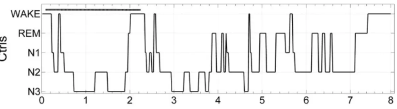
Sleep is not a unitary process, but is classified into non-rapid-eye movement (NREM) sleep and rapid-eye movement (REM) sleep. NREM sleep is further divided into three stages (1-3), corresponding to increasing depth of sleep (Rechtschaffen and Kales, 1968; Iber et al., 2007). Stage 1 is the sleep onset. Stage 2 is characterized by spindles (11-15 Hz) and K-complexes (Steriade and Amzica, 1998). Stage 3 corresponds to slow-wave sleep (SWS), representing a mass cortical synchrony (Amzica and Steriade, 1995) (Figure 3).
8
Figure 3. Hypnogram showing sleep cycles (Gemignani et al., 2012).
Sleep serves different functions: energy conservation (Berger and Phillips, 1995), brain thermoregulation (McGinty and Szymusiak, 1990), brain detoxification (Inoue et al., 1995), and tissue restoration (Adam and Oswald, 1977). As described above, another interesting hypothesis is that sleep periods are favorable for memory consolidation (Maquet, 2001). In 1924, Jenkins and Dallenbach for the first time provided evidence that sleep favors memory consolidation. They found less forgetting of nonsense syllables after sleep periods compared to wakefulness (Jenkins and Dallenbach, 1924). This hypothesis is later confirmed by a growing body of data from different studies (Maquet et al., 2003). At the behavioral level, sleep following learning has a beneficial effect on declarative memory consolidation in humans (Gais et al., 2006). One explanation might be that sleep protects declarative memories from subsequent interference (Ellenbogen et al., 2006).
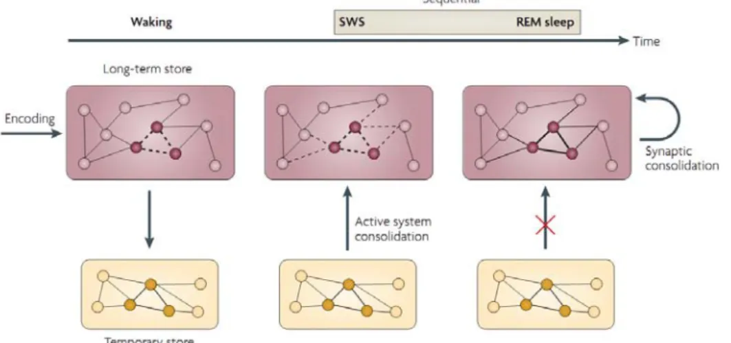
More direct evidence comes from demonstration that patterns of brain activity, previously associated with learning, are selectively replayed during subsequent sleep. In 1994, Wilson and McNaughton demonstrated that information acquired during active behavior is re-expressed in hippocampal circuits during SWS in rats (Wilson and McNaughton, 1994). In humans, sleep-dependent reactivation during SWS of brain regions implicated in prior learning was shown in a declarative spatial memory task using PET (Peigneux et al., 2004). Interestingly, in this study the amount of hippocampal replay during SWS positively correlated with the improvement of performance on the next day. Furthermore, Rasch (2007) reported the first evidence for a causal role of reactivation during SWS in memory consolidation in humans. Object-locations were learned in the presence of an odor; re-exposure of the odor during SWS, but not REM sleep, improved later retrieval. Likewise, Rudoy (2009) showed similar sleep-dependent reactivation of item-specific information using auditory cues.
Building on these results, declarative memory consolidation seems to be based on an iterative replay of newly encoded memory traces, especially during SWS (Plihal and Born, 1997). Additional, low acetylcholine level during SWS is known to facilitate memory consolidation (Gais and Born, 2004; Hasselmo and McGaughy, 2004). Recent memory traces are thereby gradually integrated into the neocortex (Diekelmann and Born, 2010). It is further proposed that these structural changes in cortical network are then stabilized during subsequent REM sleep (Giuditta et al.,
9
1995) (Figure 4). In fact, up-regulation of ZIF268 expression, a transcription factor that regulates long-term plasticity, occurs during REM sleep (Ribeiro et al., 2002).
Figure 4. Model of declarative memory consolidation during sleep: sequential contribution of SWS and REM
(Diekelmann and Born, 2010). For declarative memory, the temporary store is the hippocampus and the long-term store the neocortex.
Finally, such a process would promote the gradual redistribution and reorganization of memory traces (i.e. system consolidation). The concept that sleep allows the transfer of information from the hippocampus to the neocortex is corroborated by human brain imaging studies (Takashima et al., 2006; Gais et al., 2007; Sterpenich et al., 2009; Takashima et al., 2009). These studies showed that three- and six-month delayed memory retests involve an increased reactivation of neocortical regions, with a central role of medial prefrontal cortex (mPFC), and a decreased hippocampal activation.
1.2.2 MODEL FOR MEMORY TRANSFER FROM HIPPOCAMPUS TO NEOCORTEX DURING SLEEP
Altogether the findings above highlight the importance of a synchronized interaction between hippocampus and neocortex during SWS, in order to reactivate and redistribute memory traces (Diekelmann and Born, 2010). This dialogue is under the control of slow oscillations, <1Hz (SO), which are generated in the neocortex during SWS (Molle et al., 2002). The depolarizing up-states of the SO repetitively drive the replay of memory traces in the hippocampus. These reactivations are accompanied by sharp wave-ripples in hippocampus, which stimulate the transfer of memory information to neocortex. The transfer induces a generation of thalamo-cortical spindles, which is associated with a massive calcium influx into neothalamo-cortical cells, which is eventually supposed to contribute to plastic synaptic changes (Born et al., 2006; Marshall et al., 2006).
Evidence of this coordinated interaction between hippocampal ripples and cortical spindles during SO comes from animal studies (Buzsaki, 1996; Siapas and Wilson, 1998) and one human study (Clemens et al., 2007). Interestingly, these
10
findings are corroborated by a study, which demonstrated that selective suppression of hippocampal ripples impairs spatial memory in rats (Girardeau et al., 2009).
Moreover, some studies support an association between NREM sleep spindles and memory: a learning-dependent increase in spindle density was demonstrated in humans (Gais et al., 2002; Schabus et al., 2004; Schmidt et al., 2006) and in animal (Eschenko et al., 2006). In addition, spindle density was positively associated with recall performance (Gais et al., 2002; Schmidt et al., 2006). Furthermore, one fMRI study demonstrated that the same cortical areas active during learning are also replayed during post-encoding spindles events (Bergmann et al., 2012). These results indicate that increased spindle activity may represent an index of sleep-dependent memory consolidation (Fogel and Smith, 2011).
Finally, our work is based on the hypothesis that sleep is involved in system consolidation, which occurs during the replay of neuronal circuits during sleep. However, another hypothesis suggests that the role of sleep in memory consolidation is to maintain synaptic homeostasis, which assumes that consolidation is the result of global synaptic downscaling that occurs during SWS (Tononi and Cirelli, 2003, 2006). Although we do not discuss this latter hypothesis in detail, both mechanisms may act in concert to optimize the sleep-dependent memory consolidation.
1.2.3 SLEEP AND RELATIONAL MEMORY
So far, ample evidence demonstrates that sleep facilitates declarative memory processing. However, most of the studies have focused on direct associative relationships, using paradigms in which participants have to memorize direct associations between two words or images. Of equal importance is whether sleep boosts the capacity to form indirect associations between events, i.e. untrained relationship between stimuli, a form of flexible memory called relational memory (Lau et al., 2010). In everyday life many judgments and decisions are not based on direct knowledge, but rather require inferences based on knowledge acquired across distinct experiences (Zeithamova et al., 2012).
Such inferential reasoning seems to crucially depend on the hippocampus, as demonstrated by human neuroimaging studies (Preston et al., 2004; Shohamy and Wagner, 2008; Zalesak and Heckers, 2009). Converging evidence from animal research indicates that hippocampal lesions impair performance on inferential memory (Bunsey and Eichenbaum, 1996; Devito et al., 2010). Moreover, additional results suggest that other brain regions are recruited together with hippocampus for successful inference, e.g. prefrontal cortex (Acuna et al., 2002).
The idea that sleep-based replay of hippocampal memory traces could enhance performance on an inference task was previously tested by Ellenbogen et al. (2007). They concluded that human relational memory develops during offline periods and that more distant inference appears to be selectively enhanced following offline time periods containing sleep. Furthermore, Lau et al. (2010) showed that even a daytime nap promotes relational memory, which positively correlated with the
11
duration of SWS. Therefore, SWS may provide an optimal brain state for the consolidation of declarative memory and also for the organization of an efficient network of relational associations.
In sum, the concept of active system consolidation during sleep implicates that (i) memory traces are reactivated and transferred from a temporary into a long-term store (Diekelmann and Born, 2010) and (ii) discrete memory traces are reorganized into a flexible relational memory networks (Lau et al., 2011).
Another interesting point discussed in the next paragraph is to determine (iii) if the reactivation and reorganization of memories during sleep is a universal or a selective process, and eventually on which features of the memory trace is based this selection (Saletin and Walker, 2012).
1.3 REWARD SYSTEM
1.3.1 THE MESOLIMBIC DOPAMINERGIC SYSTEM
Reward is a reinforcer and is considered as a positive stimulus that produces a change in behavior, also called learning (Pavlov, 1927). Reward-related stimuli are perceived as salient and relevant and their motivational valence is increased (Schultz, 2006; Berridge, 2007).
Reward processing depends on the mesolimbic-dopaminergic system (ML-DA) (Schultz, 1998; Knutson et al., 2000), which promotes adapted, goal-directed behaviors and incentive-based learning by mediating different aspects of reward processing (Haber and Knutson, 2010).
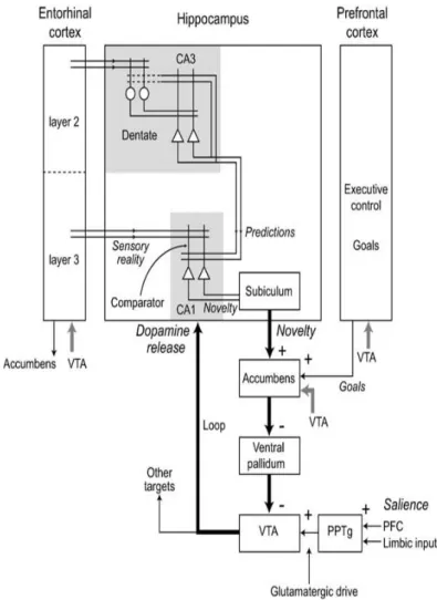
The ML-DA system originates from the ventral tegmental area (VTA) and projects to various structures, like the nucleus accumbens (Nacc) of the ventral striatum (VS), the hippocampal complex and the prefrontal cortex (Alcaro et al., 2007) (Figure 5). The key neurotransmitter of the ML-DA is dopamine, which is closely associated with reward-seeking behaviors (Wise, 2004). VTA bursting activity (phasic signal) is strongly related to reward processing (Yun et al., 2004), especially during reward anticipation (Carter et al., 2009). VTA bursting activity is defined as a brief increase in dopamine concentration in terminal mesolimbic regions and differs from the tonic neural signal, which is a slow change in dopamine concentration (Floresco et al., 2003). It has been shown that dopaminergic bursts also occur when a reward is associated to learning, suggesting an interaction between the ML-DA and memory systems (Sajikumar and Frey, 2004).
1.3.2 MESOLIMBIC-DOPAMINERGIC SYSTEM AND MEMORY CONSOLIDATION
Recent functional imaging studies demonstrated that reward improves long-term memory formation in monetary incentive encoding paradigms, via a
co-12
activation of midbrain related structures like VTA, substantia nigra (SN), Nacc and the hippocampus (HP) (Wittmann et al., 2005; Adcock et al., 2006). These two neural systems, the mesolimbic-dopaminergic circuit involved in reward anticipation and the medial temporal lobe (MTL) circuit involved in memory formation, support motivated learning (Adcock et al., 2006). Moreover, these two systems are also anatomically closely connected and form a functional loop: the Hippocampus-VTA loop (Lisman and Grace, 2005).
Lisman and Grace proposed a model, according to which the novelty signal from the HP converges to the VTA through the Nacc, via a polysynaptic pathway (Figure 5). This is the downward arc of the hippocampus-VTA loop. But not all novel events are of sufficient importance to enter into long-term memory; indeed, the novelty signal from the HP interacts with salience information from pedunculopontine tegmental nucleus (PPT) and goal-related motivation from the prefrontal cortex (PFC). Thus, the activation of VTA is induced by novel salient stimuli from the HP, PPT and PFC. The confluence of this information at the level of the VTA controls the direct dopaminergic input to the HP, via the upward arc of the loop. The released dopamine enhances long-term-potentiation (LTP) in the hippocampus, thereby promoting consolidation of relevant information (Lisman and Grace, 2005).
13
Given that reward-predicted stimuli induce a dopaminergic response (Schultz, 2007), dopamine seems to be the key neurotransmitter of these patterns. Interestingly, hippocampal LPT is dependent on dopamine (Kandel, 2001): hippocampal LTP is blocked by a dopamine D1 receptor antagonist (Bach et al., 1999) and facilitated by D1 receptor agonist (Li et al., 2003).
Overall the HP-VTA loop suggests a strong interaction between reward and memory systems during encoding. We will now discuss the interaction between the ML-DA system and memory consolidation, specifically during sleep.
1.3.3 MESOLIMBIC-DOPAMINERGIC SYSTEM AND MEMORY CONSOLIDATION DURING SLEEP
Evidence suggests that some memories are privileged for consolidation during sleep, such as those with high emotional value (Sterpenich et al., 2007; Sterpenich et al., 2009) or with high future relevance (Fischer and Born, 2009; Saletin et al., 2011; Wilhelm et al., 2011). Such associations would effectively label specific recently encoded information with contextually salient tags, causing a selective reactivation and subsequent consolidation during sleep (Saletin and Walker, 2012).
Interestingly, several studies in animal and human demonstrated an activation of many components of the ML-DA system during sleep.
Pennartz ( IN ANIMAL
2004) demonstrated for the first time a replay of ventral striatum neurons (VS) during slow-wave sleep (SWS) in the rat, after a reward-searching task on a T-maze. Importantly, the reactivation of VS occurred especially in neuronal subgroups whose firing rate was modulated in close temporal association with hippocampal ripples (Figure 6). In 2008, the same group confirmed the preferential reactivation of reward-related information in VS during SWS and quiet wakefulness, but not during rapid eye movement sleep (REM) (Lansink et al., 2008).
NB: the ventral striatum contains the nucleus accumbens, which is reported above in figure 5.
Figure 6. Coherent cross-structural reactivation in hippocampal-ventral striatum circuitry (Lansink et al., 2009).
14
striatal units during periods of active behavior and rest in a single session. Individual neurons are represented as dots around the perimeter of a circle (filled dots: hippocampal CA1 units, n = 10; open dots: ventral striatal units, n = 13). Lines indicate a significant firing correlation between two neurons (red: positive, yellow: negative). A pattern of correlations emerges during track running and is reinstated in postbehavioral rest, whereas it was largely absent in rest preceding behavior.
These results indicated that the HP initiates and orchestrates via ripples the replay in the directly connected VS, which typically fires in relation to expected or delivered reward, as well as cues predictive of reward (Apicella et al., 1992). In turn, VS neurons could reactivate the VTA, eliciting dopamine release (Pennartz et al., 2004). In this way, the reactivation of motivational information in ventral striatum during offline periods contributes to the consolidation of memory by strengthening intrastriatal connections but also by affecting the reactivation in other subcortical and cortical structures (Lansink et al., 2008).
Another important result in animal was the evidence that VTA has an increased bursting activity, especially during REM sleep (Dahan et al., 2007). That could be explained by the activation during SWS of structures that project directly or indirectly to the VTA, for example the HP and PPT. HP through VS, the downward arc of the HP-VTA loop, projects to VTA carrying a novelty signal (Lisman and Grace, 2005) and PPT projects directly to VTA, carrying a saliency information (Holmstrand and Sesack, 2011). In fact, the simultaneous HP-VTA and PPT-VTA activation can lead to a four-fold increase the quantity of dopaminergic neurons firing in bursts (Lodge and Grace, 2006).
Fischer and Born ( IN HUMAN
2009) demonstrated that the expectancy of a reward enhances offline learning during sleep. Their behavioral study clearly showed that the benefit from post-training sleep was impacted by motivational aspects: sleep improved performance of a motor finger sequence that was associated with reward, compared to a non-rewarded sequence.
Another study demonstrated that sleep may selectively enhance memory (both declarative and procedural) expected to be of future relevance (Wilhelm et al., 2011). The common denominator of these studies seems to be that sleep does not benefit all memories with no distinction, instead it operates a selection based on salience.
1.3.4 THE REWARD ACTIVATION MODEL (RAM) FOR SLEEP
According to these results, a suitable explication for the hypothesis that reward processing and memory consolidation interact during sleep is the Reward Activation Model (RAM). The model proposes that ML-DA activation during sleep contributes to memory consolidation by prioritizing the processing of information with high motivational relevance (Perogamvros and Schwartz, 2012).
The RAM is based on the HP-VTA loop, elaborated by Lisman and Grace (2005) (Figure 7).
15
- Downward arc: HP VTA
The coordinated reactivation during NREM sleep of HP and VS triggers novel salient information to VTA (Pennartz et al., 2004). The result is an activation of dopaminergic neurons of VTA, especially during REM (Dahan et al., 2007). - Upward arc: DA input VTA HP
Dopamine release from VTA to HP contributes to synaptic plasticity and learning (Adcock et al., 2006; Fischer and Born, 2009). In HP, dopamine enhances long-term potentiation (LTP) (Kandel, 2001).
Finally, joint and coordinated reactivation of HP and ML-DA may enable the consolidation of a memory trace comprising both contextual and motivational aspects (Lansink et al., 2009).
Figure 7. Schematic illustration of the activation of the ML-DA system during sleep, according to the Reward
Activation Model (RAM) (Perogamvros and Schwartz, 2012).
1.4 HYPOTHESES
In this study, we aim to test whether reward signals may guide sleep-related memory consolidation in healthy human adults. A second main aim is to test whether sleep favors the linkage of discrete events into more integrated (and possibly more abstract) representations.
We investigate the consolidation and generalization of recent information associated with a highly or lowly reward and compare performance for groups with and without a period of sleep (i.e. nap) between encoding and testing.
Predictions:
1.) At the behavioral level, (i) memory improvement for highly rewarded sequences will be better than for lowly rewarded ones at test, and especially in
16
the sleep group. (ii) Generalization of knowledge is enhanced for highly rewarded sequences, and especially in the sleep group.
2.) At the brain level, we expect that (i) activation and connectivity in mesolimbic and hippocampal regions will increase for highly rewarded sequences at delayed testing for the sleep compared to the wake group. (ii) Furthermore, retest after three months will elicit larger responses in the neocortex and a progressive reduction of hippocampal regions activation.
3.) We expect that changes in post-training sleep EEG oscillations, particularly spindle activity and SWS amount, will correlate with memory and generalization performance at test, with distinct effects as a function of reward values.
4.) Consolidation of reward related memories via the dopaminergic system may lead to a form of more explicit memory and therefore higher confidence judgments.
The predictions above concerning fMRI will not be discussed in this Master thesis.
2 METHODS
2.1 PARTICIPANTS
Thirty-three healthy young volunteers (17 women and 16 male, age range = 18-30) gave written informed consent and received financial compensation for their participation in this study, which was approved by the Ethics Committee of Geneva University Hospitals. All participants were right-handed, non-smokers, free from psychiatric and neurological history, and had a normal or corrected-to-normal vision. They were within the normal ranges on self-assessed questionnaires for depression (Beck depression) and anxiety (STAI-T). Importantly, in order to exclude major sleep disturbance and extreme sleep-wake habits and as part of the inclusion procedure, we checked that all participants had normal habitual sleep (PSQI) and circadian typology (Horne & Osberg), no excessive daytime sleepiness (ESS) and regular naps. Additionally, they also filled in the Impulsive Behavior Scale (UPPS) and the Sensitivity to Punishment and Sensitivity to Reward Questionnaire (SPSRQ). All selected participants had to come in the afternoon for a habituation nap under polysomnography and sleep analysis for these naps did not show any abnormality. They then kept a regular sleep-wake schedule five days prior to the experimental day. Compliance was documented by actigraphy (Actiwatch, Cambridge Neuroscience, Cambridge, UK) and sleep diary. Moreover, they were requested to refrain from all caffeine and alcohol-containing beverages and intense physical activity for the 48h preceding the experience. Two volunteers were discarded and thus thirty-one volunteers were included in the final analyses. Participants were
17
randomly assigned to either a ‘sleep’ group and invited to take a nap of 1h30 after the first fMRI session (n = 16, 8 male), or to a ‘wake’ group (n= 15, 7 male), which instead had a 1h30 of quiet rest, both under polysomnography.
2.2 PROTOCOL
An overview of the study is shown in Figure 8.
Participants arrived in the lab at 12h45 and they got acquainted with the task. During the first fMRI session, they performed a memory encoding phase directly followed by a training phase. Then, they took a nap or stayed quietly awake during 1h30 depending on group assignment. At 16h30, participants underwent the second fMRI session with a test phase, where memory was tested across three different levels of inference. Finally, a retest was conducted three months later, in which the test phase was repeated. Before each fMRI session, a psychomotor vigilance task (PVT) was performed.
Figure 8. Experiment timeline. NB: the first fMRI session was comprised of an encoding and training part,
followed by a 9 min resting state (not explicited here). The fMRI test session also comprised the structural acquisitions, which are not detailed here.
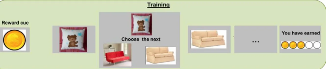
We developed a sequential association memory task that would strongly engage hippocampal regions (Kumaran and Maguire, 2006). Participants were told that they had to learn 8 picture sequences, half of which was highly rewarded, the other half lowly rewarded. Each sequence was formed of 6 pictures presented in the following order: pillow (A), sofa (B), kitchen (C), bedroom (D), house (E), landscape (F) (Figure 9). To promote motivation towards the highly rewarded sequences, we told the participants that the final gain depended on their performance for highly rewarded sequences. However, the final outcome was fixed and the same for all the participants to minimize potential effects of reward size on subsequent memory. Moreover, participants were informed in the instructions that they would have to do an inference task, and were explicitly asked to memorize sequences as wholes rather than direct pairwise associations only.
18
Participants were installed in the MRI scanner and saw the 8 sequences once each, in a randomized order. At the beginning of the sequence, the reward value associated with the sequence was indicated by a picture of one dollar or one cent (XXsec) (Figure 9).
Encoding
Figure 9. Encoding phase: the reward cue was presented for 1.5 sec. Then each picture was presented for 2.5
sec with a 1.5 sec blank between two pictures.
The training phase directly followed the encoding. Participants had to learn all the premise pairs (i.e. inference 0) of the 8 sequences, i.e. (AB), (BC), (CD), (DE), and (EF). Participants were shown the first image (pillow) of the sequence and had to choose the correct second item (sofa) among two options by pressing on an MRI compatible response button box (HH-1×4-CR, Current Designs Inc., USA) (Figure 10). After their answer, the correct sofa was presented and they then had to choose the correct kitchen among two options, and so on. At the end of each sequence, participants were shown how much they earned in dollars or cents, depending on whether this was a highly or lowly rewarded sequence.
Training
Figure 10. Training phase. Timing was as follows: reward cue 1.5 sec, blank 1.5 sec. Presentation of the pillow 2
sec, blank 1.5 sec, presentation of the pillow with the two sofas 2 sec. Then the “choose the next” banner appeared and participants had 6 sec to make a decision. Then 1.5 sec of blank and presentation of the correct sofa for 2 sec. etc. At the end, gain presentation for 2.5 sec.
All volunteers underwent 3 blocks of training; in each block, each of the 8 sequences was presented once, in a randomized order. At the end of the training phase, participants stayed in the scanner 9 minutes more for a resting-state acquisition.
Half of the participants took a nap (sleep group), the other half stayed awake (wake group), both for 1h30. The wake group was allowed to read in dim light a book or magazine not involving any type of memorizing or high cognitive load. For both Nap time
19
groups the temperature of the room was controlled (~21°C). Polysomnographic data were continuously recorded during each 1h30 nap or wake session (see below EEG acquisition). Before starting the test phase, the sleep group participants completed the St. Mary's Hospital sleep questionnaire and took a shower to ensure that they were awake and alert.
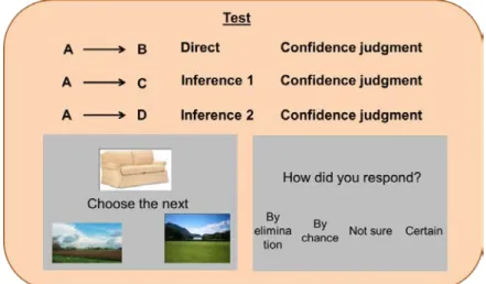
Participants were scanned while being tested on all possible previously learnt premise pairs of inference 0 (e.g. AB) and first inference (e.g. AC) or second inference (e.g. AD) pairs (Figure 11). Furthermore, after each response participants gave a confidence judgment on how they responded using 4 possible options: certain, not sure, by chance, or by elimination.
Test
Figure 11. Timing as for training; for confidence judgment: the four judgment’s possibilities were presented for 2
sec and then when the “how did you respond?” banner appeared participants had 6 sec to make a decision
At the end of the experiment, participants were asked about their strategies to memorize the sequences, the relative difficulty of the inference tasks, and whether they paid more attention to the highly rewarded sequences.
Three months after the experiment, volunteers were retested on the test part, again with simultaneous fMRI measurements (will not be detailed here).
Retest
2.3 ANALYSIS OF BEHAVIORAL DATA
To ensure that incorrect responses during the test part reflected forgetting (or failure in memory consolidation), rather than insufficient encoding of the images during the training phase, we defined the following rule: an image was considered to be properly encoded whenever the participant made at least two correct responses when asked to choose it: one during block 1 or block 2, and one during block 3 of the training phase. Images not sufficiently well learned were removed from the test phase (2.16 ± 1.85 picture per participant). After this transformation, we removed one outlier
20
participant, because of memory performance below two standard deviations of the group mean.
2.4 EEG DATA ACQUISITION AND ANALYSIS
Both sleep and quiet wakefulness period were monitored using a V-Amp recorder (Brain Products, Gilching, Germany). Standard polysomnography included 6 EEG (Fz, Cz, Pz, Oz, C3, C4, reference: mastoids), chin EMG, and vertical and horizontal EOG recordings (sampling rate: 250 Hz).
For PSG analyses, we used FASST (fMRI Artifact rejection and Sleep Scoring Toolbox; Cyclotron Research Centre, University of Liège, Belgium), implemented in Matlab (The Mathwork, MATLAB R2009.b ).
The recordings were converted from .eeg to .edf. Data from one participant could not be included because of a technical problem with the recording. Finally fifteen naps and fifteen quiet wakefulness periods were visually scored on a 20 s epoch basis by two independent scorers, according to standard criteria by the AASM Manual for the Scoring of Sleep (Iber et al., 2007). EEG artifacts and arousals were visually detected. Statistics were then computed to determine the duration of the different sleep stages and a spectral power analysis was conducted together with a mean power spectrum for each specific sleep stage (based on Cz) (Figure 12).
Figure 12. Power spectrum of one nap (left). Mean power spectrum during stage 2 for channel Cz (right).
Additionally, automatic detection of slow wave and spindle was performed (Figure 13). The automatic detection of slow waves was performed on averaged signals over 4 ROIs: frontal, central left, central right and parietal (Fz, C3, C4, Pz), and the detection was based on Massimini’s criteria (Massimini et al., 2004).
Sleep spindles were detected based on their amplitude as in Molle (Molle et al., 2002). The signal from three reference channels (Fz, Cz, Pz) was extracted and filtered. A frequency threshold of spindle detection (percentile 95) was computed for each reference channel signal in sleep stage 2. A root mean square (RMS) signal was also computed from the previously extracted signals. A binary signal was then
21
computed: whenever the RMS signal was larger than the corresponding threshold, the value was 1, otherwise 0. The average of the 3 binary signals was computed and ceiled. Finally using this ceiled average, one could detect bursts of activity in the frequency band of interest (here recommended 8–20 Hz), lasting at least 400 ms and separated by at least 1000 ms, which were then considered as spindles. Furthermore, the predominant frequency of each detected spindle was assessed using wavelet analysis.
Figure 13. An example of detected spindles (here 4 posterior spindles) and one slow wave (delta) in a scoring
window of stage 2.
2.5 fMRI DATA ACQUISITION AND ANALYSIS
MRI data was acquired on a 3 Tesla MRI scanner (Siemens Trio). Multislice T2*-weighted fMRI 2D images were obtained with a gradient echo-planar sequence using axial slice orientation (36 slices; voxel size, 3.2 x 3.2 x 3.2 mm; repetition time (TR) = 2100 ms; echo time (TE) = 30 ms; flip angle (FA) = 80°).
Structural images were acquired at the end of the test part: a whole brain structural image was acquired with a T1-weighted 3D sequence (192 contiguous sagittal slices; voxel size, 1.0 x 1.0 x 1.0 mm; repetition time (TR) = 1900 ms; echo time (TE) = 2.27 ms; flip angle (FA) = 9°). An additional structural image was acquired with a proton-density weighted sequence (20 axial slices; voxel size, 0.8 x 0.8 x 3.0 mm; repetition time (TR) = 6000 ms; echo time (TE) = 8.4 ms; flip angle (FA) = 149°). This acquisition served for the localization of the VTA in the midbrain (D'Ardenne et al., 2008).
22
Gaze direction and pupil diameter were recorded using an MRI compatible eye tracker (EyeTrac 6 LRO, Applied Science Laboratories, USA), to get additional measures of emotional physiological response.
All stimuli for fMRI were designed and delivered using a MATLAB Toolbox (Cogent 2000).
A short overview of fMRI analysis is illustrated below; however please note that this aspect of the study has not been my principal task during the master.
Functional images were analysed using SPM8 (Wellcome Department of Imaging Neuroscience, London, UK). This analysis included standard preprocessing procedures: realignment, unwarping, slice timing to correct for differences in slice acquisition time, normalization (images were normalized to an EPI template that was aligned to the MNI T1 template), and smoothing (with an isotropic 8-mm FWHM Gaussian kernel).
A standard general linear model (GLM) approach was then used to compare conditions of interest at the individual level and then these contrasts from each participant were entered in a second-level random-effects analysis. We reported activations surviving an uncorrected statistical threshold of p < 0.001. Coordinates of brain regions were reported in MNI space.
3 PRELIMINARY RESULTS
3.1 MEMORY ACCURACY AND SLEEP
3.1.1 BEHAVIORAL RESULTS
A 2-way repeated measures analysis of variance (ANOVA) was performed on memory performance (% good response) with reward (high and low) and learning (block 1, block 2, block 3) as within-subjects factors. We observed a main effect of reward (F(1,30) = 35.86; p < 0.001), and also a main effect of learning (F(2,30) = 20.68; p < 0.001). Furthermore, there was a significant interaction between the reward and learning effect (F(2,30) =4.72; p = 0.012) (Figure 14).
TRAINING PERFORMANCE
As expected, reward influenced performance during the training phase, with highly rewarded sequences better remembered than lowly rewarded ones. However, after the third training section, the volunteers were able to remember both highly and lowly rewarded sequences in the same manner (criterion of ~90 % good response). Please note that both highly and lowly rewarded sequences might thus have the same potential for subsequent offline consolidation.
23
Figure 44. Training performance for the 3 blocks of 8 sequences each, showing a reward effect for the first 2
blocks. Error-bars indicate standard error.
After removal of not learnt images during the training phase (see methods) TEST PERFORMANCE
1
This analysis revealed a main effect of group (F(1,14) = 4.19 ; p = 0.044), that is participants who slept showed increased memory performance at the test phase. Furthermore, there was a main effect of reward (F(1,29) = 16.60; p < 0.001) and also a main effect of inference (F(2,29) = 4.64; p = 0.012) (Figure 15). There were no significant interactions.
, we computed a repeated measures ANOVA on memory performance (% good response) with reward (high reward “HR” and low reward “LR”) as repeated measure, inference (direct, inference 1, inference 2) as within-subjects factor, and group (sleep and wake) as between-subjects factor.
1
24
Figure 15. Test performance for the direct, inference 1 and inference 2 trials, showing the difference between
highly (HR) and lowly (LR) rewarded associations and between the sleep and wake groups. Error-bars indicate standard error.
In summary, even though at the end of the training phase highly and lowly rewarded sequences were equally well learnt, after a nap or quiet wakefulness, memory performance diverged. The nap-group achieved a better memory performance. However, both the nap and wake group better remembered highly rewarded associations and as expected there was an effect of task difficulty (inference effect).
3.1.2 EEG MEASURES: SLEEP SPINDLES
We then examined the precise role of sleep in memory consolidation, also encouraged by the ANOVA results of memory test performance.
The amount of sleep time spent in each sleep stage and additional information about spindles are summarized in Table 1.
Table 1. Mean duration of each sleep stage in minutes, sleep efficiency and total number of spindles (n = 14,
mean ± s.e.m).
Sleep stages Nap Group
W (min) 21.67 ± 2.92 N1 (min) 9.05 ± 1.62 N2 (min) 17.67 ± 2.24 N3 (min) 31.55 ± 3.70 REM (min) 5.05 ± 1.99 N2 Latency (min) 14.29 ± 2.04
Total sleep time (min) 54.26 ± 4.39
25
Naps’ profile was normal with a good duration of N2 and N3. That was extremely important for the hypotheses we wanted to test. Moreover, eleven out of fourteen participants evaluated at the end of the nap their sleep as deep and just one found that it was very light.
We correlated memory test performance with these sleep parameters. We found that the total number of sleep spindles positively correlated with memory performance for highly rewarded items in the direct (R = 0.62; p = 0.017) and inference 1 (R = 0.60; p = 0.022) tasks (Figure 16).
Figure 16. Positive correlation between sleep spindles (tot number) and highly rewarded direct and inference 1
tasks (% good response).
Furthermore, we separated sleep spindles into slow and fast ones and we found that the total number of slow spindles positively correlated with highly rewarded associations in the direct (R = 0.54; p = 0.045) and inference 2 (R = 0.53; p = 0.049) tasks, and a positive trend was shown for the inference 1 task (R = 0.41; p = 0.140) (Figure 17).
Figure 17. Positive correlation between slow spindles (tot number) and highly rewarded direct and inference 2
tasks (% good response). 0 20 40 60 80 100 120 0 100 200 300 400 HR Di re c t (% good r e s pons e )
Sleep Spindles (total number)
0 20 40 60 80 100 120 0 100 200 300 400 H R I n fer en ce 1 (% good r e s pons e )
Sleep Spindles (total number)
0 50 100 150 0 100 200 300 HR Di re c t (% g o o d r e s p o n s e )
Slow Spindles (total number)
0 50 100 150 0 100 200 300 H R I n fer en ce 2 ( % g o o d r e s p o n s e )
Slow Spindles (total number)
Total number of spindles 210.93 ± 22.08
Total number of slow spindles 104.00 ± 15.09 Total number of fast spindles 106.93 ± 14.31
26
Finally, we found another positive correlation between the total number of slow spindles and the reward-associated improvement of the memory performance in the inference 2 task (R = 0.56; p = 0.039) (Figure 18).
Figure18. Positive correlation between slow spindles (tot number) and the reward-associated gain of the memory
performance for inference 2 task.
3.2 CONFIDENCE JUDGMENT AND SLEEP
3.2.1 BEHAVIORAL RESULTS
During the test, after each response, subjects gave a confidence judgment, i.e. how they chose between the two images: certain, not sure, by chance, by elimination. Two participants were excluded from this analysis, one because of a response bias toward the “certain” response category, the other because of a response bias toward the “by elimination” response category (selection of those respective response categories were over two standard deviations above the group mean). A repeated-measures ANOVA was performed on the % of good responses with reward (high and low) as repeated measure, inference (direct, inference 1, inference 2) and confidence judgment type (certain, not sure, by chance, by elimination) as within-subjects factors, and group (sleep and wake) as between-subjects factor.
We observed a main effect of reward (F(1,28) = 1224.15; p < 0.001) and of confidence (F(3,28) = 120.413; p < 0.001), as well as reward by confidence (F(3,28) = 67.441; p < 0.001) and a group by confidence (F(3,28) = 3.378; p = 0.018) interaction. Importantly, there was an effect of group selectively for the inference 2 trial types (T(1,28) = 2.219, p=0.035).
In summary, all subjects were more confident in their responses for the highly rewarded trials than for the lowly rewarded trials, but importantly for the most difficult inference 2 task the sleep-group was more confident than the wake group (Figure 19). Finally, all T-tests performed on the other confidence judgments did not give significant results. -40 -20 0 20 40 0 100 200 300 D if fer n ce b et w een H R L R Inf e re nc e 2 ( % good resp o n se)
27
Figure 19. Certain judgment (% of good response) for direct, inference 1 and inference 2 trials, showing an effect
of reward and particularly for the inference 2 task an effect of group too.
3.2.2 EEG MEASURES: STAGE 2
In order to test whether increased confidence ratings would relate to specific sleep parameters, we performed correlation analyses. We found a positive correlation between stage 2 (% of the nap) and the “certain” judgment for highly rewarded inference 1 (R = 0.58; p = 0.028) and inference 2 (R = 0.61; p = 0.020) (Figure 20). There was also a trend for the direct trials (R = 0.49; p = 0.078).
Figure 20. Positive correlation between stage 2 (% of the nap) and certain judgment for highly rewarded
inference 1 and inference 2 trials (% of good response).
So far, it seems that the “certain” judgment for highly rewarded inference 1 and 2 trials may be modulated at least in part by the amount of stage 2 sleep.
0 20 40 60 80 100 0 10 20 30 cer ta in fo r H R i nf 1 (% g oo d re sp on se ) S2 (% of the nap)
28
3.3 FUNCTIONAL MRI RESULTS
Below, we report imaging results at an uncorrected threshold of p = 0.001 (n = 20). Coordinates of peak activation are in the MNI (Montreal Neurological Institute) space.
During the training phase, preliminary fMRI results showed a significant activation among other regions: of the hippocampus (x, y, z, mm: 33, -25, -8; Z-score, 3.28; side: R) for the choice of the good image versus the choice of a control image, the VTA (x, y, z, mm: -9, -28, -14; Z-score, 3.78; side: L) for the choice a highly rewarded image versus the choice of a lowly rewarded image, and Nacc (x, y, z, mm: 3, 20, -5; Z-score, 4.37; side: R) for the correct choice of a highly rewarded image versus the incorrect choice of a highly rewarded image. These preliminary results suggest that, as expected, this task highly engaged the hippocampal memory regions and the dopaminergic reward circuit (Figure 21).
Figure 21. Brain regions activated during the training phase. In particular, Nucleus accumbens activated in case
of reward expectation (Spicer et al., 2007) while VTA depended especially on reward prevision (Carter et al., 2009).
4 DISCUSSION
In this study, we assessed whether highly rewarded stimuli are prioritized by sleep-dependent memory consolidation and also whether relational memory is facilitated by offline sleep periods, in particular for highly rewarded information. Eventually, we investigated if these changes in memory representation yield higher confidence judgment at test. We used a transitive inference paradigm, i.e. a set of overlapping pairs forming a sequence of pictures with hierarchically-ordered relationships (Heckers et al., 2004). Some of these sequences were highly rewarded and others were lowly rewarded. To test the effect of sleep on memory, we measured behavioral performance and whole-brain fMRI activity before and after a nap or a corresponding period of wakefulness monitored by EEG. As predicted, we found that people who slept had a better overall memory performance. Moreover, for both the sleep and wake groups, there was an effect of reward, with increased performance for the highly rewarded sequences, and an effect of task difficulty (inference effect). A
Training: choice vs control
Hippocampus
Training: high vs low reward
Ventral Tegmental Area
Training: high reward good vs bad
29
second important finding was that memory performance for the highly rewarded elements in both the direct and inference tasks positively correlated with sleep spindles. Finally, we showed that the nap group reported being more confident than the wake group when selecting the good option, specifically for the most difficult task, i.e. the inference 2 trials, suggesting that sleep decreased uncertainty associated with decision making on the task. Importantly, this effect was associated with the percentage of the nap spent in N2 sleep.
In our study, participants who took a nap had better direct and relational associative memories (Figure 15). The data confirm that performance is superior following a sleep period (
Global sleep effect on memory performance
Maquet et al., 2003), even for a 1h30 daytime nap. There is growing evidence supporting the benefit of a short daytime nap (Mednick et al., 2003), and especially on declarative memory (Schmidt et al., 2006; Tucker et al., 2006). As proposed by Nishida and Walker (2007), one explanation could be that daytime naps may trigger an accelerated consolidation or alternatively that for some specific memory types an entire night may not be required. Moreover, in our study the sleep group took the nap directly after the training phase, which may have limited potential interferences from waking activities (Ellenbogen et al., 2006) and thus let memory maximally benefit from sleep (Gais et al., 2006; Talamini et al., 2008). Overall, our findings support the idea that sleep, even in a form of nap, plays an active role in declarative memory consolidation by promoting the reprocessing of previously encoded memory traces (Maquet, 2001; Diekelmann and Born, 2010).
Besides a global sleep-dependent gain, highly rewarded associations were better remembered in both the sleep and wake groups compared to lowly rewarded ones, despite the fact that, at the end of the training session, both highly and lowly rewarded sequences were associated with the same level (near ceiling) of performance. A possible explanation is that an enhanced activation of reward-relevant structures such as VTA during encoding and training for the highly rewarded sequences promotes memory formation via dopamine release in the hippocampus (
Rewarded memory
Wise, 2004; Wittmann et al., 2005; Adcock et al., 2006). However, the sleep group reached a higher performance for the highly rewarded sequences compared to the wake group, suggesting that the mesolimbic-dopaminergic system is not only activated during wakeful behaviors, but also reactivated during sleep (Pennartz et al., 2004). This finding is consistent with the RAM model proposed by Perogamvros and Schwartz (2012): the engagement of the mesolimbic-dopaminergic system during sleep contributes to memory processes by prioritizing high motivational relevance for reprocessing.
Our results are difficult to reconcile with those of Tucker et al (2011), who did not find that sleep boosts rewarded information compared to wake, but in this study reward
30
effect was measured between groups and not within subjects as half of the subjects got rewarded stimuli and half got non rewarded stimuli.
Furthermore, our data confirm that sleep selectively enhances memory expected to be of future relevance (Wilhelm et al., 2011). Indeed, we explained to participants at the beginning that the final gain depended on how good they would perform at the test (reward-motivated learning), i.e. more correct highly rewarded answers leading to overall higher gain. In sum, anticipated high reward seems to be one of the factors determining access to sleep-dependent memory consolidation (Fischer and Born, 2009). We anticipate that the neural mechanisms underlying these effects of reward could be revealed by our fMRI imaging results (not discussed here), in particular if functional connectivity strength between mesolimbic and hippocampal regions is enhanced after sleep and if it is positively associated with enhanced memory.
Our results confirm that sleep not only strengthens memory for learned item pairs (
Generalization versus reconstructive theory for relational memory
Diekelmann and Born, 2010) but also boosts the emergence of relational knowledge (Ellenbogen et al., 2007; Lau et al., 2010; Lau et al., 2011). Before discussing our results in more detail, we briefly summarize the two main theories of inferential reasoning. Both theories converge to suggest a critical role of the hippocampus; however, they diverge concerning the underlying mechanisms. According to the reconstructive theory, the hippocampal retrieval process supports the inferential reasoning (Preston et al., 2004; Zalesak and Heckers, 2009). That means retrieval of inferential memories is based on recall and recombination of individual memories, i.e. the retrieval of previously encountered memories (AB) and (BC) could then be recombined to support successful inferential judgment (AC). However, according to the generalization theory, it is the hippocampal encoding processes that support inferential reasoning (Shohamy and Wagner, 2008). This would involve the integration of overlapping events during encoding, promoting a single hierarchical representation of overlapping stimuli (ABC), rather than (AB) and (BC). However, both models are not mutually exclusive; indeed, they may coexist and optimize relational memory processes. Importantly, the nature of learning could be an important factor in determining the relative contribution of these mechanisms. Here, we hypothesize that our task preferentially engages the hippocampal encoding processes, firstly because we explicitly asked participants to try to memorize the sequence as a whole (ABC), rather than just separated premise pairs (AB) and (BC). Secondly, we speculate that the sequential association episodic memory task that we developed may promote the integration of the elements within a scene (e.g. in this landscape there is this house. Within, there is this pillow on the sofa and beside this kitchen etc.) rather than a set of single independent associations. Therefore, our instructions and type of task may have facilitated generalization processes occurring as early as during encoding (Shohamy and Wagner, 2008). Subsequently, both direct and more distant associations, already formed during encoding, may be reactivated
31
during the sleep period (Peigneux et al., 2004; Rasch et al., 2007), explaining the fact that both direct and inference tasks are better in the sleep group.
However, sleep itself could promote relational memory, regardless of whether a generalization process already started during the encoding. Ellenbogen et al. (2007) speculated that offline hippocampal reprocessing not only strengthens memories but also favors the binding of items that are not learned together, reorganizing them for a subsequent flexible use. Another study showed that the duration of SWS predicts relational memory performance (Lau et al., 2010). So far, our results suggest a role of sleep in relational memory, which is also reflected in higher confidence ratings for correct responses on inference 2 trials, suggesting that sleep facilitates generalization.
In our study, we found that the total number of sleep spindles was associated with a better memory performance for the highly rewarded elements of the direct and inference 1 tasks. Secondly, in a more fine-grained analysis, the total number of slow spindles positively correlated with the highly rewarded items of the direct and inference 2 tasks, and a positive trend was shown for the inference 1 task. Thirdly, the number of slow spindles determined the memory performance gain for rewarded and more remote associations (inference 2 trials).
Slow spindles contribute to reward-associated memory consolidation
In recent years, sleep studies have started to differentiate slow and fast spindles according to their dominant frequency power (De Gennaro and Ferrara, 2003; Schabus et al., 2007; Molle et al., 2011) and examined their respective contribution to memory processes (Gais et al., 2002; Schabus et al., 2004; Schmidt et al., 2006). Slow spindles are typically associated with slower frequency (~ 11-13.5 Hz) and are dominant over frontal cortical sites. Because of this anterior topographic scalp distribution, they are also called anterior spindles. Fast spindles have a faster frequency (~ 13.5-15 Hz) and a more widespread distribution over parietal and central sites. Because of this posterior topographic scalp distribution, they are also called posterior spindles. Although it is still unclear which characteristic of sleep spindles (total number, density, frequency, duration) is specifically related to the memory consolidation, we found that the total number of sleep spindles correlated with a better memory performance for the highly rewarded items for the direct and inference 1 tasks. Additionally, a more fine-grained analysis of our results showed that specifically slow spindles supported a better memory performance for highly rewarded items in the direct task but also, more interesting, in the inference 2 task, which was the most difficult. This result is in agreement with Schmidt et al. (2006), who used a declarative episodic verbal memory task with two different encoding difficulties and a nap to test sleep-dependent memory consolidation. They found that spectral power in the anterior spindle frequency range (11.25 – 13.75 Hz) and the density of anterior spindles (analyses based on left frontal location) were both significantly increased after the difficult encoding condition. Moreover, they found positive correlations between these changes and the memory performance for the
32
difficult condition at the recall session. These data suggest a critical role of the nature of the learning materials, in particular task difficulty and/or the potential for improvement, in determining sleep-related changes during a nap.
Concerning the putative underlying neural mechanisms, a strong temporal correlation between ripples in the hippocampus and spindles in the neocortex was observed in animals (Siapas and Wilson, 1998) and humans (Clemens et al., 2007), suggesting that spindles are involved in the hippocampal-neocortical dialogue necessary for declarative memory consolidation (Molle et al., 2002). Spindle activity may induce long-term synaptic plastic changes via a Ca2+ entry in neocortical cells (Born et al., 2006; Marshall et al., 2006). Thus, spindles are supposed to be involved in the process of consolidating newly learned information into a more stable form of long-term memory (Fogel and Smith, 2011).
Finally, our third result intriguingly suggests an interaction between slow spindles and reward. In fact, the total number of slow spindles positively correlated with the reward- dependent improvement of memory performance for the inference 2 trials. So far, if we assume that cortical sleep spindles reflect hippocampal ripples, our finding suggests a possible link between hippocampus and reward-associated regions. Eventually, this link could be the base for the formation of a memory trace comprising both contextual and motivational components. The idea of such a coordinated replay was convincingly demonstrated in animal studies (Pennartz et al., 2004; Lansink et al., 2009). A simultaneous EEG-fMRI acquisition could possibly provide further support to the existence of this joined reactivation of contextual and motivational features of recent memories.
While several studies have linked fast spindles with hippocampal activity and suggested that fast spindles play a key role in sleep-dependent memory processing (Schabus et al., 2007), here we did not find any significant correlations between fast spindles and memory performance. Instead, our study and that of Schmidt et al. (2006) link slow spindles with declarative memory processing during a midday nap, and for more difficult conditions. One plausible interpretation for our results could follow a recent suggestion proposed by Mölle et al. (2011): “whereas the fast spindles coinciding with hippocampal sharp wave ripples may represent a mechanism that facilitates the transfer of memory-related information from the hippocampus to the neocortex, subsequent slow spindles may be related to a cortico-cortical cross-linking of transferred information with prefrontal circuitry. […] Although thalamic activity cannot be excluded as a common source for slow and fast spindles, slow spindles could alternatively be considered primarily of cortical origin, developing in the aftermath of a strong network depolarization.” We would like to speculate that the association between slow spindles and cortico-cortical interactions, and in particular with prefrontal cortex, is especially important for the consolidation of the most difficult and complex information. More studies are needed to prove this speculation.
33
Our results showed that sleep led to more “certain” judgments for correct highly rewarded inference 2 responses than wakefulness. We ruled out that this effect could be due to overall higher confidence ratings after sleep as there was no main effect of sleep. This finding may suggest that sleep promotes the conversion of implicit forms of memory into more explicit and conscious memory. A possible
explanation could be that the generalization process based on integrative encoding (
Gaining confidence while asleep
Shohamy and Wagner, 2008) and reinforced by subsequent sleep (Ellenbogen et al., 2007; Lau et al., 2010) makes indirect learned relationships readily available during test phase. This form of representation could paradoxically make inference 2 associations as easy as direct and inference 1 ones (Zeithamova et al., 2012). This could explain why the participants in the sleep group were as confident when responding direct, inference 1 and inference 2 trials.
Furthermore, we can also think that sleep plays an independent pivotal role in this conversion. The study of Wagner et al. (2004) showed that sleep promotes insight into regularities that remain out of awareness before sleep. Further,
Yordanova et al. (2012) found that slow spindle activity during SWS may indicate the transformation of pre-sleep implicit knowledge to post-sleep explicit knowledge. In our study, we not only did that the sleep group provided overall increased confidence ratings for correct answers, in this group we found a positive correlation between the number of ‘certain’ confidence responses for highly rewarded inference 1 and 2 and the percentage of the nap in stage N2. This latter finding could possibly reflect a sleep-induced gain of explicit knowledge, leading to an increase in confidence. The emergence of explicit knowledge in the waking brain involves activity in prefrontal cortical areas in connection with hippocampal and closely connected medial temporal lobe areas (Rose et al., 2010). The importance of such interplay between the
hippocampus and prefrontal regions during NREM sleep for insightful behavior was suggested by Darsaud et al.(2011). Eventually, we can speculate that during stage 2 these areas are reactivated during processes of memory consolidation and thus facilitate the generation of explicit knowledge. This view is supported by evidence relating stage N2 rather than N3 with memory consolidation (Gais et al., 2002;
Schabus et al., 2004; Fogel and Smith, 2006). Moreover, a study in rodents identified a replay of relevant information in the hippocampus and also in the prefrontal cortex during subsequent sleep (Peyrache et al., 2009).
Our results show that sleep has an effect on both memory reorganization and generalization, by prioritizing highly rewarded information for consolidation. Moreover, they suggest a role of sleep in enhancing explicit knowledge and consequently confidence judgment. Further information concerning associated changes in brain activity will be provided by our fMRI data. At this point, preliminary fMRI results showed that reward brain regions are activated in our original Conclusion