CURRENT OPINION
Clinical trial design principles and endpoint
definitions for transcatheter mitral valve repair and
replacement: part 1: clinical trial design principles
A consensus document from the mitral valve academic research
consortium
Gregg W. Stone
1,2*
, Alec S. Vahanian
3, David H. Adams
4, William T. Abraham
5,
Jeffrey S. Borer
6, Jeroen J. Bax
7, Joachim Schofer
8, Donald E. Cutlip
9,
Mitchell W. Krucoff
10, Eugene H. Blackstone
11, Philippe Ge´ne´reux
1,2,12,
Michael J. Mack
13, Robert J. Siegel
14, Paul A. Grayburn
13, Maurice Enriquez-Sarano
15,
Patrizio Lancellotti
16, Gerasimos Filippatos
17, and Arie Pieter Kappetein
18, for the
Mitral Valve Academic Research Consortium (MVARC)
1Columbia University Medical Center/New York-Presbyterian Hospital, New York, New York;2Cardiovascular Research Foundation, New York, New York;3Hoˆpital Bichat, Paris,
France;4
Mount Sinai Health System, New York, New York;5
The Ohio State University, Columbus, Ohio;6
SUNY Downstate Medical Center, Brooklyn, New York;7
Leiden University
Medical Center, Leiden, the Netherlands;8Hamburg University Cardiovascular Center, Hamburg, Germany;9Beth Israel Deaconess Medical Center, Boston, Massachusetts;
10
Duke University Medical Center, Durham, North Carolina;11
Cleveland Clinic, Cleveland, Ohio;12
Hoˆpital du Sacre´-Coeur de Montre´al, Montreal, Quebec, Canada;13
Baylor
University Medical Center, Dallas, Texas;14Cedars-Sinai Medical Center, Los Angeles, California;15Mayo Clinic, Rochester, Minnesota;16University Hospital of Lie`ge, Lie`ge, Belgium;
17
Athens University Hospital Attikon, Athens, Greece; and18
Erasmus University Medical Center, Rotterdam, the Netherlands. For complete information on the MVARC members and participants, please see the Online Appendix
Received 5 March 2015; revised 5 May 2015; accepted 21 May 2015
See page 1849 for the editorial comment on this article (doi:10.1093/eurheartj/ehv334)
Mitral regurgitation (MR) is one of the most prevalent valve disorders and has numerous aetiologies, including primary (organic) MR, due to underlying degenerative/structural mitral valve (MV) pathology, and secondary (functional) MR, which is principally caused by global or regional left ventricular remodelling and/or severe left atrial dilation. Diagnosis and optimal management of MR requires integration of valve disease and
*Corresponding author. Columbia University Medical Center, Cardiovascular Research Foundation, 111 East 59th Street, 11th Floor, New York, New York 10022. Email:gs2184@
columbia.edu
The MVARC initiative was funded by unrestricted grant support from Abbott Vascular, Boston Scientific, Cardiac Dimensions, Cordis, Edwards Lifesciences, Guided Delivery Systems Inc., Mitralign, Medtronic, and Valtech. Dr. Stone has served as a consultant for AGA Medical, AstraZeneca, Atrium, Boston Scientific, Cardiovascular Systems, Inc., Eli Lilly/Daiichi Sankyo, InfraReDx, InspireMD, Miracor, Osprey, Reva, TherOx, Thoratec, Velomedix, and Volcano; and has equity in the Biostar and MedFocus family of funds, Caliber, Guided Delivery Systems, MiCardia, and Vascular Nanotransfer Technologies. Dr. Vahanian has received personal fees from Abbott Vascular, Edwards Lifesciences, and Valtech; has served as a con-sultant for Abbott Vascular and Valtech; and has received honoraria from Edwards Lifesciences. Dr. Adams has received royalties for intellectual property paid to his medical institution from Edwards Lifesciences and Medtronic. Dr. Abraham has served as a consultant for Abbott Vascular, Novartis, and St. Jude Medical/CardioMEMS; and was coprincipal investigator for the COAPT trial of MitraClip sponsored by Abbott Vascular. Dr. Borer has served as a consultant for Amgen, ARMGO, BioMarin, Boehringer Ingelheim, Celgene, Cleveland BioLabs, JenaValve, Salix, Sanofi, and Servier; and serves as a committee member for Cardiorentis, Celladon, the National Heart, Lung, and Blood Institute, Novartis, Pfizer, Somahlution, and Takeda USA. Dr. Cutlip has received research support from Medtronic, Boston Scientific, and Abbott Vascular. Dr. Krucoff has received grants and personal fees from and served as a consultant for Abbott Vascular, Boston Scientific, and Medtronic. Dr. Blackstone has received grants from Edwards Life-sciences. Dr. Ge´ne´reux has received grants from Cardiac Dimensions, Inc.; and serves as a consultant/speaker for Abbott Vascular, Cardiovascular Systems, Inc., and Edwards Lifesciences. Dr. Mack has served as a coprincipal investigator of trials on transcatheter mitral valve replacement and MitraClip therapy. Dr. Grayburn has served as a consultant for Abbott Vascular, Bracco Diagnostics, and Tendyne Holdings; has received grants from Abbott Vascular, Edwards Lifesciences, Guided Delivery Systems, Medtronic, Tendyne Holdings, and Valtech Cardio; has received personal fees from Abbott Vascular, Bracco Diagnostics, and Tendyne Holdings; and has served as a consultant to Tendyne Holdings. Dr. Filippatos has received grants from Abbott Vascular; and serves as a committee member or principal investigator of trials sponsored by Bayer, Cardiorentis, European Union, Medtronic, and Novartis. All other authors have reported that they have no relationships relevant to the contents of this paper to disclose. Saibal Kar, MD, served as Guest Editor for this paper.
Published on behalf of the European Society of Cardiology. All rights reserved.&American College of Cardiology 2015. For permissions please email: journals.permissions@oup.com.
This article is being published concurrently in Journal of the American College of Cardiology [1]. The articles are identical except for minor stylistic and spelling differences in keeping with each journal’s style. Either citation can be used when citing this article.
[1] Stone GW, Vahanian AS, Adams DH, Abraham WT, Borer JS et al. Clinical trial design principles and endpoint definitions for transcatheter mitral valve repair and replacement: Part 1: Clinical Trial Design Principles. J Am Coll Cardiol 2015;66:278 – 307. doi: 10.1016/j.jacc.2015.05.046
European Heart Journal (2015) 36, 1–27 doi:10.1093/eurheartj/ehv281
by guest on July 14, 2015
heart failure specialists, MV cardiac surgeons, interventional cardiologists with expertise in structural heart disease, and imaging experts. The introduction of transcatheter MV therapies has highlighted the need for a consensus approach to pragmatic clinical trial design and uniform endpoint definitions to evaluate outcomes in patients with MR. The Mitral Valve Academic Research Consortium is a collaboration between leading academic research organizations and physician-scientists specializing in MV disease from the United States and Europe. Three in-person meetings were held in Virginia and New York during which 44 heart failure, valve, and imaging experts, MV surgeons and interventional car-diologists, clinical trial specialists and statisticians, and representatives from the U.S. Food and Drug Administration considered all aspects of MV pathophysiology, prognosis, and therapies, culminating in a 2-part document describing consensus recommendations for clinical trial design (Part 1) and endpoint definitions (Part 2) to guide evaluation of transcatheter and surgical therapies for MR. The adoption of these recommen-dations will afford robustness and consistency in the comparative effectiveness evaluation of new devices and approaches to treat MR. These principles may be useful for regulatory assessment of new transcatheter MV devices, as well as for monitoring local and regional outcomes to guide quality improvement initiatives.
-Keywords Heart failure † Mitral regurgitation † Mitral valve † Valve intervention † Valve surgery (or cardiac surgery)
Abbreviations and acronyms
FDA U.S. Food and Drug Administration
GDMT guideline-directed medical therapy
LA left atrial
LV left ventricular
LVEF left ventricular ejection fraction
MR mitral regurgitation
MV mitral valve
MVARC Mitral Valve Academic Research Consortium
TEE transoesophageal echocardiography
TTE transthoracic echocardiography
Mitral regurgitation (MR) is the most prevalent valvular disease in the United States and Europe, and along with aortic stenosis, is one of the most frequent valve disorders referred for surgical
cor-rection.1–4In contrast to aortic stenosis, which is typically
charac-terized by severe and homogenous cusp calcification, MR is heterogeneous in aetiology, mechanisms, and pathoanatomy. MR may develop either from primary pathology involving any of the components of the mitral valve (MV) apparatus (primary MR, also known as organic MR, usually due to degenerative MV disease) or arise secondarily to left ventricular (LV) dysfunction or occasionally from left atrial (LA) dilation (secondary MR, also known as
function-al MR).1,2,5–7Surgical MV repair is the recommended approach for
severe primary MR, with a recently accepted role for transcatheter repair for patients who are at very high or prohibitive surgical
risk.1,2,8Conversely, secondary MR is typically treated with
medica-tions and (if indicated) biventricular pacing for heart failure, and cor-onary revascularization when appropriate, with the utility of MV surgery and transcatheter devices representing active areas of
inves-tigation.8Few randomized trials, however, have been performed to
evaluate the safety and efficacy of MV therapies. The introduction of transcatheter MV devices and the performance of a randomized trial
comparing 1 such device to MV surgery8have exposed the
com-plexities required to properly evaluate MR therapies, specifically re-garding the appropriate study population and control group, background medications and procedures, efficacy and safety end-points, learning curve issues, and analysis cohort and statistical
considerations.8,9Moreover, although the outcomes of patients
with MV disorders are sometimes tracked at single centres10,11or
in national databases,12,13no standardized endpoints and definitions
have been proposed to provide consistency and uniform interpret-ability of reported results.
The Academic Research Consortium was organized as a collect-ive endeavour between leading academic research organizations and physician-scientists to reach consensus as to what constitutes meaningful clinical endpoints and definitions for evaluation of
cardiovascular devices.14In collaboration with the U.S. Food
and Drug Administration (FDA) and supported by device manu-facturers, prior Academic Research Consortium initiatives have addressed consensus endpoints for events following percutaneous coronary intervention and transcatheter aortic valve replacement
(TAVR),15–17as well as bleeding definitions,18and have been
adopted to improve the uniformity and interpretation of clinical
studies.19 The Mitral Valve Academic Research Consortium
(MVARC) working group was therefore assembled to develop endpoint definitions for clinical studies of MR therapies. In add-ition, given the complexity of issues that must be considered for MV trials, MVARC has also developed design principles for clinical trials and registries investigating transcatheter device therapies to treat MR, which may also be applied to surgical and other ap-proaches. Three inperson meetings were held in 2012 to 2014 in which stakeholders and experts in MV disease and therapeutics from the United States and Europe convened to comprehensively review the principles and elements required to successfully inves-tigate and evaluate the relative risks versus benefits of MV therap-ies. As listed in the Online Appendix, these multidisciplinary gatherings included specialists in general cardiology and valve dis-orders, heart failure, cardiac surgery, inter-ventional cardiology, imaging, statistics and epidemiology, and clinical trials. Representa-tives from the FDA Center for Devices and Radiological Health participated in an advisory role. MVARC was funded by multiple in-dustry sponsors who did not participate in either the sessions or document preparation, but were provided a copy of the report be-fore submission. No fees or honoraria were provided to the writ-ing group or participants.
The present document that resulted from this effort is meant to summarize the current state of knowledge and consensus expert
G.W. Stone et al.
2
by guest on July 14, 2015
opinion for MR therapies and is organized in 2 parts: recommenda-tions for clinical trial design principles (Part 1), and consensus end-point definitions (Part 2). We acknowledge that the field of MV therapeutics is highly dynamic and evolving, and we anticipate regu-lar revisions to these recommendations. Finally, we have concen-trated our current effort on therapies for primary and secondary MR; however, many of the principles in this document may also be applied to other MV conditions, including treatment of mitral stenosis, degenerated mitral bioprostheses, and failed surgical valvuloplasty.
Overview: investigative and
regulatory perspectives
Clinical trials that are intended to support device regulatory approval or expansion of indications must have clearly documen-ted objectives and be performed in a highly rigorous manner. In Europe, the CE mark process requires demonstration that the device is safe and functions both medically and technically as the manufacturer intends. Effectiveness is usually investigated after CE mark approval, and post-marketing surveillance is an inte-gral part of ongoing clinical evaluation. Either randomized trials or well-performed registries may support CE mark approval. For ex-ample, both the MitraClip edge-to-edge device (Abbott Vascular, Santa Clara, California) and the Carillon coronary sinus annulo-plasty device (Cardiac Dimensions, Kirkland, Washington) received a CE mark to treat MR on the basis of registry data dem-onstrating safety.
For U.S. FDA regulatory approval, high-risk class III devices must demonstrate “reasonable assurance” of both safety and effective-ness in a well-defined population for its intended use. Pivotal evalua-tions of breakthrough technologies such as transcatheter mitral repair systems or percutaneous implantable valves will, in most cases, necessitate randomized controlled trial designs wherein the new device is compared with the currently established standard of care therapy, unless approval for a very limited patient cohort is desired for which randomization is not feasible. For example, the MitraClip was approved in the U.S. to treat symptomatic pa-tients with severe primary MR at prohibitive surgical risk on the basis of high-quality registry data.
For U.S. approval trials, depending on the comparator group, either a superiority or non-inferiority design for the primary end-point may be appropriate. Although superiority in either safety and/or effectiveness is typically preferred for FDA regulatory ap-proval, a new device may demonstrate non-inferiority for both and still be approvable as an alternative therapy to the existing standard of care, depending on the benefit-risk balance. In studies addressing an unmet clinical need for a severe disease in which the available therapeutic alternatives are suboptimal, the benefit-risk profile of an investigational device may also be favourable even if effectiveness is somewhat less than that of the comparator if treatment with the investigational device shows evidence of sub-stantial safety benefits (and is more effective than a putative
pla-cebo).20As knowledge accumulates and technology matures,
non-inferiority designs (e.g., comparing a new design to a
previously approved transcatheter device) and even non-randomized comparisons to performance goals or objective per-formance criteria may become reasonable to evaluate device iterations and to expand the indications for use (label expansion) of existing approved devices.
Primary effectiveness should be evaluated with a clinically rele-vant endpoint, either a single event type (e.g., hospitalization for heart failure) or a composite measure (e.g., death or hospitaliza-tion for heart failure). Addihospitaliza-tional support for effectiveness can be obtained through the use of validated instruments demonstrating improved quality-of-life, improvement in symptom status (e.g., New York Heart Association [NYHA] functional classification), and improved exercise performance. Although at the present time these measures are not usually sufficient for principal FDA regulatory device approval, increasing attention is being paid to patient-centred benefit-risk metrics in device approval decisions. Evidence of meaningful MR reduction by the device that is sus-tained over time is important to demonstrate, and improvement in ventricular volumes and function during follow-up are addition-al supportive secondary effectiveness endpoints that should be assessed. Safety assessments may include both short- and long-term procedural and device-related complications, and a primary safety endpoint (separate from the primary effectiveness end-point) should be pre-specified (see Primary and Secondary End-points). Finally, the duration of follow-up must be sufficient to ensure adequate device durability, relevant to the population being studied and comparable to alternative therapies, if available. Late device failures may occur after the primary endpoint of pre-market studies, necessitating robust postmarket surveillance to monitor long-term device performance after regulatory approval.
Identifying the intended population for use (e.g., primary vs. sec-ondary MR, high vs. low surgical risk, and so on) may importantly af-fect decisions on comparator therapies (e.g., medical, surgical, or other transcatheter devices; see Control Group Therapies). As a general principle, because the pathophysiology, prognosis, control groups, and response to therapies for primary and secondary MR vary greatly, these 2 conditions should be studied in separate inves-tigations unless randomization is stratified and each cohort is indi-vidually powered for both safety and effectiveness. As a corollary, inclusion and exclusion criteria must be carefully selected to define the population of use (see Inclusion and Exclusion Criteria). Because transcatheter devices for MR are likely to be evaluated over a range of disease severity and comorbidities, detailed anatomic and clinical characterization is required, in addition to key surrogates such as MR quantification and structural cardiac evaluation using imaging techniques (see Assessment of Mitral Regurgitation: Role of Non-invasive Imaging).
Determining operative risk is central to defining the population for intended use of a new device as well as selecting the appropriate comparator arm. Current scoring systems such as the Society of
Thoracic Surgeons (STS) and EuroSCORE II indexes21–23may not
by themselves be sufficient to define risk or operability in all pa-tients. Assessment of patient operability (which may define clinical trial eligibility) should be determined by a local multidisciplinary heart team after comprehensive patient evaluation (including risk
by guest on July 14, 2015
score assessment). For MR studies, the heart team should usually in-clude valve and heart failure specialists, MV surgeons, interventional cardiologists experienced in transcatheter MV procedures, imaging experts, and potentially others depending on the specific population and device being studied (see also the subsection Role of the Heart Team).
Several trials may now be cited wherein the use of a sham control helped to demonstrate a lack of device efficacy, contrary to the
re-sults of prior unblinded investigations.24–26Use of sham controls
(if possible) are thus desirable and, in most cases, are ethically justi-fiable (see also discussion on sham controls in Control Group Ther-apies). When a sham control is not feasible, additional efforts should be considered to blind the patient and participants involved in data collection to the extent possible (e.g., the use of patient headphones to mask device allocation during the procedure; not recording ran-domization allocation in the chart; and using separate research co-ordinators and physicians for device implantation and follow-up). Patient-related outcomes, such as quality-of-life, are considered more robust in studies that can be blinded. For pivotal device trials, the use of independent core laboratories and event adjudication and data safety and monitoring committees are mandatory to ensure pa-tient safety, reduce reporting bias, and enhance credibility, accuracy, and interpretability of study findings, especially when patient and physician blinding is not possible.
For both randomized trials and registry studies of MR therapies, written informed consent must be obtained from all patients un-less waivers are provided with specific ethical oversight. Within the framework of a randomized trial, study-eligible patients who decline randomization should ideally be followed in a separate registry to provide additional insights into potential study selection bias and the natural history of the control population. If explora-tory comparison with randomized trial arms is contemplated, the statistical methodology must be pre-specified and justified (e.g., propensity scoring analysis with appropriate covariates, and so on).
Finally, although randomized trials with primary clinical endpoints are strongly recommended, given the logistical, time and cost con-straints, MVARC acknowledges that many investigations of MV ther-apeutics will collect observational or registry data only (preferably compared with either a concurrent or historical control group), or if randomized, will not be powered for clinical endpoints. Potential ef-ficacy endpoints for these studies may include reduction in MR grade, improvement in LV pressures and chamber dimensions, im-proved quality of life, and enhanced functional capacity (see Primary and Secondary Endpoints). However, currently none of these end-points have been sufficiently linked to a major clinical outcome such as death or heart failure hospitalization to be considered a true surrogate, especially as procedural risks must be taken into account when considering the benefit-risk profile of a novel therapy. As such, these studies should be considered hypothesis generating with regard to clinical utility. Nonetheless, such investigations are valuable in their own right, and they provide important support-ive data when considering the utility of a new device or approach. Further studies are warranted to strengthen the association be-tween these nonclinical endpoints and clinical outcomes such that, in the future, they might serve as primary endpoints in FDA regulatory trials.
Primary versus secondary mitral
regurgitation: similarities,
differences, and implications for
trial design
Classification of mitral regurgitation and
implications for mitral valve therapies
Accurate diagnosis of the underlying MV anatomy and pathophysi-ology is essential to understand the aetipathophysi-ology, mechanism, lesion lo-calization, and severity of MR; to establish its prognosis; and to design appropriate trials of MR therapies. The MV complex is a dy-namic structure including the annulus, the anterior and posterior leaflets and commissures, different level chordae tendineae, the papillary muscles, the underlying LV myocardium, and the LA. Pathological changes in any of the components of the MV can lead to MR, and often lesions are present in more than 1 structural com-ponent of the valve. Assessment of MR involves comprehensive evaluation of its aetiology and mechanism (the lesion or deform-ation resulting in valve dysfunction), including the dysfunction type
(leaflet motion abnormality).27–29Of note, annular dilation is
al-most universally present in patients with severe MR, regardless of other structural abnormalities, although it typically develops late. One exception is MR arising secondary to LA dilation (often in the setting of atrial fibrillation), in which annular dilation may be
the principal mechanism of MR.5,6Comprehensive characterization
of the underlying aetiology and MV lesion(s) in each patient is espe-cially critical in the new device era, as many transcatheter devices mechanistically target only a single component of the MV or a single mechanism of MR.
The mechanism of MR may be described by Carpentier’s classifica-tion of leaflet moclassifica-tion: type I: normal leaflet moclassifica-tion (e.g., annular dila-tion, leaflet perforadila-tion, or clefts), type II: excessive leaflet motion (e.g., chordal elongation or rupture), and type III: restricted leaflet
motion (Figure1).30Type III dysfunction is further subclassified
ac-cording to restricted leaflet motion predominantly in diastole but also in systole (type IIIa [e.g., rheumatic disease]) versus only in systole (type IIIb [e.g., ischaemic or non-ischemic LV remodelling with leaflet tethering due to local or diffuse ventricular dilation]). Carpentier’s segmental leaflet anatomy classification is a useful construct when
de-scribing MV disease and planning and performing an intervention.30
Primary versus secondary mitral
regurgitation
The first and most important distinction that must be drawn is to classify the underlying aetiology as either predominantly: (1) primary MR (also commonly known as organic MR), which is due to under-lying degenerative/structural MV pathology; or (2) secondary MR (also known as functional MR), which is principally caused by global or regional LV remodelling and/or severe LA dilation, in which case the MV structures are usually normal or exhibit only secondary late fibrosis and/or annular dilation. As discussed in the following text, this distinction currently serves as the central basis for selecting standard of care therapies, which will dictate the choice of control group in randomized trials.
G.W. Stone et al.
4
by guest on July 14, 2015
Primary MR usually implies Carpentier type II dysfunction, but may be type I in endocarditis and type IIIa in cases of rheumatic ori-gin. Primary MV disease is the most common form of MR referred for surgical correction and covers a large spectrum of lesions, ran-ging from abnormalities in an isolated scallop to multisegment (or generalized) prolapse, and from thin/non-redundant leaflets to
thickened leaflets with excess tissue (Barlow’s disease).28Prolapse
location, the presence of valvular/annular calcification, and the se-verity of annular dilation may affect the feasibility and choice of
sur-gical and transcatheter mitral repair techniques.31
Secondary MR usually implies a Carpentier type IIIb dysfunction, although type I dysfunction with isolated annular dilation may occur secondary to LA dilation. Secondary MR most commonly develops despite a structurally normal MV due to mitral leaflet tethering sec-ondary to ventricular deformation/ remodelling, annular dilation/ dysfunction, and insufficient LV-generated closing forces. Assessing global LV function and dilation (diameters, volumes, sphericity, mass) and local remodelling (displacement of papillary muscles) as well as MV deformation (coaptation depth, tenting area, and tenting volume in 3 dimensions) is of paramount importance in evaluating
the potential for reparability and results of treatment.32,33Tethering
may be limited to an isolated leaflet segment on the basis of “localized” ventricular remodelling or be present along the entire
MV closure line in end-stage and diffuse ventricular remodelling. The degree of secondary MR may vary greatly depending on loading conditions (more so than in primary MR).
Secondary type IIIb MR can further be sub-classified as arising from underlying ischaemic heart disease (usually prior myocardial infarction) versus non-ischaemic dilated cardiomyopathy (whether idiopathic or due to specific causes such as hypertension). The mitral jet is typically eccentric or commissural in the setting of ischaemic disease and posterior infarction, resulting in posterior leaflet tethering with medial commissural gap, and is central in most cases when the LV is globally dilated due to anterior infarction or non-ischaemic cardiomyopathy, resulting in more symmetric dis-placement of both papillary muscles.
It is particularly important to differentiate and separate popula-tions of patients with primary versus secondary MR in clinical trial
design (Table1), as the comorbidities, prognosis, and therapeutic
ap-proaches in these patients vary greatly. Most patients with primary MR due to degenerative MV disease achieve long-term event-free survival similar to an age-matched population after MV surgery, pro-vided MR correction is achieved through valve repair surgery rather than valve replacement, and before significant deterioration in LV
geometry or function.1In contrast, patients with secondary MR
have varying degrees of myocardial remodelling and dilation, and
Figure 1 Mitral valve anatomy and carpentier classification of mitral regurgitation. (Top) The middle scallop of the posterior leaflet is designated as P2 and the adjacent lateral and medial segments are PI and P3. The opposing segments of the anterior leaflet are designated as A1, A2, and A3. AC and PC represent the anterolateral and posteromedial commissures. (Bottom) Leaflet dysfunction (Carpentier type I, type II, type III) is clas-sified on the basis of motion of the free margin of the leaflet in relation to the annular plane.
by guest on July 14, 2015
usually have significant LV dysfunction. Most patients with second-ary MR are treated with heart failure therapies (guideline-directed medical therapy [GDMT] + cardiac resynchronization therapy [CRT] when appropriate) as well as coronary revascularization if substantial ischaemia is present. For patients failing those initial treatments, advanced therapies including LV assist devices and heart transplantation may be considered. In patients with severe LV dys-function, the long-term prognosis may be dictated more by the ex-tent of ventricular dysfunction and remodelling than the severity of secondary MR. There is currently little evidence that survival or the natural history of the underlying myocardial disease are affected by mitral intervention in patients with secondary MR, although
reduc-tion or correcreduc-tion of MR may provide symptomatic relief.34–36
Assessment of mitral
regurgitation: role of non-invasive
imaging
Echocardiographic evaluation of mitral
regurgitation
Echo-cardiography is fundamental in evaluating the aetiology, me-chanisms, and severity of MR, and its effect on cardiac structures and function. In addition, serial echocardiography is essential to demonstrate the effects of medical therapy, devices, and surgical MV repair and replacement over time. Routine 2-dimensional (2D) transthoracic echocardiography (TTE) differentiates whether MR is due to primary valve degeneration or is secondary to LV dys-function or LA dilation. For primary MR, 2D TTE discriminates the specific pathological changes in the MV complex. In the presence of mixed pathologies, classification can be more difficult (e.g., second-ary MR with notable annular calcification or leaflet thickening), although usually a predominant aetiology can be assigned.
Specific anatomical measurements are also useful in assessment
of secondary MR (Figure2), including leaflet length, leaflet angles
(particularly the posterolateral angle, indicating posterior leaflet tethering), coaptation distance (apical displacement of the coapta-tion point), coaptacoapta-tion length, and tenting area. Asymmetric tenting indicates posterior leaflet restriction, whereas symmetric tenting in-dicates bileaflet restriction. Measurements of global LV remodelling include LV diameters/volumes and the sphericity index.
Measurements of local LV remodelling include apical displacement of the posteromedial papillary muscle, second order chords, and
the interpapillary muscle distance (Figure2).29,37Finally,
echocardio-graphic measures of annular dimensions (anterior-posterior diam-eter .35 mm or the ratio of the anterior-posterior diamdiam-eter to mid-diastolic anterior MV leaflet length .1.3) due to LV
dysfunc-tion, diladysfunc-tion, or dyssynchrony have prognostic significance.37,38
Quantification of mitral regurgitation
Three echocardiography grades of MR severity are generally recog-nized: mild, moderate, and severe. Whereas this 3-group classifica-tion is preferred, a 4-group quantitative scale is sometimes used as
well, wherein 1+ ¼ mild MR, 2 + ¼ moderate MR, 3 + ¼
moderate-to-severe MR, and 4+ ¼ severe MR. Because each
echocardiographic measurement has specific limitations and lack of precision, an integrated approach incorporating multiple variables should be used to assess MR severity, with somewhat different
cri-teria for primary and secondary MR (Tables2and3).29,39These
in-clude qualitative findings (MV morphology, colour flow, and continuous wave signals of the MR jet), semiquantitative measures (vena contracta width, pulmonary vein flow, mitral inflow), and quantitative measures (regurgitant volume [RVol] and effective regurgitant orifice area [EROA]), as well as supportive findings (enlarged LV and/or LA, increased pulmonary artery pressure
[PAP]) (Figures3and4). MR severity should be evaluated by 2D
TTE in the non-sedated, non-anaesthetized patient, although 2D and 3-dimensional (3D) transoesophageal echocardiography (TEE)
may improve assessment, particularly in secondary MR (Figure5).
Moreover, for consideration of patient eligibility for a trial evaluating treatment of chronic MR, the echocardiographic severity of MR must be evaluated during a period of clinical stability. If the patient presents with decompensated LV failure, the degree of MR should not be assessed until at least 30 days after the patient has stabilized on a maximal medical regimen.
Colour flow imaging is not solely used for grading MR severity. Localization, duration, timing, and direction of the regurgitant jet into the LA may be useful to evaluate MR, both at baseline and dur-ing follow-up after device or surgical intervention. When feasible, the vena contracta width and the flow convergence method (prox-imal isovelocity surface area [PISA], which permits assessment of RVol and EROA) are strongly recommended. Inherent limitations of the PISA method should be appreciated, however, including
. . . . Table 1 Implications of the aetiology of mitral regurgitation
Primary Mitral Regurgitation Secondary Mitral Regurgitation
Prognosis Primarily dependent on the severity of mitral regurgitation and secondarily on left and right ventricular function and pulmonary pressures
Primarily dependent on the degree of underlying left ventricular dysfunction and secondarily on the severity of mitral regurgitation
Principal management strategy (standard of care)
Mitral valve surgery when severe (repair preferred to replacement); MitraClip may be considered in patients at prohibitive surgical risk with appropriate anatomy
GDMT for heart failure + cardiac resynchronization therapy + coronary revascularization when indicated; mitral valve surgery (repair or replacement) is not common clinical practice but may be considered in selected cases
GDMT, guideline-directed medical therapy.
G.W. Stone et al.
6
by guest on July 14, 2015
reduced accuracy with eccentric or multiple jets (especially com-mon in secondary MR or after transcatheter MV repair with certain devices), changes in PISA radius throughout systole, and difficulty in precisely locating the regurgitant orifice. In addition, the assumption that the proximal flow convergence is hemispheric (vs. ellipsoidal or
irregularly shaped, as in secondary MR,40leading to underestimation
of MR severity) and that it occurs over a flat surface (requiring angle correction in some cases, including post-MitraClip) are important limitations. By permitting direct planimetry of the vena contracta (as well as multiple jets), 3D-TEE may provide a more accurate
as-sessment of MR severity, especially in secondary MR.41,42However,
both 2D and 3D colour flow Doppler may overestimate the orifice area due to aliasing and blooming artefacts. Despite these limitations, PISA is a practical method that correlates well with the severity of MR and prognosis.
Importantly, systolic regurgitant flow lasts only as long as mitral leaflet malcoaptation persists; therefore, EROA and RVol are dynam-ic. For example, in MV prolapse, the EROA appears or increases in mid-to-late systole, whereas in secondary MR, it decreases in mid sys-tole. EROA is usually holosystolic in severe MR. In the current valve
guidelines from both the United States and Europe,1,2an EROA≥40
mm2(RVol≥60 ml) indicates severe primary MR, whereas an EROA
≥20 mm2
(RVol≥30 ml) indicates severe secondary MR. These
dif-ferent thresholds for severe MR due to primary and secondary MV dysfunction have been largely derived from outcome studies demon-strating the prognostic effect of varying degrees of quantitatively
mea-sured MR in the 2 conditions.29,43 In both cases, however, the
regurgitant fraction is≥50%. Of note, however, a regurgitant fraction
≥50% can be produced by different values of EROA and RVol, de-pending on LV volumes and ejection fraction, which can vary widely
Figure 2 Echocardiographic measurements in secondary mitral regurgitation. (A) Global left ventricular (LV) remodelling (LV diameter, LV vol-ume, sphericity index [SI] [SI ¼ L/1, where L is the major axis and 1 is the minor axis]). (B) Local LV remodelling (1, apical displacement of the posteromedial papillary muscle; 2, second order cords; 3, interpapillary muscle distance). (C) Mitral valve deformation (1, systolic tenting area [TA]; 2, coaptation distance [CD]; 3, posterolateral angle [PLA]). The single-headed arrows are pointing to structures. The double-headed arrows re-present length measurements. Reproduced with permission from Lancellotti et al.29PLL ¼ posterior leaflet length.
by guest on July 14, 2015
. . . . . . . .
Table 2 Grading the severity of primary mitral regurgitation by echocardiography
MR Severity*
Mild Moderate Severe
Qualitative
MV morphology Mildly abnormal leaflets (e.g., mild rheumatic
thickening, limited prolapse)
Moderately abnormal leaflets
(e.g., moderate thickening or prolapse)
Severe valve lesions (e.g., flail leaflet, ruptured papillary muscle, severe retraction, large perforation)
Colour flow MR jet Small LA penetration or not
holosystolic
Moderate LA penetration or large penetration and late systolic Deep LA penetration and holosystolic jet
Flow convergence zone† Not visible, transient or small Intermediate in size and duration Large throughout systole
CW signal MR jet Faint/partial/parabolic Dense but partial or parabolic and light density Holosystolic and dense or triangular
Semiquantitative
Vena contracta width, mm <3 Intermediate ≥7 (>8 for biplane)‡
Pulmonary vein flow Systolic dominance Systolic blunting§ May be normal with low LA pressure.
Systolic flow reversal
Mitral inflow|| A-wave dominant Variable E-wave dominant (.1.5 cm/s)
TVI mitral/TVI aortic ratio ,1.0 1.0 – 1.4 .1.4
Quantitative
EROA, mm2 ,20 20 – 29; 30 – 39}
≥40
Regurgitant volume, ml ,30 30 – 44; 45 – 59} ≥60
LV and LA size Usually normal Usually normal or mild dilation Usually dilated#
PA systolic pressure, mm Hg Usually normal Usually normal May be normal; .50 at rest without
other cause
General considerations: All measurements have limitations, and an integrated approach must be used that weighs the strength of each echocardiographic measurement. All signs and measures should be interpreted in an individualized manner that accounts for body size, sex, and all other patient characteristics. Finally, there may be uncertainty in classifying mild versus moderate and moderate versus severe MR. Further differentiation may be obtained with additional testing (e.g., exercise echocardiography, cardiac magnetic resonance imaging, right and left heart catheterization) if clinically indicated or needed for clinical trial classification. Bolded qualitative and semi-quantitative signs are considered specific for their MR grade. *Mild MR ¼ 1+; moderate MR ¼ 2+; moderate-severe MR ¼ 3+; and severe MR ¼ 4+.†
With Nyquist limit .50 to 60 cm/s.‡For average between apical 2- and 4-chamber views.§Signs are non-specific and are influenced by many other factors (LV diastolic function, atrial fibrillation, LA pressure). kSigns are non-specific, are most valid in patients .50 years of age, and are influenced by other causes of elevated LA pressure.}
The 2 ranges indicate mild/moderate and moderate/severe MR respectively. EROA 30 to 39 mm2or RVol 45 to 59 ml may be consistent with severe MR in individuals of small body size, particularly women.#LV and LA can be within the “normal” range for patients with acute severe MR or with chronic severe MR who have small body size, particularly women, or with small LV size preceding the occurrence of MR. Modified with permission from Lancellotti et al.29
and Zoghbi et al.39
CW, continuous wave; EROA, effective regurgitant orifice area; LA, left atrium; LV, left ventricular; MR, mitral regurgitation; MV, mitral valve; PA, pulmonary artery; TVI, time velocity integral.
G.W . Stone et al .
8
in secondary MR. Therefore, defining severe MR requires careful
in-tegration of all echocardiography data (Tables2and3).44
Exercise echocardiography can demonstrate the dynamic nature of MR (mild-moderate MR increasing to severe MR during exercise) and
exercise-induced pulmonary hypertension.45In asymptomatic
pa-tients with primary MR and borderline normal values of LV function and size, worsening of MR (with increasing systolic PAP) and lack of contractile reserve during exercise echocardiography are associated
with worse outcomes.46In patients with secondary MR and chronic
LV dysfunction, worsening MR with increase in EROA by≥13 mm2
with exercise is associated with a poor prognosis.47,48Increasing LV
dyssynchrony with increased MR can also occur during exercise and may improve after CRT. Improved regional wall motion during (low-level) exercise indicates residual viability, whereas worsening regional wall motion indicates ischaemia. Although exercise echocardiography is increasingly used, the accurate assessment of MR severity during peak exercise remains technically challenging. Pharmacological stress alone is incapable of comprehensively evaluating dynamic changes in
MR. Further studies are warranted to evaluate the role of exercise echocardiography in the risk stratification of patients with MR.
Assessing the consequences of mitral
regurgitation
LV diameters are derived from M-mode echocardiography or 2D imaging. LV end-systolic diameter .40 to 45 mm and left ven-tricular ejection fraction (LVEF) ,60% are indicators of LV systol-ic dysfunction/dilation in the patient with severe MR. The 2D-based biplane Simpson’s method is recommended for estima-tion of LV volumes and LVEF; 3D assessment of LV funcestima-tion is gen-erally more accurate than 2D imaging. The LA dilates in chronic volume and pressure overload; the biplane area-length method using apical 2- and 4-chamber views is recommended for assessing
LA size. An LA volume index .60 ml/m2predicts a poor
progno-sis in primary MR. However, LA dilation is more non-specific than LV dilation, as LA enlargement can also occur in the setting of
. . . . . . . . Table 3 Grading the severity of secondary mitral regurgitation by echocardiography
MR Severity*
Mild Moderate Severe
Qualitative
MV morphology Normal leaflets with mild tenting Leaflets with moderate tenting Severe tenting and movement restriction with leaflet coaptation reduced to leaflet tips or locally absent Colour flow MR jet Small Moderate penetration of the
aliasing jet
Large jet with profound LA penetration of the aliasing jet Flow convergence zone† None or small Intermediate Large
CW signal MR jet Low density or incomplete duration May be dense or holosystolic Dense and holosystolic, low velocity and triangular Semiquantitative
Vena contracta width, mm ,3 Intermediate ≥7 (.8 for biplane)‡ Pulmonary vein flow§ Systolic dominance§(may be absent
with restrictive filling or atrial fibrillation)
Systolic blunting is non-specific§ Systolic flow reversal§
Mitral inflow§ A-wave dominant§|| Variable§ E-wave dominance (non-specific§) Quantitative
EROA, mm2 Not established Not established ≥20 Regurgitant volume, ml Not established Not established ≥30 LV and LA size and systolic PAP# Variable Variable Variable
General considerations: All measurements have limitations, and an integrated approach must be used that weighs the strength of each echocardiographic measurement. All signs and measures should be interpreted in an individualized manner that accounts for body size, sex, and all other patient characteristics. These recommendations are for holosystolic MR. The values of EROA and RVol associated with severe MR (regurgitant fraction .50%) should be consistent with LV end-diastolic volume, LVEF, and LV forward stroke volume
calculated by other methods. The values presented here are rough guides.44
Functional MR is dynamic, and EROA changes during systole (may be limited to early and late systole) and over time (depending on loading conditions). In such circumstances, single-frame PISA or 3-dimensional measurements may overestimate MR severity. There may be uncertainty in classifying mild versus moderate and moderate versus severe MR. Further differentiation may be obtained with additional testing (e.g., exercise echocardiography, cardiac magnetic resonance imaging, right and left heart catheterization) if clinically indicated or needed for clinical trial classification. *Mild MR ¼ 1+; moderate MR 2+;
moderate-severe MR ¼ 3+; and severe MR ¼ 4+.†
At a Nyquist limit of 50 to 60 cm/s.‡
For average between apical 2- and 4-chamber views.§
Pulmonary venous flow and mitral inflow are indirect signs of MR and are influenced by many other factors such as LV systolic and diastolic function, LA size and pressure, atrial arrhythmias, and the presence of mitral inflow obstruction. Pulmonary venous flow reversal, which is specific to severe primary MR, is rarely observed in severe functional MR. kUsually in patients .50 years of age.
}
EROA and regurgitant volume by PISA may be substantially underestimated in secondary MR if the regurgitant orifice is elliptical or has multiple jets, as is often the case. Several but
not all studies have shown an adverse prognosis with EROA≥20 mm2
or regurgitant volume≥30 ml in secondary MR. It is not clear what the cut-off values for mild vs moderate
MR should be, in part because of absence of a clear gold standard. 3-dimensional imaging of EROA should be considered in such patients, although it tends to overestimate actual
EROA.42#
In secondary MR, LV and LA size and PAP may be increased by the underlying LV systolic and diastolic dysfunction and, therefore, may be increased in all grades of MR.
PAP, pulmonary artery pressure; PISA, proximal isovelocity surface area; other abbreviations as in Table2.
by guest on July 14, 2015
atrial fibrillation or secondary to an increase in LV end-diastolic pressure, whether due to LV diastolic or systolic dysfunction. A systolic PAP (obtained as the sum of the transtricuspid pressure gradient and the estimated right atrial pressure) .50 mm Hg at rest or .60 mm Hg with exercise is strongly as-sociated with adverse outcomes in primary MR. Elevated PAP re-sults in right ventricular pressure overload, which may induce right ventricular failure, and echocardiographic signs include right ventricular hypertrophy, dilation, impaired function, and
in-creased tricuspid regurgitation ( peak jet velocity .3 m/s).49
Tricuspid annular dilation (≥40 mm or .21 mm/m2) contributes
to tricuspid regurgitation after MV surgery, in which case tricuspid annuloplasty may be considered concomitant with
MV surgery.29.
Echocardiographic eligibility criteria for
surgical and transcatheter mitral valve
repair or replacement
Surgical and transcatheter MV repair or replacement is generally
re-served for severe MR (3+ to 4+).1,2,50Echocardiography eligibility
criteria must also carefully consider the likelihood of procedural success for surgery and the experimental transcatheter device. For example, in primary MR,successful surgical MV repair may be
compromised in the setting of multiple complex regurgitant jets,
ex-tensive leaflet or valve calcification, and/or when≥3 scallops
(par-ticularly affecting the anterior leaflet) are involved.51,52In secondary
MR, the risk of unsuccessful surgical repair or MR recurrence is in-creased with the presence of severely altered geometry of the MV apparatus, severe global LV remodelling, and/or extensive basal LV
scar or aneurysm (Table4).29
Given their varying mechanisms of action, the echocardiographic determinants of successful transcatheter repair of MR are likely to be device specific. For example, the MitraClip reduces MR by
grasp-ing and approximatgrasp-ing the anterior and posterior mitral leaflets.8
Echocardiography is indispensable in determining the complexity of the anatomic lesion, and whether the amount of leaflet tissue and coaptation depth and length are sufficient to afford leaflet
grasp-ing and approximation by the MitraClip (Table5).52The precise
echocardiographic features for procedural success or failure for transcatheter devices that reduce MR by other mechanisms, such as direct or indirect annuloplasty and MV replacement, are notably different and unique to each device.
Imaging during and after mitral valve
repair and replacement procedures
Echocardiography is vital for assessing the acute and late results of both surgical and transcatheter mitral interventions. As regards
Figure 3 Echocardiographic measurements to quantify severe mitral regurgitation. A 72-year-old patient with ischaemic cardiomyopathy and severe secondary mitral regurgitation visualized with colour Doppler showing a jet regurgitant area occupying the entire left atrial area (A). On a zoomed view of the mitral leaflet coaptation (B), colour Doppler acquisition permits measurement of the vena contracta width (7 mm, double arrowhead). Continuous wave Doppler along the regurgitant jet shows the dense, holosystolic and triangular shape of the spectral signal (C, arrow). Using pulsed wave Doppler, the early diastolic transmitral velocity (E-wave) is prominent (1.25 m/s) (D), and the pulmonary venous flow shows systolic reversal of the flow (E, arrow). A ¼ late diastolic velocity; D ¼ diastolic pulmonary vein flow; E ¼ early diastolic velocity.
G.W. Stone et al.
10
by guest on July 14, 2015
surgery, 2D TEE (complemented by 3D TEE when available) is performed acutely in the operating theatre after surgical MV repair to exclude more than mild residual valvular MR (e.g., vena contracta
width .3 mm). Adequate leaflet coaptation (length ≥8 mm)
should be verified. Leakage due to anatomic/technical problems
or ring dehiscence and MV stenosis (MV area ,1.5 cm2, mean
transmitral gradient≥5 mm Hg) should be excluded.53Systolic
anterior motion of the MV and injury to the left circumflex artery (expressed as wall motion abnormalities in the basal and mid
inferolateral LV segments) due to the close proximity of sutures needed for annuloplasty ring fixation or compression by the ring
it-self should also be excluded.53
LV function may worsen after surgical MV repair and should thus be evaluated in the immediate post-operative period. Historically, this has been attributed to the increase in LV afterload due to re-duction in MR. However, after MitraClip repair, cardiac output generally increases, LV filling pressures tend to normalize, and sig-nificant LV dysfunction is uncommon, even in patients with severe
baseline LV dysfunction.54This suggests that the LV dysfunction
observed in some patients after MV surgery may be attributable to myocardial oxidative stress, systemic inflammation and free rad-ical injury from cardiopulmonary bypass, cardiac arrest, and cardi-oplegia, rather than to increased afterload due to the reduction
in MR.55
In addition to assessing the acute results of transcatheter device repair or replacement of MR, echocardiography is essential to guide most transcatheter MV procedures. For example, 2D and 3D TEE
are used to guide each step of MitraClip implantation,49
comple-menting fluoroscopy. Immediate post-procedural echocardiograph-ic evaluation includes assessment of residual MR, potential MV stenosis, and exclusion of complications (e.g., pericardial effusion/ tamponade, thrombus formation on clips, [partial] clip detachment, and entrapment of chordae by the clip).
Depending on the device, echocardiographic assessment of MR severity after transcatheter MV procedures may pose unique chal-lenges. For example, MR quantification with colour flow Doppler is complex in the setting of a double MV orifice after the MitraClip, and artefacts from the clip(s) hamper quantification. Pulse wave Doppler of the pulmonary veins is useful to corroborate a reduction in MR. Specifically, pulmonary vein flow reversal should be elimi-nated, and there is often a conversion from the pulmonary vein flow from a “D” dominant pattern (consistent with an elevated LA pressure) to an “S” dominant pattern, reflecting a drop in LA pressure secondary to MR reduction. MV stenosis should be ex-cluded, as evidenced by mean transmitral valve gradient ,5 mm
Hg and MV area≥1.5 cm2. Assessment of paravalvular leak is
par-ticularly important in patients undergoing transcatheter and surgical
MV replacement.56,57
Figure 5 Evaluation of secondary mitral regurgitation with 3-dimensional transoesophageal echocardiography. (A) Full volume of a dilated left ventricle and tethered mitral leaflets. The left atrial (LA) “en face“ view of the mitral valve shows normal mitral leaflets with lack of coaptation between the central scallops (B, arrows). With 3-dimensional transoesophageal echocardiography colour Doppler data, the regurgitant flow is observed from the LA “en face” view of the mitral valve (C, arrow). Post-processing software permits reconstruction of a 3-dimensional model (D) of the mitral valve showing flattening of the mitral annulus and tethering of the mitral leaflets (blue). A ¼ anterior; AL ¼ anterolateral; Ao ¼ aorta; P ¼ posterior; PM ¼ posteromedial.
Figure 4 Quantification of mitral regurgitation using the prox-imal isovelocity surface area method. To calculate the effective re-gurgitant orifice area (EROA), the radius (r) of the hemispheric convergence flow is measured on a zoomed apical 4-chamber view. To better visualize the largest flow convergence, the colour scale baseline (Nyquist) is reduced to velocities around 35 cm/s. The Nyquist limit is considered the velocity of aliasing (Va) and is introduced in the formula. From the continuous wave Doppler of the regurgitant jet, the peak velocity (Vmax) is measured and the velocity time integral (VTI) is calculated. Regurgitant volume (RVol) is then calculated from the EROA and the VTI.
by guest on July 14, 2015
For both surgical and transcatheter MV procedures, evaluation of the immediate post-repair/replacement results should be per-formed when the patient’s blood pressure is at least equal to the basal state (and after the effects of anaesthesia have worn off). Comprehensive follow-up TTE is typically recommended at 1 month, at 6 months, and then annually to serially assess MR severity, chamber volumes and pressures, and structural and func-tional device performance (including the detection of specific device-related technical failure issues and complications as discussed in part 2 of this document).
For clinical trials using serial echocardiographic imaging to assess device performance, study-specific site training and certification in imaging quality before enrolment are recommended, and should be conducted in collaboration with an independent echocardio-graphic core laboratory.
Role of novel imaging technologies: 3D
transoesophageal echocardiography,
intracardiac echocardiography,
cardiacmagnetic resonance, and
multidetector row computed tomography
In MV disease, 2D TTE and 2D TEE are the standard imaging modalities. Three-dimensional TEE has substantially improved visu-alization of MV anatomy and function, and the spatial relation of the
valve with its surrounding structures (Figure6). Superior diagnostic
accuracy for MV prolapse (with anterior leaflet and commissural
in-volvement) (Figure6), perforations, and clefts has been reported.53
Three-dimensional TEE improves MR quantification (specifically in eccentric or multiple jets), improves vena contracta width as-sessment, and permits direct measurement of the anatomic
EROA.58Post-processing precisely delineates the mitral annulus,
leaflet lengths, leaflet angles, coaptation length, and tenting area
(Figure7).59Three-dimensional TEE may also be useful to guide
transcatheter MV repair procedures, such as the MitraClip (Figure8).
Conversely, intracardiac echocardiography is rarely used to guide MitraClip procedures, as acquisition of the different views needed during the procedure can be challenging, but may be useful for other
transcatheter MV applications.60
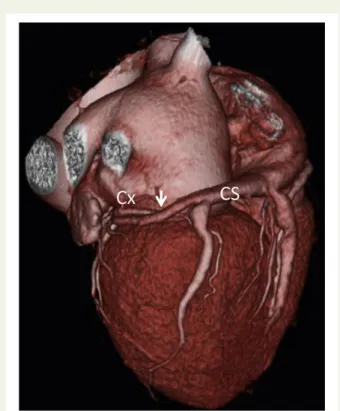
Advanced imaging techniques, including cardiac magnetic resonance (CMR) and multidetector row computed tomography (MDCT), can provide complementary information in patients with MR. Both CMR and MDCT permit assessment of LA and LV volumes, function, sphericity, and scar tissue. Given its high spatial resolution, MDCT
can accurately delineate MV anatomy (Figure9)59,61and is uniquely
useful in demonstrating the size and course of the coronary sinus in relation to the mitral annulus and circumflex coronary artery
(Figure10), which is an important consideration for some
transcath-eter MV devices.62CMR may have particular value in the precise
quantification of MR (Figure11)63; however, like all other imaging
modalities, the accuracy of CMR in assessing MR severity is reduced
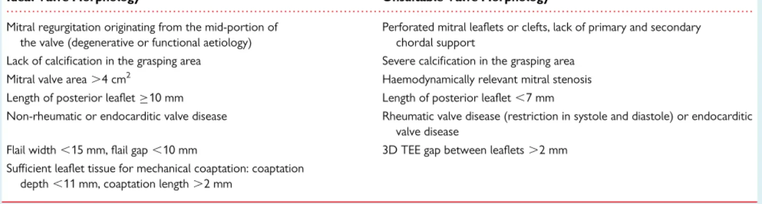
. . . . Table 5 Relationship between the morphological characteristics of the mitral valve and suitability for the mitraclip procedure
Ideal Valve Morphology Unsuitable Valve Morphology
Mitral regurgitation originating from the mid-portion of the valve (degenerative or functional aetiology)
Perforated mitral leaflets or clefts, lack of primary and secondary chordal support
Lack of calcification in the grasping area Severe calcification in the grasping area Mitral valve area .4 cm2 Haemodynamically relevant mitral stenosis Length of posterior leaflet≥10 mm Length of posterior leaflet ,7 mm
Non-rheumatic or endocarditic valve disease Rheumatic valve disease (restriction in systole and diastole) or endocarditic valve disease
Flail width ,15 mm, flail gap ,10 mm 3D TEE gap between leaflets .2 mm Sufficient leaflet tissue for mechanical coaptation: coaptation
depth ,11 mm, coaptation length .2 mm
Adapted with permission from Wunderlich et al.49
3D, 3-dimensional; TEE, transoesophageal echocardiography.
Table 4 Unfavourable transthoracic
echocardiographic characteristics for surgical mitral valve repair in secondary mitral regurgitation
1. Mitral valve remodelling † Coaptation distance≥10 mm † Tenting area .2.5– 3.0 cm2
† Complex regurgitant jets † Posterolateral angle .458 2. Local left ventricular remodelling
† Interpapillary muscle distance .20 mm † Posterior papillary-fibrosa distance .40 mm † Lateral wall motion abnormality
3. Global left ventricular remodelling † End-diastolic diameter . 65 mm
† End-systolic diameter .51 mm (end-systolic volume .140 ml) † Systolic sphericity index .0.7
Adapted with permission from Lancellotti et al.29
G.W. Stone et al.
12
by guest on July 14, 2015
in the setting of atrial fibrillation. In the future it is likely that CMR and MDCT will be increasingly used for pre-procedural assessment and planning of both surgical and transcatheter MR repair and replacement procedures, and post-intervention surveillance.
Control group therapies
Selection of the appropriate control group is essential to inter-preting the benefit-risk profile of a new device. For randomized MR clinical device trials, 3 control groups may be considered:
Figure 6 Assessment of mitral valve morphology with 3-dimensional transoesophageal echocardiography in primary mitral regurgitation. (A) LA “en face” view of the normal mitral valve with anterior and posterior mitral leaflets divided in 3 scallops (A1-P1: lateral; A2-P2: central; A3-P3: medial). (B) Prolapse of the anterior mitral leaflet with flail of the A2 scallop (arrow). (C) Isolated prolapse of the P2 scallop. (D and E) Examples of prolapse of the anterior and posterior commissures (arrows), respectively. The aortic valve (Ao) and the left atrial appendage (LAA) are landmarks for orientation of the LA “en face” view of the mitral valve. Abbreviations as in Figure5.
Figure 7 Measurement of mitral leaflets and annulus dimensions from 3-dimensional transoesophageal echocardiography. Accurate measure-ments of the mitral leaflets and annulus can be obtained by creating 3-dimensional (3D) reconstructions of the mitral valve from 3D transoeso-phageal echocardiography data. The multiplanar reformation planes are aligned across the mitral annulus (A) providing LV outflow tract, bicommissural, and cross-sectional views of the mitral valve. (B) By tracing the leaflets and determining the mitral annulus landmarks, the 3D models are created, and the post-processing software provides semiautomatic measurements of the mitral leaflets and annulus. Reproduced with permission from Shanks et al.59LA ¼ left atrium; LV ¼ left ventricle; other abbreviations as in Figure5.
by guest on July 14, 2015
(1) GDMT alone (with or without a sham control) when GDMT is standard of care; (2) GDMT plus surgical therapy when surgical therapy is standard of care; and (3) GDMT plus an active compara-tor device if an alternative device is available and is considered a standard of care.
Ensuring the use of appropriate GDMT is a requirement for all patients enrolled in randomized controlled trials and registries. It is the basis upon which the safety and incremental efficacy of pro-cedural therapies may be judged. GDMT in symptomatic patients with severe MR includes treatments for heart failure (for all patients with secondary MR due to LV dysfunction, and for those with primary MR with symptoms of heart failure or volume overload (class D), especially those in whom surgery is not performed or
will be delayed).1GDMT includes not only the use of specific
recommended therapies, but also titration of those therapies to re-commended target doses, as tolerated. Optimal GDMT use before study enrolment minimizes the likelihood of major changes in medi-cation dosing during the course of a trial, defined for each drug class
as an increase in dose by≥100% or decrease in dose by ≥50% from
baseline. Thus, patients should meet pre-defined GDMT dosing sta-bility criteria before randomization, as the initiation, discontinuation, or titration of therapies after randomization (in either the treatment of control groups) may otherwise seriously confound interpretation of the study results. Although it may not be possible to always prevent major changes in drug dosing (e.g. after improvement in hemodynamics with effective MR therapy), in general such changes should be minimized to isolate the effect of the randomized treatment, unless they are pre-specified and considered as part of
the treatment arm strategy (including, for instance, a prospective approach to reduction of heart failure medications).
Achieving and maintaining maximally tolerated guideline recommended doses of beta-blockers, angiotensin-converting enzyme inhibitors or angiotensin receptor blockers, and mineralo-corticoid receptor antagonists is especially important before enrolment in secondary MR trials, as reduction in LV dimensions and LV remodelling with effective medical therapy in heart failure may substantially reduce MR in individual patients, obviating the need for advanced or experimental therapies. MR severity and appropriateness for study eligibility should be reassessed at least 30 days (and preferably 90 days) after any major change in GDMT.
Compliance with optimal GDMT in individual patients is often challenging and should be documented at baseline and throughout the course of the study. Before enrolment, the adequacy of GDMT in individual patients (including drug class, dose, and patient compli-ance) should be verified by a central eligibility committee to reduce bias associated with subjects changing their behaviour under obser-vation post-enrolment (Hawthorne type effect) (see also Role of the Central Eligibility Committee). Intolerance to a drug or drug class or limitation in drug dosing should be on the basis of objective clinical criteria, according to the known adverse effects of specific agents, and must be well-documented in the medical chart and study case report form. Examples include symptomatic hypotension with angiotensin-converting enzyme inhibitors, hyperkalemia with min-eralocorticoid receptor antagonists, and symptomatic bradycardia with beta-blockers.
Figure 8 Transoesophageal echocardiogram evaluation of mitraclip implantation in a patient with severe secondary mitral regurgitation. From the midesophageal 4-chamber (A) and bicommissural (B) views, the vena contracta width of the central regurgitant jet can be measured. The 3D LA “en face” view shows lack of coaptation between the anterior and posterior mitral leaflets at the central level (C, arrows). With 3D colour Doppler data, the convergence flow can be observed along the coaptation line from the LV view (D, arrows). Three MitraClip devices were suc-cessfully implanted with significant reduction of MR as observed from the colour Doppler biplane views of the MV (E). On 3D transoesophageal echocardiogram full volume of the mitral valve, the LA “en face” view shows a double orifice mitral valve after MitraClip implantation (F). The clips were positioned at the central and anterolateral levels (arrow) leading to a large orifice at the posteromedial level and a small anterolateral orifice (F, asterisks). (G) The colour Doppler 3D “en face” view of the mitral valve with 2 residual mild regurgitant jets. Abbreviations as in Figures5and7.
G.W. Stone et al.
14
by guest on July 14, 2015
In addition to GDMT for heart failure, appropriate patients should also be treated with biventricular pacing (CRT) and coronary revascularization when substantial ischaemia is present, according to contemporary clinical practice guidelines, such as those from the American College of Cardiology Foundation/American Heart
Asso-ciation1,50and the European Society of Cardiology/European
Asso-ciation for Cardio-Thoracic Surgery.2CRT is indicated (Class I) in
patients with NYHA functional class II to IV symptoms on GDMT
with LVEF≤35%, sinus rhythm, a left bundle branch block pattern,
and QRS duration≥150 ms.50In such patients, CRT may
substan-tially decrease LV dimensions and reduce MR in as many as 50% of
patients.64–67CRT may also be considered (Class IIa) for selected
patients with a left bundle branch block pattern and QRS duration ,150 ms, and for those with a non-left bundle branch block and
QRS duration≥150 ms (Class IIa).50Surgical or percutaneous
cor-onary revascularization in patients with substantial ischaemia may also, on occasion, reduce secondary MR and should be performed
in appropriate patients before study enrolment.68,69After CRT or
coronary revascularization, at least 30 days (and preferably 90 days) should pass, after which TTE or other relevant imaging tests are repeated to assess MR severity and appropriateness for study
eligibility. Similar to optimal GDMT use, whether CRT and/or cor-onary revascularization are indicated and utilized should be verified by the central eligibility committee before study enrolment.
Appropriate scenarios for
guideline-directed medical therapy alone (with or
without a sham) as the control group
GDMT should be used alone as the comparator (control) group when a surgical comparator is either not indicated (i.e., is not stand-ard of care) or is contraindicated due to high surgical risk, and no other active comparator exists. Examples of this scenario are seen in recent studies of TAVR for critical aortic stenosis in extreme
sur-gical risk patients70and from a single arm registry of the MitraClip
for primary MR in prohibitive surgical risk patients.35Another
ex-ample comes from the ongoing COAPT (Cardiovascular Outcomes Assessment of the MitraClip Percutaneous Therapy for Heart Fail-ure Patients with Functional Mitral Regurgitation) trial of the Mitra-Clip for severe secondary MR in chronic heart failure patients (NCT01626079). Although some practice variability exists in this setting, GDMT (rather than MV surgery) is considered the default
Figure 9 Multidetector row computed tomography for assessment of mitral valve geometry in secondary mitral regurgitation. From the recon-structed short-axis view of the mitral valve, orthogonal planes can be placed across the anterolateral, central, and posteromedial levels of the MV leading to the left ventricular outflow tract view at each level. The angles (Aaand Pa) and tenting (MVTht) of the mitral leaflets can be measured at the anterolateral (A1-P1), central (A2-P2), and posteromedial (A3-P3). Reproduced with permission from Delgado et al.61AC ¼ anterior com-missure; Ao ¼ aorta; PC ¼ posterior comcom-missure; RA ¼ right atrium; RVOT ¼ right ventricular outflow tract.
by guest on July 14, 2015