O
pen
A
rchive
T
oulouse
A
rchive
O
uverte
(OATAO)
OATAO is an open access repository that collects the work of some Toulouse researchers and makes it freely available over the web where possible.This is
an author'sversion published in:
https://oatao.univ-toulouse.fr/23066Official URL : https://doi.org/10.1007/s00402-017-2796-6
To cite this version :
Any correspondence concerning this service should be sent to the repository administrator: [email protected]
Bayle, Xavier and Pham, Thuy-Trang and Faruch, Marie and Gobet, Aurelie and Mansat, Pierre and Bonnevialle, Nicolas No difference in outcome for open versus arthroscopic
rotator cuff repair: a prospective comparative trial. (2017) Archives of Orthopaedic and
Trauma Surgery, 137 (12). 1707-1712. ISSN 0936-8051
OATAO
Open Archive Toulouse Archive Ouverte
No difference in outcome for open versus arthroscopic rotator cuff
repair: a prospective comparative trial
Xavier Bayle1 · Thuy‑Trang Pham1 · Marie Faruch2 · Aurelie Gobet3 ·
Pierre Mansat1 · Nicolas Bonnevialle1,4
p = 0.3), ASES score (88 vs 91 points; p = 0.3), and SSV
(81 vs 85%). The overall rate of re-tear (Sugaya type IV or V) reached 7 and 9%, respectively, in group A and O (p = 0.8).
Conclusion This study did not prove any difference of
arthroscopic over open surgery in case of rotator cuff repair regarding clinical outcome and cuff integrity at 1-year follow-up.
Level II Prospective comparative study.
Keywords Open surgery · Arthroscopy · Rotator cuff
repair · Shoulder · Re-tear · Clinical outcomes
Introduction
Currently all arthroscopic technique trends to establish itself as the gold standard for rotator cuff repair. In 2005, it was reported that AAOS surgeons performed cuff repairs with arthroscopic, mini-open, and open technique in 14.5, 46.2, and 36.6%, respectively [1]. In 2012, only 15.7% of sur-geons’ respondent to ASES survey still used an open tech-nique [2].
Only few studies compared open and arthroscopic rota-tor cuff repair and it remains unclear that arthroscopic is superior to open technique in term of clinical outcomes and tendon healing [3–8].
Therefore, the purpose of the study was to compare the postoperative clinical results and structural integrity of rota-tor cuff repairs performed either open or arthroscopically. We hypothesized that both techniques would achieve equiva-lent clinical outcomes and tendon repair integrity.
Abstract
Purpose Arthroscopic techniques tend to become the
gold standard in rotator cuff repair. However, little data are reported in the literature regarding the improvement of post-operative outcomes and re-tear rate relative to conventional open surgery. The aim of this study was to compare clinical outcomes and cuff integrity after arthroscopic versus open cuff repair.
Methods We prospectively assessed clinical outcomes and
cuff integrity after an arthroscopic or open rotator cuff repair with a minimum follow-up of 12 months. Clinical evaluation was based on Constant score, Simple Shoulder Value (SSV) and American Shoulder and Elbow Score (ASES). Rotator cuff healing was explored with ultrasound.
Results 44 patients in arthroscopic group A (mean age
56-year-old) and 43 in open group O (mean age 61-year-old) fulfilled the inclusion criteria. Tendons were repaired with a single row technique associated with biceps tenodesis and subacromial decompression. All objective clinical scores significantly improved postoperatively in both groups. No statistical difference was identified between group A and O regarding, respectively, Constant score (72 vs 75 points;
IRB approval: Approved study on number 17-0313. * Nicolas Bonnevialle
[email protected]; [email protected] 1 Department of Orthopaedic Surgery, Hôpital Riquet, Place
Baylac, 31059 Toulouse, France
2 Department of Radiology, Hôpital Riquet, Place Baylac, 31059 Toulouse, France
3 Department of Epidemiology and Public Health, Hôpital Pellegrin, Place Amélie Raba Leon, 33076 Bordeaux, France 4 Centre Hospitalier Universitaire de Purpan, Place Baylac,
Methods
Study design and patients
The study was a prospective comparative cohort analysis of rotator cuff repairs between 2012 and 2013. Inclusion criteria were: patients operated on for a primary rotator cuff repair of supraspinatus and/or infraspinatus tendons tears, and reviewed with clinical examination and ultrasound eval-uation with a minimum follow-up of 12 months.
Exclusion criteria were: prior surgical procedure on the shoulder, cuff tear extended to subscapularis tendon (full thickness tear), irreparable tears, and degenerative or inflam-matory arthritis of the glenohumeral joint.
The institutional review board approved the research pro-tocol (number 17-0313).
Eighty-seven patients were consecutively included and enrolled in an arthroscopic surgery (group A) or open sur-gery group (group O) according to the surgeon who per-formed the procedure (NB for arthroscopic and PM for open surgery).
Surgical technique
Patients received a preoperative interscalene block (single injection of Ropivacaine 7.5 mg/mL) and were placed in a beach-chair position under general anaesthesia in both group.
Arthroscopic technique: after intra-articular examina-tion, a biceps tenotomy or tenodesis was done in every case because of tenosynovitis or instability. A subacromial decompression was performed systematically with a burr and the footprint on the greater tuberosity was abraded before tendon fixation. The cuff repair was performed with a single row of double-loaded threaded suture anchors placed lateral to the greater tuberosity with a tension band technique as described by Boileau et al. [5].
Open technique
Through a superior approach the anterior body of the deltoid was detached from the acromion to perform a subacromial decompression with an osteotome. The long head of the biceps was fixed to the transverse ligament in the groove, after the intra-articular portion has been resected. Then, the cuff was repaired onto the abraded greater tuberosity with trans osseous number 2 non absorbable sutures and Masson-Allen stitches. At the end of the procedure the deltoid was reattached to the acromion with trans osseous sutures.
Postoperative protocol
In both group a sling was placed for 6 weeks and passive range of motion as well as pendulum exercises were author-ized from the day after the surgery. A physiotherapist con-trolled the rehabilitation program from the 2nd week to the 3rd month. Active motion started at 6 weeks. No heavy labour or activities were allowed before the 6 months.
Clinical assessment
All the patients were evaluated by an independent observer (XB or TTP) preoperatively, at 3 months, 6 months and 1 year after the surgery. At last follow-up, Constant score, American Shoulder and Elbow Surgeons (ASES) score and Simple Shoulder Value (SSV) were used [9, 10]. For active range of motion, elevation was measured with a goniometer (patient in a seated position) in the sagittal plane, external rotation was measured in the coronal plane with the arm by the patient’s side, and internal rotation was measured by level when the patient was asked to reach behind to the highest vertebral level.
Radiological assessment
Preoperatively, tendon retraction was staged on MRI or arthro-CT scan according to Patte’s classification and fatty degeneration according to Gouttalier [11, 12]. At follow-up, ultrasonographic evaluation was performed by two radiolo-gists specialized in musculoskeletal disorders in a blinded double-check. Cuff repair integrity was classified into five types following Sugaya classification adapted for ultrasound [13]: type I, sufficient and homogeneous thickness; type II, sufficient, but heterogeneous thickness; type III, less than half the thickness without discontinuity; type IV, minor discontinuity; and type V, major discontinuity. In case of disagreement between the radiologists, the worst grade of healing was taken.
Statistical analysis
Descriptive statistical analysis consisted of means, maxi-mum, minimum and standard deviations for continuous variables and percentages for discrete variables. Statistical comparisons between groups were performed with paired t tests or Wilcoxon test for continuous variables and the χ2 or
Fisher test for categorical variables. The significant differ-ence was set at p < 0.05. All analyses were performed with the use of SAS software (SAS Institute, Cary, NC).
Results
Patients demographics
One hundred and eighteen patients were operated on for rotator cuff repair during the period of the study. Eighteen were excluded and 13 were lost before the minimum follow-up of 12 months, leaving 44 patients in grofollow-up A and 43 in group O available for analysis.
As documented in Table 1, there was no statistical differ-ence between group A and O regarding gender, trauma inci-dence, work compensation context, tobacco use, diabetes,
dominance side involved, active range of motion, preop-erative Constant score, ASES score, and SSV. However, patients of group O were older and with smaller tears in group A (Table 2).
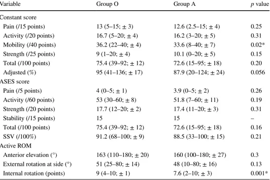
Clinical assessment
At mean follow-up of 13 months (12–17), Constant score, ASES score and SSV significantly improved post-operatively in both groups. No significant difference was detected between the two groups regarding total Constant score, ASES score, SSV (Table 3). However, active internal
Table 1 Preoperative analysis
of group O and group A
* Significant difference at p < 0.05 Group O (n = 43) Group A (n = 44) p Age (years) 61.6 (43–78; ± 8) 55.6 (37–72; ± 7) 0.0026* Gender (female/male) 17/26 20/24 0.57 Worker compensation 7 (16%) 10 (23%) 0.45 Traumatic etiology 13 (30%) 13 (30%) 0.94 Tobacco use 6 (14%) 10 (23%) 0.29 Diabetes 3 (7%) 4 (7%) 0.77
Dominant side involved 30 (70%) 36 (82%) 0.19
Mobility
Elevation 121 (60–180; ± 34) 147 (60–180; ± 36) 0.65
External rotation at side (°) 39 (10–90; ± 15) 42 (10–80; ± 15) 0.38 Internal rotation (points) 6 (2–10; ± 3) 6 (2–10; ± 3) 0.9 Functional scores
Total constant (points) 42 46 0.27
Adjusted constant (%) 52.1 55.5 0.42
ASES (points) 61.2 61.4 0.95
SSV (%) 42.4 45.9 0.45
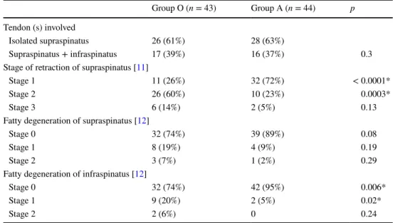
Table 2 Preoperative analysis
of tear patterns in group O and group A * Significant difference at p < 0.05 Group O (n = 43) Group A (n = 44) p Tendon (s) involved Isolated supraspinatus 26 (61%) 28 (63%) Supraspinatus + infraspinatus 17 (39%) 16 (37%) 0.3
Stage of retraction of supraspinatus [11]
Stage 1 11 (26%) 32 (72%) < 0.0001*
Stage 2 26 (60%) 10 (23%) 0.0003*
Stage 3 6 (14%) 2 (5%) 0.13
Fatty degeneration of supraspinatus [12]
Stage 0 32 (74%) 39 (89%) 0.08
Stage 1 8 (19%) 4 (9%) 0.19
Stage 2 3 (7%) 1 (2%) 0.29
Fatty degeneration of infraspinatus [12]
Stage 0 32 (74%) 42 (95%) 0.006*
Stage 1 9 (20%) 2 (5%) 0.02*
rotation was significantly better in group O, as well as the mobility according the Constant score.
Tendon integrity
The rate of re-tear (stage IV and V) was 7 and 9%, respec-tively, for group A and O (p = 0.8) (Table 4).
Complications
One patient in each group developed capsulitis. Neither patient required further surgical intervention. No compli-cation related to the deltoid detachment was observed in group O. No patient needed revision surgery (even in case of tendon re-tear) at the last follow-up.
Discussion
This study compared open and arthroscopic cuff repair in a prospective case series. Because arthroscopic technique became the gold standard over the past decade, the debate is to know if open surgery is still warranted or not. Therefore, we demonstrated that there was not any significant difference
between both techniques, regarding functional outcomes and cuff integrity at the minimum follow-up of 12 months.
This study has some limitations. Firstly, patients were not randomly included in arthroscopic or open group. Thereby, two different surgeons performed two different technique of cuff repair. However, each of them had a wide experi-ence in his own technique and could attest of its reproduc-ibility. This allowed to compare properly the outcomes of both technique. Secondly, the number of patients lost of follow-up was high (13 patients). The main reason was the lack of clinical and ultrasound examination at 12 months for many of them. Because the number of patient lost was equivalent in each group (group O n = 7; group A n = 6), this bias should not impact our comparative analysis and as far as the 6th postoperative month, none of them needed a revision surgery or complained of unsatisfactory results. In addition, both group were not perfectly similar in terms of age and tear size, which could have specifically an impact on tendon healing and clinical outcomes. Finally, the follow-up for clinical and ultrasound evaluation was only 1 year. How-ever, it was previously reported that re-tears mainly occur during the first postoperative year [14, 15].
Previously several studies compared arthroscopic and mini-open surgery, and did not find any statistical differences
Table 3 Postoperative
outcomes
* Significant difference at p < 0.05
Variable Group O Group A p value
Constant score Pain (/15 points) 13 (5–15; ± 3) 12.6 (2.5–15; ± 4) 0.25 Activity (/20 points) 16.7 (5–20; ± 4) 16.2 (3–20; ± 5) 0.31 Mobility (/40 points) 36.2 (22–40; ± 4) 33.6 (8–40; ± 7) 0.02* Strength (/25 points) 9 (1–20; ± 4) 10.1 (0–20; ± 5) 0.15 Total (/100 points) 75.4 (39–92; ± 12) 72.6 (15–95; ± 18) 0.20 Adjusted (%) 95 (41–136; ± 17) 87.9 (20–124; ± 24) 0.056 ASES score Pain (/5 points) 4 (0–5; ± 1) 3.9 (0–5; ± 2) 0.26 Activity (/60 points) 53 (30–60; ± 8) 51.8 (7–60; ± 11) 0.19 Strength (/20 points) 17.7 (12–20; ± 2) 17.4 (11–20; ± 3) 0.31 Stability (/15 points) 15 15 – Total (/100 points) 75.4 (39–92; ± 12) 72.6 (15–95; ± 18) 0.16 SSV (/100%) 91.2 (68–100; ± 9) 88.5 (33–100; ± 15) 0.21 Active ROM Anterior elevation (°) 163 (110–180; ± 20) 160 (100–180; ± 27) 0.3 External rotation at side (°) 51 (25–80; ± 14) 48 (10–80; ± 16) 0.13 Internal rotation (points) 9 (4–10; ± 1) 7.6 (2–10; ± 3) 0.001*
Table 4 Ultrasound
evaluation in both group and results according Sugaya’s classification
Type I Type II Type III Type IV Type V
Group O (n = 43) 26% (n = 11) 63% (n = 27) 2% (n = 1) 9% (n = 4) 0 Group A (n = 44) 43% (n = 19) 50% (n = 22) 0 7% (n = 3) 0
in outcomes measurement [16–22]. Only few studies com-pared clearly open surgery to all arthroscopic procedure. Buess et al. [6] compared clinical results after open and arthroscopic rotator cuff repair. They concluded that arthro-scopic cuff repair yielded equal or better results than open. However, the open group mixed conventional technique and mini-open (12 and 18 patients, respectively). In the same way, Bishop et al. [4] constructed in their prospective study a heterogeneous cohort of 24 open and 8 mini-open surgery compared to an arthroscopic cohort. They suggested that open technique should be chosen for large tears because of a lower rate of re-tears. Walton and Murrel [8] published a large series of 200 true open and 200 arthroscopic cuff repair. They excluded lesion that exceeded 16 cm2 and
did not report how they managed long head of the biceps’ pathologies. A decompression was performed systematically in open group compared to 76% in arthroscopic group, and two different configurations of single row arthroscopic repair were used. However, they concluded that arthroscopic repair was superior to open regarding operative time, cuff integrity, and speed of postoperative recovery.
In our study, the clinical evaluation at follow-up did not highlight any difference between the two groups, except a greater active internal rotation after open surgery. However, this parameter did not influence overall clinical scores. This finding was contrary to many studies which were in favour of better range of motion after arthroscopic technique [6, 8]. However, it was observed that passive range of motion con-verging at 2 years of follow-up between both technique [8]. One relevant information of this study is that open sur-gery did not increase the rate of postoperative complications. We did not identify clinical deltoid impairment despite the detachment of its proximal origin [23]. This finding con-firmed the study of Cho et al. [24] who explored with MRI postoperative thickness of the deltoid after open and arthro-scopic surgery and did not find out any difference between both techniques.
However, in a large series of more than 1800 cuff repairs, Vopat et al. [25] reported that open technique was more at risk of infection than arthroscopic surgery, as well as to be a male and to have a high body mass index. Owens et al. [26] confirmed a higher incidence of complication and hospital readmission after open cuff repair in a veteran population.
Conclusion
This study did not demonstrate any difference of arthro-scopic over open surgery in case of rotator cuff repair regard-ing clinical outcome and cuff integrity at 1-year follow-up. Moreover, the rate of postoperative complication remained low in both group and this cannot be an argument to support one technique.
Compliance with ethical standards
Financial biases None.
Conflict of interest The authors declare that they have no conflict
of interest.
References
1. Dunn WR, Schackman BR, Walsh C et al (2005) Variation in orthopaedic surgeons’ perceptions about the indications for rotator cuff surgery. J Bone Jt Surg Am 87:1978–1984
2. Acevedo DC, Paxton ES, Williams GR, Abboud JA (2014) A survey of expert opinion regarding rotator cuff repair. J Bone Jt Surg Am 16(96):e123
3. Adla DN, Rowsell M, Pandey R (2010) Cost-effectiveness of open versus arthroscopic rotator cuff repair. J Shoulder Elbow Surg 19:258–261
4. Bishop J, Klepps S, Lo IK, Bird J, Gladstone JN, Flatow EL (2006) Cuff integrity after arthroscopic versus open rotator cuff repair: a prospective study. J Shoulder Elbow Surg 15:290–299 5. Boileau P, Brassart N, Watkinson DJ, Carles M, Hatzidakis AM,
Krishnan SG (2005) Arthroscopic repair of full-thickness tears of the supraspinatus: does the tendon really heal? J Bone Jt Surg Am 87:1229–1240
6. Buess E, Steuber KU, Waibl B (2005) Open versus arthroscopic rotator cuff repair: a comparative view of 96 cases. Arthroscopy 21:597–604
7. Ide J, Maeda S, Takagi K (2005) A comparison of arthroscopic and open rotator cuff repair. Arthroscopy 21:1090–1098 8. Walton JR, Murrell GA (2012) A two-year clinical outcomes study
of 400 patients, comparing open surgery and arthroscopy for rota-tor cuff repair. Bone Jt Res 1:210–217
9. Constant CR, Murley AH (1987) A clinical method of functional assessment of the shoulder. Clin Orthop Relat Res 214:160–164 10. Richards RR, An KN, Bigliani LU, Friedman RJ et al (1994) A standardized method for the assessment of shoulder function. J Shoulder Elbow Surg 3:347–352
11. Patte D (1990) Classification of rotator cuff lesions. Clin Orthop Relat Res 254:81–86
12. Goutallier D, Postel JM, Bernageau J, Lavau L, Voisin MC (1994) Fatty muscle degeneration in cuff ruptures. Pre- and postoperative evaluation by CT scan. Clin Orthop Relat Res 304:78–83 13. Sugaya H, Maeda K, Matsuki K, Moriishi J (2007) Repair
integ-rity and functional outcome after arthroscopic double-row rota-tor cuff repair. A prospective outcome study. J Bone Jt Surg Am 89:953–960
14. Kim JH, Hong IT, Ryu KJ, Bong ST, Lee YS, Kim JH (2014) Retear rate in the late postoperative period after arthroscopic rota-tor cuff repair. Am J Sports Med 42:2606–2613
15. Iannotti JP, Deutsch A, Green A et al (2013) Time to failure after rotator cuff repair: a prospective imaging study. J Bone Jt Surg Am 95(11):965–971
16. Ji X, Bi C, Wang F, Wang Q (2015) Arthroscopic versus mini-open rotator cuff repair: an up-to-date meta-analysis of rand-omized controlled trials. Arthroscopy 31:118–124
17. Kim SH, Ha KI, Park JH, Kang JS, Oh SK, Oh I (2003) Arthro-scopic versus mini-open salvage repair of the rotator cuff tear: out-come analysis at 2 to 6 years’ follow-up. Arthroscopy 19:746–754 18. Lindley K, Jones GL (2010) Outcomes of arthroscopic versus open rotator cuff repair: a systematic review of the literature. Am J Orthop (Belle Mead NJ) 39:592–600
19. van der Zwaal P, Thomassen BJ, Nieuwenhuijse MJ, Lindenburg R, Swen JW, van Arkel ER (2012) Clinical outcome in all-arthro-scopic versus mini-open rotator cuff repair in small to medium-sized tears: a randomized controlled trial in 100 patients with 1-year follow-up. Arthroscopy 29:266–273
20. Youm T, Murray DH, Kubiak EN, Rokito AS, Zuckerman JD (2005) Arthroscopic versus mini-open rotator cuff repair: a com-parison of clinical outcomes and patient satisfaction. J Shoulder Elbow Surg 14:455–459
21. Osti L, Papalia R, Paganelli M, Denaro E, Maffulli N (2010) Arthroscopic vs mini-open rotator cuff repair. A quality of life impairment study. Int Orthop 34:389–394
22. Kasten P, Keil C, Grieser T, Raiss P, Streich N, Loew M (2011) Prospective randomised comparison of arthroscopic versus
mini-open rotator cuff repair of the supraspinatus tendon. Int Orthop 35:1663–1670
23. Gumina S, Di Giorgio G, Perugia D, Postacchini F (2008) Deltoid detachment consequent to open surgical repair of massive rotator cuff tears. Int Orthop 32:81–84
24. Cho NS, Cha SW, Rhee YG (2015) Alterations of the deltoid muscle after open versus arthroscopic rotator cuff repair. Am J Sports Med 43:2927–2934
25. Vopat BG, Lee BJ, DeStefano S et al (2016) Risk factors for infec-tion after rotator cuff repair. Arthroscopy 32:428–434
26. Owens BD, Williams AE, Wolf JM (2015) Risk factors for surgi-cal complications in rotator cuff repair in a veteran population. J Shoulder Elbow Surg 24:1707–1712