O
pen
A
rchive
T
oulouse
A
rchive
O
uverte
(OATAO)
OATAO is an open access repository that collects the work of some Toulouse
researchers and makes it freely available over the web where possible.
This is
an author'sversion published in: https://oatao.univ-toulouse.fr/23073
Official URL :
https://doi.org/10.1007/s00264-017-3456-8
To cite this version :
Any correspondence concerning this service should be sent to the repository administrator:
Elia, Fanny and Azoulay, Vadim and Lebon, Julie and Faraud, Amélie and Bonnevialle,
Nicolas and Mansat, Pierre Clinical and anatomic results of surgical repair of chronic
rotator cuff tears at ten-year minimum follow-up. (2017) International Orthopaedics, 41 (6).
1219-1226. ISSN 0341-2695
OATAO
Open Archive Toulouse Archive Ouverte
Clinical
and anatomic results of surgical repair of chronic
rotator
cuff tears at ten-year minimum follow-up
Fanny Elia1 & Vadim Azoulay1 & Julie Lebon1 & Amélie Faraud1 & Nicolas Bonnevialle1 &
Pierre Mansat1
Abstract
Purpose The purpose of this study was to evaluate long-term clinical and anatomic results after open rotator cuff repair. Materials and method Fifty-three patients were included in this study. Rotator cuff tears were limited to one tendon in 34, and two tendons or more in 19. The supraspinatus tendon was involved alone in 30 cases, and the subscapularis in four. Clinical, radiographic and MRI evaluations were performed for all patients at ten-year minimum follow-up.
Results At 11.4-years average follow-up, satisfactory results were obtained with improvement on pain level, in range of motion with a gain in active anterior elevation and in external rotation as well as in strength in abduction. Absolute Constant score reached 74.7 points and adjusted Constant score 99.6%. SSV was 82.5% with a SST score of 10 points. Osteoarthritis changes increased with follow-up with osteoarthritis lesion in 69% of the cases. MRI evaluation found a re-tear of the repair
in 42% of the cases: 30% when only the supraspinatus tendon was initially involved, and 63% when two or more tendons were repaired. Tear size and quality of the repair were the most important prognosis factors for re-tear of the repair. No corre-lation was found between the occurrence of a new tear and the clinical outcome.
Conlcusion Surgical repair of chronic rotator cuff tendon tear can produce consistent and lasting pain relief and improve-ment in range of motion. Re-tear of the repair was not corre-lated with degradation of clinical results. Best results were obtained for isolated supraspinatus tear with a complete sur-gical repair.
Keywords Rotator cuff tear . Fatty infiltration . Muscle atrophy . Prognosis factors
Introduction
Surgical repair of chronic rotator cuff tears is one of the most common surgical procedures for shoulder surgeons. Performed open or under arthroscopy clinical results are often satisfactory. However, anatomical studies have shown in-creased incidence of tendon re-tear with follow-up. Re-tear rate varied from 20 to 50% depending of the studies [1–4]. Failure of tendon healing or tendon re-tear is not always cor-related to functional impairment, with often a painless or slight painful shoulder, with a functional range of motion [5]. However, shoulder strength is often decreased [1].
In literature, there are only a few studies already published evaluating clinical and anatomical results of surgical repair of rotator cuff tendon lesions with long-term follow-up [6–8]. In 2001, Cofield et al. [9] published the results of 105 shoulders with rotator cuff repairs at 13.4-year follow-up. However, on-ly the clinical results were evaluated, and no anatomic
* Pierre Mansat
mansat.p@chu toulouse.fr; pierre.mansat@univ tlse3.fr Fanny Elia [email protected] Vadim Azoulay [email protected] Julie Lebon julie [email protected] Amélie Faraud [email protected] Nicolas Bonnevialle [email protected]
1 Département d’Orthopédie et Traumatologie Urgences Mains,
Hôpital Pierre Paul RIQUET, Hôpital Universitaire de Toulouse, Place du Dr Baylac, 31059 Toulouse Cedex, France
evaluation of the repair was performed. More recently in 2015, Saraswat et al. [10] reported the results of 59 open cuff tendon repairs at ten-year follow-up. Similarly, only the clin-ical results and quality of life were evaluated.
A retrospective study has been performed in our depart-ment in order to evaluate patients who underwent a surgical repair of chronic rotator cuff tears in 2003, to analyze clinical results but also anatomical evolution of the repair at minimum year follow-up. Our hypothesis was that at more than ten-year follow-up, repair of rotator cuff tendon tears gave reliable tendon-healing rate, with preservation of satisfactory func-tional range of motion.
Material and methods
Patients demographics
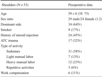
A retrospective study was conducted in our Upper Limb Surgery Department of the University Hospital. Approval was obtained from the local ethics committee under the num-ber: 07–1015. All patients were informed about utilization of their personal data for the study and all approved. Inclusion criteria were all patients that underwent open rotator cuff ten-don repair in our department in 2003, that were reviewed clinically with a shoulder MRI control at minimum ten-year follow-up. Exclusion criteria were patients who had a revision surgery for rotator cuff repair in 2003, or primary open rotator cuff repair in 2003 with less than ten-year follow-up, or with-out MRI shoulder control, or with history of shoulder trauma, shoulder infection, proximal humerus tumour, avascular ne-crosis of the humeral head, muscle transfer, or an isolated acromioplasty without tendon repair. In 2003, open surgical repair of chronic rotator cuff lesions has been performed on 114 patients (115 shoulders). Thirty-two patients were lost for follow-up. Twenty-one were contacted by phone but did not want to come back for evaluation. Nine patients were de-ceased at the time of the study. Finally, 53 patients (53 shoul-ders) were reviewed clinically with a radiographic analysis and MRI evaluation at ten-year minimum follow-up. There were 29 men and 24 women of 59 ± eight years on average at surgery (range, 38–75). The dominant side was involved in 34 cases (64%). All patients’ characteristics are summarized in Table1.
Surgical procedure
Open rotor cuff repair was performed under combined region-al and generregion-al anaesthesia by three senior surgeons. The pa-tient was positioned in the beach chair position and a superior-lateral approach was used. The deltoid was incised between the anterior and lateral part. After section of the coraco-acromial ligament, an acromioplasty was performed.
Resection of the lateral clavicle was performed for treatment of degenerative arthritic changes in only ten cases. After eval-uation of the tendon tear, mobilization of the tendon was per-formed and repair was done by Mason-Allen trans-osseous non-absorbable number two sutures or with anchors. The tears were repaired totally in 45 and partially in eight cases. The long head of the biceps was preserved in 37 and tenodesed in 16. The deltoid was then repaired with non-absorbable sutures.
Post-operative management
Post-operatively patients were immobilized in a sling for 45 days. Pendulum exercises were started at day two, and pas-sive motion was started at day 15 until day 45. Active motion was started at day 45, and strengthening exercises at day 90. Outcome measures
Clinical and radiographic evaluations were performed for all patients. Outcome measures included Constant score, SST, and simple shoulder value (SSV). Range of motion was mea-sured using a goniometer. Strength was evaluated with the shoulder at 90° abduction with a dynamometer.
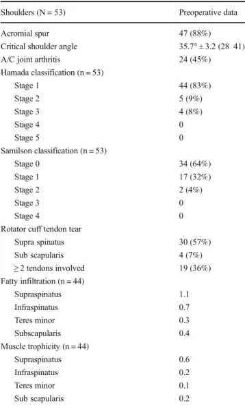
Radiographic analysis was performed pre-operatively and post-operatively using an A/P view as well as a lateral view of the shoulder. The type of the acromion, the acromio-humeral distance (AHD), the critical shoulder angle (CSA), as well as the osteoarthritis changes according to Samilson and Prieto classification were analyzed [11]. Hamada classification was also used pre-operatively [12]. An MRI or an arthro-CT were also performed pre-operatively and were available for 44 patients to evaluate the tear size, the fatty infiltration index (FI) according to Goutallier classification [13], as well as the trophicity of the rotator cuff muscles according to Warner classification [14]. Muscles were considered as
Table 1 Patients’ epidemiology characteristics
Shoulders (N = 53) Preoperative data
Age 59 ± 8 (38 75)
Sex ratio 29 male/24 female (1.2)
Dominant side 34 (64%)
Smoker 9 (17%)
History of steroid injection 24 (45%)
ATC trauma 17 (32%)
Type of activity
Sedentary 31 (58%)
Light manual labor 7 (13%)
Heavy manual labor 12 (23%)
Repetitive activities 3 (6%)
functional when FI was equal or inferior to 2, and non-functional for FI greater than 2. All patients sustained an MRI of the operated shoulder at follow-up. The analyses of the imaging data, to evaluate the cuff integrity and muscle infiltration, were analyzed by one radiologist and one surgeon. The patients’ data and clinical findings were blinded during evaluation of the imaging data. The quality of the repaired tendons was staged according to Sugaya classification [15]. Stages 1, 2 and 3 were considered as intact repair whereas stages 4 and 5 were considered as re-tear. Fatty infiltration index as well as the muscle trophicity were also evaluated at follow-up.
Statistical analysis
Statistical analyses were performed using SPSS 20.0 (128 IBM SPSS, Atlanta, GA). Tests used were related to the type of variable analyzed. Univariate analyses consisted in treating each variable in an isolated way. Bivariate analyses then made it possible to study the links between two variables. Four types of tests could then be considered: chi-square test was carried out to study the link between two variables of the qualitative type, Pearson’s correlation to study the link between two var-iables of the quantitative type, and Student’s t-test and Fisher’s exact or ANOVA to study the link between variables of a quantitative type and variables of a qualitative type. Post-hoc test was also performed to evaluate the link between the two modalities in an isolated manner. The whole of the tests car-ried out at the time of bivariate statistics generated an indicator of significance. Statistical tests were considered significant at p < 0.05.
Results
Pre-operative lesions
All lesions characteristics are summarized in Table2. Rotator cuff tears were limited to one tendon in 34, and two tendons or more in 19. The supraspinatus tendon was involved alone in 30 cases, and the subscapularis in four. According to Goutallier et al. [13], the fatty infiltration index was 1.1 for the supraspinatus, 0.7 for the infraspinatus, 0.3 for the teres minor, and 0.4 for the subscapularis. According to Warner et al. [14], muscle trophicity of the supraspinatus was 0.6, 0.2 for the infraspinatus, 0.1 for the teres minor, and 0.2 for the subscapularis.
Overall results
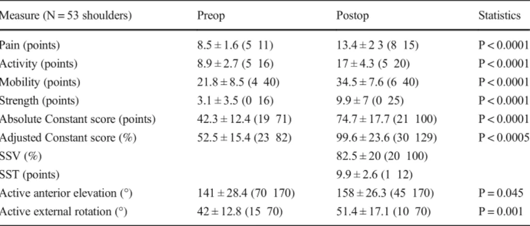
At 11.4-year follow-up (range, 10.8–12), patients were still satisfied from the surgery with SSV of 82.5% ± 20 (range, 20–100), and SST score of 9.9 ± 2.6 points (range, 1–12).
The absolute Constant score was 74.7% ± 17 (range, 21– 100) with an adjusted score of 99.6% ± 23 (range, 30–129), significantly better than before surgery (p < 0.0001 and p < 0.0005, respectively). Overall results are summarized in Table3.
Clinical results
Satisfactory pain relief was still remaining with pain score of 13.4 ± 2.3 points (range, 8–15) compared to 8.5 ± 1.6 points (range, 5–11) pre-operatively (p < 00001). Range of motion was significantly better at follow-up than before surgery with active anterior elevation of 158° ± 26 (range, 45–170) com-pared to 140° ± 28 (range, 70–170) (p = 0.045) and with ex-ternal rotation of 51.4° ± 17 (range, 10–70) compared to 42° ± 13 (range, 15–70) (p = 0.001). Strength in abduction was im-proved as well from 3.1 kg ± 3.5 (range, 0–16) pre-operatively
Table 2 Patients’ shoulder imaging analysis characteristics
Shoulders (N = 53) Preoperative data
Acromial spur 47 (88%)
Critical shoulder angle 35.7° ± 3.2 (28 41)
A/C joint arthritis 24 (45%)
Hamada classification (n = 53) Stage 1 44 (83%) Stage 2 5 (9%) Stage 3 4 (8%) Stage 4 0 Stage 5 0 Samilson classification (n = 53) Stage 0 34 (64%) Stage 1 17 (32%) Stage 2 2 (4%) Stage 3 0 Stage 4 0
Rotator cuff tendon tear
Supra spinatus 30 (57%) Sub scapularis 4 (7%) ≥ 2 tendons involved 19 (36%) Fatty infiltration (n = 44) Supraspinatus 1.1 Infraspinatus 0.7 Teres minor 0.3 Subscapularis 0.4 Muscle trophicity (n = 44) Supraspinatus 0.6 Infraspinatus 0.2 Teres minor 0.1 Sub scapularis 0.2
to 9.9 kg ± 7 (range, 0–25) post-operatively (p < 0.00001). All clinical results are summarized in Table3.
Radiographic results
Radiographic analysis showed preservation of a normal acromio-humeral distance in most cases with a distance aver-aging 7.8 ± 2.2 mm (range, 2–13). Osteoarthritis changes in-creased with follow-up with osteoarthritis lesion in 69% of the cases (p = 0.041). Radiographic results are summarized in Table4.
MRI analysis
MRI evaluation found a re-tear of the repair in 42% of the cases. It was 30% when only the supraspinatus tendon was initially involved, and 63% when two or more tendons were initially repaired (p = 0.031). Fatty infiltration index increased significantly between the pre-operative evaluation and the analysis at follow-up. Fatty degeneration index increased in 28 cases out of 53 (53%) for the supraspinatus and the teres minor and in 32 cases out of 53 (60%) for the infraspinatus and the subscapularis. The muscles were considered as func-tional in 82% of the cases (FI≤ 2), and poorly functional in 18% (FI > 2). Muscle trophicity did not worsen between the pre-operative evaluation and the analysis at follow-up with a normal trophicity in 63% of the cases, and a decreased trophicity on 37% of the cases. Trophicity was normal in 19 cases for the supraspinatus, 37 cases for the infraspinatus, 44 cases for the teres minor and 37 cases for the subscapularis. MRI results are summarized in Table4.
Prognosis factors Re tear rate
Tear size (p = 0.031) and quality of the repair (p = 0.002) were the most important prognosis factors for re-tear of the repair.
Pre-operative strength (p = 0.02), as well as muscle trophicity of the supraspinatus (p = 0.030) and the infraspinatus (p = 0.002) have also influenced significantly the re-tear rate at follow-up. Age at surgery, had also influenced the incidence of re-tear, with a rate of 34.5% for patients under 60 years (29 patients), 37% for patients between 60 and 65 years (11 pa-tients), 72% for patients between 65 and 70 years (5 patients) and 73% for patients above 70 years (8 patients). However, the difference was not statistically significant (p = 0.078). Gender, smoking habits, steroid injections, dominant side, history of trauma, work compensation, Hamada classification, pre-operative osteoarthritis, pre-operative Constant score, fat-ty infiltration index, and surgical technique did not influence the re-tear rate at follow-up.
Post operative Constant score
There was no correlation between the occurrence of a new tear and the clinical outcome as most of the patients kept a satis-factory functional score (p = 0.512). Post-operative Constant score was only correlated with pre-operative active anterior elevation (p = 0.004), pre-operative Hamada classification (p = 0.002), and to quality of tendon repair (complete or par-tial) (p = 0.004).
Best results were obtained for isolated supraspinatus tear with normal pre-operative trophicity with a complete surgical repair.
Discussion
Results of surgical repair of chronic rotator cuff tears are often satisfactory. However, anatomical studies have shown in-creased incidence of tendon re-tear with follow-up not always correlated with functional impairment [1–5]. In our study, we found that surgical repair of chronic rotator cuff tendon tear can produce consistent and lasting pain relief and improve-ment in range of motion. However, a 42% re-tear rate was
Table 3 Comparison of clinical data preoperatively and at follow up
Measure (N = 53 shoulders) Preop Postop Statistics
Pain (points) 8.5 ± 1.6 (5 11) 13.4 ± 2 3 (8 15) P < 0.0001 Activity (points) 8.9 ± 2.7 (5 16) 17 ± 4.3 (5 20) P < 0.0001 Mobility (points) 21.8 ± 8.5 (4 40) 34.5 ± 7.6 (6 40) P < 0.0001 Strength (points) 3.1 ± 3.5 (0 16) 9.9 ± 7 (0 25) P < 0.0001 Absolute Constant score (points) 42.3 ± 12.4 (19 71) 74.7 ± 17.7 (21 100) P < 0.0001 Adjusted Constant score (%) 52.5 ± 15.4 (23 82) 99.6 ± 23.6 (30 129) P < 0.0005
SSV (%) 82.5 ± 20 (20 100)
SST (points) 9.9 ± 2.6 (1 12)
Active anterior elevation (°) 141 ± 28.4 (70 170) 158 ± 26.3 (45 170) P = 0.045 Active external rotation (°) 42 ± 12.8 (15 70) 51.4 ± 17.1 (10 70) P = 0.001
observed at 11-year average follow-up not correlated with degradation of clinical results. Tear size, quality of the repair, and muscle trophicity, were the main prognosis factors of this re-tear rate. Best results were obtained for isolated supraspinatus tear with normal pre-operative trophicity with a complete surgical repair.
There are only a few series in literature reviewing the re-sults of rotator cuff repair at ten year or more follow-up [6–10,
16–21]. Most of the series reported only functional results but rarely anatomic evaluation of the repair. Even the type of tendon tears and the surgical techniques were different. Satisfactory outcomes at ten years or more follow-up have been reported in all series varying between 69 and 80% of the cases, with Constant score ranging from 62 to 74 points, adjusted Constant score from 73 to 99.6%, active anterior evaluation from 131 to 154°, and active external rotation at side from 49 to 61°. In all series, the patients were still im-proved compared to pre-operative values, validating the prin-ciple of surgical rotator cuff repair (Table5).
In 2001, for Cofield et al. [9], tear size was the most important determinant outcome with regard to active mo-tion, strength, rating of the result, patient satisfacmo-tion, and need for a reoperation. Older age, less pre-operative active
motion, preoperative weakness, and distal clavicular exci-sion were all associated with larger tear size. In 2007, Gladstone et al. [22] showed that muscle atrophy and fatty infiltration of the rotator cuff muscles, particularly of the infraspinatus, played a significant role in determining functional outcome after cuff repair. The satisfactory functional results at follow-up were not correlated with the quality of the repair, since in the few studies with an anatomic evaluation of the tendons, the healing rate was obtained in only 6 to 58% of the cases (Table 5).
Many factors have been outlined to influence tendon healing. In 1991 and 1995, Harryman et al. [23] and Gazielly et al. [3] showed that re-tear rate was correlated with the number of tendons involved with 10–20% re-tear rate for isolated supraspinatus tendon tear and more than 50% if two or more tendons were involved. In 2006, Postel et al. [24] found in their series a 9% retear-rate at 8.8 years follow-up. Prognosis factors were more than two tendons involved, mus-cular degeneration equal or greater than 2, and acromio-humeral distance less than 5 mm. In our series, at more than ten-year follow-up, the re-tear rate was only 30% for isolated supraspinatus tendon repair, whereas it was 63% if two or more tendons were involved.
Table 4 Comparison of imaging analysis of the shoulder pre operatively and at follow up
Measure Pre operative At follow up Statistical analysis
Acromio humeral distance (mm) 7.8 ± 2.2 (2 13)
Critical shoulder angle (°) 35.7 ± 3.2 (28 41) 35.7 ± 3.2 (28 41) P = 1 Samilson classification Stage 0 34 (64%) 11 (21%) P = 0.041 Stage 1 17 (32%) 26 (49%) Stage 2 2 (4%) 10 (19%) Stage 3 0 0 Stage 4 0 6 (11%)
Fatty infiltration index
Supraspinatus 1.1 2 P = 0.530 Infraspinatus 0.7 1.9 P = 0.004 Teres minor 0.3 1.3 P = 0.149 Subscap 0.4 1.5 P = 0.006 Muscle trophicity Supraspinatus 0.6 0.9 P = 0.427 Infraspinatus 0.2 0.4 P = 0.04 Teres minor 0.1 0.2 P = 0.028 Subscapularis 0.2 0.5 P = 0.00005
Tendon healing according to Sugaya
Stage 1 5 (9%)
Stage 2 15 (28%)
Stage 3 11 (21%)
Stage 4 11 (21%)
Stage 5 11 (21%)
In 2003, Goutallier et al. [25] outlined the influence of muscular fatty infiltration on the prognosis of rotator cuff sur-gical repair with worse results if there was muscle fatty infil-tration of the supra and infraspinatus. Same results were ob-tained by Postel et al. [24], Gerber et al. [26], and Thomazeau et al. [27]. In our series, no influence of the fatty infiltration index was found on the re-tear rate. However, it was correlated with Constant score at follow-up. Muscle atrophy was corre-lated with the re-tear rate for the supra-spinatus (p = 0.03) and for the infra-spinatus (p = 0.002). In 2006, Fuchs et al. [5] found significant subjective and objective improvement and very high patient satisfaction. However, muscular atrophy or fatty infiltration did not decreased after tendon repair. In fact, fatty infiltration in the supraspinatus and infraspinatus mus-cles increased significantly. In 2007, same results were ob-served by Gladstone et al. [22]. A successful repair did not lead to improvement or reversal of muscle degeneration and a failed repair resulted in significantly more progression. However, healed repairs seemed to demonstrate minimal pro-gression. Similarly, in our study we observed increased fatty infiltration index with follow-up, especially for the infraspinatus muscle. However, the muscles were considered as functional in 82% of the cases (FI≤ 2) and poorly function-al in 18% (FI > 2).
On the other hand, muscle trophicity did not worsen sig-nificantly between the pre-operative evaluation and the anal-ysis at follow-up with a normal trophicity in 63% of the cases, and a decreased trophicity in 37% of the cases.
Osteoarthritis seems also to increase with follow-up. In 2006, Postel et al. [24] found increased osteoarthritis changes were correlated with pre-operative FI of the infraspinatus and subscapularis as well as the pre-operative acromio-humeral distance. In our study, osteoarthritis changes increased with follow-up with osteoarthritis lesion in 69% of the cases com-pared to 36% pre-operatively (p = 0.041).
In 2005, using an arthroscopic technique, Boileau et al. [2] found a re-tear rate at 29-month follow-up of 29% after repair of isolated supraspinatus tendon tear. Factors that were nega-tively associated with tendon healing were increasing age and associated delamination of the subscapularis or infraspinatus tendon. Only ten (43%) of 23 patients over the age of 65 years had completely healed tendons. The same influences were observed by Cofield et al. [9]. In our study, older patients had a higher re-tear rate than younger patients.
Additional prognosis factors have been found by other au-thors like preoperative acromio-humeral distance [26, 28], smoking status [29,30], use of anti-inflammatory drugs [31], use of steroid injections [32,33], and quality of vascu-larization of the footprint [34, 35]. However, we could not demonstrate any statistical influence of these parameters in our study.
Weakness of our study was related to the number of patients lost for follow-up, as 114 patients (115 shoulders)
Ta b le 5 R esults of literature for rotator cuf f tendon repa irs for series with fo llow-up of ten y ears o r m ore Au thors P ati ents (N) Age (mean, years) Fol low-up (mean, years ) T ype of tendon tear Su rgical techni que Functional score Healing rate Adamson et al [ 16 ] 3 0 1 0 8 0% sa tisf acto ry results Cofie ld et al [ 9 ] 105 5 8 13 4 -O pe n 8 0 % sa ti sfa ct or y (AAE 1 54° /ER 69° ) -Ga la tz et al [ 17 ] 3 3 5 5 1 0 C ons ta nt 81 2 p o in ts/9 1% Sperling et al [ 18 ] 2 9 4 4 7 1 6 2 3 sm all , 15 m ed ium, 6 la rg e, 5 m assi ve Op en 5 5 % sa tisf acto ry results Zums tein et al [ 6 ] 2 3 -9, 9 M assiv e Ope n Adj Cons tan t sc or e 8 5 % , S SV 82% 4 3 % (MRI ) Bor g ma sta rs et al [ 19 ] 7 5 5 2 2 0 -Ope n Cons ta nt 64 4 p o in ts, A d j C ons tan t 81 3% , SST 8 (AEE 13 9° /ER 4 9° ) -Ma rr er o et al [ 20 ] 3 3 -12 6 -A rt hro sc opic U CLA 31 8 p o ints , 87 7% ex ce lle nt an d go od o u tc ome s -V astam äk i et al [ 7 ] 6 7 5 2 2 0 -Ope n Cons ta nt 61 9 p o in ts, SST 8 1 6 % (MRI) B ell et al [ 21 ] 4 9 5 4 5 1 5 6 8 la rg e, 40 med ium , 1 sm al l M in i-o p en 69 % sati sfa ct or y, 3 re ope ra ti ons -Paxt on et al [ 8 ] 1 5 6 4 6 1 0 M assiv e Art h ro sc opic A SES 7 9 4 poin ts, V A S 2 2 p o in ts, SST 9 2 , Adj Cons tan t 73 2% 6% (Ultrasound) Sa ra swat et al [ 10 ] 5 9 5 3 1 0 -Min i-o pe n A SES 5 6, WORC 8 8 7 (A EE 131 °/ ER 5 1 °) -Our se rie s 53 5 9 1 1 5 30 S up ra sp, 4 Su bsc ap, 19 ≥ 2 tend ons Ope n Cons ta nt 74 7 p o in ts, A d j C ons tan t 99 6% , SSV 8 2 5 % , SST 9 9 (AAE 15 8° /ER 5 1° ) 58 % (M R I)
were operated in 2003 and only 53 patients (53 shoulders) were reviewed. Weak statistical power may explain that some usual prognosis factors could not be found correlated statisti-cally with the re-tear rate and the post-operative Constant score. However, the main strength of this study was that these 53 patients were reviewed clinically with a radiographic anal-ysis and MRI evaluation at ten-year minimum follow-up. It is one of the longer studies published in literature with long-term evaluation of open rotator cuff repair with functional outcome and anatomic analysis.
Conclusion
At more than ten-years follow-up, open surgical repair of chronic rotator cuff tendon tear gave satisfactory results with consistent and lasting pain relief and improvement in range of motion compared to poperative values. However, a 42% re-tear rate was observed at 11-year average follow-up not cor-related with degradation of clinical results. Tear size, quality of the repair, and muscle trophicity were the main prognostic factors of this re-tear rate. The best results were obtained for isolated supraspinatus tear with normal pre-operative trophicity with a complete surgical repair. We recommend early rotator cuff repair to get the better results with follow-up.
Compliance with ethical standards
Conflict of interest No benefits in any form have been received or will be received from a commercial party related directly or indirectly to the subject of this article.
Funding statement No funding has been received related to this study.
References
1. Bellumore Y, Mansat M, Assoun J (1994) Results of the surgical repair of the rotator cuff. Radio clinical correlation. Rev Chir Orthop 80:582 94
2. Boileau P, Brassart N, Watkinson DJ, Carles M, Hatzidakis AM, Krishnan SG (2005) Arthroscopic repair of full thickness tears of the supraspinatus: does the tendon really heal? J Bone Joint Surg Am 87:1229 40
3. Gazielly DF, Gleyze P, Montagnon C, Bruyère G, Prallet B (1995) Functional and anatomical results after surgical treatment of rup tures of the rotator cuff. 1: Preoperative functional and anatomical evaluation of ruptures of the rotator cuff. Rev Chir Orthop 81:8 16 4. Mansat P, Cofield RH, Kersten TE, Rowland CM (1998) Complications of rotator cuff repair. Orthop Clin North Am 28: 205 213
5. Fuchs B, Gilbart MK, Hodler J, Gerber C (2006) Clinical and struc tural results of open repair of an isolated one tendon tear of the rotator cuff. J Bone Joint Surg Am 88:309 16
6. Zumstein MA, Jost B, Hempel J, Hodler J, Gerber C (2008) The clinical and structural long term results of open repair of massive tears of the rotator cuff. J Bone Joint Surg Am 90:2423 2431
7. Vastamäki M, Lohman M, Borgmästars N (2013) Rotator cuff in tegrity correlates with clinical and functional results at a minimum 16 years after open repair. Clin Orthop 471:554 561
8. Paxton ES, Teefey SA, Dahiya N, Keener JD, Yamaguchi K, Galatz LM (2013) Clinical and radiographic outcomes of failed repairs of large or massive rotator cuff tears minimum ten year Follow up. J Bone Joint Surg Am 95:627 632
9. Cofield RH, Parvizi J, Hoffmeyer PJ, Lanzer WL, Ilstrup DM, Rowland CM (2001) Surgical repair of chronic rotator cuff tears. A prospective long term study. J Bone Joint Surg Am 83:71 7 10. Saraswat MK, Styles Tripp F, Beaupre LA, Luciak Corea C, Otto
D, Lalani A, Balyk RA (2015) Functional outcomes and health related quality of life after surgical repair of full Thickness rotator cuff tears using a mini open technique: A concise 10 year follow up of a previous report. Am J Sports Med 43:2794 9
11. Samilson RL, Prieto V (1983) Dislocation arthropathy of the shoul der. J Bone Joint Surg Am 65:456 60
12. Hamada K, Fukuda H, Mikasa M, Kobayashi Y (1990) Roentgenographic findings in massive rotator cuff tears. A long term observation. Clin Orthop 254:92 6
13. Goutallier D, Postel JM, Bernageau J, Lavau L, Voisin MC (1994) Fatty muscle degeneration in cuff ruptures. Pre and postoperative evaluation by CT scan. Clin Orthop 304:78 83
14. Warner JJ, Higgins L, Parsons IMT, Dowdy P (2001) Diagnosis and treatment of anterosuperior rotator cuff tears. J Shoulder Elbow Surg 10:37 46
15. Sugaya H, Maeda K, Matsuki K, Moriishi J (2007) Repair integrity and functional outcome after arthroscopic double row rotator cuff repair. A prospective outcome study. J Bone Joint Surg Am 89:953 60
16. Adamson GJ, Tibone JE (1993) Ten year assessment of primary rotator cuff repairs. J Shoulder Elbow Surg 2:57 63
17. Galatz LM, Griggs S, Cameron BD, Iannotti JP (2001) Prospective longitudinal analysis of postoperative shoulder function: a ten year follow up study of full thickness rotator cuff tears. J Bone Joint Surg Am 83:1052 6
18. Sperling JW, Cofield RH, Schleck C (2004) Rotator cuff repair in patients fifty years of age and younger. J Bone Joint Surg Am 86: 2212 2215
19. Borgmastars N, Paavola M, Remes V, Lohman M, Vastamaki M (2010) Pain relief, motion, and function after rotator cuff repair or reconstruction may not persist after 16 Years. Clin Orthop 468: 2678 2689
20. Marrero LG, Nelman KR, Nottage WM (2011) Long term follow up of arthroscopic rotator cuff repair. Arthroscopy 27:885 888 21. Bell S, Lim Y J, Coghlan J (2013) Long term longitudinal follow
up of mini open rotator cuff repair. J Bone Joint Surg Am 95:151 157
22. Gladstone JN, Bishop JY, Lo IK, Flatow EL (2007) Fatty infiltra tion and atrophy of the rotator cuff do not improve after rotator cuff repair and correlate with poor functional outcome. Am J Sports Med 35:719 28
23. Harryman DT 2nd, Mack LA, Wang KY, Jackins SE, Richardson ML, Matsen FA 3rd (1991) Repairs of the rotator cuff. Correlation of functional results with integrity of the cuff. J Bone Joint Surg Am 73:982 9
24. Postel JM, Goutallier D (2006) Résultats anatomiques et fonctionnels à long terme d’une série de réparations sans tension de la coiffe des rotateurs avec résection des berges (recul: 8,8 ans). Rev Chir Orthop 92:36 37
25. Goutallier D, Postel JM, Gleyze P, Leguilloux P, Van Driessche S (2003) Influence of cuff muscle fatty degeneration on anatomic and functional outcomes after simple suture of full thickness tears. J Shoulder Elbow Surg 12:550 4
26. Gerber C, Fuchs B, Hodler J (2000) The results of repair of massive tears of the rotator cuff. J Bone Joint Surg Am 82:505 15
27. Thomazeau H, Boukobza E, Morcet N, Chaperon J, Langlais F (1997) Prediction of rotator cuff repair results by magnetic reso nance imaging. Clin Orthop 344:275 83
28. Walch G, Marechal E, Maupas J, Liotard JP (1992) Surgical treat ment of rotator cuff rupture: Prognostic factors. Rev Chir Orthop 78:379 388
29. Mallon WL, Misamore G, Snead DS, Denton P (2004) The impact of preoperative smoking habits on the results of rotator cuff repair. J Shoulder Elbow Surg 13:129 32
30. Neyton L, Godenèche A, Nové Josserand L, Carrillon Y, Cléchet J, Hardy MB (2013) Arthroscopic suture bridge repair for small to medium size supraspinatus tear: healing rate and retear pattern. Arthroscopy 29:10 17
31. Cohen DB, Kawamura S, Ehteshami JR, Rodeo SA (2006) Indomethacin and Celecoxib impair rotator cuff tendon to bone healing. Am J Sports Med 34:362 369
32. Wei AS, Callaci JJ, Juknelis D, Marra G, Tonino P, Freedman KB, Wezeman FH (2006) The effect of corticosteroid on collagen ex pression in injured rotator cuff tendon. J Bone Joint Surg Am 88: 1331 8
33. Mikolyzk DK, Wei AS, Tonino P, Marra G, Williams DA, Himes RD, Wezeman FH, Callaci JJ (2009) Effect of corticosteroids on the biomechanical strength of rat rotator cuff tendon. J Bone Joint Surg Am 91:1172 80
34. Bonnevialle N, Bayle X, Projetti F, Wargny M, Gomez Brouchet A, Mansat P (2015) Variations of the micro vascularization of the greater tuberosity in patients with rotator cuff tears. Int Orthop 39: 371 6
35. Bonnevialle N, Bayle X, Faruch M, Wargny M, Gomez Brouchet A, Mansat P (2015) Does microvaslularization of the footprint play a role in rotator cuff healing of the shoulder? J Shoulder Elbow Surg 24:1257 62