Auditory and visual event-related potential alterations in fragile X
syndrome
par Inga Sophia Knoth
Département de psychologie Faculté des arts et des sciences
Mémoire présenté à la Faculté des études supérieures en vue de l’obtention du grade de maîtrise (M.Sc.) en psychologie
Août, 2013
Résumé
Le syndrome du X fragile (SXF) est la première cause héréditaire de déficience intellectuelle et également la première cause monogénique d’autisme. Le SXF est causé par l'expansion de la répétition du nucléotide CGG sur le gène FMR1, ce qui empêche
l’expression de la protéine FMRP. L’absence du FMRP mène à une altération du
développement structurel et fonctionnel de la synapse, ce qui empêche la maturation des synapses induite par l’activité et l’élagage synaptique, qui sont essentiels pour le
développement cérébral et cognitif. Nous avons investigué les potentiels reliés aux
événements (PRE) évoqués par des stimulations fondamentales auditives et visuelles dans douze adolescents et jeunes adultes (10-22) atteints du SXF, ainsi que des participants
contrôles appariés en âge chronologique et développemental. Les résultats indiquent un profil des PRE altéré, notamment l’augmentation de l’amplitude de N1 auditive, par rapport aux deux groupes contrôle, ainsi que l’augmentation des amplitudes de P2 et N2 auditifs et de la latence de N2 auditif. Chez les patients SXF, le traitement sensoriel semble être davantage perturbé qu’immature. En outre, la modalité auditive semble être plus perturbée que la
modalité visuelle. En combinaison avec des résultats anatomique du cerveau, des mécanismes biochimiques et du comportement, nos résultats suggèrent une hyperexcitabilité du système nerveux dans le SXF.
Mots-clés : Syndrome du X Fragile, déficience intellectuelle, traitement des informations
Abstract
We investigated early auditory and visual information processing in Fragile X
Syndrome (FXS), the most common form of X-linked Intellectual Disability (ID) and the only known monogenetic cause of autism. FXS is caused by a trinucleotide repeat expansion in the FMR1 (‘Fragile X mental retardation 1’) gene, which prevents expression of the ‘fragile X mental retardation protein’ (FMRP). FMRP absence leads to altered structural and functional development of the synapse, while also preventing activity-based synapse maturation and synaptic pruning, which are essential for cerebral and cognitive development. We review the contribution of electrophysiological signal studies for the understanding of information processing in FXS and compare event-related potential (ERP) findings to those concerning other clinical populations that share symptoms with FXS. In our research project, we
investigated ERPs evoked by basic auditory and visual stimulation in twelve adolescents and young adults (10-22) with FXS, as well as healthy chronological- and developmental- age matched controls. We found an altered ERP profile in FXS, including increased auditory N1 amplitude, relative to both control groups, as well as increased auditory P2 and N2 amplitudes and increased auditory N2 latencies. Rather than being immature, sensory processing appears to be specifically disrupted in FXS. Furthermore, the auditory modality seems to be more affected than the visual modality. In combination with brain anatomical, biochemical and behavioural findings, our results suggest a hyperexcitable nervous system in FXS.
Key words: Fragile X Syndrome, intellectual disability, sensory information processing,
Table of Contents
Résumé ... i
Abstract ... ii
List of tables ... v
List of figures ... vi
List of abbreviations ... vii
Acknowledgements ... x
General introduction ... 1
Learning mechanisms ... 1
Intellectual disability ... 2
Fragile X Syndrome ... 3
Genetic mechanisms underlying FXS ... 4
Brain anatomy in FXS ... 5
Functional neuroimaging in FXS ... 7
Electrophysiology in FXS ... 9
ERP alterations in FXS ... 10
Implications for our study design ... 11
Hypotheses ... 13
Contributions to the articles ... 14
First article ... 15
Abstract ... 16
Introduction ... 17
Intellectuel Disability and Fragile X Syndrome ... 17
Cognitive impairments found in FXS ... 18
ERP findings in FXS ... 19 N1 ... 24 P2 ... 33 Mismatch Negativity – MMN ... 36 N2 ... 38 P3 ... 42
Associations between alterations in early stages of information processing and later stages of
stimulus categorization in FXS ... 46
General discussion and conclusion ... 48
References ... 51 Second article ... 63 Abstract ... 64 Introduction ... 65 Method ... 68 Participants ... 68
Apparatus and Stimuli ... 69
Auditory and Visual Event-related Potential Analysis ... 70
Statistical analysis ... 71
Results ... 72
Male vs. female FXS patients ... 72
IQ, abnormal and repetitive behaviour ... 72
Event-related potentials ... 73 Auditory ERPs ... 73 Visual ERPs ... 76 Discussion ... 78 Conclusion ... 82 Acknowledgements ... 83 References ... 84 General discussion ... 88
Implications for treatments of FXS ... 94
Limitations ... 95
Perspectives ... 96
Conclusion ... 99
General References ... 100
List of tables
First article
Table I. Symptoms frequently found in FXS patients sorted by domains. ... 19
Table II. Study population characteristics in the reviewed studies. ... 20
Table III. Comparison of the experimental procedures used in the reviewed studies. ... 22
Table IV. EEG/MEG registration and analysis in the reviewed studies. ... 23
Table V. Main ERP component findings in FXS patients compared with healthy controls. ... 48
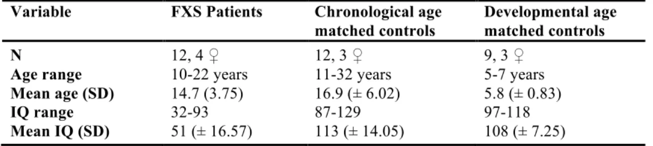
Second article Table I. Demographics of the study population... 69
Table II. Mean values (SD) of abnormal and repetitive behaviour in participants as reported by their parents/caregivers... 72
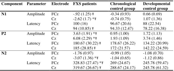
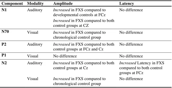
Table III. Mean amplitudes in µV and latencies in ms (SD) for auditory ERP components in FXS patients, chronological control group and developmental control group... 73
Table IV. Mean amplitudes in µV and latencies in ms (SD) for visual ERP components in FXS patients, chronological control group and developmental control group... 74
List of figures
Second article
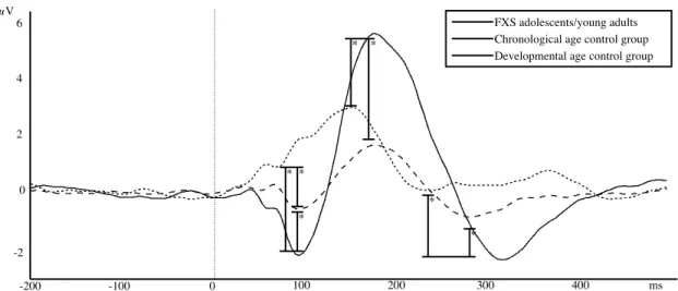
Figure 1. Group Averages of Auditory ERPs at electrode FCz for the FXS group and the two
control groups. 0ms marks stimulus onset. Significant differences are only marked for
differences between FXS and control groups... 73
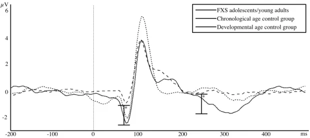
Figure 2. Group Averages of Auditory ERPs at electrode Cz for the FXS group and the two
control groups. 0ms marks stimulus onset. Significant differences are only marked for
differences between FXS and control groups... 74
Figure 3. Group averages of visual ERPs at electrode Oz for the FXS group and the two
control groups. 0ms marks stimulus onset. Significant differences are only marked for
List of abbreviations
ADHD: Attention Deficit Hyperactivity Disorder
DS: Down Syndrome
FXS : Fragile X Syndrome ID : Intellectual Disability IQ: Intelligence Quotient
DTI: Diffusion Tensor Imaging EEG: Electroencephalography ERP: Event-Related Potential
fMRI: functional Magnetic Resonance Imaging MEG: Magnetoencephalography
MMN: Mismatch Negativity
MRI: Magnetic Resonance Imaging PET: Positron Emission Tomography
FMR1: Fragile X Mental Retardation 1 FMRP: Fragile X Mental Retardation Protein GABA: Gamma-Aminobutyric Acid
mGluR: metabotropic Glutamate Receptors mRNA: messenger Ribonucleic Acid
Ag/AgCl: Silver chloride electrode ANOVA: Analysis of Variance cd/m2
: candela per square meter
cm: centimeter
Hz: Hertz
dB: decibel
kΩ: kiloohm
M: mean ms: millisecond N.A.: not available n.s.: not significant
s: second
‘Everything makes sense a bit at a time. But when you try to think of it all at once, it comes out wrong.’
Acknowledgements
First of all I thank my supervisor Sarah Lippé, without whom I would have not been able to realize my dream of studying and doing research in Québec. I thank her for having seen my potential from afar and fully supporting me for the whole duration of my stay. I thank her for sharing her academic wisdom with me, for her patience and generosity, for her
confidence and extraordinary flexibility. Without a supervisor as passionate and dedicated as her, this project would not have been possible. A very special thanks is due to Phetsamone Vannasing, without whose magic social skills and kindness I would have hardly succeeded in testing as many patients. I also thank her for helping me with every imaginable problem I encountered and for cheering me up when needed.
I thank my colleagues for being amazingly inspiring people who gave me valuable advice and encouraged me no matter how insurmountable the obstacle seemed to be. I especially thank all of you who looked over my French and helped me progress.
I thank J. Arthur White, not only for correcting my English in every important piece of writing over the past two years, but also for all the fascinating conversations, hilarious moments and adventurous trips we had together.
I thank the Montreal Improv theatre and its community for becoming my family away from home. I cannot say enough good things about these witty, brilliant and caring people who are now among my best friends. Learning and performing at the Montreal Improv theatre is a delightful pleasure for me that I am very grateful for. Improv is my therapy.
My studies in Canada were financed by excellence scholarships from the German National Academic Foundation, the German Academic Exchange Service and an International Fee Exemptions scholarship awarded by the government of Québec.
Learning mechanisms
Learning mechanisms and their underlying brain functions are a major field of
interest and research in cognitive neuroscience. One of the most influential attempts to account for the neuronal processes underlying associative learning was introduced in the late 1940s by Donald Hebb in his book The Organization of Behaviour (Hebb, 2002). His theory explains the formation of memory traces through synaptic plasticity. Frequent reverberatory activity between neurons is believed to stabilize the connection between these neurons through growth or metabolic changes. Thus, memory traces are represented through neural networks (Hebb, 2002). Modern neuroimaging techniques enable us to non-invasively investigate information processing and learning mechanisms in the human brain. However, not all of these techniques have a sufficient temporal resolution to allow the investigation of rapid neural processes like postsynaptic potentials, which usually last between tens and hundreds of milliseconds (ms) (Luck, 2005). Hemodynamic measures like functional magnetic resonance imaging (fMRI) and positron emission tomography (PET), for example, are limited to a resolution of several seconds and are thus not suited for the investigation of synaptic processes. The method of choice to address neural processes is electroencephalography, the measurement of electrical activity in the brain through electrodes placed on the scalp, which is plotted in voltage over time. Neural responses associated with specific sensory, cognitive and motor events can be extracted from the electroencephalogram (EEG) using averaging techniques in order to discard random brain activity from the specific event-related potential (ERP) (Luck, 2005). ERPs are currently believed to reflect cerebral local field potentials, which are summarized postsynaptic potentials from large groups of neurons (Luck, 2005). ERP waveforms consist of a series of positive and negative voltage deflections, which are called ‘ERP components’ and named according to the order or latency-window in which they occur. However, it is important to note that some authors refer to underlying cerebral generator processes, which contribute to the polarity of the recorded voltage deflection, when they talk about ERP components
(Näätänen & Picton, 1987). Usually, the early components are associated with basic sensory processing and differ according to modality, whereas the later components (starting with N2) are expected to reflect more cognitive phenomena, like face (N170) and language (N400, P600) processing or error detection (error related negativity). It is possible to investigate sensory and cognitive processing using ERPs since an extensive body of research identifies a number of factors that influence the amplitude and latency of specific ERP components and can be manipulated in experiments. Further, ERP components have been found to specifically change with brain development, which makes them valuable instruments in the investigation of brain maturation (Lippé, Roy, Perchet, & Lassonde, 2007).
Intellectual disability
A complementary approach to the study of brain mechanisms in healthy individuals is to investigate pathologies presenting deficits in these mechanisms. Results obtained from pathology research can provide insight toward understanding the proper functioning found in healthy individuals, while also serving as a basis for developing treatments for the condition in question. In the study of information processing and learning mechanisms, Intellectual
Disability (ID) (formerly mental retardation) is thus a disorder of particular interest, since it is ‘characterized by significant limitations both in intellectual functioning and in adaptive behavior as expressed in conceptual, social and adaptive skills.’ (Schalock et al., 2007). Generally, ID can be assessed using Standard Intelligence Quotient (IQ) tests with a mean of 100 and a standard deviation of 15 in combination with the Vineland adaptive Behavior Scales, and must be diagnosed before the age of 18 (Schalock et al., 2007). In this context, ID is diagnosed when IQ is assessed as <70 (i.e., at least 2 standard deviations below the mean) (Ropers, 2010) and a significant deficit in adaptive functioning is identified (Perry & Factor, 1989). However, ID cannot be considered a homogeneous condition, since it can be caused by numerous genetic and environmental factors. In 30 to 50% of cases, the cause of ID remains unknown (Daily, Ardinger, & Holmes, 2000). In order to study the underlying mechanisms of a disorder, a certain extent of phenotypic, genetic and mechanistic homogeneity is required. Thus, it is reasonable to investigate a condition with an identified cause. Among genetic
causes, X-linked recessive gene defects are believed to be accountable for approximately 10-12% of ID found in males (Ropers & Hamel, 2005). The most common form of X-linked intellectual disability is Fragile X Syndrome (FXS), which affects about 2% of male ID patients (Ropers & Hamel, 2005). Since FXS is caused by a single gene mutation, it is regarded as an important pathology in the investigation of gene-brain-behavior relationships.
Fragile X Syndrome
The physical phenotype of FXS is rather subtle, including a long face, prominent ears and hyperextensible joints (Hull & Hagerman, 1993). Over 90% of male and over 50% of female FXS individuals meet the criteria for an ID, ranging from mild to severe for male patients and from mild to moderate in females (Hessl et al., 2009). FXS patients who do not meet the criteria for ID often present learning disabilities (Loesch et al., 2003). Since cognitive growth in children with FXS is significantly slower than in typical developing children (Hall, Burns, Lightbody, & Reiss, 2008), the intellectual discrepancy increases with age, resulting in an age-dependent gradual decline in IQ (Schneider, Hagerman, & Hessl, 2009). The most severely impaired cognitive domain in FXS is executive functions, including deficits in working memory, planning and set shifting, attentional control and inhibition (K. M. Cornish et al., 2008; Schneider et al., 2009). Further, visual-spatial cognition is often impaired, including visual-spatial reasoning, object occlusion and arithmetical problem solving (K. M. Cornish et al., 2008; Farzin & Rivera, 2010; Loesch et al., 2003). Over 50% of male FXS patients meet the behavioral criteria for Attention-Deficit/Hyperactivity Disorder (ADHD), as reported by parents and teachers (Sullivan et al., 2006). Hyperarousal to sensory stimuli is especially common in FXS, while hyperactivity, impulsivity, impairment of inhibitory control and short attention span are also frequently found (Schneider et al., 2009; Sullivan et al., 2006). Many of the symptoms found in FXS are typical of the autistic spectrum; about 30% of male individuals with FXS meet the full diagnostic criteria for autism, with FXS considered the only known monogenetic cause of autism (Rogers, Wehner, & Hagerman, 2001). The main symptoms shared between FXS and autism concern abnormal behavior, lack of social cognition and language deficits (Schneider et al., 2009). Abnormal behavior in FXS includes
stereotyped behavior, self-injury, perseverative preoccupations and interests (Bregman, Leckman, & Ort, 1988), as well as delayed socialization and avoidance (Budimirovic et al., 2006). Delayed echolalia, idiosyncratic responses, abnormalities in intonation and rhythm, verbal perseveration, tangential language and cluttering of speech are examples of language deficits found in FXS (Bregman et al., 1988; K. M. Cornish et al., 2008; Sudhalter & Belser, 2001). Even though many FXS males show a broad spectrum of anxiety symptoms, they often do not meet the criteria for an established anxiety disorder enumerated in the Diagnostic and Statistical Manual of Mental Disorders. However, anxiety symptoms most frequently observed in FXS, such as poor eye contact, gaze aversion and excessive shyness are reminiscent of social phobia (Tranfaglia, 2012). Thus, a broad spectrum of functioning is found to be impaired in FXS, even though the specific symptoms, as well as their intensity, vary
considerably from case to case (Schneider et al., 2009). On the other hand, vocabulary, verbal working memory and long-term memory for meaningful information appear to be well
preserved in most cases (K. Cornish et al., 2005b).
Genetic mechanisms underlying FXS
FXS is caused by a CGG trinucleotide repeat expansion in the fragile X mental retardation 1 (FMR1) gene, which is located on the X-chromosome. Generally, it follows the hereditary transmission of X-chromosomal inheritance, but with some particular features. Firstly, despite their existing non-mutated X-chromosome, women can also be affected (approximately half of the prevalence found in men) but with greater variation in the
phenotype expression (Bennetto, Pennington, Porter, Taylor, & Hagerman, 2001). Besides the full mutation of more than 200 trinucleotide repeats (compared to the normal length of
approximately 30 triplets), a premutation with an intermediate length of between 55 and 200 repeats also exists. This premutation leads to non-penetrant carriers, who may pass on a full mutation to their child, due to the instability of the premutation in meiosis (Bassell & Warren, 2008). Normally, the FMR1 gene codes for the ‘fragile x mental retardation protein’ (FMRP) (Verheij et al., 1993). FMRP is a messenger ribonucleic acid (mRNA)-binding protein, which has been shown to strongly inhibit translation of various mRNAs (Laggerbauer, Ostareck,
Keidel, Ostareck-Lederer, & Fischer, 2001). The FMR1 mutation found in FXS silences the transcription of FMRP, resulting in an FMRP absence (Laggerbauer et al., 2001). According to the mGluR theory of FXS, FMRP deficit results in an exaggerated mRNA translation and thus causes continuous enhanced mGluR-dependent Long Term Depression. Consequently, the protein-synthesis in the synapses is not modified specifically to stimuli induction, resulting in a loss of protein synthesis-dependent plasticity (Bassell & Warren, 2008). Further, FMRP absence leads to altered axonal development, including increased density of dendritic spines, weak, elongated dendritic spines and immature synaptic connections (Comery et al., 1997). Based on the assumed molecular mechanisms, mGluR5 inhibitors were investigated as
possible medical treatments for the FXS phenotype in several animal models (Krueger & Bear, 2011). Subsequent to findings that a number of phenotypes were reversed in animal models, a clinical pilot with human patients was carried out. No clinically significant adverse effects were detected in FXS patients after the administration of a single dose of the mGluR5 inhibitor fenobam and potentially beneficial clinical effects were discovered in half of the patients (Berry-Kravis et al., 2009). However, to this day no double-blind randomized trial with fenobam in FXS patients has been completed.
Brain anatomy in FXS
A number of structural studies have investigated brain anatomy in FXS. One of the most frequently and consistently found differences between FXS and age-matched controls is a significantly enlarged caudate nucleus in FXS patients (Lightbody & Reiss, 2009). The caudate nucleus is a structure located in the basal ganglia believed to be involved in movement, learning and memory, notably in associative learning (Packard & Knowlton, 2002), as well as in transferring information to the frontal lobe (Ring & Serra-Mestres, 2002). The alterations observed in the caudate nucleus might therefore be connected with deficits in learning, motor coordination and attention found in FXS (Lightbody & Reiss, 2009). The enlarged caudate nucleus is found early on in FXS and not only in comparison to healthy controls, but also when compared to children with idiopathic developmental delay and autism (Hazlett et al., 2009). Some authors found a difference in relative volume increase between
male and female FXS patients (Eliez, Blasey, Freund, Hastie, & Reiss, 2001; Gothelf et al., 2008), whereas others did not find these gender differences (Lee et al., 2007). Further, a positive correlation between caudate nucleus volume and aberrant behavior, as assessed by the Aberrant Behavior Checklist and the Stereotypy subscale of the Autism Behavior Checklist, has been found (Gothelf et al., 2008). The second structure in which volume alterations have been detected is the cerebellar vermis, which has been found to be consistently smaller in FXS (Lightbody & Reiss, 2009). While the cerebellum has traditionally been mainly associated with motor functioning, evidence has accumulated that it may also play a role in cognitive processes, especially spatial cognition, language production and executive functions
(Rapoport, van Reekum, & Mayberg, 2000), all of which have been found to be disturbed in FXS. Since the reduced volume of the cerebellar vermis has been found early on and
consistently in FXS, it has been suggested as a distinguishing feature of brain anatomy for the disorder (Hoeft et al., 2008). Further, a positive relationship between cerebellar vermis size and IQ has been found in FXS, but not in healthy controls (Gothelf et al., 2008). However, results concerning the cerebellar vermis and autistic behavior in FXS have been inconsistent, possibly due to differences in group size and diagnostic criteria (Lightbody & Reiss, 2009). Studies of FXS brain anatomy have also found a smaller superior temporal gyrus (Gothelf et al., 2008), which is involved in auditory processing, including language, and also in social cognition (Bigler et al., 2007). The amygdala has been found to be significantly smaller in children affected with FXS, even at very young ages (Hazlett et al., 2009; Kates, Abrams, Kaufmann, Breiter, & Reiss, 1997). The amygdala is known to play a central role in the mediation of emotions, particularly fear (LeDoux, 1995), and in the organization of social behavior (Adolphs, Tranel, & Damasio, 1998). Considering the autistic symptoms often found in FXS, such as social avoidance and gaze aversion, it is not surprising that the amygdala has been found to be reduced in FXS patients with autism diagnosis (Lightbody & Reiss, 2009). However, the amygdala also appears to be reduced in patients with FXS who do not show signs of autism (Hazlett et al., 2009). Alterations found in the size of the hippocampus, a structure important for memory and learning, were too inconsistent in the case of FXS to establish any general conclusions, since the hippocampus of FXS patients has been found to be larger, smaller and not different from that of controls (Lightbody & Reiss, 2009). Hoeft and colleagues found an enlarged fusiform gyrus and a decreased insula in young children with
FXS in comparison to normally developing and developmentally delayed control children (Hoeft et al., 2008). The fusiform gyrus is a cortical region specialized in face processing (Kanwisher & Yovel, 2006), while the insula is believed to be involved in interoceptive awareness, emotional responses, empathetic processes, as well as salience and cognitive control (Menon & Uddin, 2010). Aberrant maturation of the prefrontal gyri has been linked to abnormal intellectual development in FXS (Bray et al., 2011). Diffusion tensor imaging (DTI), an MRI method that maps molecular diffusion in the brain, showed a decreased white matter tract connectivity in frontostriatal pathways and parietal sensory-motor tracts in FXS females, relative to healthy controls (Barnea-Goraly et al., 2003). These pathways are believed to be involved in the mediation of sensory processes, while also affecting regulation, executive functions and motor programming (Hessl, Rivera, & Reiss, 2004) - domains that have been found to be impaired in FXS.
Functional neuroimaging in FXS
Given that, in contrast to structural studies, functional studies do not allow for sedation of participants, it is more difficult to obtain functional data from FXS patients. The behavioral phenotype of FXS patients, often including hyperactivity, anxiety, impulsivity and stereotyped behavior, makes it nearly impossible for some of them to stay motionless and attentive in an unknown and somewhat intimidating setting, as required in fMRI. Thus, most of the earlier studies focus on the more functional female FXS patients (Lightbody & Reiss, 2009). However, advantages in behavioral training and pre-test preparation in recent years have made it possible to test some male patients as well (Lightbody & Reiss, 2009). Given the gaze aversion frequently found in FXS, face and gaze processing are of particular interest in functional imaging studies. While female FXS patients appeared to process face stimuli in a relatively appropriate manner, they did not show a preference for the more socially relevant forward faces in terms of brain activation, as found in healthy controls (Garrett, Menon, MacKenzie, & Reiss, 2004). In a follow-up study with male patients, it was found that face processing was accompanied by less prefrontal cortex activity, while activity in the insula and amygdala were enhanced (Watson, Hoeft, Garrett, Hall, & Reiss, 2008). Thus, FXS patients
showed an increased sustained activation in brain regions related to emotion perception and arousal, in comparison to typically developing controls and controls with non-syndromic developmental delay. A study investigating anxiety and face processing in FXS showed that FXS patients with high reported anxiety recruited encoding, social cognition and memory related areas of the brain significantly less during face processing than FXS patients with lower levels of anxiety (Holsen, Dalton, Johnstone, & Davidson, 2008). The relationship between autism and FXS with regard to face processing has been examined by Dalton and colleagues (Dalton, Holsen, Abbeduto, & Davidson, 2008). While activation in the fusiform gyrus was comparable across FXS and autism groups, FXS patients showed more activation in the left hippocampus, the right insula, left postcentral gyrus and superior temporal gyrus than healthy and idiopathic autism control groups. These findings suggest that, despite similar behavioral outcomes, the underlying cerebral mechanisms in FXS and autism might differ. Working memory is one of the executive functions that has been investigated in FXS. Two studies with female FXS patients showed that brain areas associated with working memory (inferior and middle frontal gyri, superior parietal lobule and supramarginal gyrus) are
activated during specific working memory tasks (N-back task and math calculations) (Kwon et al., 2001; Rivera, Menon, White, Glaser, & Reiss, 2002). However, unlike the healthy control group, the FXS females in both studies did not show increased brain activation in response to greater task difficulty, suggesting a failure to recruit additional resources when demanded. With regards to attention and impulse control, Hoeft and colleagues carried out a study investigating inhibition in male adolescent FXS patients using a Go/No Go fMRI task (Hoeft et al., 2007). While developmentally delayed and normally developed controls recruit a right fronto-striatal network during the response inhibition task, the FXS group demonstrated what may have been a compensatory strategy through increased ventrolateral pre-frontal cortex activity.
Electrophysiology in FXS
The recording of spontaneous electrical activity over a short period of time is
generally used in a clinical context in order to diagnose epileptic activity. Epilepsy describes a set of neurologic syndromes whose predominant feature is a predisposition to recurrent
unprovoked seizures (Chang & Lowenstein, 2003). In FXS, epilepsy is reported in 10 to 20% of cases, an incidence significantly larger than in the general population (<1%) (Berry-Kravis, 2002). The seizure pattern most frequently resembles benign focal epilepsy of childhood, an idiopathic age-specific epileptic syndrome that usually goes into remission in adolescence. The EEG pattern in benign childhood epilepsy features centrotemporal epileptiform foci with wide spikes that appears bi- or triphasic with a relatively high amplitude (Kramer, 2008). Thus, FMRP absence seems to cause increased neuronal excitability and susceptibility to epilepsy (Berry-Kravis, 2002).
Van der Molen and Van der Molen investigated the oscillatory dynamics during resting-state EEG in male FXS patients (M. J. Van der Molen & Van der Molen, 2013). They found an increased relative theta power and a decreased relative upper alpha power in FXS when compared to healthy controls. This is a pattern also typically found in children and adults with ADHD (Barry, Clarke, & Johnstone, 2003). Alpha rhythm in EEG is believed to play a pulsed inhibition role in cognitive processing, which gates information by reducing the processing capabilities of a given area (Jensen & Mazaheri, 2010). The reduction of alpha activity in FXS may thus be a neural marker of a hyperexcitable nervous system, since the neural inhibitory mechanism that regulates incoming sensory information is aberrant (M. J. Van der Molen & Van der Molen, 2013). The pulsed inhibition reflected by the alpha rhythm is believed to be influenced by gamma-Aminobutyric acid (GABA)ergic input from the interneural network. This supports the observation of reduced alpha oscillations in FXS, since the GABAergic system appears to be dysfunctional in FXS patients and in animal models (Paluszkiewicz, Martin, & Huntsman, 2011). The alpha/theta power abnormalities may underlie further information processing deficits, since alpha/theta synchronization has also been associated with cognitive and memory performance (Klimesch, 1999). The results obtained during resting state in FXS already indicate that electrophysiology is a promising method of investigating information processes in FXS.
After having described the behavioral and cognitive phenotype of FXS, the underlying genetic mechanisms, as well as alterations in brain anatomy and activation, the question arises: how does FXS affect the synaptic mechanism underlying information processing? How does the reduction in synaptic plasticity caused by FMRP absence affect basic sensory processing and more sophisticated cognitive processing? As described in the beginning of this section, ERPs enable us to non-invasively investigate sensory and cognitive processing in humans. A review presenting and discussing relevant ERP studies conducted with full mutation FXS patients is the first of two articles presented within the framework of this master’s thesis and logically precedes the research article, which is presented second.
ERP alterations in FXS
The aim of writing the review article was to collect and discuss all results obtained thus far in relevant ERP studies investigating information processing in FXS. We then
designed a study based on what has previously been found, while also adding new elements, in order to broaden knowledge of specific brain responses in FXS. Surprisingly, only five
relevant ERP studies with FXS full mutation patients have been published since the 1980s. The explanation for this is most likely found in the difficulty of testing FXS patients due to the behavioral phenotype they present, a problem we also encountered in our own study. In
contrast to clinical EEG, the participant cannot be sedated during EEG if brain responses to specific sensory events are to be evoked and recorded. The installation of electrodes often poses a problem, since most of the patients present social anxiety and do not like to be touched by a stranger or restricted by an EEG net. Further, participants need to remain still during the testing, since movement artifacts distort the data and recorded segments during which the participant has moved must be rejected in the analysis.
The review first gives a brief description of FXS and its underlying genetic
mechanisms, as well as its cognitive profile. After a short introduction of the ERP method, the five selected studies are presented and a detailed comparison of their methods is given. In the main part of the review, each of the following ERP components is presented in detail: N1, P2, MMN, N2 and P3. A general description is given for each component, followed by the
findings of each study concerning them and possible alterations in FXS. These are then compared to findings in syndromes sharing symptoms with FXS, namely other IDs and autism. Further, the maturation of every component is described, in case the alterations found in FXS reflect an immature brain response resembling that of a younger child with the same level of cognitive functioning. Possible factors that might have influenced or caused the deviances are discussed and hypotheses concerning underlying neuronal mechanisms are proposed. Finally, controversies between studies are addressed. In the discussion and conclusion the ERP profile specific to FXS is presented, including all reported alterations. While parameters of the more cognitive components MMN, N2 and P3 appear to be generally altered in ID, the basic sensory components N1 and P2 seem to be altered more specifically in FXS. In conclusion, the review article suggests that basic stimulus processing, attentional processing, and memory formation are impaired, which is consistent with symptoms found in FXS.
Implications for our study design
Since basic sensory processing seemed to be especially impaired in FXS, we decided to choose a simple auditory and visual stimulation paradigm in order to evoke basic stimulus processing brain responses. The tasks involved were used in two earlier studies investigating the maturation of infant auditory and visual processing realized by my supervisor, Sarah Lippé (Lippé, Martinez-Montes, Arcand, & Lassonde, 2009; Lippé et al., 2007). So far, only oddball paradigms have been studied in FXS patients with full mutation. Furthermore, only one study investigated visual ERPs and the results seemed to suggest that stimulus processing in the visual modality is less affected than in the auditory modality in FXS, implying an important modality difference. Thus, we wanted to further examine visual processing in FXS by investigating basic visual ERPs.
While a number of imaging studies contains a control group with non-syndromic ID or with younger controls matching the developmental age of the patients in order to control for general effects of ID or brain immaturity, this has only been done in one of the ERP studies of FXS. It is therefore difficult to determine whether brain development in FXS remains
immature, causing ERP profiles to appear similar to younger children with the same level of cognitive functioning as the patients, or if the absence of FMRP further disrupts sensory processing, leading to an ERP profile specific to FXS. In order to differentiate these two possibilities, we not only tested a healthy age and gender matched control group, but also healthy controls in the age of cognitive functioning (developmental age) of the patients tested.
The ethics, scientific and administrative committee at the CHU Sainte-Justine Mother and Child University Hospital Center reviewed the research protocol and asked for minor specifications concerning recruitment methods, data access, neuropsychological testing, statistical analysis and number of participants as well as group sizes. In consequence, we modified the protocol and the consent form in order to obtain final permission from all committees.
Considering the expected difficulties in testing FXS patients, we decided to administer the IQ test during a home visit preceding the EEG recording at the CHU Sainte-Justine Mother and Child University Hospital Center. This allowed the patient to feel safe while meeting us first in a well-known environment and thus to develop a positive relationship with us, which was especially important for the second visit when the EEG was recorded. During the first visit, we prepared the patients for the EEG by showing them pictures and explaining the procedure in simple words. During the EEG testing we created a pleasant environment, by playing a movie in the beginning and offering the patient snacks and toys while they got used to the environment. While installing the electrodes, we proceeded effectively but carefully in order to avoid disturbing the patient more than necessary.
Whenever the patient was continuously dissatisfied with the situation, we stopped the testing. Given the high prevalence of epilepsy in FXS, we presented all patient EEG data to a neurologist. In the case of epileptic activity, data was excluded from analysis and the family doctor of the patient was contacted in order to inform the patient and schedule a follow-up meeting for an accurate diagnosis at the hospital.
Hypotheses
Based on the structural and functional alterations in neurons and synaptic plasticity caused by the FMRP absence, the cognitive phenotype, as well as the alterations in brain anatomy and brain activation described above, and in particular the ERP alterations
summarized in our literature review, we assume that basic neuronal information processing is impaired in FXS.
Therefore, we expect to find an auditory and visual ERP profile in our FXS patient population that differs in several components, notably in auditory N1, P2 and N2 and visual N70, P1 and N2 amplitude, as well as in N2 latency, from the healthy control group matched to the patient group on the basis of chronological age and gender.
Since the absence of FMRP is believed to lead to altered neurodevelopment, and since aberrations in brain anatomy are found as early as one year of age, we expect that at least some of the components will not only appear immature, but specifically altered, in FXS. Thus, we expect that the auditory components N1 and P2 will not only be altered relative to the chronological control group, but also relative to a healthy control group that is matched to the developmental age of cognitive functioning of patients with ID. Component N2, however, might not differ from the developmental control group, since it is typically altered in ID and could therefore reflect an immature brain response associated with the level of cognitive functioning.
A modality difference in basic processing impairments between the auditory and visual modality in FXS has been suggested in a previous ERP study. This modality difference seems to gain further support through language deficits often found in FXS, which could be partially explained by impairments in auditory processing. Thus, we expect visual ERPs to be less aberrant in FXS than auditory ERPs, meaning that fewer components differ significantly from the control groups in the visual compared to the auditory modality.
Contributions to the articles First article
The first article is a literature review presenting all relevant ERP studies that have thus far been conducted with patients with FXS full mutation. The objective of this article was to unveil the contribution of electrophysiological signal studies for the understanding of the information processing impairments in FXS. The literature review, as well as the initial draft of the article, were entirely carried out by Inga Sophia Knoth. Sarah Lippé’s corrections and commentaries were taken into account before the manuscript was submitted to Frontiers in
Human Neuroscience in April 2012. The article was accepted and published in November
2012.
Second article
The second article describes the research project that was realised by Inga Sophia Knoth in the framework of her Master’s degree. The initial idea of the project originated from Sarah Lippé and Jacques Michaud. The EEG paradigm was created by Sarah Lippé in the framework of her PhD studies. Jacques Michaud provided his database of FXS patients for the project. Recruitment and screening of patients and controls, IQ testing and evaluation, EEG recording, EEG pre-treatment, ERP and statistical analysis were mainly carried out by Inga Sophia Knoth. Phetsamone Vannasing helped with patient EEG recording and EEG pre-treatment. Some patients were tested by neuropsychologist Domitille Malfait. Bachelor students Maude Joannette and Patricia Laniel helped with recruitment and EEG recording of control participants. The first draft of the article was entirely written by Inga Sophia Knoth. Corrections and commentaries of the co-authors have been taken into account and the manuscript is ready for submission to the Journal of Neurodevelopmental Disorders.
First article
Event-related potential alterations in fragile X syndrome
Published in Frontiers in Human Neuroscience, volume 6, september 2012
Inga Sophia Knoth1,2 & Sarah Lippé1,2
1Centre de Recherche CHU Ste-Justine, University of Montreal, Montreal, QC, Canada
2Centre de Recherche en Neuropsychologie et Cognition, University of Montreal, Montreal, QC, Canada
Abstract
Fragile X Syndrome (FXS) is the most common form of X-linked intellectual
disability (ID), associated with a wide range of cognitive and behavioral impairments. FXS is caused by a trinucleotide repeat expansion in the FMR1 gene located on the X-chromosome. FMR1 is expected to prevent the expression of the “fragile X mental retardation protein (FMRP)”, which results in altered structural and functional development of the synapse, including a loss of synaptic plasticity. This review aims to unveil the contribution of electrophysiological signal studies for the understanding of the information processing impairments in FXS patients. We discuss relevant event-related potential (ERP) studies conducted with full mutation FXS patients and clinical populations sharing symptoms with FXS in a developmental perspective. Specific deviances found in FXS ERP profiles are described. Alterations are reported in N1, P2, Mismatch Negativity (MMN), N2, and P3 components in FXS compared to healthy controls. Particularly, deviances in N1 and P2 amplitude seem to be specific to FXS. The presented results suggest a cascade of impaired information processes that are in line with symptoms and anatomical findings in FXS.
Key words: fragile X syndrome, event-related potential, cognition, intellectual disability,
Introduction
Intellectuel Disability and Fragile X Syndrome
Intellectual disability (ID) is among the most common and severe handicaps of childhood. It is defined as “a condition of arrested or incomplete development of the mind, which is especially characterized by impairment of skills manifested during the developmental period, skills which contribute to the overall level of intelligence, i.e., cognitive, language, motor, and social abilities” (World Health Organization, 2004). Generally, Standard
Intelligence Quotient (IQ) tests with a mean of 100 and a standard deviation of 15 are used for diagnosis. In this context, ID is determined by assessing an IQ <70 (i.e., less than 2 standard deviations below the mean) (Ropers, 2010). Numerous genetic and environmental factors can cause ID. They remain unknown in 30–50% of cases (Daily et al., 2000). Among genetic causes, X-linked recessive gene defects are believed to be responsible for approximately 10– 12% of ID found in males (Ropers and Hamel, 2005). The most common form of X-linked mental retardation is the Fragile X Syndrome (FXS), which affects about 2% of male ID patients (Ropers and Hamel, 2005). FXS is caused by a trinucleotide repeat expansion in the FMR1 gene, which is located on the X-chromosome. Generally, it follows the hereditary transmission of X-chromosomal inheritance, but with some particular features. Firstly, despite their existing non-mutated X- chromosome, women can also be affected (approximately half of the prevalence found in men) but with greater variation in the phenotype expression (Bennetto et al., 2001). Besides the full mutation of more than 200 repeats which underlies FXS in comparison to the normal length of 30 triplets, there also exists a premutation with an intermediate length between 55 and 200 repeats. This premutation leads to non-penetrant carriers, who may pass on a full mutation to their child, due to the instability of the
premutation in meiosis (Bassell and Warren, 2008). According to the mGluR theory of FXS, the FMR1 gene prevents expression of the encoded “fragile X mental retardation protein (FMRP)” (Bear et al., 2004). Normally, FMRP is known to repress the translation of specific mRNAs in response to the activation of metabotropic Glutamate Receptors (mGluRs). In turn, mGluRs are regulated by the inhibitory GABAergic system presynaptically, a putative altered mechanism in FXS. In Fragile X patients, the absence of FMRP leads to altered structural and
functional development of the synapse. On the structural level, altered dendritic development, including increased density of dendritic spines, weak, elongated dendritic spines, and
immature synaptic connections, are found in FXS patients and FXS animal models (Comery et al., 1997). Functionally, the FMRP deficit results in an exaggerated mRNA translation and thus causes continuous enhanced mGluR-dependent long-term depression. Consequently, the protein-synthesis in the synapses is not modified specifically to stimuli induction and therefore a loss of protein synthesis-dependent plasticity occurs (Bassell and Warren, 2008). The FMRP absence might therefore prevent activity-based synapse maturation and synaptic pruning, which is essential for normal brain development (Weiler and Greenough, 1999) and cognitive development (Schneider et al., 2009). In this context, the mGluR5 inhibitors were investigated as possible medical treatments for the FXS phenotype in several animal models (Krueger and Bear, 2011). Subsequent to the finding of a number of reversed phenotypes in animal models, clinical trials with human patients have been initiated and show promising preliminary results (Berry-Kravis et al., 2009).
In this review, we aim at unveiling the contribution of electro- physiological signal studies for the understanding of information processing impairments of a common intellectual deficiency syndrome, FXS.
Cognitive impairments found in FXS
The ID in FXS does not globally extend to all cognitive domains, but concerns abilities within and across specific domains, which show stability into adulthood (Cornish et al., 2008). In most cases, vocabulary, verbal working memory and long-term memory for meaningful information are well preserved (Cornish et al., 2005), whereas the cognitive and behavioral domains listed in table I tend to be affected frequently. Since the FXS phenotype shows great variability from case to case, the mentioned symptoms occur in some, but not all, FXS patients. In addition, the intensity of the symptoms ranges from mild to severe (Schneider et al., 2009). The deficits shown in behavior and social cognition, marked in gray within the table, are shared with disorders belonging to the autistic spectrum; about 30% of male individuals with FXS meet the diagnostic criteria for autism (Rogers et al., 2001).
Although non-exhaustive, table I shows a wide range of cognitive impairments in FXS patients. Most studies have investigated patients with FXS full mutation; however, it is worth mentioning that a recent study found attentionally based enumeration impairments in premutation carriers (Goodrich-Hunsaker et al., 2011). Premutation carriers may thus also present subtle cognitive impairments.
Table I. Symptoms frequently found in FXS patients sorted by domains.
Domain Symptoms frequently found in FXS patients
Behavior Pervasive hyperactivity & Impulsivity (Baumgardner, Reiss, Freund, & Abrams, 1995;
Bregman et al., 1988; Schneider et al., 2009)
Stereotyped behavior, self injury, perseverative preoccupations and interest (Bregman et
al., 1988)
Poor fine and gross motor coordination (Loesch et al., 2003) Delayed socialization and avoidance (Budimirovic et al., 2006) Social cognition Gaze aversion (Bregman et al., 1988; Schneider et al., 2009)
Impaired face recognition & emotion perception (Turk & Cornish, 1998) Theory of mind (Garner, Callias, & Turk, 1999)
Language Delayed echolalia (Bregman et al., 1988; K. Cornish et al., 2005a; Schneider et al., 2009) Idiosyncratic responses (Bregman et al., 1988)
Abnormalities in intonation & rhythm (Bregman et al., 1988) Verbal perseveration (Bregman et al., 1988; Schneider et al., 2009) Cluttering of speech (K. Cornish et al., 2005a)
Tangential language (Sudhalter & Belser, 2001) Executive
functions
Working memory (K. Cornish et al., 2005a; K. M. Cornish et al., 2008; Schneider et al., 2009) Planning & set shifting (Schneider et al., 2009)
Deficits in attentional control (Bregman et al., 1988; K. Cornish et al., 2005a) Inhibition (K. Cornish et al., 2005a)
Sequential processing (Loesch et al., 2003) Emotional
stability
Anxiety disorders (Bregman et al., 1988; K. Cornish et al., 2005a; Schneider et al., 2009) Social avoidance (K. Cornish et al., 2005a; Schneider et al., 2009)
Aggression (Schneider et al., 2009) Visual-spatial
cognition
Impairments in visual-spatial reasoning (K. M. Cornish et al., 2008; Schneider et al., 2009) Object occlusion (Farzin & Rivera, 2010)
Arithmetic problems (Loesch et al., 2003)
Hyperarousal Hyperarousal to sensory stimuli (Schneider et al., 2009)
ERP findings in FXS
In order to address maturational abnormalities in FXS, cortical and subcortical morphology have been studied and were found to be associated with alterations in cognition (Meguid et al., 2012). Given the availability of the Event Related Potential technique and its
capacity to record local field potentials, which are summarized postsynaptic potentials from large groups of neurons (Luck, 2005), it is surprising that only a few ERP studies have addressed FXS, in which synaptic plasticity is assumed to be impaired. Indeed, five relevant ERP studies conducted with full mutation FXS patients have been published since the 1980s (St. Clair et al., 1987; Rojas et al., 2001; Castrèn et al., 2003; Van der Molen et al., 2012a,b). After a short description of the applied study designs, their findings will be presented in an order corresponding to the investigated ERP components.
Study design
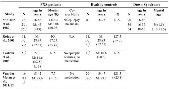
Table II shows the study population characteristics in the reviewed studies. Samples varied
between 5 and 28 individuals, from children to adults, and male and female frequency varied between studies’ samples1.
Table II. Study population characteristics in the reviewed studies.
Study
FXS patients Healthy controls Down Syndrome
N Age in years Mental age /IQ Co-morbidity N Age in years IQ N Age in years Mental age St. Clair et al., 1987 28, 2♀, 26♂ 16-66 M: 43 (±13) 1.8-4.6 M: 3.08 (±0.08) No epilepsy, no autism 83 18-75 N.A. 90 36 54 16-66 16-37 38-66 3(±1.5) 2.17(±1.5) Rojas et al., 2001 6♀, 11, 5♂ M: 28.95 (±2.51) IQ: 67.55 (±5.47) N.A. 11, 6♀, 5♂ M: 28.83 (±2.51) 127.5 (±2.9) Castrèn et al., 2003 5♂ 7-13 M: 11.6 (±2.8) 1x 28 N.A. No epileptic seizures, no medication 4♂ M: 10.6 (±0.6) N.A. Van der Molen et al., 2011/12 16 /11 ♂ 18-42 M: 29.6 7.7 (±1.6) No medication 22♂ 20/ M: 29.2 19-47 (±25.8) 121.5
1 Some of the listed specifications for St. Clair’s study population were detailed elsewhere (St.
All researchers investigated full mutation FXS patients and age-matched healthy controls. However, St. Clair and colleagues included an additional control group with ID, i.e., Down syndrome (DS). This control group enabled differentiation between obtained effects that rely on the level of brain development and effects that are specific for brain mechanisms underlying FXS. Therefore, the developmental level of the FXS patients has to be considered as a confounding variable to the results of the other four studies. Both chronological and mental age show considerable variation among the reported studies, ranging from children in Castrèn’s study to patients in retirement age in St. Clair’s study. This variation has to be kept in mind when results between the studies are compared, since both chronological and
developmental age is expected to influence ERP waves (Courchesne, 1990). The IQs reported for the control subjects in Rojas and Van der Molen’s studies are strikingly high, which probably reflects the tendency to recruit controls in the university setting, since years of education are positively correlated with IQ (Rowe et al., 1998).
The higher prevalence of FXS full mutations in men is reflected in the gender
distribution in the majority of the studies. By contrast, Rojas and colleagues investigated more female FXS patients (Rojas et al., 2001), which might account for the rather moderate ID found in their population compared to the other three studies which provide maturational age for their FXS patients (St. Clair et al., 1987; Van der Molen et al., 2012a,b), since the female FXS phenotype shows more variability (Bennetto et al., 2001).
The authors reported little on possible comorbidities in the investigated patients. Only St. Clair and colleagues specifically mentioned the absence of autism in their population (Primrose et al., 1986), whereas most of the other studies mainly controlled for epilepsy and medication. All participants were tested for sufficient hearing.
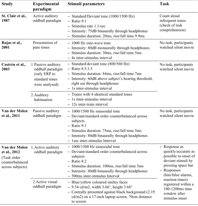
Experimental procedures used in the reviewed studies are listed in table III. All studies investigated the auditory modality. However, Van der Molen and colleagues (2012b) investigated the visual modality in their second study. Except for Rojas and colleagues (2001), all other studies made use of oddball paradigms—active in half of the cases and passive in the other half. St. Clair’s group did not report the behavioral outcomes of their task, nor did they connect them with the recorded brainwaves, since they used it predominantly to check if the participants were able to perceive the difference between the standard and the deviant tone.
Table III. Comparison of the experimental procedures used in the reviewed studies. Study Experimental
paradigm
Stimuli parameters Task
St. Clair et al., 1987
Active auditory
oddball paradigm − Standard/Deviant tone (1000/1500 Hz) − Ratio 9:1 − Stimulus rate 1.1/sec
− Intensity: 75dB binaurally through headphones − Stimulus duration: 20ms, rise/fall time 9.9ms
Count aloud infrequent tones (check of task comprehension)
Rojas et al.,
2001 Presentation of pure tones − 1000 Hz sine-wave tone − Intensity: 80dB monaurally through headphones − Stimulus duration: 30ms, rise/fall time 5ms − 4s inter-stimulus interval
No task, participants watched silent movie
Castrèn et al., 2003 1. Passive auditory oddball paradigm (only ERP to standard tones were analysed) − Standard/deviant tone (800/560 Hz) − Ratio 8.5:1.5
− Stimulus duration: 84ms, rise/fall time 7ms − Intensity: 60dB above subject’s hearing threshold,
right ear through headphones − 1s inter-stimulus interval
No task, participants watched silent movie
2. Auditory habituation
− Trains with 4 identical standard tones − 1s inter-stimulus interval
− 12s inter-train interval
Van der Molen et al., 2011
Passive auditory
oddball paradigm − 1000/1500 Hz sinusoidal tone − Deviant/standard order counterbalanced across subjects
− Ratio 9:1
− Stimulus duration: 75ms, rise/fall time 5ms − Intensity: 80dB binaurally through headphones − 1sec inter-stimulus Interval
No task, participants watched silent movie
Van der Molen et al., 2012 (Task order counterbalanced across subjects) 1. Active auditory oddball paradigm − 1000/1500 Hz sinusoidal tone
− Deviant/standard order counterbalanced across subjects
− Ratio 8:2
− Stimulus duration: 100ms, rise/fall time 5ms − Intensity: 80dB binaurally through headphones − 500ms inter-stimulus Interval
− Response as quickly/accurate as possible to onset of deviant stimuli by pressing space bar − Responses (hits/false alarms, reaction times) registered within a 100-1200ms time window after stimulus onset 2.Active visual
oddball paradigm − Blue/yellow coloured smiley faces − 9.34 cd/m2, width 3.66°, height 3.68°
− Centrally presented against black background (2.19 cd/m2) on a 17-inch laptop screen, 70cm distance to screen
Rojas and colleagues (2001) used Magnetoencephalography (MEG) as opposed to EEG. Their study is nevertheless considered in this review, since MEG signals are expected to originate from the same neurophysiological processes as EEG and offer evoked field
potentials equivalent to ERPs. The details of the conducted EEG/MEG recording and analysis in the reviewed studies are summarized in table IV. Obviously, the time span between the first study reviewed in this article, published by St. Clair and colleagues in 1987, and the most recent studies by Van der Molen and colleagues (2012a,b) has an influence on the technical
sophistication of EEG recording and analysis equipment. The number of recording electrodes has increased as well as the computational possibilities to remove artifacts. Moreover, St. Clair and colleagues did not report separate results according to standard and deviant tones, even though they claim to have analyzed them separately.
Table IV. EEG/MEG registration and analysis in the reviewed studies.
Study Electrodes Processing Component Analysis
St. Clair et al., 1987
1 Ag/AgCl-electrode at Cz, earlobe electrode as reference
− Separated average for standard/deviant tones − 500 trials total
− N1, P2, N2, P3 determined through 2 independent rater − Latencies/ amplitudes calculated
separately for each FXS patient
Rojas et al., 2001 4D Neuroimaging Magnes I neuro-magnetometer system, 37 axially-wound, first-order gradiometers, right-handed Cartesian coordinate system as reference
− Signal averaged separately for each hemisphere to obtain averaged auditory evoked magnetic field
− Min. 150 trials/ear
− P50m, N100m, P200m observed in auditory evoked field data − Source analysis
Castrèn et
al., 2003 19 Ag/AgCl electrodes, 10-20 system, right mastoid electrode as reference
− Signal averaged for standard
tones − N1, N2 determined at the highest peak amplitude site (Fz) − Global field power
Van der Molen et al., 2011
EasyCap electrode cap with 28 Ag/AgCl ring electrodes, left & right mastoid electrode as linked references
− Average: 895/99 resp. 892/99 (standard/ deviant) trials in controls resp. FXS patients
− N1, P2, MMN, N2b, P3a at F3, Fz, F4, FC1, FCz, FC2, C3, Cz, C4, P3, Pz, P4, O1, Oz, and O2 − Peak amplitude defined by the
method of local peak amplitude measurement (Luck, 2005),
relative to the pre-stimulus baseline
Van der Molen et al., 2012
− Average:
Auditory task: 236/58 resp. 234/59 (standard/deviant) trials for controls resp. FXS Visual task: 216/48 resp. 212/48 trials for controls resp. FXS
ERP components investigated
ERPs enable us to extract neural responses associated with specific sensory,
cognitive, or motor events from the overall EEG (Luck, 2005). Currently, ERPs are believed to reflect cerebral local field potentials, which are summarized postsynaptic potentials from large groups of neurons (Luck, 2005). Whereas the ERP technique enables an excellent temporal solution of 1ms or better under optimal conditions, the spatial solution has to be studied with caution since the voltage measured at an electrode always reflects the
The reviewed studies compare ERP components between FXS patients and control groups. The term “ERP component” can either simply describe the positive and negative voltage deflections within an ERP waveform according to the order or latency-window in which they occur (Luck, 2005) or it can refer to underlying cerebral generator processes, which contribute to the polarity of the recorded voltage deflection (Näätänen and Picton, 1987). Usually, the early components are related to sensory events and thus differ among modality, whereas the later components (starting with N2) are expected to reflect more
cognitive phenomena. The reviewed studies reported results regarding auditory N1 and N2 (St. Clair et al., 1987; Castrèn et al., 2003; Van der Molen et al., 2012a,b), auditory P2 and P3 (St. Clair et al., 1987; Van der Molen et al., 2012a,b) and auditory and visual MNN, visual N1, P2, N2, and P3 (Van der Molen et al., 2012b). This covers most of the commonly investigated auditory components and some of the cognitive components; however, it should be mentioned that other components exist, which might also allow interesting contributions to FXS research. Some of the predominantly cognitive ones will be addressed in the discussion toward the end of this article.
N1
Description of N1
The N1 is usually not the first major sensory response. In the auditory modality, brainstem evoked responses occur within the first 10ms after stimulus onset, which are followed by midlatency components at around 10–50ms and finally an auditory P1 at about 50ms before the auditory N1 (Luck, 2005). In the visual modality, the first ERP component, the C1 wave, typically arises 40–60ms after stimulus onset and shows a positive or negative deflection depending on which part of the visual field the stimulus is presented in (Luck, 2005). So far, no study has investigated the very early sensory components in FXS patients. Nevertheless, the main purpose of studying N1 in FXS is detecting alterations in early sensory stimulus processing. The auditory N1 peaks frontocentral at around 100ms after the onset of an auditory stimulus, whereas the visual N1 peaks 30–40ms later, at about 135ms after the onset of a visual stimulus (Näätänen and Picton, 1987). Näätänen and Picton (1987) conclude
in their review that the auditory N1 consists of three “true” components upon which three other stimulus-dependent components overlap. The first subcomponent is supposed to be a frontocentral negativity generated in the auditory cortex on the superior part of the temporal lobe. The second subcomponent, the T-complex, which peaks at temporal sites and consists of a positive wave at around 100ms and a negative wave at 150ms, probably stems from the auditory association cortices in the superior temporal gyrus. Lastly, there is a subcomponent of unknown source, generating a negative wave at the vertex at around 100ms after stimulus onset, which is believed to reflect an unspecific reaction to sensory stimulation and often overlaps with the first described subcomponent.
The visual N1 was decomposed by Di Russo et al. (2002) into four subcomponents to find pairs of generator dipoles which fit the N1 complex. They suggest an occipital source for the early N150, which peaks at occipito–parietal sites and has a centro-parietal source for the fronto-central N155. The later temporo-parietal N180 and occipito-parietal N200 are expected to be associated with the early P1 sources in the lateral extrastriate cortex and the late P1 source in the ventral occipito–temporal cortex (Di Russo et al., 2002). Research interest has been focused on the effects of spatial attention (Luck et al., 2000) and discrimination processing (Vogel and Luck, 2000).
N1 findings in FXS
St. Clair and colleagues (1987) reported that N1 latency in FXS did not differ from that in healthy controls, whereas it has been found to be significantly longer in patients with DS, during the active auditory oddball paradigm. N1 amplitude was found to be generally enhanced at vertex electrode Cz in response to both standard and deviant tones in FXS
patients, compared to patients with DS and healthy controls. Rojas et al. (2001) considered the N1 equivalent in MEG, the N100 m auditory-evoked field potential, in response to pure tones and also found a significantly higher amplitude in FXS patients than in healthy aged matched controls. They further observed a difference in the lateralization of the N100 m source. While healthy adults show N100 m source location asymmetry (right anterior to left), a reduction in lateralization is found in FXS patients. The authors proposed that the reduced asymmetry either reflects a non-specific neurodevelopmental disturbance which occurs during prenatal
development of cerebral asymmetry, since the phenomenon has also been found in schizophrenia (Reite et al., 1989, 1997), or stems from postnatal influences of the FXS mutation on the temporal lobe (Reiss et al., 1994). In either case, reduced N100 m source location asymmetry would be an outcome of disrupted brain development. Castrén and colleagues (2003) also found significantly larger auditory N1 amplitudes in FXS patients compared to healthy age matched controls in response to standard tones in their auditory oddball paradigm. This difference in N1 amplitude was most prominent in the frontal site Fz and was confirmed through global field power analysis. Van der Molen and colleagues (2012a) did not find any group differences of N1 latency at FCz. As for amplitude, they reported a significantly larger N1 amplitude to standard tones in FXS in a passive auditory oddball paradigm. This difference could be observed at electrodes Fz, the fronto-central FCz and Cz, whereas no differences were found for posterior sites. Further, the N1 amplitudes in controls were significantly larger for deviant than for standard tones, a difference which could not be found in FXS. Using an active oddball paradigm, a second study of Van der Molen et al. (2012b) again did not find any differences in N1 latency, neither in the auditory, nor in the visual modality. In the active auditory oddball paradigm, they reported larger N1 amplitudes for standard and deviant tones in FXS. In the active visual oddball paradigm, they found N1 peak amplitudes to be maximal at occipito-central electrode Oz in controls, but at FCz in FXS patients. At FCz the visual N1 amplitude was significantly larger for both stimuli in FXS than in controls. In both groups, visual N1 amplitude was larger at FCz than at Oz.
In addition, two groups tested habituation of N1 in response to stimulus repetition (Castrèn et al., 2003; Van der Molen et al., 2012a). Castrèn and colleagues (2003) tested short-term habituation of N1 to trains of four identical standard tones. Van der Molen and
colleagues compared N1 response to late standard tones with N1 response to early standards. In both studies, controls showed a reduction of N1 amplitude after several presentations of the same tone, whereas no N1 habituation could be found in FXS patients.
Regarding behavioral results, Van der Molen’s group (Van der Molen et al., 2012b) reported less accuracy, more false alarms and an increase in reaction time in FXS patients in both auditory and visual task compared to controls.