HAL Id: dumas-02561940
https://dumas.ccsd.cnrs.fr/dumas-02561940
Submitted on 4 May 2020HAL is a multi-disciplinary open access archive for the deposit and dissemination of sci-entific research documents, whether they are pub-lished or not. The documents may come from teaching and research institutions in France or abroad, or from public or private research centers.
L’archive ouverte pluridisciplinaire HAL, est destinée au dépôt et à la diffusion de documents scientifiques de niveau recherche, publiés ou non, émanant des établissements d’enseignement et de recherche français ou étrangers, des laboratoires publics ou privés.
cryo-ablation de fibrillation atriale
Camille Pichard
To cite this version:
Camille Pichard. Anatomie atypique des veines pulmonaires droites et cryo-ablation de fibrillation atriale. Sciences du Vivant [q-bio]. 2019. �dumas-02561940�
THÈSE D'EXERCICE / UNIVERSITÉ DE RENNES 1
sous le sceau de l’Université Bretagne Loire
Thèse en vue du
DIPLÔME D'ÉTAT DE DOCTEUR EN MÉDECINE
présentée parCamille Pichard
Née le 9 Août 1990 à Rennes (35)
Anatomie atypique
des veines
pulmonaires droites
et cryo-ablation de
fibrillation atriale.
Thèse soutenue à Rennes Le Mardi 10 Septembre 2019
devant le jury composé de :
Philippe MABO
PU-PH – CHU de Rennes – Président du jury
Christophe LECLERCQ
PU-PH – CHU de Rennes – Juge
Mathieu LEDERLIN
PU-PH – CHU de Rennes – Juge
Dominique PAVIN
PH – CHU de Rennes – Juge
Raphaël MARTINS
Professeurs des Universités - Praticiens Hospitaliers
Nom Prénom Sous-section CNU
ANNE-GALIBERT Marie-Dominique Biochimie et biologie moléculaire
BARDOU-JACQUET Edouard Gastroentérologie; hépatologie; addictologie
BELAUD-ROTUREAU Marc-Antoine Histologie; embryologie et cytogénétique
BELLISSANT Éric Pharmacologie fondamentale;
pharmacologie clinique; addictologie BELOEIL Hélène Anesthésiologie-réanimation; médecine
d'urgence
BENDAVID Claude Biochimie et biologie moléculaire
BENSALAH Karim Urologie
BEUCHEE Alain Pédiatrie
BONAN Isabelle Médecine physique et de réadaptation
BONNET Fabrice Endocrinologie, diabète et maladies métaboliques; gynécologie médicale
BOUDJEMA Karim Chirurgie générale
BOUGET Jacques
Professeur des Universités Emérite
Thérapeutique; médecine de la douleur; addictologie
BOUGUEN Guillaume Gastroentérologie; hépatologie; addictologie
BRASSIER Gilles Neurochirurgie
BRISSOT Pierre
Professeur des Universités Emérite
CARRE François Physiologie
CATROS Véronique Biologie cellulaire
CATTOIR Vincent Bactériologie-virologie; hygiène hospitalière
CHALES Gérard
Professeur des Universités Emérite
Rhumatologie
CORBINEAU Hervé Chirurgie thoracique et cardiovasculaire
CUGGIA Marc Biostatistiques, informatique médicale et technologies de communication
DARNAULT Pierre Anatomie
DAUBERT Jean-Claude
Professeur des Universités Emérite
Cardiologie
DAVID Véronique Biochimie et biologie moléculaire
DAYAN Jacques Professeur associé
Pédopsychiatrie; addictologie
DE CREVOISIER Renaud Cancérologie; radiothérapie
DECAUX Olivier Médecine interne; gériatrie et biologie du vieillissement; addictologie
DESRUES Benoît Pneumologie; addictologie
DEUGNIER Yves
Professeur des Universités Emérite
Gastroentérologie; hépatologie; addictologie
DONAL Erwan Cardiologie
DRAPIER Dominique Psychiatrie d'adultes; addictologie
ECOFFEY Claude Anesthésiologie-réanimation; médecine péri-opératoire
EDAN Gilles Consultant
Neurologie
FERRE Jean Christophe Radiologie et imagerie Médecine
FEST Thierry Hématologie; transfusion
FLECHER Erwan Chirurgie thoracique et cardiovasculaire
GANDEMER Virginie Pédiatrie
GANDON Yves Radiologie et imagerie Médecine
GANGNEUX Jean-Pierre Parasitologie et mycologie
GARIN Etienne Biophysique et médecine nucléaire
GAUVRIT Jean-Yves Radiologie et imagerie Médecine
GODEY Benoit Oto-rhino-laryngologie
GUGGENBUHL Pascal Rhumatologie
GUILLÉ François Urologie
GUYADER Dominique Gastroentérologie; hépatologie; addictologie
HAEGELEN Claire Anatomie
HOUOT Roch Hématologie; transfusion
JEGO Patrick Médecine interne; gériatrie et biologie du vieillissement; addictologie
JEGOUX Franck Oto-rhino-laryngologie
JOUNEAU Stéphane Pneumologie; addictologie
KAYAL Samer Bactériologie-virologie; hygiène hospitalière
LAMY DE LA CHAPELLE Thierry Hématologie; transfusion
LAVIOLLE Bruno Pharmacologie fondamentale;
pharmacologie clinique; addictologie LAVOUE Vincent Gynécologie-obstétrique; gynécologie
médicale
LE BRETON Hervé Cardiologie
LE GUEUT Mariannick Consultant
Médecine légale et droit de la santé
LE TULZO Yves Médecine intensive ; réanimation
LECLERCQ Christophe Cardiologie
LEDERLIN Mathieu Radiologie et imagerie Médecine
LEGUERRIER Alain
Professeur des Universités Emérite
Chirurgie thoracique et cardiovasculaire
LEJEUNE Florence Biophysique et médecine nucléaire
LEVEQUE Jean Gynécologie-obstétrique; gynécologie
médicale
LIEVRE Astrid Gastroentérologie; hépatologie; addictologie
MABO Philippe Cardiologie
MALLEDANT Yannick
Professeur des Universités Emérite
Anesthésiologie-réanimation et médecine péri-opératoire
MENER Éric
Professeur associé
Médecine générale
MEUNIER Bernard Chirurgie digestive
MICHELET Christian
Professeur des Universités Emérite
Maladies infectieuses; maladies tropicales
MOIRAND Romain Gastroentérologie; hépatologie; addictologie
MORANDI Xavier Anatomie
MOREL Vincent Professeur associé
Epistémologie clinique
MOSSER Jean Biochimie et biologie moléculaire
MOURIAUX Frédéric Ophtalmologie
MYHIE Didier Professeur associé
Médecine générale
ODENT Sylvie Génétique
OGER Emmanuel Pharmacologie fondamentale;
pharmacologie clinique; addictologie PARIS Christophe Médecine et santé au travail
PERDRIGER Aleth Rhumatologie
PLADYS Patrick Pédiatrie
RAVEL Célia Histologie; embryologie et cytogénétique
RICHARD de LATOUR Bertrand Professeur associé
Chirurgie thoracique et cardiovasculaire
RIFFAUD Laurent Neurochirurgie
RIOUX-LECLERCQ Nathalie Anatomie et cytologie pathologiques
ROBERT-GANGNEUX Florence Parasitologie et mycologie
ROPARS Mickaël Chirurgie orthopédique et traumatologique
SAINT-JALMES Hervé Biophysique et médecine nucléaire
SAULEAU Paul Physiologie
SEGUIN Philippe Anesthésiologie-réanimation; médecine péri-opératoire
SEMANA Gilbert Immunologie
SIPROUDHIS Laurent Gastroentérologie; hépatologie; addictologie
SOMME Dominique Médecine interne; gériatrie et biologie du vieillisement; addictologie
SOULAT Louis Professeur associé
Médecine d'urgence
SULPICE Laurent Chirurgie générale
TADIÉ Jean Marc Médecin intensive-réanimation
TARTE Karin Immunologie
TATTEVIN Pierre Maladies infectieuses; maladies tropicales
TATTEVIN-FABLET Françoise Professeur associé
THIBAULT Ronan Nutrition
THIBAULT Vincent Bactériologie-virologie; hygiène hospitalière
THOMAZEAU Hervé Chirurgie orthopédique et traumatologique
TORDJMAN Sylvie Pédopsychiatrie; addictologie
VERHOYE Jean-Philippe Chirurgie thoracique et cardiovasculaire
VERIN Marc Neurologie
VIEL Jean-François Epidémiologie, économie de la santé et prévention
VIGNEAU Cécile Néphrologie
VIOLAS Philippe Chirurgie infantile
WATIER Eric Chirurgie plastique, reconstructrice et esthétique; brûlologie
WODEY Eric Anesthésiologie-réanimation; médecine
Maîtres de Conférences des Universités - Praticiens Hospitaliers
Nom Prénom Sous-section CNU
ALLORY Emmanuel
(Maître de conférence associé des universités de MG)
Médecine générale
AME-THOMAS Patricia Immunologie
AMIOT Laurence (Baruch) Hématologie; transfusion
ANSELMI Amédéo Chirurgie thoracique et cardiovasculaire
BEGUE Jean-Marc Physiologie
BERTHEUIL Nicolas Chirurgie plastique, reconstructrice et esthétique ; brûlologie
BOUSSEMART Lise Dermato-vénéréologie
CABILLIC Florian Biologie cellulaire
CAUBET Alain Médecine et santé au travail
CHHOR-QUENIART Sidonie
(Maître de conférence associé des universités de MG)
Médecine générale
DAMERON Olivier Informatique
DE TAYRAC Marie Biochimie et biologie moléculaire
DEGEILH Brigitte Parasitologie et mycologie
DROITCOURT Catherine Dermato-vénéréologie
DUGAY Frédéric Histologie; embryologie et cytogénétique
EDELINE Julien Cancérologie; radiothérapie
FIQUET Laure
(Maître de conférence associé des universités de MG)
Médecine générale
GARLANTEZEC Ronan Epidémiologie, économie de la santé et prévention
GOUIN Isabelle épouse THIBAULT Hématologie; transfusion
GUILLET Benoit Hématologie; transfusion
JAILLARD Sylvie Histologie; embryologie et cytogénétique
KALADJI Adrien Chirurgie vasculaire; médecine vasculaire
LAVENU Audrey Sciences physico-chimiques et technologies pharmaceutiques
LE GALL François Anatomie et cytologie pathologiques
LEMAITRE Florian Pharmacologie fondamentale;
pharmacologie clinique; addictologie
MARTINS Pédro Raphaël Cardiologie
MATHIEU-SANQUER Romain Urologie
MENARD Cédric Immunologie
MOREAU Caroline Biochimie et biologie moléculaire
NAUDET Florian Thérapeutique ; médecine d'urgence ; addictologie
PANGAULT Céline Hématologie; transfusion
RENAUT Pierric
(maître de conférence associé des universités de MG)
Médecine générale
ROBERT Gabriel Psychiatrie d'adultes; addictologie
SCHNELL Frédéric Physiologie
THEAUDIN Marie épouse SALIOU Neurologie
TURLIN Bruno Anatomie et cytologie pathologiques
VERDIER Marie-Clémence (Lorne)
Pharmacologie fondamentale;
pharmacologie clinique; addictologie
SERMENT D’HIPPOCRATE
Au moment d'être admis à exercer la médecine, je promets et je jure d'être fidèle aux lois de l'honneur et de la probité.
Mon premier souci sera de rétablir, de préserver ou de promouvoir la santé dans tous ses éléments, physiques et mentaux, individuels et sociaux.
Je respecterai toutes les personnes, leur autonomie et leur volonté, sans aucune discrimination selon leur état ou leurs convictions. J'interviendrai pour les protéger si elles sont affaiblies, vulnérables ou menacées dans leur intégrité ou leur dignité. Même sous la contrainte, je ne ferai pas usage de mes connaissances contre les lois de l'humanité.
J'informerai les patients des décisions envisagées, de leurs raisons et de leurs conséquences. Je ne tromperai jamais leur confiance et n'exploiterai pas le pouvoir hérité des circonstances pour forcer les consciences.
Je donnerai mes soins à l'indigent et à quiconque me le demandera. Je ne me laisserai pas influencer par la soif du gain ou la recherche de la gloire.
Admis dans l'intimité des personnes, je tairai les secrets qui me seront confiés. Reçu à l'intérieur des maisons, je respecterai les secrets des foyers et ma conduite ne servira pas à corrompre les mœurs.
Je ferai tout pour soulager les souffrances. Je ne prolongerai pas abusivement les agonies. Je ne provoquerai jamais la mort délibérément.
Je préserverai l'indépendance nécessaire à l'accomplissement de ma mission. Je n'entreprendrai rien qui dépasse mes compétences. Je les entretiendrai et les perfectionnerai pour assurer au mieux les services qui me seront demandés.
J'apporterai mon aide à mes confrères ainsi qu'à leurs familles dans l'adversité.
Que les hommes et mes confrères m'accordent leur estime si je suis fidèle à mes promesses ; que je sois déshonoré et méprisé si j'y manque.
REMERCIEMENTS
Au Professeur Philippe Mabo,
Qui me fait l’honneur de présider cette thèse. Merci de m’avoir transmis votre passion pour la cardiologie et en particulier pour la rythmologie, et ce dès mon stage d’externat. Merci pour la qualité de notre formation médicale à Rennes, merci pour votre bienveillance et votre disponibilité. Veuillez trouver dans ce travail ma plus profonde estime.
Au Professeur Christophe Leclercq,
Qui me fait l’honneur de juger cette thèse. Merci d’avoir contribué à ma formation médicale, notamment en salle technique. Merci de me faire confiance pour le post-internat. Veuillez trouver dans ce travail mon sincère respect.
Au Professeur Mathieu Lerderlin,
Qui me fait l’honneur de juger cette thèse. Merci d’avoir contribué à l’élaboration de ce travail. J’espère continuer à collaborer avec votre service pour de nouveaux travaux.
Au Docteur Dominique Pavin,
Qui me fait l’honneur de juger cette thèse, merci d’avoir contribué à ma formation médicale et d’enseigner avec passion les tracés ECG. Merci pour votre aide durant mon internat et pour votre sympathie. Ce travail n’aurait pu avoir lieu sans toutes ces ablations de FA !
Au Docteur Raphaël Martins,
Qui me fait l’honneur d’être mon Directeur de thèse, merci de m’avoir confié ce travail. Un grand merci pour ton aide et ton implication dans l’écriture de cette thèse. Ton énergie et ta motivation sont toujours au rendez-vous. Merci pour ton soutien tout au long de mon internat.
A Amélie Nicolas,
Merci d’avoir contribué à la réalisation de ce travail, en relisant en aveugle les scanners cardiaques.
A Charlène et Vincent, merci pour votre aide dans le suivi des patients.
A l’ensemble des médecins qui se sont impliqués dans ma formation de médecin et de cardiologue. Je remercie les médecins du service de Pneumologie du CHBS de Lorient pour
avoir encadrer mes premiers pas dans l’internat, les médecins du service de Réanimation polyvalente du CH de Saint-Malo pour leur accueil chaleureux et leur formation, ainsi que l’ensemble des médecins du service de Cardiologie du CHU de Rennes pour la qualité de leur formation, leur motivation et leur écoute. Je remercie plus particulièrement les indispensables CCA qui ont accompagné mon parcours et grandement participé à ma formation, les
Docteurs Nelly Amara, Vincent Auffret, Albin Behaghel, Arnaud Hubert et Baptiste Polin. Merci pour les bons moments passés ensemble !
A toutes les équipes de soins, vous êtes une aide précieuse à vos chers internes !
A mes co-internes et compagnons de stage,
Yann et Arnaud ; Etienne et Céline ; Anne et Clément ; Emilie, Bastien et Valentin ; Mathilde ; Louis, Adrien et Jérôme ; Vincent, Arnaud, Baptiste, Benoît, Claire, Aurélie, Mathurin, Béa, Nicolas, Kader, Auriane, Marion et tous les autres.
Pour ces belles années d’internat passées à vos côtés.
A mes amis,
Claire, Laëtitia, Claire et Nolwenn, les bancs de l’amphi de première année sont bien loin maintenant, mais votre amitié m’est toujours très chère. Merci pour votre présence.
Marie-Hélène, Mélanie, Aline, Elodie et Agathe, ça y est c’est enfin la fin ! Merci pour votre soutien, que notre belle et longue amitié de plus de 15 ans ( !) dure encore longtemps.
Vincent et Valentine, Grégoire et Elise, Stéphanie et Guillaume, Arthur, Chloé, Raphaëlle et Guillaume, Blandine, Stan, merci pour votre amitié, votre humour et votre énergie, que les vacances à St Clair deviennent un rituel annuel obligatoire !
A ma belle-famille, Patrick et Anne-Aymone, et aussi à Caroline et Aurélien, Maud et Julien, et les enfants
Merci de m’avoir accueillie dans votre famille et de prendre part à chaque moment important de nos vies.
A ma grand-mère, Agnès,
Qui je pense serait fière de sa petite-fille aujourd’hui, comme elle l’a toujours été pour tous ses petits-enfants.
A mes frère et sœurs, Jeanne, François et Anne-Laure, les fratés,
Merci pour votre soutien pendant ces études, mais surtout merci pour tous les moments passés ensemble, les fous rires, les déguisements chez les grands-parents, les parties de foot, les cabanes, les duos (solo ?) de chant, les vacances à la mer, la visite de l’Elysée, les essayages de jeans, les anniversaires, les chasses au trésor, les filtres Snapchat, les tartes Tatin et les muffins, les repas du dimanche et j’en passe : la vie est vraiment très joyeuse à vos côtés, j’espère que vous réussirez chacun dans vos projets. Je vous aime très fort.
A Constance et Alexandre, c’est un plaisir d’agrandir notre famille pour vous. Surtout
« gardez la pêche » !
A mes parents, Fabienne et Jacques,
Merci pour votre soutien tout au long de ces années d’études. Maman, je crois que ce sont tes histoires d’urgences et de cardio qui m’ont donné envie de m’intéresser à la médecine. Papa, tes études, ta curiosité et ton amour des connaissances ne sont pas indifférents à ce parcours non plus. Merci de m’avoir transmis les bonnes valeurs, le respect, la curiosité, le goût des études et du travail. Merci pour votre amour. Vous êtes à la tête d’une sacrée famille, qui j’espère n’a pas fini de s’agrandir !
Ce travail vous est dédié.
A mon mari, François,
Pour son soutien inconditionnel, merci de m’avoir soutenue dans mes choix et de m’avoir suivie dans cette escapade parisienne. Merci pour tout ce que tu m’apportes au quotidien, et pour ton amour.
Je t’aime.
A mon fils, Gautier,
Cryoballoon ablation of atrial fibrillation in patients with atypical right pulmonary vein anatomy using a standard approach and neglecting accessory veins: a safe and efficient technique
Abstract
Background: Cryoballoon (CB) ablation is widely used for pulmonary vein (PV) isolation in
patients with atrial fibrillation (AF). There are no data regarding clinical efficacy of CB ablation in patients with atypical right PV anatomy. We aimed to evaluate the impact of right PV anatomy on safety and efficacy of CB ablation.
Methods: Patients referred for CB ablation of paroxysmal AF were prospectively enrolled. A
left atrial computed tomography was performed prior to CB ablation to determine whether right PV anatomy was “normal” or “atypical”. For patients with atypical anatomy, CB ablation was only performed for right superior (RSPV) and right inferior PV (RIPV), neglecting accessory PVs.
Results: 303 patients were included, 254 (83.8%) with normal and 49 (16.2%) with atypical
right PV anatomy. First freeze isolation for RSPV and RIPV occurred in 44 (89.8%) and 37 (75.5%) and in 218 (85.8%) and 217 (85.4%) patients with atypical and typical PV anatomy, respectively (p=NS). Phrenic nerve palsies (PNPs) were only observed in patients with normal anatomy (0 vs. 26, 8.6%, p=0.039). Mid-term survival free from atrial arrhythmia was similar, regardless of right PV anatomy.
Conclusion: A significant proportion of patients have atypical right PV anatomy. Procedural
characteristics, acute PV isolation success, and mid-term procedural efficacy are similar, regardless of right PV anatomy. In addition to left-side PVs isolation, cryoballoon ablation of RSPV and RIPV only, neglecting accessory PVs, is sufficient to obtain acute right-side PV isolation and mid-term sinus rhythm maintenance in patients with atypical anatomy.
Abbreviations:
AF: atrial fibrillation CB: cryoballoon
CT: computed tomography LA: left atrium
PNP: phrenic nerve palsy PV: pulmonary vein
RIPV: right inferior pulmonary vein RMPV: right middle pulmonary vein RSPV: right superior pulmonary vein
Introduction
Cryoballoon (CB) ablation has become a widely used technique to achieve pulmonary vein (PV) isolation in patients with symptomatic atrial fibrillation (AF). Similar mid- and long-term results compared to radiofrequency ablation have been reported. (1,2) However, PV isolation using the CB is critically dependent on a circumferential and homogeneous freezing all around PV ostia, and anatomical factors may affect catheter stability, ablation efficacy and long-term sinus rhythm maintenance. Thus, PV anatomy is an important factor influencing CB ablation. Indeed, the efficacy of CB ablation in patients with a left common trunk was analyzed in few studies, reporting either a similar (3,4) or lower long-term efficacy (5,6) compared to patients with a normal left PV drainage.
A considerably more variable anatomy has been described for right PV drainage, since up to 26% of patients may present a middle lobe vein directly connected to the left atrium. (7) However, to the best of our knowledge, there are no studies analyzing the efficacy of CB ablation in such patients. Thus, in this study, we prospectively evaluated mid-term efficacy and procedural characteristics of CB ablation in patients with typical and atypical PV anatomy, and hypothesize that an ablation of the right superior (RSPV) and right inferior PV (RIPV), neglecting accessory PVs, is sufficient to obtain acute PV isolation and mid-term sinus rhythm maintenance.
Methods
Patients and Design
Consecutive patients referred in our tertiary center from January 1, 2015 to October 31, 2018 for a CB ablation of paroxysmal AF were prospectively enrolled. Patients were included if they presented symptomatic drug refractory AF with at least one recurrence on antiarrhythmic drug therapy. Patients were not included if left atrial (LA) computed tomography (CT) scan could not be realized prior to the procedure. This study was approved by institutional ethics committee and all patients provided informed consent.
Cardiac computed tomography acquisition and analysis of right PV anatomy
A left atrial CT scan was performed the day prior to CB ablation to analyze PV anatomy. Image acquisition was performed on a 64-row multidetector CT scanner (GE discovery CT
750 HD, GE Healthcare, Milwaukee, WI, USA) during a single breath-hold, without ECG gating. Typical acquisition parameters were: field of view 20-25 cm, tube voltage 100-120 kV, tube current 350 mAs with a dose modulation protocol, slice thickness 0.625 mm, pitch 0.984. Z-axis coverage was limited to the cardiac volume, from the carina to the caudal part of the left atrium. A bolus of 60-100 mL of iobiditridol (Xenetix 350, Guerbet, Roissy, France) was injected at 4 mL/s, followed by a 40-mL saline chaser bolus. An automated bolus tracking system was used to synchronize the arrival of the contrast material with the initiation of the scan, with a threshold set at 150 Hounsfield units in the LA.
A blind analysis of right PV anatomy was performed by two independent observers (1 thoracic radiologist and 1 cardiologist) using Telemis PACS-software, version 4.7 (Telemis SA, Louvain-la-Neuve, Belgium) and through a post-processing 3D reconstruction using CARTO-Merge software (Biosense Webster, Diamond Bard, CA, USA). Discordance between readers was adjudicated by a third reader to determine the exact type of anatomy. Ostial architecture using both external and endoluminal projections were analyzed, identifying PV drainage pattern. For each patient, PV drainage pattern, number of PV ostia and maximal and minimal right PV ostial diameter were assessed. To classify right PV anatomy, the classification proposed by Marom et al. (7) was used (Figure 1). Thus, R1, R2, R3, R4 and R5 types of right PV anatomy were used to describe patients with a single, 2, 3, 4 or 5 right PV ostia draining to the LA, respectively. Then, each pattern was differentiated depending on the draining pattern of the middle pulmonary lobe (e.g. R2a, R2b or R2c, as shown in Figure 1). In our study, a “normal” PV anatomy was defined as patients having two PV ostia (i.e. R2a, R2b, R2c patterns), while “atypical” PV anatomy was defined as those having 1, 3, 4 or 5 PV ostia (i.e. R1, R3a, R3b, R3c, R4a, R4b and R5 patterns).
Cryoballoon ablation procedural characteristics
Before the procedure, a transesophageal echocardiography was performed to exclude the presence of a LA thrombus. Vitamin K antagonists were continued (international normalized ratio of 2-3) if prescribed, while non-vitamin K antagonist oral anticoagulation were discontinued and replaced by intravenous heparin prior to ablation. Procedures were performed under conscious sedation using midazolam and fentanyl as necessary.
Venous access was obtained via the femoral vein. A 6F Xtrem® quadripolar catheter (Sorin
SPA, Milan, Italy) was placed in the coronary sinus via the right femoral vein. A single transseptal puncture was performed under fluoroscopic and pressure guidance without use of peri-procedural transesophageal or intracardiac echocardiography. A “single big cryoballoon”
approach, using a 28-mm balloon (Medtronic, Minneapolis, MN) was performed, as previously described. (8)
The CB catheter was introduced into the LA through a steerable, 12-F inner diameter FlexCath® sheath (Medtronic, Minneapolis, MN) constantly flushed with heparinized saline. An Achieve™ mapping catheter (Medtronic, Minneapolis, MN) was advanced over the CB to the PV orifice and positioned inside the PV. The CB was inflated and advanced to the ostium of each PV. The quality of vein occlusion was assessed by the injection of diluted iodinated contrast agent into the PV using a semi-quantitative grading, as previously described, from grade 4 (excellent with full retention of contrast medium) to grade 1 (very poor occlusion leading to rapid leakage from the PV) (9). Ablation duration was decided depending on the time-to-PV isolation: 180 or 240 seconds if PV isolation was documented prior to or after 30 seconds of freezing, respectively; if the Achieve catheter was pushed into the PV to obtain a better stability of the CB, leading to the inability to observe real time PV isolation, a 240 seconds freezing was performed.
Ablation was first performed for the left superior and then the left inferior PVs. Before ablation of right-sided PVs, the quadripolar catheter was relocated to the superior vena cava to constantly pace the right phrenic nerve at a 1,500-2000 ms cycle length and 10 mA/2ms output during freezing. The pacing was started 30 seconds after the initiation of freezing in order to avoid CB dislodgment due to the intense right hemi-diaphragmatic contractions. In case of cessation or weakening of the contraction, freezing was immediately discontinued and the CB deflated.
For patients with atypical right PV anatomies, CB ablation was only performed for the RSPV and the RIPV. Indeed, since a 28mm CB was used, and to avoid increasing the risk of phrenic nerve palsy (PNP), we assumed that the RSPV and RIPV applications would have isolated these supplementary PVs.
At all, the accessory right PVs were eventually catheterized with the Achieve catheter to assess PV isolation prior CB removal.
Post ablation management and follow-up
After the procedure, all antiarrhythmic drugs were stopped and patients underwent a continuous in-hospital ECG monitoring for 48 hours. First out-patient clinic visit was 4-6 weeks after the procedure. Subsequent follow-up visits consisted of a clinical interview, surface-ECGs and 24-hours Holter monitoring at 3, 6 and 12months. Recurrence of atrial arrhythmia was defined as any AF, atrial tachycardia or flutter lasting longer than 30 seconds
recorded, after 3 months of blanking period, according to the latest guidelines. (10) Procedural success was defined as freedom from any documented arrhythmia recurrence without administration of any antiarrhythmic drugs.
Endpoints
The primary endpoint was the survival free from any documented atrial arrhythmia. The secondary endpoints were procedural characteristics during first freezing application (minimal temperature, time to PV isolation, number of pulmonary vein potentials recorded during freezing), in both groups (normal and atypical right PV anatomy), and the occurrence of PNP.
Statistical analysis
Normally distributed variables were expressed as means ± standard deviation and compared using Student’s t-test. Non-normally distributed variables were expressed as median and interquartile ranges and compared using Mann-Whitney’s U-test. Categorical variables were expressed as counts and percentages and were compared using the Chi-square test or exact Fisher test when needed. A p value <0.05 was considered statistically significant. The analyses were performed with the SPSS statistical package, version 11.0 (SPSS Inc., Chicago, IL).
Results
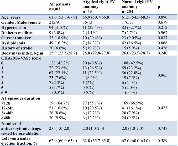
Population characteristics
From January 1, 2015 to October 31, 2018, 303 consecutive patients had a CB ablation for symptomatic paroxysmal AF and were prospectively included. Clinical characteristics are described in Table 1. Patients were mainly men, aged 61.0 (53.8-67.9) years old, and had rare comorbidities reflected by the low CHA2DS2-VASc score of the overall population (66.0% of all patients had a score of 0 or 1). Left ventricular ejection fraction was 62.0% (60.0-65.0). A median of 2.0 (1.0-2.0) antiarrhythmic drugs were tested before the ablation was scheduled.
Anatomical considerations
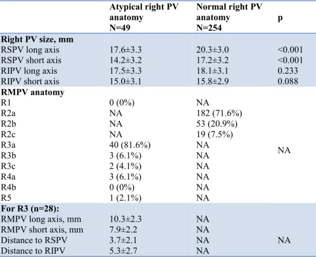
Right PV drainage pattern were classified as stated above (Figure 1). As shown in Table 2, most patients (n= 254, 83.8%) had the expected normal right PV anatomy consisting of two right atrial ostia for upper and lower lobe veins, respectively, with the middle lobe vein
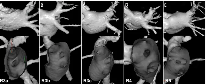
joining the superior lobe vein. The remaining 49 (16.2%) patients had an atypical right PV anatomy. The most common atypical drainage was a R3a pattern (n=40, 81.6%) i.e. three atrial ostia for the superior, middle and lower lobe veins, respectively. R3b and R4 anatomies were found in 3 patients, R3c anatomy in 2 patients and one patient had R5 anatomy. None of the patients presented a R1 anatomy. Examples of atypical right PV anatomies are depicted in Figure 2.
RSPV short and long axis diameters of patients with “atypical” right PV anatomy were significantly smaller than those with R2 pattern (p<0.001). For patients with R3 anatomy, the right middle pulmonary vein (RMPV) long and short axis were 10.3±2.3mm and 7.9±2.2mm respectively, and its distance to the RSPV and the RIPV was 3.7±2.1 and 5.3±2.7mm, respectively.
Procedural characteristics
After ablating left-side PVs, CB applications were performed in the RSPV and RIPV only, neglecting accessory right PVs. First freeze isolation for RSPV occurred in 44 (89.8%) and 218 (85.8%) patients with atypical and typical right PV anatomy, respectively (p=0.606, Table 3). Similar results were observed for the RIPV, with 37 (75.5%) and 217 (85.4%) inferior veins isolated during the first freeze (p=0.130). There was no difference in terms of occlusion degree during freezing. Minimal temperature was lower in both RSPV and RIPV in normal anatomy group (-47.6±5.7 vs -49.5±9.5, p=0.006 and 46.2±7.2 vs -47.7±8.1, p=0.046), and more real-time PV potentials recording were observed in the RSPV for patients with normal anatomy. Lastly, there was no difference in procedure duration between both groups.
To note, once RSPV and RIPV isolation were performed, the accessory right PV isolation was assessed and found to be isolated in all patients with a R3 pattern anatomy. Catheterization of accessory PVs in patients with R4 and R5 anatomies could not be obtained due to their small diameter.
Complications
Twenty nine transient PNP occurred in 26 patients (8.6%), including 20 PNP during RSPV isolation, 3 PNP during RIPV isolation and 3 PNP during both superior and inferior CB isolation. Interestingly, all PNPs occurred in patients with normal right PV anatomy, while none of the patients with atypical anatomy had this complication (p=0.039). To note, all the PNPs resolved before the end of the procedure. No other major complication occurred.
Follow-up
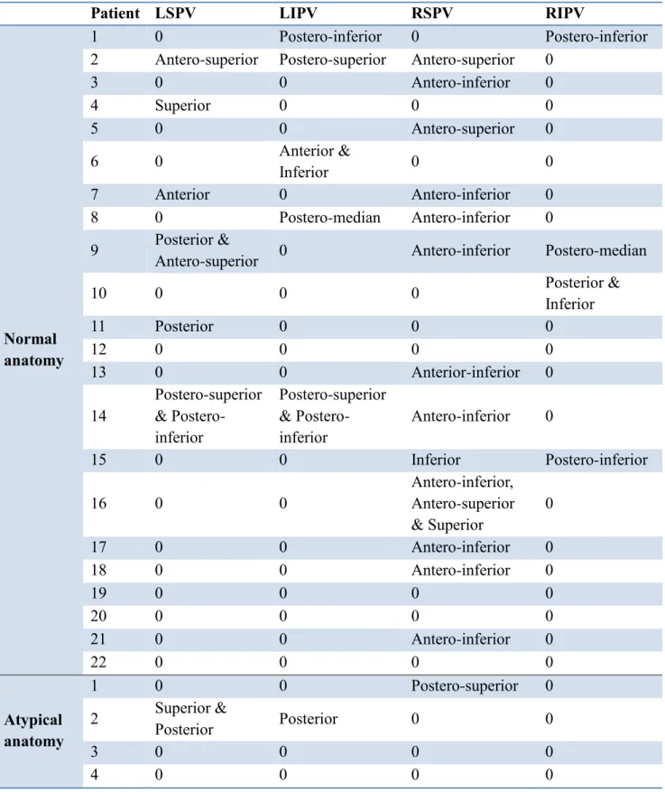
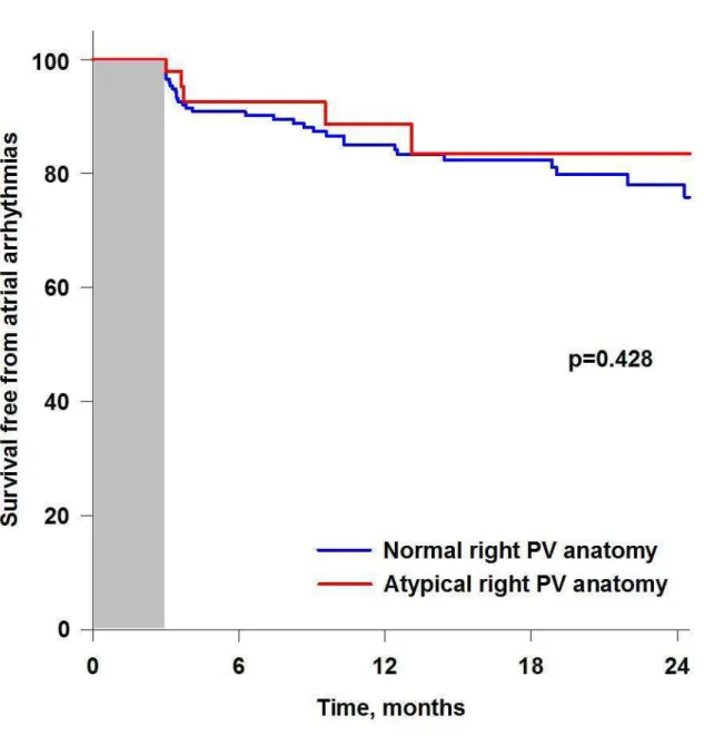
After a follow-up of 12.6±0.2 months, 42 (13.9%) patients had symptomatic documented recurrences of atrial arrhythmias, including 37 (14.6%) and 5 (10.2%) patients with normal and atypical right PV anatomy, respectively (p=0.560). Kaplan-Meier curves of survival free from atrial arrhythmias are shown in Figure 3. No difference was observed among groups (Log rank p=0.428). Among these patients, 22 and 4 patients were referred for a redo ablation, respectively. The localization of gaps encountered during redo cases are detailed in Table 4. Among the 4 patients with atypical anatomy, only 1 had a gap in the RSPV, in its’ postero-superior region, i.e. not close to the accessory PV.
Discussion
Main results
The main findings of the study are: 1) A significant proportion of patients have atypical right PV anatomy (16.2% in our study); 2) Procedural characteristics and acute PV isolation success are similar in all patients, regardless of their right PV anatomy, although a less negative temperature was observed in patients with atypical anatomy; 3) The isolation of RSPV and RIPV, neglecting accessory PVs, is sufficient to obtain acute right-side PV isolation and mid-term sinus rhythm maintenance in patients with atypical anatomy; 4) PNPs were only observed in patients with normal anatomy.
Variations in pulmonary vein anatomy
Normal PV anatomy is defined as the presence of four PVs presenting well-defined ostia. The RSPV usually drains the right upper and middle pulmonary lobes while the left superior PV drains the left upper lobe, including the lingula. Inferior PVs drain their respective lower lobes. This configuration is observed in 57-82% of the population. (7,11–16) Anatomical variants of this typical drainage have been described in 18-53% (11,12,14,17,18) of the patients, the most common being the presence of a left common trunk (8-59%) (3,4,6,7,12,17–19). Anatomical variants on the right PVs are less common (6-32%) (7,12– 15,17,18) but tend to be more complex because of the presence of a third PV coming from the middle pulmonary lobe, draining either in the superior or the inferior PV, or directly in the LA through a separated ostium. The presence of a RMPV has been described in 11-26% of
individuals (7,13–15,17,20). Tsao et al. (21) reported that the RMPV might drain directly into the LA in 23% of patients. In our study, 16.2% of the patients had an atypical right PV drainage, mainly a R3a pattern, consisting on a separate PV coming from the middle lobe and draining directly into the LA. Other types of atypical right PV anatomy were less common, and only 4 patients had 4 or 5 PV ostia.
Cryoballoon ablation in patients with atypical pulmonary vein anatomy
Data regarding the efficacy of CB ablation in patients with a left common trunk are controversial. Some studies have demonstrated a similar AF free-survival rate compared to patients with normal left PV anatomy (3,4) whereas some others have reported a worse arrhythmia free-survival (5,6). Shigeta et al have (6) demonstrated that the presence of a left common trunk was an independent predictor of atrial tachyarrhythmia recurrence after a follow up of 454 ± 195 days (hazard ratio 3.0, 95% confidence interval 1.32-6.83, p<0.01). Further studies will be needed to investigate whether a critical size of the common trunk may impair CB ablation efficacy.
However, to the best of our knowledge, there are currently no reports regarding specifically the efficacy of CB ablation in patients with atypical right PV anatomy. In our study, we did not find any difference in terms of mid-term arrhythmia-free survival, although some procedural parameters differed among groups. Indeed, there was no difference in terms of degree of PV occlusion, successful first freeze and total number of applications per PV. However, the nadir temperature was significantly more negative in those patients with normal PV anatomy, probably explained by their larger short and long-axis diameters compared to patients with atypical drainage. Indeed, the middle lobe vein is usually connected to the RSPV, which drains the blood coming from the upper and the middle pulmonary lobes. In patients with a middle lobe vein directly connected to the LA, the RSPV only drains the upper pulmonary lobe, explaining its smaller size, and subsequently, the less negative nadir temperatures. Surprisingly, more applications with PV potentials were recorded in patients with normal right PV drainage during RSPV isolation compared to those with atypical PV anatomy (p=0.005). This observation can possibly be explained by a difference in terms of myocardial PV sleeve anatomy or to a more distal positioning of the Achieve catheter during ablation.
Interestingly, as previously stated, since a 28mm CB was used, and to avoid increasing the risk of PNP, we decided to not perform a freezing application specifically for the accessory PVs, assuming that the proximity to RSPV and RIPV ostia (3.7±2.1 and 5.3±2.7mm,
respectively) would have led to an isolation of these supplementary PVs. Despite their smaller size (10.3±2.3mm x 7.9±2.2mm for RMPV in R3 patients), accessory PVs could be catheterized in R3 patients to prove PV isolation, demonstrating that a specific freezing application is unnecessary for these PVs, and that they can be neglected without compromising ablation outcome. Indeed, the similar mid-term efficacy observed in our study, regardless of the right PV pattern, is a plea to avoid performing supplemental freezing applications in patients with atypical PV drainage. As described, none of the two patients requiring a redo ablation had a conduction gaps in region close to the supplemental PV.
Clinical impact of pre-procedural imaging
The aim of this study was to show whether a difference in clinical outcomes after CB ablation of paroxysmal AF was observed between patients with normal and atypical right PV anatomy, ablating RSPV and RIPV only and neglecting ablation of accessory PVs. Indeed, some studies (22) have showed that CB ablation efficacy was critically dependent on venous occlusion degree, suggesting that it would be more difficult to obtain a perfect occlusion in patients with atypical PV drainage. Conversely, in this study, we did not show any difference in terms of clinical outcomes depending on PV anatomy, and patients with atypical right PV drainage should not be denied a CB ablation only based on this anatomical pattern. However, we believe that this finding should not weaken the value of pre-procedural 3D imaging of PV anatomy. Indeed, many studies have demonstrated the benefit of cardiac CT scan or Magnetic Resonance Imaging prior to AF ablation (11,15,23), allowing the recognition of anatomical variants and providing an indication about PV orientation, shape and size. Furthermore, as described above, the efficacy of CB ablation in patients with a left common trunk is still a matter of debate (3–6). Thus, we believe that 3D reconstruction is essential for procedural planning and may help to decide whether a CB- or a radiofrequency-based ablation should be performed in a given patient.
Impact of pulmonary vein anatomy on phrenic nerve palsy
PNP is one of the most common complications during CB ablation, occurring in 1.5-13.6% of patients (24–29). A similar proportion of 8.6% of PNP was observed in our study. Several predictors of PNP occurrence have been described, including the presence of a right common ostium, large PV dimensions, early branching pattern originating from the same ostium, or shorter distance from PV to superior vena cava (25,26). More precisely, these studies described predictors of PNP occurrence for RSPV freeze (RSPV area, RSPV-LA angle) and
RIPV freeze (temperature drop velocity from basal to -20°C). Saitoh et al. (30) have found that the strongest predictor for phrenic nerve injury at RSPV was the position of the balloon ≥ 1/3 outside the cardiac shadow (p=0.001, OR 119.9, 95%,CI 11.6-1234.7). Interestingly, in our study, all PNPs occurred in patients with a normal PV anatomy and none of the patients with atypical right PV drainage had this complication. Different hypotheses may explain such finding. First, one may hypothesize that atypical right PV anatomy may be associated with an atypical phrenic nerve position, located further from PV ostia, making it less prone to freezing. This hypothesis was not specifically analyzed in this study and would require further investigations. Second, as demonstrated in our study, patients with normal PV anatomy have larger RSPVs, and cryoenergy may be delivered further inside the PV ostium, closer to the phrenic nerve position. Indeed, Casado-Arroyo et al (31) showed that a less vigorous wedging maneuver in the RSPV ostia might reduce the occurrence of PNP. Authors recommended in their study to slightly retrieve the inflated CB until a small leak is observed before repositioning it at the beginning of the cryoenergy application to create a more proximal lesion without compromising the quality of the occlusion. Patients with a typical anatomy, having smaller RSPVs, are probably at less risk since the lesion created by a big 28-mm CB is possibly more proximal. Lastly, the nadir temperature was less negative in patients with atypical anatomy, a parameter possibly explaining why no PNPs were observed in this group. Further studies will be needed to analyze the effect of PV shape, orientation and anatomy on PNP occurrence.
Limitations
This study has several limitations that must be considered. First, it is a single center study. The clinical endpoint of sinus rhythm maintenance evaluates the global efficacy of CB ablation for both right and left PVs, and AF recurrence in a given patient with typical or atypical right PV anatomy may be due to a reconnection of left PVs while right PVs remain disconnected. Most of the patients with atypical anatomy had a R3 pattern, only a limited number of patients had a R4/R5 anatomy, and none had a R1 pattern, somehow limiting the results observed to patients with this type of anatomy. Lastly, PV reconnection was only assessed in those patients referred for a redo ablation, and one may argue that reconnection rate could differ between asymptomatic or symptomatic patients non-referred for a second procedure. However, Miyazaki et al proved that regardless of clinical recurrences, the incidence and characteristics of PV reconnections were similar between both groups (32).
Conclusion
To the best of our knowledge, this is the first study specifically reporting the safety and efficacy of CB ablation in patients with atypical right PV drainage. In addition to the isolation of left-side PVs, the isolation of RSPV and RIPV, neglecting accessory PVs, is sufficient to obtain acute right-side PV isolation and mid-term sinus rhythm maintenance in patients with atypical anatomy. To note, the risk of PNP seems to be negligible in these patients, and further studies will be needed to explain whether anatomical or procedural characteristics explain such finding.
References
1. Mugnai G, Chierchia G-B, de Asmundis C, Sieira-Moret J, Conte G, Capulzini L, et al. Comparison of pulmonary vein isolation using cryoballoon versus conventional radiofrequency for paroxysmal atrial fibrillation. Am J Cardiol. 2014;113:1509‑13. 2. Kuck K-H, Brugada J, Fürnkranz A, Metzner A, Ouyang F, Chun KRJ, et al.
Cryoballoon or Radiofrequency Ablation for Paroxysmal Atrial Fibrillation. N Engl J Med. 2016;374:2235‑45.
3. Heeger C-H, Tscholl V, Wissner E, Fink T, Rottner L, Wohlmuth P, et al. Acute efficacy, safety, and long-term clinical outcomes using the second-generation cryoballoon for pulmonary vein isolation in patients with a left common pulmonary vein: A multicenter study. Heart Rhythm. 2017;14:1111‑8.
4. Ströker E, Takarada K, de Asmundis C, Abugattas J-P, Mugnai G, Velagić V, et al. Second-generation cryoballoon ablation in the setting of left common pulmonary veins: Procedural findings and clinical outcome. Heart Rhythm. 2017;14:1311‑8.
5. Beiert T, Lodde PC, Linneborn LPT, Werner J, Prinz L, Stöckigt F, et al. Outcome in patients with left common pulmonary vein after cryoablation with second-generation cryoballoon. Pacing Clin Electrophysiol PACE. 2018;41:22‑7.
6. Shigeta T, Okishige K, Yamauchi Y, Aoyagi H, Nakamura T, Yamashita M, et al. Clinical assessment of cryoballoon ablation in cases with atrial fibrillation and a left common pulmonary vein. J Cardiovasc Electrophysiol. 2017;28:1021‑7.
7. Marom EM, Herndon JE, Kim YH, McAdams HP. Variations in pulmonary venous drainage to the left atrium: implications for radiofrequency ablation. Radiology. 2004;230:824‑9.
8. Chun K-RJ, Schmidt B, Metzner A, Tilz R, Zerm T, Köster I, et al. The « single big cryoballoon » technique for acute pulmonary vein isolation in patients with paroxysmal atrial fibrillation: a prospective observational single centre study. Eur Heart J. 2009;30:699‑709.
9. Chierchia GB, Sorgente A, Sarkozy A, de Asmundis C, Brugada P. The Use of Cryoballoon Ablation in Atrial Fibrillation: Simplifying Pulmonary Vein Isolation? J Atr Fibrillation. 2010;3:294.
10. Calkins H, Hindricks G, Cappato R, Kim Y-H, Saad EB, Aguinaga L, et al. 2017 HRS/EHRA/ECAS/APHRS/SOLAECE expert consensus statement on catheter and surgical ablation of atrial fibrillation: Executive summary. Europace. 2018;20:157‑ 208.
11. Kato R, Lickfett L, Meininger G, Dickfeld T, Wu R, Juang G, et al. Pulmonary vein anatomy in patients undergoing catheter ablation of atrial fibrillation: lessons learned by use of magnetic resonance imaging. Circulation. 2003;107:2004‑10.
12. Sohns C, Sohns JM, Bergau L, Sossalla S, Vollmann D, Lüthje L, et al. Pulmonary vein anatomy predicts freedom from atrial fibrillation using remote magnetic navigation for circumferential pulmonary vein ablation. Europace. 2013;15:1136‑42.
13. Hof I, Chilukuri K, Arbab-Zadeh A, Scherr D, Dalal D, Nazarian S, et al. Does left atrial volume and pulmonary venous anatomy predict the outcome of catheter ablation of atrial fibrillation? J Cardiovasc Electrophysiol. 2009;20:1005‑10.
14. Cronin P, Kelly AM, Desjardins B, Patel S, Gross BH, Kazerooni EA, et al. Normative analysis of pulmonary vein drainage patterns on multidetector CT with measurements of pulmonary vein ostial diameter and distance to first bifurcation. Acad Radiol. 2007;14:178‑88.
15. Mansour M, Holmvang G, Sosnovik D, Migrino R, Abbara S, Ruskin J, et al. Assessment of pulmonary vein anatomic variability by magnetic resonance imaging: implications for catheter ablation techniques for atrial fibrillation. J Cardiovasc Electrophysiol. 2004;15:387‑93.
16. Scharf C, Sneider M, Case I, Chugh A, Lai SWK, Pelosi F, et al. Anatomy of the pulmonary veins in patients with atrial fibrillation and effects of segmental ostial ablation analyzed by computed tomography. J Cardiovasc Electrophysiol. 2003;14:150‑ 5.
17. McLellan AJA, Ling L, Ruggiero D, Wong MCG, Walters TE, Nisbet A, et al. Pulmonary vein isolation: the impact of pulmonary venous anatomy on long-term outcome of catheter ablation for paroxysmal atrial fibrillation. Heart Rhythm. 2014;11:549‑56.
18. Wannasopha Y, Oilmungmool N, Euathrongchit J. Anatomical variations of pulmonary venous drainage in Thai people: multidetector CT study. Biomed Imaging Interv J. 2012;8:e4.
19. Kubala M, Hermida J-S, Nadji G, Quenum S, Traulle S, Jarry G. Normal pulmonary veins anatomy is associated with better AF-free survival after cryoablation as compared to atypical anatomy with common left pulmonary vein. Pacing Clin Electrophysiol PACE. juill 2011;34(7):837‑43.
20. Mulder AAW, Wijffels MCEF, Wever EFD, Boersma LVA. Pulmonary vein anatomy and long-term outcome after multi-electrode pulmonary vein isolation with phased radiofrequency energy for paroxysmal atrial fibrillation. Europace. 2011;13:1557‑61. 21. Tsao HM, Wu MH, Yu WC, Tai CT, Lin YK, Hsieh MH, et al. Role of right middle
pulmonary vein in patients with paroxysmal atrial fibrillation. J Cardiovasc Electrophysiol. 2001;12:1353‑7.
22. Sorgente A, Chierchia GB, de Asmundis C, Sarkozy A, Namdar M, Capulzini L, et al. Pulmonary vein ostium shape and orientation as possible predictors of occlusion in patients with drug-refractory paroxysmal atrial fibrillation undergoing cryoballoon ablation. Europace. 2011;13:205‑12.
23. Mlcochová H, Tintera J, Porod V, Peichl P, Cihák R, Kautzner J. Magnetic resonance angiography of pulmonary veins: implications for catheter ablation of atrial fibrillation. Pacing Clin Electrophysiol. 2005;28:1073‑80.
24. Su W, Orme GJ, Hoyt R, Baker J, Compton S, Fellows C, et al. Retrospective review of Arctic Front Advance Cryoballoon Ablation: a multicenter examination of second-generation cryoballoon (RADICOOL trial). J Interv Card Electrophysiol. 2018;51:199‑ 204.
25. Abugattas J-P, de Asmundis C, Iacopino S, Salghetti F, Takarada K, Coutiño H-E, et al. Phrenic nerve injury during right inferior pulmonary vein ablation with the second-generation cryoballoon: clinical, procedural, and anatomical characteristics. Europace. 2018;20:e156-r163.
26. Ströker E, de Asmundis C, Saitoh Y, Velagić V, Mugnai G, Irfan G, et al. Anatomic predictors of phrenic nerve injury in the setting of pulmonary vein isolation using the 28-mm second-generation cryoballoon. Heart Rhythm. 2016;13:342‑51.
27. Mugnai G, de Asmundis C, Ciconte G, Irfan G, Saitoh Y, Velagic V, et al. Incidence and characteristics of complications in the setting of second-generation cryoballoon ablation: A large single-center study of 500 consecutive patients. Heart Rhythm. 2015;12:1476‑ 82.
28. Fürnkranz A, Bordignon S, Schmidt B, Perrotta L, Dugo D, De Lazzari M, et al. Incidence and characteristics of phrenic nerve palsy following pulmonary vein isolation with the second-generation as compared with the first-generation cryoballoon in 360 consecutive patients. Europace. 2015;17:574‑8.
29. Martins RP, Hamon D, Césari O, Behaghel A, Behar N, Sellal J-M, et al. Safety and efficacy of a second-generation cryoballoon in the ablation of paroxysmal atrial fibrillation. Heart Rhythm. 2014;11:386‑93.
30. Saitoh Y, Ströker E, Irfan G, Mugnai G, Ciconte G, Hünük B, et al. Fluoroscopic position of the second-generation cryoballoon during ablation in the right superior pulmonary vein as a predictor of phrenic nerve injury. Europace. 2016;18:1179‑86. 31. Casado-Arroyo R, Chierchia G-B, Conte G, Levinstein M, Sieira J, Rodriguez-Mañero
M, et al. Phrenic nerve paralysis during cryoballoon ablation for atrial fibrillation: a comparison between the first- and second-generation balloon. Heart Rhythm. 2013;10:1318‑24.
32. Miyazaki S, Taniguchi H, Hachiya H, Nakamura H, Takagi T, Hirao K, et al. Clinical recurrence and electrical pulmonary vein reconnections after second-generation cryoballoon ablation. Heart Rhythm. 2016;13:1852‑7.
Table 1: Patients’ characteristics All patients n=303 Atypical right PV anatomy n=49 Normal right PV anatomy n=254 p Age, years 61.0 (53.8-67.9) 56.9 (50.7-66.8) 61.5 (54.5-68.3) 0.090 Gender, Male/Female 212/91 36/13 176/78 0.679 Hypertension 94 (31.0%) 11 (22.4%) 83 (32.7%) 0.212 Diabetes mellitus 9 (3.0%) 2 (4.1%) 7 (2.7%) 0.967 Current smoker 33 (10.9%) 10 (20.4%) 23 (9.05%) 0.037 Dyslipidemia 49 (16.2%) 7 (14.3%) 42 (16.5%) 0.866 History of stroke 20 (6.6%) 5 (10.2%) 15 (5.9%) 0.426
Body mass index, kg.m² 25.9 (23.5-28.7) 25.8 (22.8-27.8) 26.0 (23.5-28.7) 0.240
CHA2DS2-VASc score
0 1 2 3 4 5 6-9 128 (42.2%) 72 (23.8%) 67 (22.1%) 23 (7.6%) 7 (2.3%) 5 (1.7%) 1 (0.3%) 20 (40.8%) 13 (26.5%) 11 (22.5%) 4 (8.2%) 1 (2%) 0 (0%) 0 (0%) 108 (42.5%) 59 (23.2%) 56 (22.0%) 19 (7.5%) 6 (2.4%) 5 (2.0%) 1 (0.4%) 0.965 AF episodes duration <12h 12-24h 24-48h >48h 196 (64.7%) 51 (16.8%) 26 (8.6%) 30 (9.9%) 27 (55.1%) 10 (20.5%) 6 (12.2%) 6 (12.2%) 169 (66.5%) 41 (16.1%) 20 (7.9%) 24 (9.5%) 0.473 Number of antiarrhythmic drugs
tested before ablation 2.0 (1.0-2.0) 2.0 (1.0-2.0) 2.0 (1.0-2.0) 0.747 Left ventricular
Table 2: Anatomical considerations. RSPV: right superior pulmonary vein; RIPV: right
inferior pulmonary vein; RMPV: right middle pulmonary vein
Atypical right PV anatomy N=49 Normal right PV anatomy N=254 p Right PV size, mm RSPV long axis RSPV short axis RIPV long axis RIPV short axis
17.6±3.3 14.2±3.2 17.5±3.3 15.0±3.1 20.3±3.0 17.2±3.2 18.1±3.1 15.8±2.9 <0.001 <0.001 0.233 0.088 RMPV anatomy R1 R2a R2b R2c R3a R3b R3c R4a R4b R5 0 (0%) NA NA NA 40 (81.6%) 3 (6.1%) 2 (4.1%) 3 (6.1%) 0 (0%) 1 (2.1%) NA 182 (71.6%) 53 (20.9%) 19 (7.5%) NA NA NA NA NA NA NA For R3 (n=28): RMPV long axis, mm RMPV short axis, mm Distance to RSPV Distance to RIPV 10.3±2.3 7.9±2.2 3.7±2.1 5.3±2.7 NA NA NA NA NA
Table 3: Procedural characteristics. PNP: Phrenic nerve palsy; LIPV: left inferior
pulmonary vein; LSPV: left superior pulmonary vein; RIPV: right inferior pulmonary vein; RSPV: right superior pulmonary vein.
Atypical right PV anatomy N=49 Normal right PV anatomy N=254 p RSPV Occlusion 4/4
Applications with PVP recording Minimal temperature, °C
PNP
Successful 1st freeze
Total number of applications
48 (98.0%) 34 (69.4%) -47.6±5.7 0 (0%) 44 (89.8%) 1.2±0.5 248 (97.6%) 220 (86.6%) -49.5±9.5 23 (9.05%) 218 (85.8%) 1±.20.5 0.702 0.005 0.006 0.058 0.606 0.939 RIPV Occlusion 4/4
Applications with PVP recording Minimal temperature, °C
PNP
Successful 1st freeze
Total number of applications
43 (87.7%) 13 (26.5%) -46.2±7.2 0 (0%) 37 (75.5%) 1.2±0.5 232 (91.3%) 74 (29.1%) -47.7±8.1 6 (2.4%) 217 (85.4%) 1.2±0.5 0.601 0.844 0.046 0.598 0.130 0.422
Procedure duration, minutes 75.0 (67.5-87.5) 75.0 (70.0-90.0) 0.742
Table 4: Localization of conduction gaps during redo procedures. LIPV: left inferior
pulmonary vein; LSPV: left superior pulmonary vein; RIPV: right inferior pulmonary vein ; RSPV: right superior pulmonary vein.
Patient LSPV LIPV RSPV RIPV
Normal anatomy
1 0 Postero-inferior 0 Postero-inferior
2 Antero-superior Postero-superior Antero-superior 0
3 0 0 Antero-inferior 0
4 Superior 0 0 0
5 0 0 Antero-superior 0
6 0 Anterior & Inferior 0 0
7 Anterior 0 Antero-inferior 0
8 0 Postero-median Antero-inferior 0
9 Posterior & Antero-superior 0 Antero-inferior Postero-median
10 0 0 0 Posterior & Inferior
11 Posterior 0 0 0
12 0 0 0 0
13 0 0 Anterior-inferior 0
14 Postero-superior & Postero-inferior Postero-superior & Postero-inferior Antero-inferior 0 15 0 0 Inferior Postero-inferior 16 0 0 Antero-inferior, Antero-superior & Superior 0 17 0 0 Antero-inferior 0 18 0 0 Antero-inferior 0 19 0 0 0 0 20 0 0 0 0 21 0 0 Antero-inferior 0 22 0 0 0 0 Atypical anatomy 1 0 0 Postero-superior 0
2 Superior & Posterior Posterior 0 0
3 0 0 0 0
Figure 1: Right pulmonary drainage pattern. Adapted from Marom et al (7)
RLL: right lower lobe; RML: right middle lobe; RUL: right upper lobe; SSRLL: superior segment right lower lobe; BSRLL: basilar segment right lower lobe.
Figure 2: Cardiac CT scan reconstructions of LA (postero-anterior view and endoluminal
view) showing: panel A: R3a, panel B: R3b, panel C: R3c, panel D: R4 and panel E: R5 patterns.
Figure 3: Kaplan-Meier curves for cumulated survival free of atrial arrhythmias in patients with normal and atypical right PV anatomy.
PICHARD Camille - Titre de la thèse : Anatomie atypique des veines pulmonaires droites et cryo-ablation de fibrillation atriale.
39 pages, 3 illustrations, 4 tableaux - Thèse : (Médecine) ; Rennes 1; 2019 ; N° .
Résumé français : Contexte : L'ablation par cryoballon est une technique largement utilisée dans l'isolation
des veines pulmonaires (VP) chez les patients avec une fibrillation atriale (FA). A ce jour, il existe peu de données concernant l'efficacité clinique de la cryo-ablation chez les patients ayant une anatomie veineuse pulmonaire droite atypique. L'objectif de cette étude était d'évaluer l'impact de l'anatomie veineuse
pulmonaire droitesur l'efficacité et la sécurité de l’ablation par cryoballon. Méthodes : Tous les patients
adressés au CHU de Rennes pour ablation de FA paroxystique par cryoballon ont été prospectivement inclus. Un scanner de l'oreillette gauche était réalisé avant l'ablation pour déterminer si l'anatomie des VP droites était "normale" ou "atypique". Chez les patients avec une anatomie atypique, la cryoablation n'était pas réalisée sur les VP surnuméraires. Résultats : Sur 303 patients inclus, 254 (83.8%) avaient une anatomie veineuse pulmonaire droite normale et 49 (16.2%) une anatomie atypique. L’isolation veineuse était obtenue à la première application de cryothermie pour 44 (89.8%) VP supérieures droites (VPSD) et 37 (75.5%) VP inférieures droites (VPID) chez les patients avec une anatomie atypique, et pour 218 (85.8%) VPSD et 217 (85.4%) VPID chez les patients avec une anatomie typique (p=NS). Les paralysies du nerf phrénique (PNP) étaient observées seulement chez les patients avec une anatomie normale (0 vs. 26, 8.6%, p=0.039). La survie à moyen terme sans récidive d’arythmie atriale était similaire, indépendamment de l’anatomie des VP droites. Conclusion : Une proportion significative de patients a une anatomie VP droite atypique. Les caractéristiques procédurales, le succès immédiat de l’isolation des VP, et l’efficacité à moyen terme sont similaires dans les deux groupes, indépendamment de l’anatomie veineuse pulmonaire droite. L’ablation par cryoballon des VPSD et VPID, en négligeant les VP droites accessoires, associée à la cryo-ablation des VP gauches est suffisante pour obtenir une isolation en aigu et un maintien à moyen terme du rythme sinusal chez les patients avec une anatomie veineuse pulmonaire droite atypique.
Résumé anglais: Background: Cryoballoon (CB) ablation is widely used for pulmonary vein (PV) isolation
in patients with atrial fibrillation (AF). There are no data regarding clinical efficacy of CB ablation in patients with atypical right PV anatomy. We aimed to evaluate the impact of right PV anatomy on safety and efficacy of CB ablation. Methods: Patients referred for CB ablation of paroxysmal AF were prospectively enrolled. A left atrial computed tomography was performed prior to CB ablation to determine whether right PV anatomy was “normal” or “atypical”. For patients with atypical anatomy, CB ablation was only performed for right superior (RSPV) and right inferior PV (RIPV), neglecting accessory PVs. Results: 303 patients were included, 254 (83.8%) with normal and 49 (16.2%) with atypical right PV anatomy. First freeze isolation for RSPV and RIPV occurred in 44 (89.8%) and 37 (75.5%) and in 218 (85.8%) and 217 (85.4%) patients with atypical and typical PV anatomy, respectively (p=NS). Phrenic nerve palsies (PNPs) were only observed in patients with normal anatomy (0 vs. 26, 8.6%, p=0.039). Mid-term survival free from atrial arrhythmia was similar, regardless of right PV anatomy. Conclusion: A significant proportion of patients have atypical right PV anatomy. Procedural characteristics, acute PV isolation success, and mid-term procedural efficacy are similar, regardless of right PV anatomy. In addition to left-side PVs isolation, cryoballoon ablation of RSPV and RIPV only, neglecting accessory PVs, is sufficient to obtain acute right-side PV isolation and mid-term sinus rhythm maintenance in patients with atypical anatomy.
Rubrique de classement :
Mots-clés : arythmie ; fibrillation atriale ; ablation , électrophysiologie
Mots-clés anglais MeSH : Arrhythmias ; Atrial Fibrillation ; Catheter Ablation ; Electrophysiology
JURY :
Président : M. Philippe Mabo
Assesseurs : M. Christophe Leclercq M. Mathieu Lederlin M. Dominique Pavin
M. Raphaël Martins (directeur de thèse)
Adresses de l’auteur : 20 rue du Sapeur Michel Jouan – 35000 Rennes
Powered by TCPDF (www.tcpdf.org) Powered by TCPDF (www.tcpdf.org) Powered by TCPDF (www.tcpdf.org)