Study the effects of cigarette smoke on gingival epithelial
cell growth and the expression of keratins
Mémoire
Ibrahim Alharbi
Maîtrise en Biologie Cellulaire et Moléculaire
Maître ès sciences (M.Sc.)
Québec, Canada
Résumé
Mon projet de recherche vise à évaluer l'effet à long terme de l'exposition à la fumée de cigarette (FC) sur la croissance des cellules épithéliales gingivales et l'expression de plusieurs kératines (K). Les cellules exposées à la FC montrent une activité métabolique et une prolifération cellulaire élevées par rapport aux cellules non exposées. Cependant, une exposition prolongée à la FC réduit le rapport Bax/Bcl-2 ce qui suggère une résistance des cellules exposées à la mort cellulaire. Nos analyses montrent aussi que l'exposition à la FC diminue le niveau d'ARNm des kératines K5, K1, K10, K16. Cependant les K14 et K6 montrent une augmentation après une certaine période d'exposition. Fait intéressant, l'analyse de la production de protéines dont les K5, K1 et K16 montre une expression stable de ces protéines après l'exposition à la FC. En revanche, les K14 et K6 sont significativement augmentées, tandis que la K10 a diminué. Ces observations peuvent expliquer l’hyperplasie observée au niveau des tissus gingivaux des fumeurs, et la fréquence des maladies parodontales.
Abstract
This research project is aimed to evaluate the effect of long-term exposure to cigarette smoke (CS) on the gingival epithelial cells growth and the expression of different keratins (K). The CS-exposed cells are showing high metabolic activity and cell proliferation as compared to non-exposed cells, suggesting an increase in the cells growth. Importantly, the prolonged exposure to CS was found to decrease Bax/Bcl-2 ratio that indicates the death resistance. Moreover, the gene expression analysis showed that the exposure to CS decreases the mRNA level of K5, K1, K10, and K16; excepting K14 and K6 were showing an increase after a certain exposure period. Interestingly, protein production analysis of K5, K1, and K16 are showing a stable expression after the exposure to CS. In contrast, K14 and K6 are significantly increased, while K10 decreased after CS exposure. These observations suggest an abnormal growth of the gingiva that may lead to the periodontal diseases.
Table of Contents
Table of Contents
Résumé ... III Abstract ... V Table of Contents ... VII Table List ... IX Figure List ... XI Abbreviations ... XIII Acknowledgement ... XIX Foreword ... XXI Chapter I ... 1 1. Introduction ... 2 1.1. Tobacco smoke ... 2 1.1.1. History ... 2
1.1.2. Cigarette smoke’s chemical composition ... 4
1.1.3. The environmental tobacco smoke ... 6
1.1.4. Cigarette smoke chemicals detoxification ... 8
1.2. Cigarette smoking widely causes several diseases ... 9
1.2.1. Cigarette smoking-related noncancerous diseases ... 10
1.2.2. Cigarette smoking-related cancerous diseases ... 11
1.2.3. Potential mechanisms for effects of cigarette smoke in causing diseases ... 12
1.2.3.1. Modulating the cellular signaling ... 13
1.2.3.1.1. Modulating the signaling pathways of the proliferation, migration, and death ... 13
1.2.3.1.2. Modulating the signaling pathways of the inflammatory response ... 16
1.2.3.2. Damaging the DNA: Mutagenesis ... 19
1.3. Gingiva ... 21
1.3.1 Gingival tissue structure ... 21
1.3.2 Keratin cytoskeleton role in the gingival tissue ... 23
1.3.3. Gingival tissue functions ... 25
1.3.4. Cigarette smoke-induced gum diseases ... 27
1.4. The hypothesis and objectives of the study ... 28
1.4.1 The hypothesis ... 28 1.4.2. The objectives ... 29 Chapter II ... 30 2. Article ... 31 2.1. ABSTRACT ... 33 2.2. Introduction ... 35
2.3.2. Cell cultures ... 36
2.3.3. Cigarette smoke exposure procedure ... 37
2.3.4. Cell viability/Growth assay ... 37
2.3.5. BrdU-DNA synthesis assay ... 38
2.3.6. Gene expression by quantitative RT-PCR ... 38
2.3.7. Western blot ... 39
2.3.8. Statistical analysis ... 40
2.4. Results ... 40
2.4.1.Whole cigarette smoke induced human gingival epithelial cell growth ... 40
2.4.2. Bax/Bcl-2 ratio decreased following exposure to whole CS ... 41
2.4.3. Basal layer keratin gene and protein expression were both affected by CS ... 41
2.4.4. Effect of CS on suprabasal keratin gene and protein expression ... 42
2.5. Discussion ... 43 2.6. Acknowledgements ... 44 2.7. Conflict of Interest ... 44 2.8. Authors’ Contribution ... 44 2.9. References ... 46 2.10. Figure Captions ... 51 2.11. Table ... 52 2.12. Figures ... 53 Chapter III ... 60
3. General conclusions and perspectives ... 61
3.1. General conclusions ... 61
3.2. Perspectives ... 63
Table List
Table 1-1: Some of the chemical compounds present in cigarette smoke. ... 7 Table 2-1: Primers used for the quantitative RT-PCR. ... 52
Figure List
Figure 1-1: Cigarette smoking and lung cancer possibility during the twentieth century. ... 4
Figure 1-2: The xenobiotic metabolism.. ... 9
Figure 1-3: The possible diseases caused by cigarette smoking, including broad types of cancers and other noncancerous conditions. ... 13
Figure 1-4: The nicotine acetylcholine receptors (nAChRs) activation by nicotine. ... 17
Figure 1-5: Summary of cigarette smoke (CS) effects on the immune system. ... 19
Figure 1-6: Anatomic schema of the periodontal tissue and its cigarette smoke-induced diseases.. ... 23
Figure 1-7: Gingival epithelium architecture. ... 26
Figure 2-1: Effect of whole cigarette smoke on human gingival epithelium cell metabolic activity and proliferation. ... 51
Figure 2-2: Effect of whole cigarette smoke on apoptotic (Bax) and anti-apoptotic (Bcl-2) protein expression. ... 51
Figure 2-3: Effect of whole cigarette smoke on mRNA keratin expression by gingival epithelial cells. ... 51
Figure 2-4: Effect of whole cigarette smoke on basal keratin expression by gingival epithelial cells. ... 51
Figure 2-5: Effect of whole cigarette smoke on suprabasal layer keratin expression. ... 51
Abbreviations
α Alpha
β Beta
BAD Bcl-2-associated death promoter Bax Bcl-2-associated X protein Bcl-2 B-cell lymphoma 2
BrdU Bromodeoxyuridine
CaMKII Ca2+/calmodulin-dependent protein-kinase II
Ca2+ Calcium ion
COPD Chronic obstructive pulmonary disease CS Cigarette smoking/Cigarette smoke CYP450 Cytochrome P450
CT C-terminal
cGMP Cyclic guanosine monophosphate
O2 Dioxygen
δ Delta
DSG Desmoglein
DSC Desmocollin
DP Desmoplakin
DMEH Dulbecco’s modified Eagle’s–Ham’s DTT Dithiothreitol
MTT 3-[4,5-Dimethylthiazol-2-yl]-2,5-diphenyl-tetrazolium bromide ETS Environmental tobacco smoke
EGFR Epidermal growth factor receptor ε Epsilon
ERK Extracellular signal-regulated kinase ECM Extracellular matrix
eNOS Endothelial nitric oxide synthase E-cadherin Epithelial cadherin
EBS Epidermolysis bullosa simplex EDTA Ethylenediaminetetraacetic acid ELISA Enzyme-linked immunosorbent assay Fe2+ Ferrous ion
Fig. Figure
FAK Focal adhesion kinase
γ Gamma
GM-CSF Granulocyte-macrophage colony-stimulating factor GSH Glutathione S-transferases H2O2 Hydrogen peroxide HO Hydroxyl radical OHG Hydroxyguanosine HCl Hydrogen chloride
HBE Human bronchial epithelial HPV Human papillomavirus
IL Interleukin
JAK/STAT Janus kinase/ Signal transducer and activator of transcription
κ Kappa
K Keratin
μg Microgram
MS Mainstream smoke
MCP Monocyte chemoattractant Protein MMP Matrix metalloproteinase NNK 4(methylnitrosamino)-1-(3-pyridyl)-1 butanone MUC1 Mucin-1 NO Nitric oxide NO2 Nitrogen dioxide NNN N′-nitrosonornicotine
nAChR Nicotinic acetylcholine receptor
NFκB Nuclear factor κ-light-chain-enhancer of activated B cells
OED Oral epithelial dysplasia OSCC Oral squamous cell carcinoma ONOO- Peroxynitrite
PKA Protein kinase A PKB Protein kinase B PKC Protein kinase C
PLC Phospholipase C
SRC Proto-oncogene tyrosine-protein kinase P-cadherin Placental cadherin
PG Plakoglobin
PKP Plakophilin
PBS Phosphate-buffered saline ROS Reactive oxygen species RNS Reactive nitrogen species
RT-PCR Reverse transcription-polymerase chain reaction
ROCK RHO-associated protein kinase O − Superoxide anion
XV H2SO4 Sulfuric acid
SDS-PAGE Sodium dodecyl sulfate-polyacrylamide gel electrophoresis TCF/LEF Transcription factor/Lymphoid enhancer-binding factor
TLR Toll-like receptor TNF Tumor necrosis factor
TSLP Thymic stromal lymphopoietin TTBS Tween-20-Tris-buffer saline UDP Uridine diphosphate glucose
WHO World Health Organization XME Xenobiotic metabolizing enzyme
Acknowledgement
I would like to thank Prof. Mahmoud Rouabhia for allowing me joining his research team. He gave me an opportunity to conduct several experimental types of research under his guidance. Indeed, during the master period, I have learnt a lot of new knowledge. Another thank is for Mr. Eric Jacques, a research assistant in Prof. Mahmoud Rouabhia laboratory. Mr. Eric Jacques was a really supportive person for doing several experiments and also he was patient for explaining the different protocols. Also, I thank my all colleagues for sharing a good time that made unforgettable memories. In addition, I would like to thank all members in groupe de recherche en écologie buccale in the dental college for providing a great environment for studying.
As well, I really thank Saudi Arabia government for the financial support during my graduate study. I am grateful to my parents, brothers, and sisters for their kindness in supporting me.
Foreword
This thesis shows the results of effects of cigarette smoke on the human gingival epithelial cells, including their growth and expression of different keratins. This thesis is divided into three chapters. In the first chapter, a general introduction involves cigarette chemicals and its effects on human health. Structure and function of the gingival tissue and cigarette-induced gum diseases are also discussed at the end of this chapter. The second chapter contains a manuscript of the master project experimental results. This manuscript has been recently submitted to a scientific journal and still currently under review. Finally, the third chapter presents general conclusions and perspectives of the project.
The article that is included in this thesis is titled with ‘Repeated exposure to whole cigarette smoke promotes primary human gingival epithelial cell growth and modulates keratin expression’. It has been submitted on 05-20 to the journal of periodontal research. This article got a major revision on 2015-06-25; revision responded by authors and the article re-submitted on 2015-07-01. The article has been conducted by thanking contributions of the following authors: Alharbi IA and Rouabhia M.
Mahmoud Rouabhia conceived the study.
Ibrahim Alharbi conducted the experiments under the supervision of Mahmoud Rouabhia and also collected the data which were analyzed by both authors.
Ibrahim Alharbi drafted the first version of the manuscript and Mahmoud Rouabhia revised the draft. All authors approved the final manuscript prior to its submission.
Chapter I
Introduction
1. Introduction 1.1. Tobacco smoke 1.1.1. History
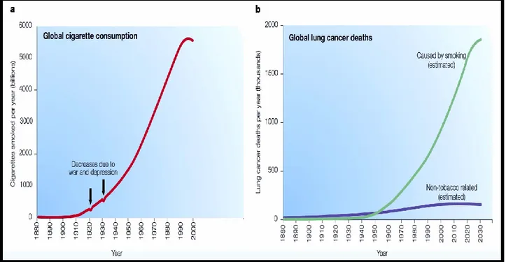
Tobacco has been utilized, originally, in America for several centuries. In the middle of the sixteenth, tobacco was exported to Europe and then spread worldwide. Initially, tobacco was smoked as cigar or in pipes, and subsequently cigarette manufacturing was started in the nineteenth century, but with limited consumption. In 1881, Bonsack JA invented a cigarette-rolling machine that speeded up cigarette production. Thereafter, the increase in manufacturing, marketing, and advertising, cigarette became a popular product e.g. from consuming 54 cigarettes per capita in 1900 to 4345 in 1963 in the United States (1). Likewise, but during the second half of the twentieth century cigarette smoking (CS) was increasing throughout the world (Figure 1-1A).
Once the cigarette became a socially popular item, some of the rare diseases turned to be common. For example during the twentieth century, the incidence of pulmonary carcinoma was low, but the deaths due to this cancer increased with the elevation in cigarette consumption (Figure 1-1B). In the beginning, cigarette was not linked to the lung cancer yet, till the epidemiological studies and statistical estimations lead to a conclusion made by Richard Doll and Richard Peto in 1981, attributing most of lung cancer cases to CS (2). However, at that time, still the molecular carcinogenesis mechanism of cigarette in causing lung cancer was not documented yet.
During the last decades of the twentieth century, more scientific research related to the molecular mechanisms was initiated showing a link between cigarette smoke and cancer. Indeed, Pfeifer et al. (1996) were first showing that the benzo[a]pyrene, one of the cigarette content, caused mutations in a gene, which encoded for a tumor suppressor protein called P53, leading to lung cancer (3). Besides lung carcinoma,
pulmonary destructions and atherosclerosis) are known to be risks of cigarette utilization. Therefore, with all these diseases, CS is considered as an environmental factor that leads to high mortality. About 6 million deaths are estimated annually throughout the world due to cigarette consumption, World Health Organization (WHO).
The statistical estimation and experimental research were useful for informing the people, and that lead to a reduction in cigarette consumption after the mid of 1990s (Figure 1-1A). However, nowadays, still the number of smokers is high globally with expansion in the population although the warnings of CS damaging effects on health are obvious. As recently reported in the United States, 273 billion cigarettes have been sold, 21.6 million men and 18.8 million women smoke cigarette daily (4-6). Hence, investigating the effects of CS on the health is important for understanding the hidden mechanisms to warn the smokers, support them for quitting and reduce or prevent CS-induced diseases development.
Figure 1-1: Cigarette smoking and lung cancer possibility during the twentieth century. A. Cigarette utilization throughout the world. In 1910 cigarette consumption was started to increase with a little drop between 1920 and 1940 because the First and Second World War, then a continuous increase reaching many billions of cigarettes per year at the end of the century. However, in the mid of 1990s, cigarette consumption slightly decreased because releasing the facts of smoking adverse effects on health. B. The annual Lung cancer deaths. Rate of deaths among smokers due to lung cancer was also increased as cigarette consumption, starting from 1950 and may continue till 2030, assuming no preventable treatment discovery. Interestingly, the deaths due to lung cancer in nonsmokers were not significantly increased as in smokers (7).
1.1.2. Cigarette smoke’s chemical composition
Burning cigarette produces mainstream and sidestream smoke. The mainstream smoke is a product of burning the tobacco that travels through cigarette column and passes by the filter then inhaled by smokers, while the sidestream smoke produced between the puffs and its directly emitted into the air from the smoldering cigarette. The mainstream and sidestream smoke that are produced from burned cigarette are in the form of an aerosol, which comprises millions of droplets containing an enormous
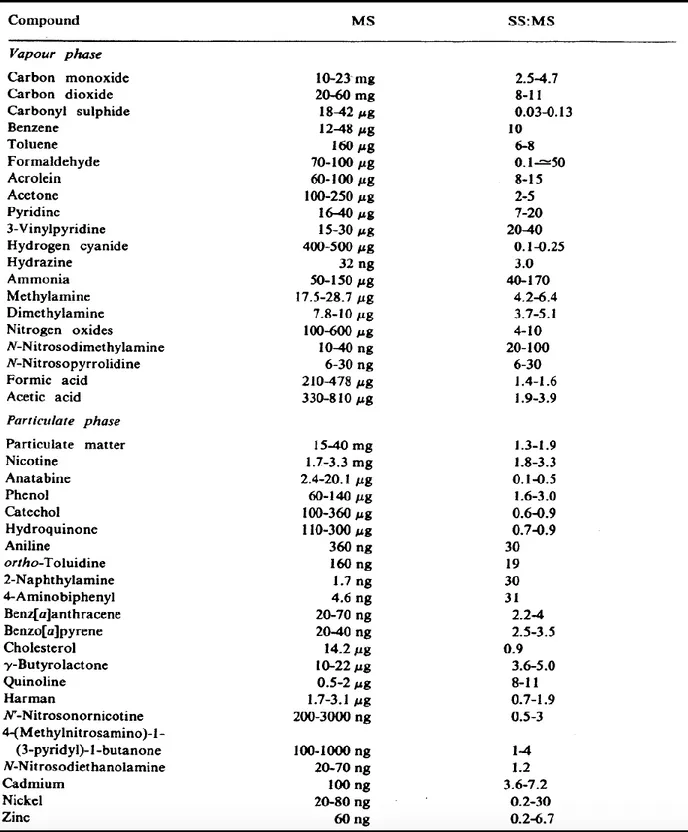
mixture of dynamic chemicals. There are approximately over than 5000 chemicals that have been isolated and identified in the total fresh smoke (8). These chemical compounds are distributed between vapor and particulate phases (Table 1-1).
Both phases contain highly toxic and carcinogenic compounds. Some of the compounds have multipotent effects such as the nicotine, which can enhance the addiction, alter normal cell functions, and induce cancer development (9, 10). Also, even though there are compounds at low levels, they are effective in mediating the cellular damage (8). For example, the benzo[a]pyrene level in the particulate phase is over 2000-fold lower than acrolein of the vapor phase, but both ultimately have similar efficiency in mediating P53 damage, leading to lung carcinogenesis (3, 11). Similarly, the particulate phase contains a trace amount of minerals, which some have been linked to a particular damage in the body (12). For instance, it has been found that cadmium concentration increased in the blood of smokers, which subsequently leads to its accumulation in the kidney and may later result in a tubular dysfunction and renal failure. (13,14).
Also, cigarette smoke (CS) of the both phases is rich in the reactive oxygen species (ROS) and reactive nitrogen species (RNS). In the particulate phase, it has been identified a highly stable ROSs, identified as various semiquinone radicals (15). Moreover, by several steps, there are other unstable ROSs, such as O2−, H2O2, and HO that can be produced from the semiquinone radicals (16). This starts with
reducing the dioxygen (O2) by the semiquinone radicals and producing the superoxide anion (O2−) that in
turn can dismutate and generate the hydrogen peroxide (H2O2). The H2O2 can be converted into a hydroxyl
radical (HO) by Fenton reaction in which the Fe2+, is one of the most available metals in the cigarette, is
the catalyzing agent. On other hand, in the vapor phase smoke, it has been found nitric oxide (NO) radical, which is classified as a RNS (17). Also, by the oxidation, or interaction with the superoxide radical, other RNSs can be generated from the nitric oxide, such as the nitrogen dioxide (NO2) and peroxynitrite (ONOO
-), respectively (18). The ROSs and RNSs are harmful oxidizing agents as they effectively can damage the cellular components and induce the oxidative stress (19).
1.1.3. The environmental tobacco smoke
The mainstream and the sidestream smoke have similar content, but in the last the substances concentrations are very high. The sidestream/mainstream ratios (SS:MS) of most of CS chemicals are increased with exception for only six compounds (carbonyl sulphide, hydrogen cyanide, anatabine, hydroquinone, catechol, and cholesterol) are showing a decrease (Table 1-1). Moreover, these compounds may spread again into the ambient air from the exhaled smoke of smokers (20). This means tremendous quantities of CS compounds are emitted into the air while puffing the cigarette and after exhaling the smoke.
Both the exhaled mainstream and sidestream smoke generate the environmental tobacco smoke (ETS). Indeed, many of CS chemicals have been detected and measured, such as nicotine (15-35 μg), acrolein (25 μg), and nitrogen oxide (50 μg) in the indoor spaces (21). The second-hand smoker (passive smoker) usually exposed to the ETS that produced from the neighbor smoker. Unfortunately, it has been reported that passive smokers can uptake nicotine (22, 23). Furthermore, nicotine and its primary metabolite, cotinine, have shown increase in the saliva, plasma, and urine of adults and infants who were exposed to CS (24). Thus, the ETS may induce diseases in the second-hand smokers, similarly, to real smokers.
Table 1-1: Some of the chemical compounds present in cigarette smoke. MS, mainstream smoke; SS:MS, sidestream/mainstream ratio (21).
1.1.4. Cigarette smoke chemicals detoxification
The cells respond to CS chemicals as foreign compounds coming from the environment and they undergo a detoxification process (Figure 1-2). The microsomal cytochrome P450 (CYP450) enzymes are a group of 57 genes forming 18 families. These enzymes are the major contributor, mainly in the liver, for metabolizing the foreign compounds that include various toxins via process known as xenobiotic metabolism (25). This pathway includes two phases, in phase I, the substrate is converted to produce a hydrophilic product. This phase is catalyzed by the CYP450, which may itself activate or inactivate the toxins (26). Then the phase I product is followed by the phase II, which is involved conjugation reactions that make the compound water soluble and ready for the exertion through the urine or feces (27). The phase II enzymes include sulfotransferases, glutathione S-transferases (GSH), UDP-glucuronosyltransferases, N-acetyltransferases, and methyltransferases that mediate the sulfation, glutathionylation, glucuronidation, acetylation, and methylation conjugations.
Interestingly, some toxins by binding to their receptors were found to up/down-regulate the xenobiotic metabolizing enzymes (XMEs), including CYP450 and conjugation enzymes. However, the CYP450 reaction itself generates active intermediates that damage the DNA as CS chemicals do (28). Moreover, it has found, in the kidney and colon, some enzymes such as glycosidases, sulphatases, and β-glucuronidases that can reverse the conjugations of phase II reactions, producing again the oxygenated forms of CS compounds, which are potent also in damaging DNA. Thereby, smokers are more susceptible for massive mutations due to the continuous deposition of CS compounds and formation their metabolites.
Figure 1-2: The xenobiotic metabolism. The chemicals of cigarette smoke are foreign for the cells and they can damage the DNA. Thus, they undergo the detoxification process that involved two steps; oxygenation by the cytochrome P450 enzymes followed by conjugation reactions. In fact, the oxygenated forms of CS compounds are mutagenic agents and can induce the carcinogenesis. XMEs, xenobiotic metabolizing enzymes; CYP, cytochrome P (28).
1.2. Cigarette smoking widely causes several diseases
The massive content of CS is accused in several adverse influences on human health. Indeed, the exposure to these harmful chemicals in CS has been well documented to induce various alterations in the normal functions of cells and also lead to oxidative damage. CS chemicals can reach and harm most of the human body organs. Therefore, CS so often ends up with numerous noncancerous and cancerous diseases in smokers (Figure 1-3). Moreover, some of these diseases have been observed in the second-hand smokers (29). Interestingly, cigarette impact on health is different between genders; women are more susceptible to risks due to CS than men (30). Although women smoking level are lower than men, they have a higher risk to get lung cancer and coronary diseases (30, 31, 32, 33). According to this fact, the
biological factor that may make women strongly affected by CS is they have a slower detoxification process against cigarette chemicals due to lower expression of some CYP450 enzymes in comparison with men (34, 35). Anyhow, this is so far controversial and needs more investigation to validate the difference in detoxifying cigarette chemicals between male and female. Generally, the progression and severity of CS-induced diseases increases with the chronic cigarette utilization or also with the continuous exposure to CS (36, 37, 38). Smokers who have started at earlier age are highly addicted, more likely to have difficulty with quitting (39). Indeed, quitting smoking at earlier age is found to be effective to reduce the progression of several CS associated diseases e.g. chronic obstructive pulmonary disease (COPD) and lung cancer at premature stage (39, 40). However, cigarette cessation might be a good strategy for inhibiting or limiting CS-induced diseases, but it may not be beneficial when these diseases developed because of deleterious mutations (41). These mutations should be eliminated through DNA repair process; otherwise, the diseases will become sustainable even after quitting cigarette, and this will be discussed in section (1.2.3.2.) (42). Therefore, in this case, the diseases may progress with continues smoking and ultimately lead to death.
1.2.1. Cigarette smoking-related noncancerous diseases
There are a bunch of chronic diseases causally associated with the smoking habit, and almost CS effects can reach almost all spots in the human body. The deposition of cigarette aerosol in lung and then the absorption of its components lead to severe damage to the respiratory tract, causing several pulmonary diseases. The prolonged smoking can result in massive injuries and chronic inflammation that subsequently increase the incidence of the chronic obstructive pulmonary disease (COPD), risk of asthma and the respiratory infections e.g. Mycobacterium tuberculosis (43-45). The damage due to cigarette chemicals can also reach the blood vessels, resulting in a swollen endothelial cell, which may due to dysregulation in muscle relaxation (46, 47). The harm of blood vessels due to CS was believed to cause the cardiovascular conditions e.g. peripheral arterial disease atherosclerosis, stroke, and coronary heart disease (48).
Subsequently, CS components are distributed by blood circulation throughout the body and then they continue insulting the various cells. Smoking cigarette was acknowledged of inducing several dysfunctions in human, causing particular illness, including pancreas (diabetes), kidney (renal failure), and reproduction (poor fertility) (49-52). As well as, CS induces gum damage and causes severe diseases and that will be discussed later in the section (1.3.4.).
The prevalence of most of the previous mentioned diseases had been increased among smokers when the cigarette has become socially popular, for instance, in 1984; the mortality from COPD has increased and the majority of the death was because of CS (53). The passive smoking may contribute to some of these illnesses, including the COPD, cardiovascular diseases, tuberculosis, and asthma (54, 55). Fortunately, it was found that smoking cessation effectively reduced the progression of COPD and most of the cardiovascular diseases (39, 56). However, smoking cessation may be sufficient just in case no deleterious mutations have occurred that make the disease not reversible by quitting cigarette.
1.2.2. Cigarette smoking-related cancerous diseases
Smoking cigarette is an avoidable environmental factor that leads to cancer. The epidemiological evidence has linked smoking cigarette with the development of several cancers. Indeed, CS can induce all of the histological types of lung cancers including small and large cell lung cancer, adenocarcinoma, and squamous carcinoma (57). Also, the exposure to ETS can promote lung cancer development in the passive smokers (58). CS also has been linked to causing cancer of, including, but not limited, the oral, liver, stomach, pancreas, and bladder (59-64). Moreover, particular carcinogenic compounds in CS, such as benzo[a]pyrene, N′-nitrosonornicotine (NNN), and 4(methylnitrosamino)-1-(3-pyridyl)-1 butanone (NNK) were found to induce tumors formation in the lung, oral, and pancreas of the rats experimental animal, the mechanisms will be explained with more details in the section (1.2.3.2.) (65-68).
The cancerous diseases that induced by CS are one of the leading causes of mortality. CS is minimally accounting for 30% of all deaths related to cancers (69). In particular this rate is higher among CS-induced lung cancer mortality, reaching about 87% of men and 70% of women deaths (69). Cessation is found to be effective in reducing death due to smoking. Indeed, smoking cessation has been documented to reduce the lung cancer mortality (70). Accordingly, CS cessation before middle age (around 30 years) can contribute to avoiding 90% of lung cancer death. However, cessation of CS is just helpful for preventing the death from lung carcinoma that diagnosed at the earlier stage, the tumor located within the lung, but not from the advanced stages, in which cancers spread to lymph nodes (40). Therefore, besides quitting cigarette that must increase early among smokers, a therapeutic breakthrough should be discovered; otherwise, CS will lead to 450 million deaths by 2050 (71).
1.2.3. Potential mechanisms for effects of cigarette smoke in causing diseases
Adversely CS can promote many modifications and damage to the cells. These effects can result in an unusual phenotype that can be due to the exposure to whole CS or by even a particular smoke compound. For example, the exposure to whole CS can lead to increase in the cell proliferation and similarly does the nicotine singly for some types of cells (72, 73). Moreover, the different types of cells may respond differently to CS. To clarify, exposing cells of the lung to CS leads to an abnormal growth, in contrast, causes apoptosis of β-cells of the pancreas (74, 75). Perhaps, this may be because the cells are differing in their expression of the receptors, signaling proteins, and detoxification enzymes. Anyhow, in general, CS induces abnormalities of the different cells by either modulating the proteins or damaging the DNA. Thereby, CS causes several diseases throughout the body by different mechanisms that will be discussed thoroughly in the following sections.
Figure 1-3: The possible diseases caused by cigarette smoking, including broad types of cancers and other noncancerous conditions. HPV, Human papillomavirus (76).
1.2.3.1. Modulating the cellular signaling
1.2.3.1.1. Modulating the signaling pathways of the proliferation, migration, and death
Effect of CS can start from its contact with the cellular surfaces. The in-vitro experiments showed that the exposure to CS activates the different receptors. The epidermal growth factor receptor (EGFR) is a
potential target for CS. The EGFR is a transmembrane glycoprotein receptor that can be activated by several ligands for regulating cellular growth, differentiation, and migration (77). The exposure to CS has been found to induce EGFR phosphorylation that in turn mediates c-terminal (CT) domain phosphorylation of mucin-1 (MUC1) (78). This subsequently leads to cleavage of β-catenin from E-cadherin for formation the β-catenin/MUC1-CT complex of the canonical Wnt/β-catenin pathway. Then, this generated complex translocates from the cytosol to the nucleus and merges with the TCF/LEF, co-transcription factors family, leading to transactivation of genes that promote the cell cycle (e.g. C-MYC, cyclin-D) (79). The EGFR activation by CS has been observed in the human bronchial epithelial (HBE) cells. The frequent exposure to CS will lead to HBE hyperproliferation by the downstream targets of the EGFR. Also, the continuous activation of EGFR may cause a poor sealing between cells, and further an abnormal migration. This is because the activation the EGFR by CS leads to losing the E-cadherin/ β-catenin complex that mediates the adherens junction between the cells (80). The decrease in E-cadherin level is known to induce the cancerous metastasis (81). Thus, CS may promote HBE over growth and malignant migration by activating the EGFR and reducing the E-cadherin, leading to lung cancer progression.
Interestingly, some CS compounds can stimulate the signaling pathways by the binding with particular receptors (82, 83). The best example is nicotine that can bind some subtypes of the nicotinic acetylcholine receptors (nAChRs). The nAChRs comprises of 17 subunits (α1-10, β1-4, γ, δ, and ε), which can organized to form heteromeric, assembly of different subunits, or homomeric, assembly of similar subunits, nAChRs subtypes (Figure 1-4A). The nAChRs are ligand-gated ion channels that are expressed on the neurons, myocytes, and epithelial cells. These nicotinic receptors are activated by the neurotransmitters (e.g. acetylcholine) to regulate the neurotransmission, metabolism, inflammatory responses, and cell growth (84). Cigarette addiction is mediated by interaction nicotine with the nAChRs subtypes (85). Also nicotine by this interaction modulates several signaling pathways and leads to cell over
stimulate JAK-2/STAT-3 signaling pathway, resulting in activating expression of genes that promote cell proliferation (Figure. 1-4B) (86). Whereas its interaction with both the homomeric α7 and α9 nAChR subtypes activates set of signaling pathways (Figure. 1-4C) that in turn induce phosphorylation of proteins involved in the cell-cell interaction, facilitating cell detaching for migration. Among those proteins are phosphorylated by action of nicotine, focal adhesion kinase (FAK) and paxillin of the focal junction, and β-catenin and desmogolin-3 of the intercellular junction (87). Collectively, through these signaling pathways, nicotine promotes cell hyperproliferation and migration leading to metastasis.
Additionally, nicotine promotes cell survival through the β-adrenergic receptor-nicotine interaction (88). β-adrenergic receptor is G-protein coupled receptors (GPCRs) that mainly involved in regulation smooth muscle contraction and relaxation (89). The catecholamine agonists (e.g. adrenaline and noradrenaline) are mediating β-adrenergic receptor signaling activation (90). Also, nicotine has been found to stimulate β-adrenergic receptor, leading to activate several kinases, including protein kinase B (PKB), protein kinase A (PKA), and extracellular signal-regulated kinases1/2 (ERK1/2). These kinases cause phosphorylation on different sites in the Bcl-2-associated death promoter (BAD) that induce its interaction with the 14-3-3 proteins (88). In this case, BAD will be sequestered from the mitochondria inhibiting apoptosis pathway. However, not all of the cells have the same response to this carcinogenic effect by the nicotine. For example, β-cells in the pancreas showed an opposite response to the nicotine-induced apoptosis. Indeed, it has been found that exposing the β-cells to the nicotine induces the apoptosis (91). This will lead to a decrease in insulin secretion, which may therefor explain why smokers are more susceptible to the type 2 diabetes mellitus (DM2), also is known as noninsulin-dependent diabetes mellitus (75). Importantly, in contrast for the activation by nicotine, it has been reported that the exposure to the CS reduces the β- adrenergic receptor response to their agonists in smokers (92, 93) Actually, this effect is known to cause dysregulation in the cardiac muscle relaxation (94). In addition to that, affecting the
β-respiratory airway (95). Thereby, CS may cause noncancerous diseases such as cardiovascular diseases and risk of asthma by affecting β-adrenergic receptors signaling, but that still needs to be proved by an additional experimental findings.
1.2.3.1.2. Modulating the signaling pathways of the inflammatory response
CS can strongly increase the cytokines secretion, causing a chronic inflammation. The cytokines are inflammatory factors that normally are expressed upon stimulating the toll-like receptors (TLRs) by the microbial interaction and also they are secreted due to a tissue injury for promoting repair process (96). Indeed, many findings have shown that the exposure to CS activates the epithelial cells, and immune cells (e.g. macrophages and cytotoxic T cell) to produce several inflammatory cytokines. Till now, the cytokines that have been found to increase after CS exposure are the different interleukins (1 β, 6, 8, and IL-10), tumor necrosis factor alpha (TNF-α), granulocyte-macrophage colony-stimulating factor (GM-CSF), monocyte chemoattractant Protein-1 (MCP-1), and thymic stromal lymphopoietin (TSLP) (97, 102). Stimulation of the inflammatory factors expression by CS also is found to be through the TLRs’ activation. For instance, incubating the HBE cells with CS extract causes stimulation in the TLR-4 signaling pathways, in particularly nuclear factor kappa-light-chain-enhancer of activated B cells (NFκB) activation that in turn transactivates IL-8 expression (103). Similarly, exposing the macrophage to CS, promotes IL-8 expression, but through activating the TLR-3 (104). Whereas, expression of TNF-α and IL-10 after incubating the cytotoxic T cell with CS extract were mediated by TLR-4 or TLR-9 activation (98).
Figure 1-4: The nicotine acetylcholine receptors (nAChRs) activation by nicotine. A. The nAChRs consists of 17 subunits that can assembly and create heteromeric, in the neurons and monocytes, or homomeric, in the epithelial cells, subtypes. B. Nicotine promotes cancerous cells growth by stimulating the α7 nAChR downstream signaling. C. Nicotine can induce the cancerous cells invasion and metastasis by activating signaling pathways of the α7 nAChR and α9 nAChR together. JAK2, Janus kinase 2; STAT, signal transducer and activator of transcription; CaMKII, Ca2+/calmodulin-dependent protein-kinase II; PLC,
phospholipase C; PKC, protein kinase C; SRC, Proto-oncogene tyrosine-protein kinase: EGFR, epidermal growth factor receptor; ROCK, RHO-associated protein kinase (73).
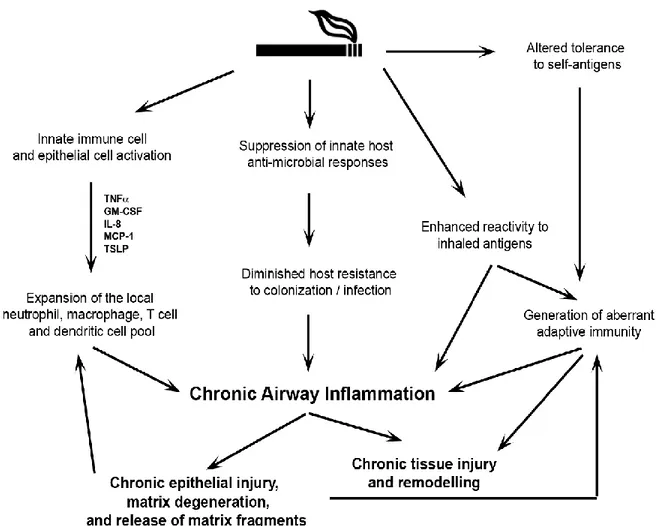
The role of the inflammatory cytokines is to recruit the immune cells during the infection and also they induce the epithelial cells proliferation and migration for the epithelization during wound healing (105). In addition, these inflammatory factors induce production of the protease enzymes, including matrix metalloproteinases (MMPs), elastase, and gelatinase, which destroy the extracellular matrix (ECM) proteins (e.g. collagen, elastin, and gelatin) and cell-cell interaction proteins (e.g. E-cadherin) during the infection or tissue repair (106, 107). Indeed, in combination with cytokines secretion, CS also has been found to increases set of the MMPs expression, including the MMP 1-3 and MMP-14 (108-110). Degrading the ECM and cellular junction proteins facilitate the immune cells (e.g. dendritic cells, T-cells, and macrophages) migration to the inflamed area and also enable epithelial cells migration for wound healing (107). However, the migrated immune cells may not be effective for killing the pathogens during the infection or clearing the damaged cells during tissue repair. This is because CS impairs the immune cells efficiency and induces their death (76). Among immune cells affected by CS are the macrophages and T cell (111, 112). This effect will increase pathogens colonization in the host cells (113). Moreover, CS exposure was found to induce T helper 17 cells of the adaptive immunity for self-antigen, recognizing the lung elastin, leading to more destruction in the lung, but the mechanism still is not fully understood (114). To sum up, CS causes an acute and chronic inflammation that result in continuous tissue destruction by the proteases, and simultaneously it promotes microbial accumulations by impairing the immune system (Figure 1-5). Such effects due to CS are believed to be the major cause for COPD development, increase in lung infections and risk of asthma in smokers (115, 116).
Figure 1-5: Summary of cigarette smoke (CS) effects on the immune system. CS exposure induces the pro-inflammatory factors secretion and leads to a chronic inflammation, on left side. In the meantime, CS affects the immune cells and weakens their abilities in clearing the pathogens, in the middle. Also, CS can cause an abnormal adaptive immunity, particularly self-antigen of lung elastin, on the right side. Altogether, CS ultimately leads to a chronic airway obstruction and, thereby, causes the chronic obstructive pulmonary disease (COPD). TNF- α, tumor necrosis factor alpha; GM-CSF, granulocyte-macrophage colony-stimulating factor; IL-8, interleukins-8; MCP-1, monocyte chemoattractant Protein-1; TSLP, thymic stromal lymphopoietin (117).
1.2.3.2. Damaging the DNA: Mutagenesis
Cigarette components can penetrate the cells and induce DNA damage, leading to various mutations (41). CS is a frequent insult that may cause huge lesions across DNA that may as well induce
process, is extremely harmful because it might produce abnormal cells, which must be removed by the apoptosis otherwise it will form a malignant tumor. Moreover, these mutations by CS can be transmitted to the subsequent generations. Indeed, an important finding has showed that after exposing the mice to CS many mutations have been found in the paternal germ cells (119). In contrast to the somatic mutations, which end up in the individual, the germline mutations spread over generations. Thus, smoking habit can cause aberrant heritable genotypes that can settle to the offspring.
There are several chemicals compounds in the fresh smoke that are produced by puffing cigarette, causing mutagenesis. The ROSs and RNSs in CS, which have been discussed in the section (1.1.2.), are potent oxidizing agents for DNA. They can attack DNA generating several breaks and oxidative damage. The ROSs (e.g. H2O2), or RNSs (e.g. ONOO-) can adduct DNA and oxidize the guanosine, and lead to the
8-hydroxyguanosine (8-OHG) formation (120, 121). The 8-OHG subsequently after DNA replication will cause a mismatched pairing, guanine to thymine transversion, which is a major result of DNA oxidative damage and contributes to carcinogenesis (122). Also, there are over 60 chemicals identified as potent carcinogenesis within CS content (123). They can bind and damage the DNA even at low concentrations e.g. benzo[a]pyrene, 2-nitropropane, and 4-aminobiphenyl, causing several mutations (Table 1-1).
CS carcinogenicity occurs when deleterious mutations occur in particular genes such those encoded for oncogene or tumor suppressor proteins. Losing the vital functions of the oncogenes (e.g. MYC or RAS) or tumor suppressor genes (P53 or P16) lead to carcinogenesis due to the losing the control of cell cycle. CS carcinogens, particularly benzo[a]pyrene, DNA adducts have been found in the lung tumors and account for the P53 mutations (3). As well, the NNK, which is one of CS carcinogen, was responsible for RAS point mutations and account of 30% of lung and 50-90% of pancreatic carcinoma (124). Thereby, mutating particular genes is a potent mechanism by which CS effectively induces cancer development. Moreover, CS-induced cancers have provided a useful model for understanding the aspects of
Other noncancerous diseases also can develop by CS-induced mutations. For example, CS is strongly associated with the cardiovascular diseases that are believed to be by dysregulating muscle relaxation. Indeed, a study showed the polymorphism in the endothelial nitric oxide synthase (eNOS) gene in smokers was associated with a decrease in its enzymatic activity (125). The eNOS is the enzyme that produces the NO. Production the NO expands the blood vessels by activating the soluble guanylyl cyclase that consequently generates the cyclic guanosine monophosphate (cGMP) in the muscle cells (126). The cGMP in turn stimulates the signaling pathways, leading to phosphorylating in several proteins that induces muscles relaxation. Hence, the low level in the eNOS because of smoking means decrease in the NO that may result in disorder in cardiac functions.
1.3. Gingiva
1.3.1 Gingival tissue structure
The gingiva, or gum, is a component of the periodontium in combination with the cementum, periodontal ligament, and alveolar bone (Figure 1-6A). The periodontal components interact together for insuring the healthy functions of the periodontium. The cementum is made up mainly of minerals, proteoglycans, and collagen fibers that form avascular matrix to cover the root of the tooth (127). After the cementum is the periodontal ligaments, which are connective tissues consist of collagens fibers and several types of cells, including fibroblasts, osteoblasts, osteoclasts, cementoblasts, epithelial cells, and stem cells (128). These ligaments mediate the attachment of the cementum with the alveolar bone. The alveolar bone, also called alveolar process, is a bony tissue of mandibular and maxillary that holds and supports the sockets of the teeth (127). The outside structure, which covers and protects the other elements of the periodontium, is the gingiva that is established during the eruption of the teeth into the oral
and underlying connective tissue. Anatomically, based on the location, the gingival epithelium is identified as oral epithelium, junctional epithelium, and sulcular epithelium that stretch along the periodontium (130).
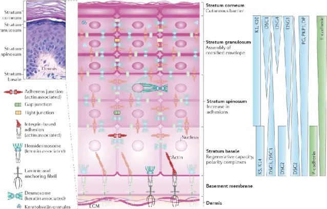
Structurally, the gingival epithelial cells, also known as keratinocytes, are the primary cells that build the gingival epithelium tissue. These cells can stratify and then organize as a multilayer structure forming basal and suprabasal (stratum spinosum, granulosum, and corneum) layers (Figure 1-7). The active proliferative cells are located in the basal layer, while the differentiated cells are the elements that build the suprabasal layers (131). The basal layer cells undergo a distinctive cell division for stratification. At the end of the cell cycle, the duplicated cells are divided symmetrically for the lateral expansion and asymmetrically for the vertical expansion, differentiation, to form the different layers (132). Moreover, the basal layer contains epithelial stem cells that can differentiate into the cell type of the basal epithelial layer to maintain the tissue homeostasis (131). The cells located in the basal layer are interacting with and lying on a basement membrane. This membrane consists of three interacting layers: lamina lucida, lamina densa, and sublamina densa that are established from the cross-linking of the ECM proteins, including mainly collagen and laminin fibers (133). The basement membrane separates the gingival epithelium from the underlying connective tissue, also termed lamina propria, which composed of fibroblasts, immune cells, ECM proteins, nerves, salivary glands, and blood vessels (128).
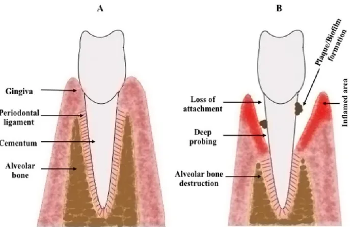
Figure 1-6: Anatomic schema of the periodontal tissue and its cigarette smoke-induced diseases. A. The periodontal components, including the gingiva, cementum, periodontal ligament and alveolar bone together form a crown-like structure for supporting and protecting the teeth. B. Some of the chronic diseases in the gum, which are associated with cigarette utilization. Reproduced from (Alharbi IA and Rouabhia M submitted review).
1.3.2 Keratin cytoskeleton role in the gingival tissue
There are a set of keratins (Ks) intermediates filaments that are expressed differentially in the basal and suprabasal epithelium, favoring many functions. Particularly, K5, K14, and K19 are the dominant keratins of the basal layer, whereas the K1, K6, K10, and K16 are predominantly expressed in the suprabasal layers (134). The keratinocytes of both the basal and suprabasal layers are tightly tied to each other through several forms of interactions. The cell-cell interactions are mediated by several intercellular proteins, including E-cadherin, and P-cadherin of adherens junction, claudins and occludins of tight
junction, connexins of gap junction, and desmogleins, desmocollin, desmoplakin, and plakoglobin, and plakophilin of desmosome junction (135-137). The keratins filaments, in addition to the actin filaments, serve as intracellular proteins that stabilize the intercellular proteins of the cell junctions (Figure 1-7). In the bottom side, the interaction of the basal layer cells with the basement membrane is mediated by the hemidesmosome and integrin adhesions (138). The basal keratins are the linker proteins for the hemidesmosome adhesion; in contrast to the integrin adhesion, which is actin-based interaction (135, 138).
The keratin filaments are also involved in defining the appearance of the epithelial cell, and the shape of its nucleus. The basal layer cells have a cuboidal shape and that is due to K5 and K14 expression. These keratins accumulate in the cytoplasm and surround the nucleus, making its shape elongated and the cell consequently becomes cuboidal (139). The decrease in K5 and K14 expression, when the basal epithelial cells undergo the differentiation for the stratification, as well, was found to influence the nucleus and cell shape. The decrease in K5 and K14 levels allows their reorganization to build a cage-like structure around the nucleus, leading to a rounded nucleus and flattened cell shape (139). Moreover, keratins have an important role in regulating the signaling pathways of cell proliferation and differentiation. It has been found that knocking out K5 and K14 expression leads to reduction in epithelial cells proliferation due to the decrease in Akt phosphorylation, but still the mechanism is not well known (140). In contrary, inhibiting K10 expression was found to induce cell hyper growth (141). Indeed, it has been found that K10 filament can interact and sequester Akt translocation and activation, which is a requirement of cell cycle progression in the epithelial cells, by that K10 promotes cell differentiation (142).
Keratins are essential proteins for tissue integrity and the defects in their expressions or structures lead to serious diseases (143). The epidermolysis bullosa simplex (EBS) is a genetically inherited disease, in which the patients are suffering from severe epidermal cytolysis and blistering (144). The molecular basis of this disease in most cases is mutations in either K5 or K14 genes (145). These mutations affect the
intermediate filament network in the basal keratinocytes (146). This results in a fragile basal keratinocyte due to loss the stable and powerful interactions between the cells and with the basement membrane that mediated by K5 and K14 (135). Also, dysregulation of keratins is documented to be associated with oral epithelial dysplasia (OED) and oral squamous cell carcinoma (OSCC) (147). Importantly, the differentiation-related keratins such as K4 and K13 are found to have reduced expression in the OED and OSCC; such effect is known to increase the basal epithetical layer thickness (147, 148). Indeed, in the OED, the cells of epithelial tissue have a high proliferative rate, and they enlarged rapidly, causing an increase in the tissue mass and it is considered as a precancerous stage (149). Moreover, the OED is believed to progress to ultimately become OSCC, but this more likely due to an accumulation of different mutations (150, 151). Finally, these diseases indicate the important role of keratins in the single cell and also to the entire tissue.
1.3.3. Gingival tissue functions
Gingiva surrounds and seals the edges of the teeth, protecting the last from accumulation of the microbes and foodstuff. Gingival structure is the first part in periodontium that encounters the environmental insults and stimuli such as physical stress, pathogens invasion, and toxin (e.g. CS compounds) (129, 152). Gingival epithelial layers function as a rigid physical barrier to protect the periodontium against the mechanical stress. Also, the overlying epithelium and underlying connective tissue are both part of the innate and adaptive immunity. The connective tissue contains within its structure various immune cells include monocyte/macrophage, granulocyte, and dendritic cells that are known in their capacity in fighting the microbes (153-155).
Figure 1-7: Gingival epithelium architecture. The gingival epithelium consists of four distinctive layers as shown in the top left by the hematoxylin-eosin staining. The stratum basal contains cells that mainly express K5 and K14 that mediate their interaction with basement membrane by the hemidesmosome in addition to integrin-based interaction. The suprabasal layers are the stratum spinosum, stratum granulosum and stratum corneum that are characterized in expressing K1 and K10, which stabilize their interactions with each other through the various junctions. The cells of different layers also differentially express set of proteins that serve as intercellular tools for supporting the cell-cell interactions, see above. Finally, the whole epithelium Seattle down on the basement membrane that is composed of extracellular matrix proteins and separate the gingival epithelia cells from the connective tissue, which is also called the dermis. K, keratin; DSG, desmoglein; DSC, desmocollin; DP, desmoplakin; PG, plakoglobin; PKP, plakophilin; E-cadherin, epithelial cadherin; P-E-cadherin, placental cadherin (135).
Besides, the gingival epithelial cell has a significant function in the innate immunity. Their roles arise from expressing the Toll-like receptors (TLRs 1 to 9), for recognizing the microbes (156). Interaction of the microbes with the TLRs launches the inflammation response by stimulating the keratinocytes to secrete the pro-inflammatory cytokines (e.g. IL-1β, IL-6, IL8, and TNF-α) that in turn activate the immune cells for killing and clearing the microbes (157, 158). Moreover, the keratinocytes produce several
β-defensins, which are antimicrobial peptides, acknowledged in inhibiting the microbial growth (159, 160). In addition to the keratinocytes, Langerhans cells, which are specialized dendritic cells, are present in the gingival epithelium (153). The Langerhans cells have a tolerogenic role arising from taking up, processing the microbial antigens, and serve as antigen-presenting cells for the T cells generating the adaptive immune response (155). Thus, gingiva is a crucial element for maintaining the protection and healthy functioning of the periodontium.
1.3.4. Cigarette smoke-induced gum diseases
Smoking cigarette is considered as one of the main causes of gum diseases. CS is strongly associated with several diseases in the gum, including, microbial accumulation, periodontitis, tooth loss, and cancer (Figure 1-6B). The prevalence of gum diseases is much greater in smokers, as well as passive smokers, than nonsmokers (161, 162). The epidemiological data suggests that the risk of gum diseases increase with the prolonged smoking and cessation has an opposite impact (163). Clinical observations showed that the smokers have a great loss in the periodontal attachment with the teeth (164). In consequence, the continuous cigarette consumption leads to an increase in the probing pocket depth due to the loss in the attachment (165). Moreover, CS was found to promote oral hyperplasia that may also lead to improper attachment between the gingiva and the teeth because of the abnormal growth in some gingival areas, leading to poor sealing (166, 167). This means the periodontium will be more susceptible for the microbial invasion and deposition of toxins. Indeed, smokers suffer from several oral infections, e.g. Porphyromonas gingivalis and Bacteroides forsythus, in comparison with nonsmokers (168, 169).
Accumulation of the microbes in the gingival tissue of smokers is believed to cause the gingivitis, which is a chronic inflammation in the gum (170). Consequently, the untreated gingivitis usually progress to affect the whole periodontium, causing an acute periodontitis. Moreover, smokers are showing a decrease
in the alveolar bone density that may therefore the severe periodontitis (171). Collectively, the increase in the attachment loss, deep pocket, chronic periodontitis, and alveolar bone destruction promote the risk of teeth loss in smokers (172). In addition to these diseases, it is well documented that CS, as it does in the most of the body parts, effectively can lead to the oral squamous cell carcinoma development in smokers than nonsmokers (173). This also has been proved in the animal model experiment in which oral cancer has occurred in rats that are exposed to CS chemicals (66). Finally, in fact, cigarette utilization is a preventable cause of various human diseases.
1.4. The hypothesis and objectives of the study 1.4.1 The hypothesis
CS-induced gum diseases suggest that the exposure to CS damages the gingival tissue components and impairs their vital functions. This has been observed in the in vitro experiments by CS-exposing of the epithelial cell of the upper gingiva, and fibroblast, which is the major constituent of the underlying connective tissue. Accordingly, the direct exposure to the whole CS was promoting the toxicity in these cells (72, 174). Also, the exposure to CS was shown to modulate the innate immunity response of the gingival epithelial cell by activating the TLRs and inducing the pro-inflammatory factors secretion (175, 176). Moreover, CS can increase the ECM proteins degradation in the gingiva by promoting the fibroblasts to secrete the different MMPs (108-110). Stimulating the immune response and proteases secretions contribute tissue damaging and cause severe periodontitis.
Here, we hypothesized that the damage in gingival tissue is accompanied by an abnormal growth in the overlying gingival epithelium. The histological staining studies suggest that as well. The hematoxylin and eosin staining of the gingival epithelium tissue from smokers showed a structural alteration in
thickness due to the exposure to CS (182, 166). Moreover, the oral hyperplasia observed in the animal model after the exposure to CS also support the thickness increase in the gingiva (183). Finally, and may most importantly, the Ki67, which is involved in the cell growth, is increased in the basal layers of gingival smokers samples (167, 184). Thus, the exposure to CS may induce the gingival epithelial cells over growth, leading to an aberrant tissue development that may subsequently promote gum diseases progression.
1.4.2. The objectives
The aim of this study was to investigate effect of the repeated long-term CS exposure on the normal human gingival epithelial cells growth.
Our specific objectives are:
1. Study the effect of CS on gingival epithelial cell proliferation and cell death resistance.
2. Investigate the impact of the CS exposure on the different keratins (K1, K5, K6, K10, K14, and K16) expression and production by gingival epithelial cells.
Chapter II
2. Article
Repeated exposure to whole cigarette smoke promotes primary human gingival epithelial cell growth and modulates keratin expression
Repeated exposure to whole cigarette smoke promotes primary human gingival epithelial cell growth and modulates keratin expression
Alharbi IA and Rouabhia M
Groupe de Recherche en Écologie Buccale, Faculté de Médecine Dentaire, Université Laval, 2420 rue de la Terrasse, Québec G1V 0A6, Canada.
Running title: Cigarette smoke affects gingival epithelial cell growth
Corresponding authors:
Dr. Rouabhia M: Groupe de Recherche en Écologie Buccale, Faculté de Médecine Dentaire, Université Laval, 2420 rue de la Terrasse, Québec G1V 0A6, Canada. (mahmoud.rouabhia@fmd.ulaval.ca)
2.1. ABSTRACT
Background and Objective: The gingiva is the first oral tissue directly exposed to cigarette smoke (CS). Exposure to CS compromises gingival tissue structure and function. Damaging or altering the gingival epithelium leads to a compromised protective barrier of the periodontium, resulting in several diseases. The aim of this study was to assess the effect of repeated exposure to CS on gingival epithelial cell growth, apoptotic protein expression, and keratin expression.
Materials and Methods: Primary human gingival epithelial cells were seeded on a collagen scaffold for 5 days to allow for growth and stratification. The cells were then exposed for 5 min to whole CS for 3, 6, and 9 days. At the end of each exposure period, the cells were used to investigate cell proliferation by (3-(4,5-dimethylthiazol-2-yl)-2,5-diphenyltetrazolium bromide) (MTT) and 5-bromo-2'-deoxyuridine (BrdU) assays, gene expression by quantitative reverse transcription polymerase chain reaction (qRT-PCR), and protein production by Western blot analysis.
Results: Greater metabolic activity was found in the CS-exposed cells than in the non-exposed cells, specifically after 3 and 6 days of exposure to CS. At 9 days there was no significant difference between CS-exposed and non-exposed cells. Metabolic activity was supported by the BrdU cell proliferation analyses showing increased cell growth at 3 days as compared to the control. However at 6 and 9 days, the cell proliferation in CS-exposed culture was comparable to the non- exposed culture. Interestingly, the Bax/Bcl-2 protein ratios decreased with increased CS exposure, suggesting cell resistance. Moreover, protein analyses showed that CS decreased keratin (K) 5 at 3, 6 and 9 days; while it increased K14 at 6 and 9 days. Finally, mRNA analyses showed significant decrease of K1, K6, K10 and K16 in CS exposed cultures correlating at times with a decrease of protein production.
Conclusion: CS was shown to increase epithelial cell proliferation, which may involve cell resistance to apoptosis. This is supported by the modulation of different keratins’ genes expression and protein
production. Altogether, these data may explain the hyperplasia reported in gingival tissue as well as periodontal disease in smokers.
2.2. Introduction
Cigarette smoke (CS) is acknowledged as a harmful factor on human health (1, 2). CS compounds, such as nicotine, carbon monoxide, and reactive oxygen species (ROS), are collectively able to damage DNA, proteins, and lipids in cells (1, 2). Exposure to CS can lead to several deleterious mutations, including but not limited to RAS and P53 genes, resulting in uncontrolled cell growth (3-5). CS also reportedly affects cell signaling, such as Akt and NFκB dysregulation, which may lead to cancer development (6).
The oral cavity and in particular the gingival mucosa are generally the first structures to be exposed to CS. This exposure contributes to deregulating periodontal tissue function and thus opening the door to several oral diseases (7-9). In the gingival mucosa, the epithelial structure and connective tissue cover the entire periodontium (10). Epithelial cells in the gingival mucosa are well-organized interconnected layers that form a protective barrier (11). These layers house proliferating (basal layer) as well as differentiated (suprabasal layer) cells. The cells in the different layers express many types of keratins (K), which are intermediate filaments that stabilize cell cohesion. Specifically, basal cells express K5 and K14, while suprabasal cells express K1 and K10 (12). As the epithelial structure is the first exposed to CS, the gingival tissues of smokers have been shown to display certain structural abnormalities, such as tissue color changes and epithelial hyperplasia (13-16). CS-induced epithelial tissue deregulation can take place through the modulation of keratin expression (17, 18). One study has shown desmoglein 3 and K10 expression to be downregulated in smokers, compared to non-smokers (18). With the effect of CS on epithelial structure, smokers may thus be more at risk of periodontal dysfunction leading to deep periodontium pocket formation (19-21). These pockets facilitate the accumulation of microbes, which in turn causes severe periodontitis due to an activation of the inflammatory pathways (22, 23). Moreover, epithelium hyperplasia observed in smokers may be the result of cell proliferation and the modulation of multiple keratin markers (24). This study was conducted to examine the effects of repeated exposure to CS
on human gingival epithelial cell proliferation and apoptosis. Also investigated was the impact of CS on the expression of keratins (K1, K5, K6, K10, K14, and K16).
2.3. Materials and Methods 2.3.1 Reagents
1R3F cigarettes were obtained from Kentucky Tobacco Research & Development (Orlando, FL, USA). CollaTape scaffolds were obtained from Zimmer Dental Inc. (Mississauga, ON, Canada). Primers were purchased from Invitrogen Life Technologies Inc. (Burlington, ON, Canada). The BrdU ELISA kit was purchased from Abcam Inc. (Toronto, ON, Canada). 3-[4,5-Dimethylthiazol-2-yl]-2,5-diphenyl-tetrazolium bromide (MTT) was obtained from Sigma (St. Louis, MO, USA). Anti-cytokeratins (-1, -5, -10, and -14), anti-Bax, and Bcl-2 antibodies were purchased from Santa Cruz Biotechnologies (Santa Cruz, CA, USA). Anti-cytokeratin-16 was obtained from Cedarlane Laboratories (Burlington, ON, Canada), while anti-cytokeratin-6 was obtained from Abcam Inc. (Toronto, ON, Canada). Anti-β-actin and peroxidase-conjugated antibodies was obtained from Sigma-Aldrich Canada Ltd. (Oakville, ON, Canada).
2.3.2. Cell cultures
To extract gingival epithelial cells, biopsies of gingival tissues were coolected from healthy nonsomkers (18-25 years old) following their informed consent in accordance Laval University Ethics Committee guidelines. The cells were cultured in Dulbecco’s modified Eagle’s medium/Ham’s F12 (3:1; DMEH) supplemented with 5 μg/mL of human transferrin, 2×10⁻⁹ M of 3,39,59-triiodo-L-thyronine,
0.4 mg/mL of hydrocortisone, 10 ng/mL of epidermal growth factor, 100 IU/mL of penicillin G, and 10% fetal bovine serum. The medium was changed daily. When the cultures reached 90% confluence, the cells were detached from the flasks with a 0.05% and 0.25% trypsin–0.1% ethylenediaminetetraacetic acid (EDTA)
solution, washed twice, and resuspended in DMEH-supplemented medium at a final concentration of 106 cells/mL. Cells at the third and fourth passages were used in the experiments. They were trypsinized
and seeded at 106 cells/mL on CollaTape patches (2 × 3 cm2) preincubated in cell culture medium.
2.3.3. Cigarette smoke exposure procedure
To help cell organization as an epithelial tissue, epithelial cells seeded at 106 cells on CollaTape
patches (2 × 3 cm2) in DMEH medium were incubated in a humid atmosphere containing 5% CO2 at 37°C.
This type of membrane also allowed for the application of CS in a long-term manner (25). The cells were then cultured for 5 days to allow for growth and stratification, with medium changing every day. To administer whole CS, the medium was replaced with 1 ml of phosphate-buffered saline (PBS) and the epithelial cell-populated CollaTape scaffold was then placed in a sterilized CS chamber. A peristaltic pump was used to transfer the smoke of one whole cigarette inside the chamber. The scaffolds were incubated for 5 min with CS, washed with warm culture medium, fed thereafter with fresh DMEH medium, and again incubated as described above. The scaffolds were thus exposed to CS for 3, 6, and 9 days. Controls followed the same protocol, without the exposure to CS. Following each exposure period, the scaffolds underwent various analyses.
2.3.4. Cell viability/Growth assay
Following exposure to CS, cell viability/growth was measured by MTT assay. Briefly, an MTT stock solution (5 mg/ml) was prepared in PBS and added to each cultured scaffold at a final concentration of 1% (v/v). The scaffolds were then incubated for 4 h at 37°C with the MTT solution, after which time the medium was removed, 2 mL of 0.04 N HCl in isopropanol were added to each culture scaffold, followed by