Université de Montréal
Advancing pain research and animal welfare: focusing on
the Rat Grimace Scale and reporting standards
par Vivian Leung
Département de sciences cliniques Faculté de médecine vétérinaire
Thèse présentée à la Faculté de médecine vétérinaire en vue de l’obtention du grade de Philosophiae Doctor (Ph. D.)
en sciences vétérinaires
Octobre, 2018 © Vivian Leung, 2018
Université de Montréal
Faculté des études supérieures et posdoctorales
Cette thèse intitulée:
Advancing pain research and animal welfare: focusing on the Rat Grimace Scale and reporting standards
Présentéé par Vivian Leung
A été évaluée par un jury composé des personnes suivantes : Dre. Elizabeth O’Toole, présidente-rapporteuse
Dr. Daniel Pang, directeur de recherche Dr. Éric Troncy, membre du jury Dre. Gilly Griffin, examinatrice externe
Résumé
Les comportements non-stimulés pour évaluer la douleur chez les animaux suscitent un intérêt grandissant. Un exemple est la « Rat Grimace Scale » (RGS), une échelle de douleur basée sur 4 unités d’action d’expressions faciales: resserrement orbital, aplatissement nez / joue, changements d’oreilles et vibrisses. Le potentiel de cette échelle et ses limites demeurent à déterminer.
La RGS standard est laborieuse à compléter (enregistrement de vidéos et extraction manuelle des images). Par conséquent, cette thèse a évalué si l'application en temps réel était possible. En comparant les résultats obtenus en temps réel à la méthode standard, il a été constaté que les résultats étaient similaires. Ainsi, la fiabilité de la RGS en temps réel élargit grandement son applicabilité en tant qu’outil clinique et de bien-être.
Toutefois, l'applicabilité de la RGS dans la douleur viscérale aiguë et chronique demeurent inexplorée. Par conséquent, cette thèse a évalué si la RGS pouvait évaluer la douleur à partir d'un modèle de colite aiguë et chronique de « dextran sulfate sodium » (DSS). Deux autres outils comportementaux (enfouissage et « Composite Behaviour Score » [CBS]) ont également été évalués. Ils ont été comparés au « Disease Activity Index » (DAI), un outil commun d'évaluation de la sévérité de la maladie. La RGS et l’enfouissage ont augmenté et diminué respectivement lorsque le DAI a augmenté. De futures études sont nécessaires pour valider le CBS. Cette étude démontre que le RGS peut évaluer la douleur viscérale et potentiellement plusieurs types de douleur.
La nécessité d'une formation avant la notation RGS a également été investiguée en évaluant la fiabilité de l'évaluateur après avoir reçu une formation ou aucune formation. Il a été constaté que la formation était bénéfique pour améliorer la fiabilité en plus de réduire la variabilité alors que la notation de plusieurs images seulement ne l’était pas. En outre, les évaluateurs obtenaient des résultats fiables après une période d'inactivité. Cette étude démontre donc le besoin de former les nouveaux évaluateurs.
Par ailleurs, cette thèse a également étudié si la publication des directives ARRIVE « Animal Research: Reporting of In Vivo Experiments » avait entraîné une amélioration des normes de déclaration. Cette étude a montré que les normes de déclaration ne s’étaient pas améliorées de manière significative, mais aussi que les articles publiés dans des revues qui soutiennent les directives ARRIVE n’ont pas de meilleurs standards. Par conséquent, cette étude souligne la nécessité d'imposer les directives ARRIVE pour assurer une amélioration significative.
Dans l'ensemble, cette thèse a démontré l'utilité de la RGS en temps réel comme outil d'évaluation clinique de la douleur viscérale chronique ainsi qu’en recherche. Elle souligne également le besoin de former les évaluateurs avant la notation RGS. Enfin, il a été démontré que les normes de déclaration restent faibles et que les directives ARRIVE doivent être imposées. Il est à espérer que ces études encourageront la progression de la recherche sur la douleur par l’amélioration des standards de déclaration ainsi que par l’utilisation de la RGS et autres comportements spontanés pour évaluer la douleur.
Mots-clés : échelles de grimace, expression faciale, douleur, comportements animaux,
développement d'outils, état affectif, rats, modèles animaux, directives ARRIVE, normes de déclaration
Abstract
There is growing interest in the use of non-evoked spontaneous behaviours to assess pain in animals. A tool that measures such behaviours is the Rat Grimace Scale (RGS), a validated facial expression pain scale consisting of four “action units”: orbital tightening, nose/cheek flattening, ear changes and whisker changes.
The strengths and limitations of the RGS are not fully explored. One limitation of the RGS (using the standard scoring method) is its time- and labour-intensive nature (video recording and manual image extraction are required). A primary goal of my research was to evaluate the feasibility of real-time RGS scoring. To accomplish this, the standard and real-time assessment methods were compared. It was found that both scoring methods were comparable and demonstrated the utility of real-time RGS scoring. This provides evidence that the RGS may be utilised not only as a research tool, but a useful clinical and welfare tool as well.
A further goal was to explore the use of the RGS in a visceral and chronic pain model. The RGS was hence tested in a dextran sulfate sodium (DSS) colitis model. Two other behavioural tools (burrowing and the composite behaviour score [CBS]) were also evaluated. These behavioural tools were compared to the Disease Activity Index (DAI), a common tool assessing disease severity in colitis models. The RGS and DAI scores increased and decreased concurrently. This study demonstrates that the RGS can be applied to assess chronic visceral pain and may be used to assess the mechanisms of different pain types.
The need for training prior to RGS scoring was explored by assessing the rater reliability after receiving training or no training (scoring of multiple images only). This study demonstrates that training is beneficial; training improved scoring reliability and reduced variability. This was not observed in raters who received no training. Additionally, trained raters could still score reliably four years later. Therefore, this study demonstrates the need for new raters to be trained in RGS use to improve reliability.
Lastly, this thesis explores whether the publication of the ARRIVE (Animal Research: Reporting of In Vivo Experiments) guidelines improved the reporting standards of animal
studies. This study found that reporting standards had not improved meaningfully, and the standard of reporting was no better in papers published in journals that support the ARRIVE guidelines. Therefore, this highlights the need for the enforcement or refinement of ARRIVE guidelines to ensure meaningful improvement of reporting standards.
Overall, this thesis demonstrates the utility of the RGS as a practical pain assessment tool, with real-time application and the ability to assess chronic visceral pain. It highlights the need for raters to be trained prior to RGS scoring. Lastly, it demonstrates that the implementation of reporting standards in line with the ARRIVE guidelines are low, and enforcement may be required to ensure widespread application. It is the hope that these studies will encourage the use of the RGS and other non-evoked spontaneous behavioural pain assessment tools and will improve reporting standards in medical literature that advance pain research.
Keywords : Grimace Scale, Facial Expression, Pain, Animal Behaviour, Tool Development,
Table of Contents
Résumé ... iii
Abstract ... v
Table of Contents ... vii
List of tables ... x
List of figures ... xii
List of acronyms ... xiii
Dedication ... xvi
Acknowledgements ... xvii
1. Introduction ... 1
1.1. What is pain?... 3
1.1.1. The basic mechanism of pain ... 3
1.1.2. Behavioural pain assessment methods in rodents ... 6
1.1.3. The ethics of pain assessments and creation of pain scales ... 32
1.1.4. Other factors to consider during pain research ... 34
1.2. The Rat Grimace Scale ... 35
1.2.1. Applications of the RGS ... 36
1.2.2. Real-time application of the RGS and potential challenges ... 43
1.3. Utilising the RGS in an untested pain model ... 59
1.3.1. Cancer type pain ... 60
1.3.2. Inflammatory bowel disease ... 63
1.3.3. Comparing pain models: bladder cancer, bone cancer and colitis ... 65
1.3.4. Comparing colitis models: TNBS- and DSS-colitis ... 67
1.3.5. Differences between acute and chronic DSS-colitis ... 73
1.4. Pain recognition training ... 77
1.4.1. The Rat Grimace Scale and training ... 78
1.5. The importance of good reporting ... 80
1.5.1. What is the reporting standard in animal research? ... 81
1.5.2. Introducing the ARRIVE (Animal Research: Reporting of In Vivo Experiments) guidelines and its… impact? ... 82
1.5.3. Items listed in the ARRIVE guidelines related to pain research ... 83
1.6. Research questions, hypotheses and objectives ... 86
1.6.1. Can the Rat Grimace Scale be utilised in real-time? ... 86
1.6.2. Can spontaneous behaviours (Rat Grimace Scale, burrowing and Composite Behavioural Score) assess visceral pain in an acute and chronic dextran sulfate sodium colitis model? ... 87
1.6.3. What is the effect of training on Rat Grimace Scale scoring? ... 87
1.6.4. What are the reporting standards of papers published five years after the ARRIVE guidelines? ... 88
2. Publications ... 90
2.1. Real-time application of the Rat Grimace Scale as a welfare refinement in laboratory rats 92 2.1.1. Abstract ... 92
2.1.2. Introduction ... 92
2.1.3. Methods and materials ... 94
2.1.4. Results ... 100
2.1.5. Discussion ... 113
2.1.6. Acknowledgements ... 117
2.2. Performance of behavioral assays: The Rat Grimace Scale, burrowing activity and a composite behavior score to identify visceral pain in an acute and chronic colitis model . 118 2.2.1. Abstract ... 118
2.2.2. Introduction ... 119
2.2.3. Methods and materials ... 120
2.2.4. Results ... 126
2.2.5. Discussion ... 135
2.2.6. Disclosures ... 137
2.2.7. Acknowledgements ... 137
2.3. The influence of rater training on inter- and intra-rater reliability when using the Rat Grimace Scale ... 138
2.3.1. Abstract ... 138
2.3.2. Introduction ... 139
2.3.3. Methods and materials ... 140
2.3.4. Results ... 144
2.3.6. Acknowledgements ... 152
2.4. ARRIVE has not ARRIVEd: Support for the ARRIVE (Animal Research: Reporting of in vivo Experiments) guidelines does not improve the reporting quality of papers in animal welfare, analgesia or anesthesia ... 153
2.4.1. Abstract ... 153
2.4.2. Introduction ... 154
2.4.3. Methods and materials ... 155
2.4.4. Results ... 164
2.4.5. Discussion ... 173
2.4.6. Acknowledgements ... 176
3. Discussion ... 177
3.1 Overview ... 177
3.2 Contributions to the field of pain, study limitations and future work ... 178
3.2.1 Real-time application of the RGS ... 178
3.1.1 Applying the RGS to a secondary model: acute and chronic visceral pain in a colitis model 182 3.1.2 The effects of training on the reliability of RGS scoring ... 183
3.1.3 Assessment of adherence to the ARRIVE guidelines five years later ... 185
3.3 Pain research: looking forward ... 186
3.3.1 Use of appropriate assessment methods ... 186
3.3.2. Use of technology to automate ... 188
3.4. How can we ARRIVE? ... 191
3.3.2. A “case-study” of the CONSORT statement ... 191
3.3.3. Improving adherence to the ARRIVE guidelines ... 192
3.5. Additional factors to improve pain research ... 195
3.6. Conclusion ... 197
4. Bibliography ... 200
5. Appendices ... i
5.1. Appendix A ... i
5.1.1. Summary of all pain models used, and action units identified for the grimace scales of different species ... i
5.1.2. Summary of the different methods of validation for the grimace scales of different species ... iii
5.3. Appendix C – composite behaviours in DSS colitis model ... xiii
List of tables
Table 1.2: Summary of all RGS papers published to date ... 45Table 1.5.2.1: Summary of the reporting rates of three basic animal demographic details that should be reported in all papers ... 85
Table 1.5.2.2: Summary of the reporting rates of four key items to the validity of a study design. ... 85
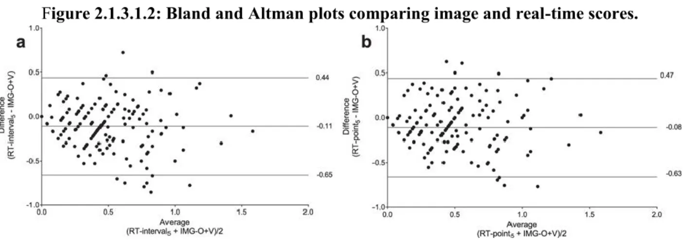
Table 2.1.1.3.1.1: Bland and Altman method comparing each real-time (RT) observation method with image (IMG) scores ... 102
Table 2.1.2.5.1: Rat Grimace Scale scores from control group. Scores generated by standard (video-based) method... 110
Table 2.1.3.3.1: Weights of petri dishes in the three treatment groups ... 111
Table 2.1.3.3.2: Weights of petri dishes in the control groups ... 112
Table: 2.2.3.5.1: Disease Activity Index scoring. ... 122
Table 2.2.3.9.1: Macroscopic scoring of colon samples ... 125
Table 2.2.3.10.1: Microscopic scoring of colon samples ... 125
Table 2.2.4.5.1: Microscopic and macroscopic scores of colon samples ... 135
Table: 2.3.4.1.1 Group Intra-class Correlation Coefficients (ICC) for each of the datasets. . 146
Tables: 2.3.4.1.2: Agreement of each individual trainee rater when compared to an experienced rater (DP). ... 146
Table 2.3.4.2.1: Intra-class Correlation Coefficients (ICC) for intra-rater reliability for each individual trainee rater four years after initial training ... 148
Table 2.3.4.3.1: Group Intra-class Correlation Coefficients (ICC) for each of the datasets for the “no training group” ... 148
Table 2.3.4.3.2: Agreement of each individual “no training” rater when compared to an experienced rater (DP). ... 149
Table 2.4.3.2.1. The ARRIVE guidelines checklist: operationalised items and sub-items to facilitate assessment of reporting (Kilkenny et al., 2010). ... 157
Table 2.4.4.1: Overall reporting quality in journals supporting (SUPP) and not supporting (nonSUPP) the ARRIVE guidelines for 2009 and 2015. ... 165 Table 2.4.4.1.1: Papers fully reporting ARRIVE checklist items in supporting (SUPP) and non-supporting (nonSUPP) journals in 2009 and 2015. ... 167 Table 2.4.1.1.2. Papers partially reporting ARRIVE checklist items in supporting (SUPP) and non-supporting (nonSUPP) journals in 2009 and 2015. ... 168 Table 2.4.4.2.1 Papers fully reporting ARRIVE checklist sub-items in supporting (SUPP) and non-supporting (nonSUPP) journals in 2009 and 2015. ... 169
List of figures
Fig. 1.2: Cartoon of the Rat Grimace Scale. ... 35
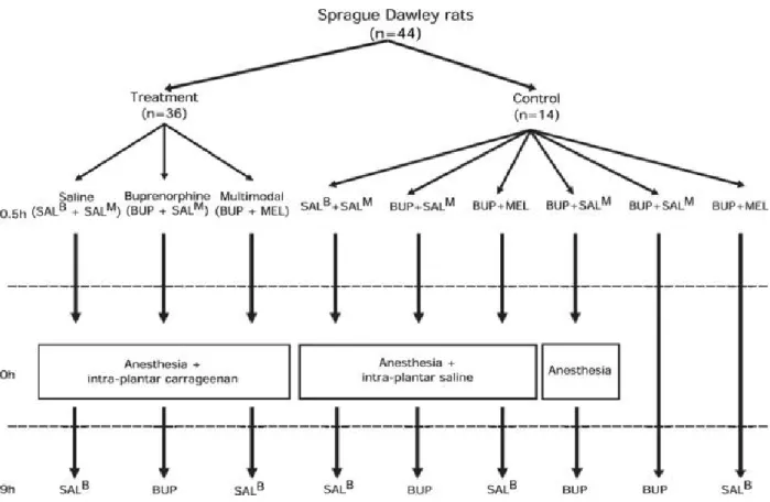
Figure 2.1.2.3.1: Flow chart depicting experimental pathway for each treatment group. ... 96
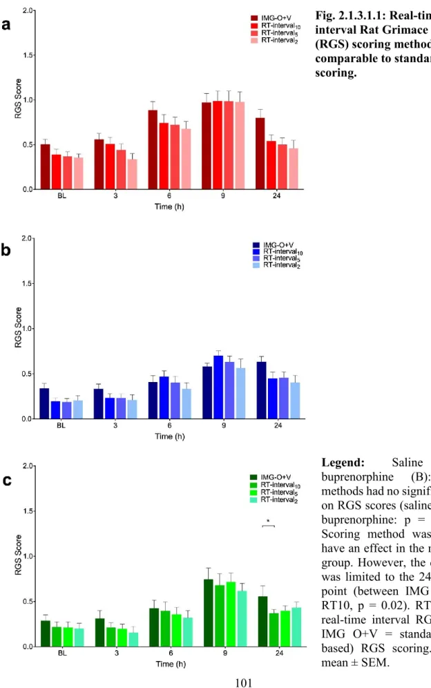
Fig. 2.1.3.1.1: Real-time interval Rat Grimace Scale (RGS) scoring methods were comparable to standard RGS scoring. ... 101
Fig. 2.1.3.1.3: Bland and Altman plots comparing real-time (RT) scoring (RT-interval10,2 and RT-point10,2) to image based (IMG) scores ... 103
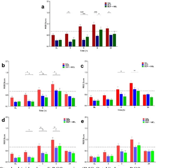
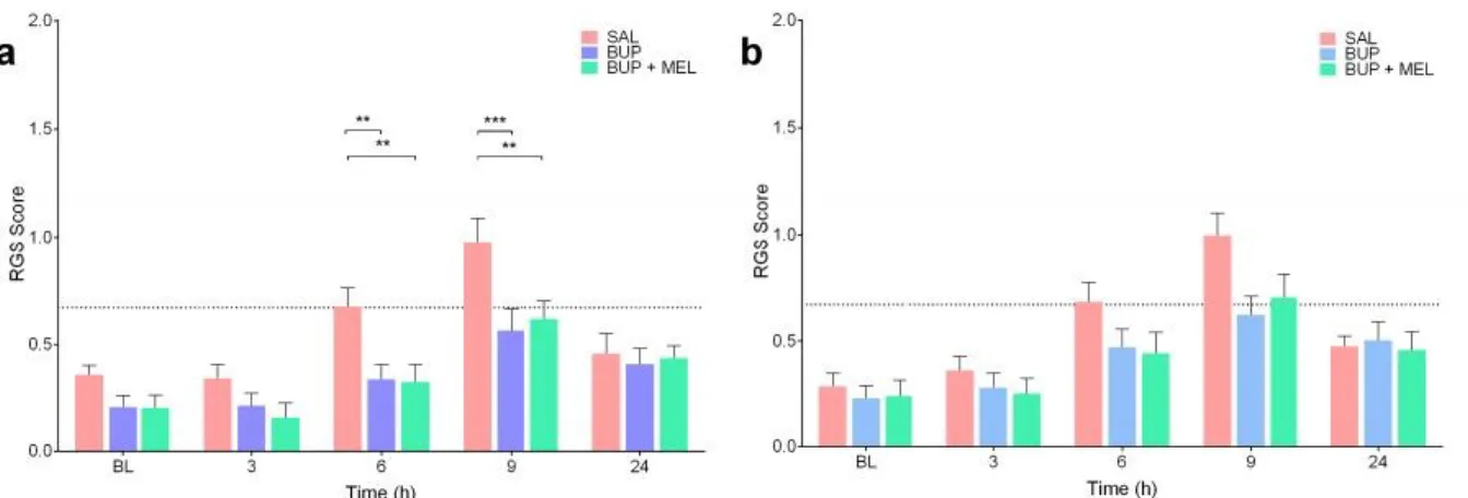
Fig. 2.1.3.1.4: Both standard Rat Grimace Scale (RGS) and real-time interval RGS scoring were able to discriminate between saline and analgesia treatment groups. ... 105
Fig. 2.1.3.1.5: Treatment effects identified with RT-interval2. ... 106
Fig. 2.1.3.1.6: Real-time (RT) point scoring methods compared to standard (video-based) Rat Grimace Scale (RGS) scoring (IMG O+V) ... 106
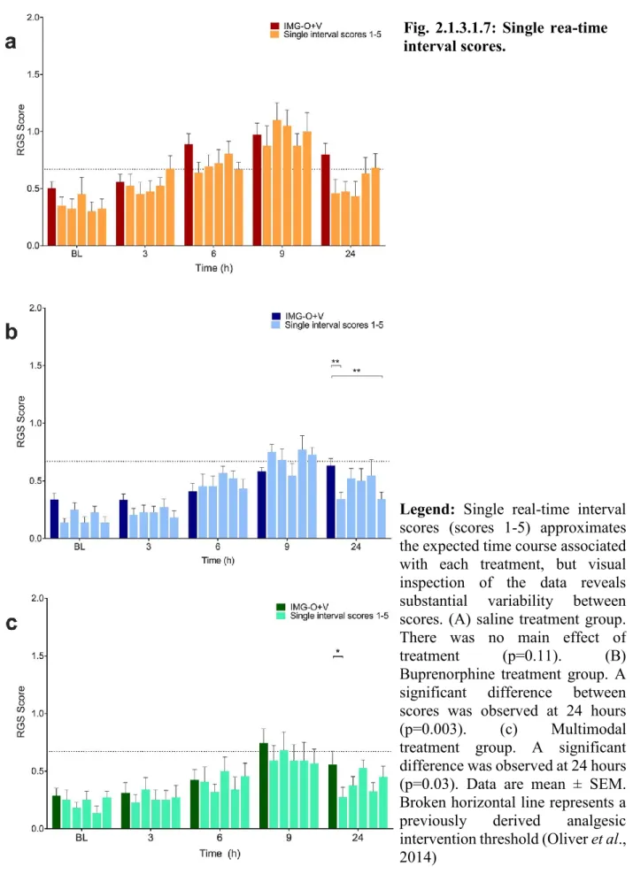
Fig. 2.1.3.1.7: Single rea-time interval scores. ... 107
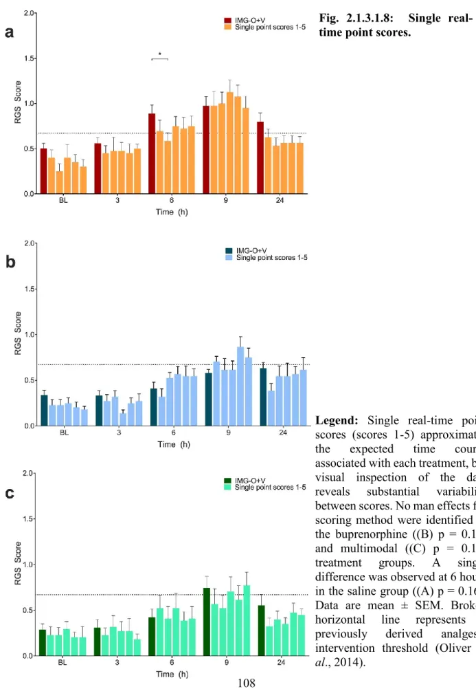
Fig. 2.1.3.1.8: Single real-time point scores. ... 108
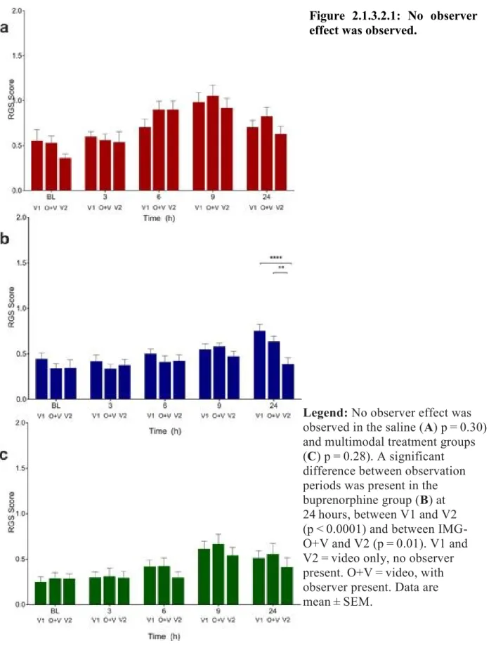
Figure 2.1.3.2.1: No observer effect was observed. ... 109
Fig. 2.2.4.1.1: Disease activity index scores during the acute and chronic phases ... 128
Fig. 2.2.4.2.2: The Rat Grimace Scale scores (real-time observations) during the acute and chronic phases (A, shaded boxes) and B) a comparison between real-time and video scores with Bland-Altman analysis for repeated measures. ... 131
Fig. 2.2.4.3.1: Mean difference in gravel displacement during acute and chronic colitis phases ... 132
Fig. 2.2.4.4.2: Breakdown of the frequency of all behaviours from the Composite Behaviour Score (CBS) ... 134
Fig. 2.1.2.3.1: Timeline of training protocol... 142
Fig. 2.3.4.1.1: Average group intra-class correlation coefficients ... 145
Fig 2.4.4.1 Flow diagram of paper selection process. ... 165
Fig 2.4.4.1.1 Bar graph of papers fully reporting individual items from the ARRIVE checklist. ... 171
Fig 2.4.4.2.1 Radar plot of ARRIVE checklist sub-items associated with bias reported in ARRIVE supporting (SUPP) and non-supporting (nonSUPP) journals in 2015. ... 172
List of acronyms
°C Celsius
ADL Active Daily Living
aMGS Automatic Mouse Grimace Scale
AMPA α-amino-3-hydroxy-5-methyl-4-isoxazolepropionic acid ARRIVE Animal Research: Reporting of In Vivo Experiments ASIC-1 Acid Sending Ionic Channel-1
ATP Adenosine Triphosphate
AU Action Unit
AWR Abdominal Withdrawal Reflex
B2 Bradykinin-2
BL Baseline
CaMkII Calmodulin-depending Protein Kinase II CBS Composite Behaviour Scale
CFA Complete Freund’s Adjuvant CGRP Calcitonin gene-related peptide CI Confidence Interval
CONSORT Consolidated Standards of Reporting Trials COX-2 Cyclooxygenase-2
CPP Conditioned Place Preference CRD Colorectal Distension
DAI Disease Activity Index DSS Dextran Sulfate Sodium e.g. exempli gratia
EMLA Eutectic Mixture of Local Anesthetics ERK Extracellular Signa-regulated Kinase et al. et alia
etc. et cetera
FACS Facial Action Coding System
GABA Gamma-Animo Butyric Acid
HARRP Harmonised Animal Research Reporting Principles HIV Human Immunodeficiency Virus
i.e. id est
IBD Inflammatory Bowel Disease ICC Intra-Class Correlation IFN Interferon IL Interleukin IMG Image kDa kilodalton kg kilogram kHz kilohertz mg milligram
MGluR Metabotropic Glutamate Receptor MGS Mouse Grimace Scale
MIA Monoiodoacetate MPO Myeloperoxidase NGF Nerve Growth Factor
NK Neurokinin
NMDA N-methyl-D-aspartate nonSUPP Non-Supporting
NSAID Nonsteroidal anti-inflammatory drugs O+V Observer + video
p.o. per os
PET Positron Emission Tomography PKA Protein Kinase A
PKB Protein Kinase B
PREPARE Planning Research and Experimental Procedures on Animal Recommendation for Excellence
PRISMA Preferred Reporting Items for Systematic Reviews and Meta-Analyses QST Quantitative Sensory Testing
RCT Randomised Controlled Trials RFF Rodent Face Finder
RGS Rat Grimace Scale
RT Real-Time
s Seconds
s.c. Subcutaneous SD Standard Deviation
SEM Standard Error of the Mean
STROBE Strengthening the Reporting of Observational studies in Epidemiology SUPP Supporting
TINT Time to Integrate Nest Test TNBS 2,4,6-trinitrobenzenesulfonic acid TrkA Tropomyosin A
TrkB Tropomyosin B
TRPA1 Transient Receptor Potential Ankyrin 1
V1 Video only V2 Video only VMR Visceromotor Response VR Vanilloid Receptor α alpha μg microgram
Dedication
This is for Daddy, Mummy and Russell. Thank you for always supporting me in whatever I do.
Acknowledgements
First and foremost, I am tremendously thankful to Dr. Daniel Pang for this PhD opportunity. Thank you for your continued support, confidence and patience throughout this journey. Thank you for being an amazing teacher and mentor, always willing to impart advice for my projects, career and life in general.
Secondly, I am grateful to my parents for their amazing love and support throughout my life. I would not be where I am if you did not encourage me to pursue my passions and interests. Thank you to all the people who have been on my supervisory committee, comité conseil and my examination committee for their invaluable feedback and encouragement throughout my PhD and with the writing of this thesis: Dr Douglas Morck, Dr. Elizabeth O’Toole, Dr. Éric Troncy, Dr. Gregory Muench, Dr. Gilly Griffin, Dr. Lee Niel and Dr. Milagros Freire Gonzalez. I would like to especially thank Dr. Lee Niel for introducing and encouraging me into the world of research during my undergraduate years.
I am very grateful to my fellow Pang Lab members: Cassandra Klune, Chelsea Schuster, Colin Laferrière, Dr. Amy Larkin, Dr. Frédérik Rousseau-Blass, Dr. Geneviève Fortin-Simard, Dr. Hayley Robbins, Dr. Julie Reimer, Dr. Katrina Frost, Dr. Maxime Rufiange. Emily Zhang and Maaria Shah. Thank you for always helping me with my projects, looking out for my rats and being fantastic company. Without all of you my PhD experience would not have been as enjoyable. I would like to especially thank Frédérik and Maxime for being abundantly patient with my poor French and always willing to lend a hand with translation.
Thank you to Auntie Audrey and Auntie Clara for supporting me all the way from Singapore by reading some parts of my thesis and instructing me on how to write better.
Thank you also to the 90 rats that were part of my actual or preliminary studies.
And last but most of all, I want to thank my Heavenly Father for guiding me through this path. It has been an enjoyable and trying experience. I know not what lies ahead, but I will do my best to continue to trust in Him.
1. Introduction
Pain is a sensation that everyone experiences at some point in their lives. In humans, the gold standard of pain assessment is by direct verbal report. We can communicate with one another and the medical staff when we are in pain and provide feedback regarding treatment efficacy. This gold standard in pain assessment reporting is not possible in animals as they cannot communicate with us directly. Therefore, pain assessment in animals has largely relied on inferences from their behaviours. Traditionally, pain assessment in preclinical animal research has largely relied on nociceptive tests which assess an animal’s reflexive response to an external stimulus. These nociceptive tests are favoured for their practicality: they can be replicated easily and reliably (Mogil and Crager, 2004). However, when the novel analgesics that had been reported as efficacious in preclinical animal trials failed during human clinical trials, it was proposed that nociceptive tests were not the appropriate pain measurement tools they were thought to be (Mogil and Crager, 2004; Rice et al., 2008 and Mogil et al., 2010). This is because nociceptive tests only assess the sensory component of pain and are unable to evaluate the affective component of pain, the primary concern reported by human patients (Backonja and Stacey, 2004; Mogil and Crager, 2004; Rice et al., 2008 and Mogil et al., 2010). This means that there is a mismatch of pain type assessed during animal and human trials (i.e. animal trials assess evoked-reflexive responses to nociceptive tests while human trials assess ongoing pain via verbal report). Therefore, it has been proposed that non-evoked spontaneous behaviours from animals should be used to assess the ongoing pain experienced (affective pain) by the animal (Mogil and Crager, 2004). Other issues with preclinical research have also been identified, one of which is the deficiencies in reporting standards in published papers (Kilkenny et al., 2009). The consequences of poorly reported published papers are the obstruction of study replication and validation of findings and will adversely skew systematic reviews and meta-analysis (Kilkenny et al., 2009; MacCallum 2010; du Sert, 2011 and Freedman et al., 2015).
At the beginning of this PhD, the Rat Grimace Scale, had just been developed by Sotocinal et al. (2011). This pain assessment tool is a promising method which utilises spontaneous changes in facial expression to assess ongoing pain experienced by rats. The studies described in this thesis explore the strengths and limitations of the RGS. Specifically, the studies explore:
1) if real-time application of the RGS is feasible, if so this would drastically reduce the time and labour required to obtain pain scores and also expand the usefulness of RGS from a research tool to a clinical tool; 2) if the RGS can be utilised to assess more pain types than originally thought (i.e. acute and chronic visceral pain); and 3) if training in RGS use prior to RGS scoring is beneficial by improving reliability.
A study was published by Kilkenny et al. (2009) which highlighted the deficiencies of reporting standards in published animal studies. This subsequently prompted the publication of the ARRIVE (Animal Research: Reporting of In Vivo Experiments) guidelines a year later by Kilkenny et al. (2010) which contained 20 key items that all papers should include for a paper to be well reported and accurate. This thesis will also assess: 1) if reporting standards have improved since the publication of the ARRIVE guidelines and 2) if journals that support the ARRIVE guidelines publish papers with higher reporting standards than journals that do not support the ARRIVE guidelines.
1.1. What is pain?
Pain has been defined by the International Association for the Study of Pain as ‘an unpleasant sensory and emotional experience associated with actual or potential tissue damage or described in terms of such damage’. Pain is an adaptive and protective response to tissue damage and maintains body integrity by preventing further contact with a noxious stimulus (nociceptive pain) that increases tissue damage (inflammatory pain; Woolf, 2010). An injured area is more sensitive to external stimuli and ongoing pain is present, making the injured individual aware of the damage and encouraging protection of the injured area to allow healing. This adaptive pain response dissipates when the injury is healed, and the protective function is no longer needed. However, pain can also be maladaptive (pathological pain) where the sensation of pain outlasts the injury due to structural damages or changes to the nervous system.
1.1.1. The basic mechanism of pain
After an injury, various inflammatory agents are released which activate and sensitise nociceptors (peripheral sensitisation; Coutaux et al., 2005). There are also alterations to neuron properties and nociceptive pathways within the central nervous system that enhance the nociceptive response (central sensitisation; Latremoliere and Woolf, 2009).
Peripheral sensitisation is mediated by nociceptors: Aδ and C fibers. Aδ fibers are lightly myelinated, small in diameter and conduct action potentials slowly (4 – 30 m/s). C fibers are unmyelinated, smaller in diameter and conduct action potentials even more slowly (0.3 – 1.5 m/s). Both fiber types have a high activation threshold and respond to both thermal and mechanical stimuli. C fibers can also be polymodal, allowing them to respond to all stimuli types (thermal, mechanical and chemical). After an injury, damaged cells, platelets, mast cells, macrophages and nerves release neuropeptides and pro-inflammatory cytokines at the injury site and within the central nervous system. This results in a cascade of events that lead to primary hyperalgesia. Damaged cells release H+ and adenosine triphosphate (ATP) which interact with
ASIC-1 (acid-sensing ionic channel), VR-1 (vanilloid receptor) and ATP receptors. These interactions cause cation channels to open and depolarise the nociceptor. Inflammatory agents (i.e. bradykinin, prostaglandins, leukotrienes, proinflammatory agents and nerve growth factor
(NGF)) sensitise other receptors, resulting in primary hyperalgesia. During platelet aggregation and mastocyte degranulation, 5-HT and histamine are released and as their concentration increases, pain develops. At the nociceptor, the inflammatory substances bind to their respective receptors and induce the phosphorylation of protein kinases A and C (PKA and PKC). This enhances the efficiency of tetrodotoxin-resistant sodium channels and lowers the thresholds of other receptors (e.g. VR-1). NGF forms a complex with TrkA (tropomyosin receptor kinase A) and, within the nociceptor, induces protein synthesis of more tetrodotoxin-resistant sodium channels. The nociceptor itself also releases substance P and calcitonin gene-related peptide (CGRP) which further activates nociceptors. Additionally, substance P induces mastocyte granules to release histamine. The histamine release causes vasodilation, resulting in further mastocyte degranulation and the release of more histamine. The increased release of histamine increases vasodilation and sensitises the nociceptors near the damaged tissues as well as in the surrounding healthy tissues (secondary or spreading hyperalgesia). Overall, there is an amplified response to a stimulus from lower thresholds resulting in the opening of voltage dependent sodium channels open and depolarising of the nociceptors.
The action potential generated spreads through the cell bodies of the nociceptors to the dorsal root ganglion (Mello and Dickenson, 2008). Most Aδ and C fibers terminate in laminae I-II and V and transmit impulses to nociceptive specific cells (in laminae I-II) and to wide- dynamic range neurones (laminae V; Mello and Dickenson, 2008). These impulses are transmitted by releasing glutamate which diffuses across the synapse to activate the AMPA (α-animo-3-hydroxy 5-methyl-4-isoxazeloproprionic acid) and NMDA (N-methyl-D-aspartate) receptors of nociceptive specific cells and wide-dynamic range neurones (Mello and Dickenson, 2008). The acute stimulation of fibers results in the activation of AMPA receptors which set the initial response to noxious and tactile stimuli or the first pain response. The influx of Ca2+ into dorsal
horn neurons activates PKC and CaMKII (calmodulin-depending protein kinase II), both of which are major effectors of central sensitisation. Repetitive and high-frequency stimulation of C fibers will cause nociceptors to release more glutamate, substance P and CGRP, causing a slow depolarisation of the neurones and removal of the Mg2+ NMDA block (long term
potentiation). Activation of the NMDA receptors with other receptors (i.e. G-coupled MGluR (metabotropic glutamate receptor), NK1 (neurokinin-1), B2 (bradykinin-2) and CGRP1
receptors) result in an increased influx of Ca2+ that amplifies and prolongs the spinal dorsal horn
neurons to subsequent input – the wind-up phenomenon. The wind-up phenomenon continues for as long as the wide dynamic range neurones receive input, resulting in elevated activation and responsiveness of the dorsal horn neurones. This is the second pain response to the ongoing unpleasant pain sensation.
The spinal neurones in laminae I-II then convey the signal to the parabrachial area, the amygdala and hypothalamus, the periaqueductal grey and the rostral ventromedial medulla, in that order. The affective component of pain (unpleasant ongoing emotional experience) is processed in the amygdala and hypothalamus. Neurones from laminae V in the spinal cord dorsal horn transmit the signal to the thalamus, then to the somatosensory cortex where the sensory component of pain is processed (location and duration of the injury). These effects can be either facilitated or inhibited. In the facilitatory pathway, there is a release of 5HT-3 from the rostral ventromedial medulla which activates 5HT-3 receptors, exerting a pronociceptive effect at the spinal cord level by allowing an influx of Na+ (Mello and Dickenson, 2008). The
inhibitory pathway begins from the release of norepinephrine into the spinal cord from the brain stem nuclei, resulting in the inhibition of transmitter releases from primary afferent terminals and the suppressed firing of projection neurones in the dorsal horn (Mello and Dickenson, 2008). Inhibition also occurs at the level of the spinal cord when inhibitory neurones in the spinal cord dorsal horn release GABA (gamma-aminobutyric acid) and glycine. These bind to their respective receptors to re-polarise the nociceptors via opening ion channels that allow negatively-charged Cl- and bicarbonate ions to go through the plasma membrane.
Pain may also be modulated via central sensitisation. This is the abnormal enhancement of neurone properties and nociceptive pathways within the central nervous system that results in increased membrane excitability, synaptic efficacy or reduced inhibition (Latremoliere and Woolf, 2009). This may only be triggered if the stimulus is intense, of a long duration and repeated. During this time, phosphorylation by protein kinases (i.e. PKA, PKC, CaMKII and ERK [extracellular signal-regulated kinases]) increases synaptic efficacy and membrane excitability by reducing the depolarization threshold and activation of NMDA and AMPA receptors. These protein kinases also recruit and insert more AMPA receptors at the membrane
and reduce the outflow of K+. Lastly, these kinases mediate transcription factors drive gene
expression (e.g. c-fos, NK1, TrkB [tropomyosin receptor kinase B], Cox2 [cyclooxygenase-2]) which causes the strengthening of the synapse to last longer. The resultant effect is an increased pain response due to increases in spontaneous activity in action potential firing, a reduction in threshold to external stimuli and the enlargement of receptive fields.
1.1.2. Behavioural pain assessment methods in rodents
Laboratory rats are frequently used in pain research as models for human pain. However, it is difficult to assess pain in animals because, unlike humans, they cannot provide verbal feedback to indicate if they experience pain or if a novel analgesic treatment is working. Therefore, pain evaluation in animals is based on inferences from their behaviours. Pain assessments in animals range from the use of nociceptive tests, reflexive responses to an external stimulus (e.g. von Frey testing), to specific and non-specific pain behaviours, behaviours that increase or decrease in response to pain or analgesic administration (e.g. grimacing). A few of these assessment methods with an emphasis on rodents are described below.
1.1.2.1. Nociceptive tests
1.1.2.1.1. von Frey test
The von Frey test is considered the gold standard method for assessing someone’s mechanical threshold (Deuis et al., 2017). It was originally designed to assess the itching sensation in humans and has now been standardised to assess mechanical sensitivity in both humans and animals (von Frey, 1922 as cited by Bove, 2006). This method involves manually applying a nylon monofilament at a right angle to the area of interest, such as the hind paw of a rat, until the filament bends (Barrot, 2012). The monofilaments are of varying diameter; a thicker filament is stiffer and applies a greater force than a thinner filament. During the test, rats are placed on a platform with a mesh bottom that allows them to move about freely, the von Frey filament is applied when the rats are stationary. Filaments of increasing force are applied until the animal withdraws its hind paw or licks or shakes its hind paw (Deuis et al., 2017). The force that evoked the response is noted as the mechanical threshold of the rat. This is performed
multiple times to ensure reliability and consistency. This usually results in 5-10 applications of von Frey filaments (Deuis et al., 2017).
The advantages of the von Frey test is that it is simple to use and inexpensive (Bove, 2006). However, the force generated by the application of a von Frey filament can be affected by differences in protocol (speed of application, degree of bending, number of applications) and biases (experimenter and environmental; Bove, 2006). The speed of application can affect the force applied. A quicker application applies the maximum force sooner or may even overshoot the intended force, thus prompting a greater response and underestimates the actual mechanical threshold. The applied mechanical threshold may also be reduced if the filament is flexed too far with over application of strength (filament tip applied at an angle) as the filament tip will apply less force. Repeated applications of the von Frey filaments may also sensitise the area and result in a reduced threshold with more applications. Experimenter bias may also be an issue when experimenters need to make a judgement about whether an observed response is a true positive because they usually know the paw that is affected and will expect a positive response (Wallas et al., 2003 and Bove, 2006). Tested animals may also be affected by environmental factors (e.g. stress) resulting in an increase of the threshold which reduces the response. Another factor that may affect the force applied is tissue compliance at the site of application because it affects the way the filament bends and the reaction may be affected by the rat’s shift in weight from its inflamed paw to the healthy one, resulting in a systematic error of requiring a greater force to evoke a response on the healthy paw (Bove, 2006).
The von Frey test may also be assessed with an electronic von Frey system. An electronic von Frey system consists of a single filament that applies an increasing force until a paw withdrawal is observed (Deuis et al., 2017). The machine records the force applied automatically and sets it as the paw withdrawal threshold. This requires fewer applications (3-4 applications) compared to the manual von Frey method. The electronic von Frey systems can also analyse the rate at which the force was applied to ensure consistency between applications (Deuis et al., 2017). Overall, the electronic von Frey test method produces fewer data variabilities and may be able to counteract many of the flaws of the manual von Frey method. However, the electronic
systems produce different values from the manual von Frey test and comparisons between the two methods is difficult.
1.1.2.1.2. Tail flick test
The tail flick test was first proposed to assess thermal nociception by D’amour and Smith (1941) by applying a radiant heat source on the tip of the rat’s tail. The heat source was turned on and the latency for the rat to twitch or move its tail (called a tail flick) was noted. An increased latency of a tail flick was described as analgesia. The intensity of the radiant heat source was set up so that the tail flick was observed after 5s at baseline. The authors performed the test 10,000 times on hundreds of rats and found individual variability to be low. The authors used this test to assess the analgesic properties of various drugs (hydromorphone, heroin, morphine, codeine and pantopon) and observed that increasing doses resulted in an increased latency to tail flick. The tail flick behaviour did not occur at higher doses (4 mg/kg hydomorphone; 4 mg/kg heroin; 12 mg/kg morphine; 30 mg/kg codeine and 24 mg/kg pantopon) even when the tails became burnt. This was described as a loss of reaction to pain by the authors. The latency to perform a tail flick is also affected by heat intensity and the area being stimulated (e.g. sensitivity increases when the most distal part of the rat’s tail is stimulated and when the heating rate is high; Le Bars et al., 2001). Interestingly, when the rate of heating was slow, the tail flick response did not occur even when the tail was burnt. Therefore, when conducting a tail flick test, a cut-off time of 10-20s must be set to avoid skin burns.
An advantage of this test is the effectiveness of assessing the activity of opioid analgesics with increasing sensitivity when the heat is applied more distally (Le Bars et al., 2001). A disadvantage of the test is that the tail flick response is considered a spinal reflex and may be affected by motor processing changes (Deuis et al., 2017). Additionally, the latency to perform the tail flick behaviour is impacted by stress and requires proper habituation and acclimatisation to ensure reliable and repeatable testing (Le Bars et al., 2001). This test is also affected by the ambient temperature during testing as the rat’s tail is important for thermoregulation and responsible for dissipating up to 20% of the rat’s body heat (Berge et al., 1988; Le Bars et al., 2001). Consequently, it has been found that a higher ambient temperature increases the temperature of the tail and reduces the tail flick latency.
1.1.2.1.3. Paw withdrawal test/ Hargreaves test
Hargreaves et al. first proposed this test in 1988. A rat was placed in a plastic box with a glass floor and a radiant heat source was aimed at the rat’s hind paws from below. The following four measurements were assessed: 1) paw withdrawal latency, 2) whether the withdrawal reflex was completed within a second; 3) licking behaviour and 4) duration of hind paw withdrawal. The authors observed that there was a decreased latency after the rat was treated with an intra-plantar carrageenan injection (decreasing from around 10s to 4s) at 1, 2.5 and 4 hours later. It was also observed that a higher carrageenan dose of 2 mg/kg had a shorter latency compared to 0.5 or 1 mg/kg of carrageenan. Withdrawal latencies returned to baseline levels when the rats were administered 3 mg/kg of morphine. It was also observed that the carrageenan-injected rats had a slower withdrawal movement, were more likely to lick their hind paw and withdraw their hind paws for a longer period of time in comparison to the saline-injected rats. The test was unaffected by repeated testing as the latency of paw withdrawal on the contralateral paw and in saline injected animals remained stable. When the heat was applied quickly (6.5 °C/s), the paw withdrawal reaction time was short and the skin surface temperature reached a higher level (Le Bars et al., 2001). This suggests that the Aδ fibers are activated when heat was applied quickly. However, when the heating process was slow, the reaction time was longer and the skin temperature increased less, thus activating only the C fibers. The effects of morphine were more evident during the second phase compared to the first phase (Le Bars et al., 2001). When the temperature increased slowly (1 °C/min) the paw and plate temperatures were close and paw withdrawal was observed at around 39-40 ˚C, which corresponds to the temperature at which thermo-nociceptors are activated (Yeomans and Proudfit, 1996).
This test is useful for assessing unilateral models of pain. The inflamed and contralateral hind paws can be compared allowing each animal to act as its own control, thus reducing variability (Barrot, 2012 and Deuis et al., 2017). Furthermore, this test allows animals to be free ranging (within the testing apparatus) and therefore reduces the possibility of stress-induced analgesia (Barrot, 2012 and Deuis et al., 2017). The disadvantage of this test is the need for the animal to acclimatise to the testing apparatus to minimise exploratory behaviours. Also, behavioural responses can differ between species and strains (Barrot, 2012 and Deuis et al.,
2017). An alternative to this test has been proposed where the radiant applied heat is increased by 2.5 °C increments every 10s until a paw withdrawal is observed (Banik and Kabadi, 2013). However, this method takes a longer time and is not available commercially (Deuis et al., 2017).
1.1.2.1.4. Hot plate test
This test was first proposed by Woolfe and MacDonald (1944). Mice were placed on a hot metal plate by trapping them within an overturned glass beaker. The mice were observed for a series of behaviours that were performed chronologically: the mice sat on their hind paws, licked their hind paws, kicked up their hind paws, and then attempted to escape. The first behaviour of paw licking was observed 30s after placement on the hot plate. When increasing doses of analgesics were administered, fewer and fewer animals reacted after 30s on the hot plate: the authors described this as analgesia. At temperatures below 50 °C, there was a large variation - some mice presented signs of discomfort while others appeared comfortable and did not attempt to escape. It was at the higher temperatures (55 °C) that all the mice reacted consistently within 30s and latency of reactions was shorter. Unlike mice, rats did not display a predictable chain of behaviour. Therefore, the sensitivity to assess the analgesic efficacy can be improved by assessing the rats’ latency to perform any behaviour (sniffing, grooming, stamping, freezing, licking and jumping; Plone et al., 1996). Further improvements in sensitivity are achieved if lower temperatures (50 °C vs 55 °C) are used (Plone et al., 1996). An alternative procedure is the dynamic hot plate test where a rat is placed on a hot plate at a comfortable temperature (< 42 °C) and the temperature is increased consistently until the licking behaviour is observed (Ogren and Berge, 1984). The temperature at which the response is observed is designated as the response temperature. This is affected by the temperature at the beginning of the study, the room temperature and the heating rate (Tjolsen et al., 1991). Overall, a variety of behaviours has been observed during the conduct of this test (sniffing, grooming, stamping, freezing, licking, leaning and jumping). However, the data was less variable if assessment simply consisted of assessing the latency to perform any of the behaviours mentioned above, and if lower temperatures were utilised (Plone et al., 1996). The licking and jumping behaviours were considered to be supraspinally mediated responses because the rats no longer performed these behaviours after a spinal transection (Le Bars et al., 2001 and Giglio et al., 2016). It was also
observed that different types of analgesics affected different behaviours (i.e. the increased latency to licking was observed with opioid administration, and the latency to jump was observed with less potent analgesics like acetylsalicylic acid or paracetamol; Le Bars et al., 2001). While these behaviours were fairly stereotypical in mice, the behaviours of rats were more irregular and did not seem to follow a specific pattern (Le Bars et al., 2001). The disadvantages of this test are the variability observed in the data generated even within a single laboratory, and that the animals learn over multiple testing sessions that the performance of certain behaviours result in their removal from the apparatus (Plone et al., 1996; Le Bars et al., 2001 and Barrot, 2012).
1.1.2.2. Non-evoked spontaneous behaviours
1.1.2.2.1. Conditioned place preference
Conditioned place preference (CPP) has traditionally been used to assess the reinforcing and associated rewarding effects of drugs (Sufka, 1994). It was proposed as an assessment for the rewarding effects of analgesics during pain by Sufka (1994). This test utilises the idea that pain is an unpleasant sensation and that alleviation of that pain with analgesia is rewarding. In this test, rats are placed in an apparatus with three compartments: two stimuli-distinct compartments that usually differ by colour (black and white) and could also differ by floor type and bedding (Sufka, 1994). The third compartment is built in between two distinct stimuli-compartments. This compartment tends to be grey in colour with doors leading to the stimuli-distinct compartments. During this test, the pain model is induced in the animals and they go through three conditioning and testing trials: 1) pre-conditioning – the rats are allowed to explore all three compartments freely for 15 minutes; 2) drug conditioning trials – the animals are administered the drug immediately before being confined to the animal’s non-preferred compartment (usually the white compartment) for 60 minutes and this is repeated with the vehicle control, but with the black compartment. These drug conditioning trials are repeated four times for each drug and vehicle control and a trial is only performed once per day. Lastly, 3) testing trials – the animals are confined in the neutral compartment and allowed to explore all the compartments. Two assessments are made: 1) time assessment – measuring the duration the animals spend in the drug or vehicle associated compartment and 2) choice assessment –
assessing the compartment that is entered first by the rat when it is allowed to leave the middle neutral compartment (Sufka, 1994). In a subsequent study, it was found that a single drug conditioning trial was sufficient to assess the efficacy of various analgesics (King et al., 2009). Animals could associate the analgesic or vehicle controls with their respective stimuli distinct compartments after one drug conditioning trial of 15 minutes for each analgesic and vehicle control. These drug conditioning trials can also be conducted on the same day and this greatly reduced the time required to perform this test.
During the initial study by Sufka (1994), rats were administered an intra-plantar CFA (complete Freund’s adjuvant) injection on day 1. They then went through the drug conditioning trials on days 2-9 and were assessed on days 10-13. It was expected that CFA-treated animals would prefer the compartments associated with the drugs (morphine, MK-801 (an NMDA receptor antagonist) and indomethacin (a non-steroidal anti-inflammatory drug)) and non-CFA treated animals would show no preference. However, it was found that CFA-treated animals were more likely to enter and preferred to spend more time in the drug associated compartment with a low dose of MK-801 (0.03 mg/kg) while non-CFA-treated animals showed no preference at this dose. Interestingly, when a high dose of MK-801 (0.3 mg/kg) was administered, both CFA- and non-CFA treated animals entered the vehicle associated compartment and also spent more time in there. This suggests that MK-801 is aversive at this dose and that any analgesic property is insufficient to offset this aversiveness. When rats were administered a high dose of morphine (10 mg/kg) both CFA- and non-CFA-treated animals entered the drug-associated compartment first and spent more time there. While CFA-treated animals were more likely to enter the morphine-associated compartment at a lower dose (3 mg/kg), they did not spend significantly more time in this compartment, and non-CFA-treated animals did not prefer this compartment. This suggests that at a high dose, morphine has rewarding effects that are not associated with its analgesic properties as non-CFA-treated animals also found it rewarding. At a lower dose, CFA-treated animals may find it rewarding enough to enter that compartment, but it was not rewarding enough for them to remain in there. When animals were administered indomethacin, a preference for the drug-paired compartment was not observed in either CFA or non-CFA-treated animals. From these results, the author concluded that assessing the animal’s choice of which compartment to enter first was more sensitive than assessing how long the
animals spent in each compartment as the animal may be influenced by other factors when choosing whether to remain in the drug-associated compartment (Sufka, 1994). However, the observed negative result with morphine and indomethacin is probably better explained by the chosen assessment times. Ongoing pain was observed to peak at 6 hours after an intra-plantar CFA injection; it decreased to below an intervention threshold 48 hours later; and it returned to baseline levels 7 days later when assessed with the Rat Grimace Scale (Sotocinal et al., 2011 and De Rantere et al., 2016). Furthermore, it was demonstrated that morphine alleviated ongoing pain at 2 and 5 mg/kg doses (Sotocinal et al., 2011). Therefore, when the CPP assessments were performed by Sufka (1994), ongoing pain was likely no longer present and therefore, the analgesic properties of drugs were not rewarding enough to keep the animals in the drug-associated compartment. This is further supported by later CPP studies that also utilised the intra-plantar CFA pain model where the animals were tested on the same day the CFA was administered. These animals consistently spent more time in the drug (lidocaine and clonidine) paired compartment (Okun et al., 2011 and He et al., 2012).
A variety of chronic pain models has since been assessed with this test (spared nerve ligation, spinal lesion, osteoarthritis via intra-articular injections of MIA (monoiodoacetate), sciatic nerve axotomy; King et al., 2009; Davoody et al., 2011; Liu et al., 2011; Qu et al., 2011; He et al., 2012 and Okun et al., 2012). The animals consistently spent more time in the drug paired compartment at doses of analgesic drugs that alleviate mechanical hyperalgesia (Clonidine, conotoxin, lidocaine; King et al., 2009). Interestingly, rats did not prefer the compartment associated with adenosine, a drug which was demonstrated to reduce hyperalgesia but not for ongoing pain in humans (King et al., 2009). Additionally, CPP no longer occurred when neurons from the rostral anterior cingulate cortex (the part of the brain that processes ongoing pain) were severed (Johansen et al., 2001). These results demonstrate that CPP is motivated by ongoing pain.
The use of CPP for pain assessment is advantageous because it is relatively reliable, can be performed readily, is quite easy to interpret, and may also assess movement-evoked pain since voluntary limb movement is required to travel within the apparatus (Li, 2013). This test is not only able to assess if a potential new drug is able to alleviate pain, it is also able to assess the
possible aversive, rewarding, or potentially abusive effects (Sufka, 1994 and Li, 2013). However, this test is quite time consuming as rats need to be trained to associate a compartment with a drug or its vehicle. Furthermore, this test requires the presence of ongoing aversive pain to assess the rewarding effects of an analgesic (Li, 2013). Lastly, this test may be useful for pain assessment in nerve injury models, but not for pain models that are short and paroxysmal (Li, 2013).
1.1.2.2.2. Composite behaviour score
The composite behaviour score (CBS) is a rat ethogram composed of multiple behaviours that are present after a laparotomy and which subside with analgesics. This pain assessment score was developed in 2000 by Roughan and Flecknell by observing the frequency and duration of 150 behaviours in rats which were moved to the surgery room and placed in an induction box with oxygen only or with oxygen and isoflurane. Rats were also observed after they were administered subcutaneous injections of saline, ketoprofen (5 mg/kg) or buprenorphine (0.05 mg/kg). Observations were over 24-hour intervals. The 150 behaviours were then reduced to 40 by identifying behaviours that were different after the above-mentioned procedures. These 40 behaviours were then categorised as: active, inactive, attentive, grooming and sleeping behaviours. At baseline, the rats displayed similar patterns of behaviours and spent a similar proportion of time performing the different categories of behaviours throughout the day. Each procedure affected how the rats spent the next 24 hours. After being moved to the surgery room with or without exposure to anesthesia, the rats displayed less active, inactive and grooming behaviours and slept more compared to their baseline. These activities were further affected by analgesics: the animals which were administered ketoprofen performed less attentive and grooming behaviours compared to the baseline; and the animals which were administered buprenorphine displayed an increase in inactive, active and attentive behaviour and a decrease in sleep behaviour compared to the other groups and to their own baseline. Animals that underwent a laparotomy surgery and administered saline performed more inactive behaviours compared to animals that received ketoprofen prior to the laparotomy procedure. When animals received buprenorphine prior to surgery, they displayed more attentive behaviours, decreased sleep and grooming behaviours compared to their baseline behaviours and to rats which received
saline or ketoprofen prior to surgery. Overall, the authors concluded that the move to the surgery room and the preparation for surgery were stressful and resulted in an alteration of behavioural patterns. They also concluded that buprenorphine caused behavioural changes in rats and that it was likely to affect any behaviour related to pain and, therefore, NSAIDs (non-steroidal anti-inflammatory drugs) like ketoprofen were probably better as they did not affect the rats’ behaviour.
In subsequent studies, many behaviours were again observed and assessed if they increased after a laparotomy surgery and decreased with analgesic administration (Roughan and Flecknell, 2001). Five key behaviours were identified: twitch, stagger, back arch, writhing and belly pressing (Roughan and Flecknell, 2001; 2003; 2006 and Thomas et al., 2016). However, behaviours were excluded if they were found to be absent (e.g. back arch) or were too variable (e.g. twitch) within a particular study (Roughan and Flecknell, 2003 and Thomas et al., 2016). In general, these behaviours were observed to increase after a laparotomy and decreased or occurred at a lower frequency when an analgesic was administered (ketoprofen (5, 10, 15 mg/kg), carprofen (5, 10, 15 mg/kg) or meloxicam (1 or 2 mg/kg)). The alteration in behaviours lasted 4-5 hours after the laparotomy surgery (Roughan and Flecknell, 2001; 2003). The frequency of these behaviours also decreased with a dose of analgesia, thus displaying a dose dependent change which could be identified with a 5- or 10-minute observation time (Roughan and Flecknell, 2003). The observers were also able to accurately distinguish if the animals received an analgesic (buprenorphine 0.05 mg/kg or carprofen 5 mg/kg) 90% of the time (Roughan and Flecknell, 2004). This is a practical approach to the assessment of pain as these behaviours are distinct and a 5-minute observation period is sufficient to differentiate between groups (Roughan and Flecknell, 2003). It is also user-friendly as inexperienced observers were able to recognise these behaviours after a short training session (Roughan and Flecknell, 2006). Lastly, there is no need to train the rats as the behaviours occur naturally. The limitations of this ethogram are that there is no single behaviour that can predict analgesic dosage or pain severity (Roughan and Flecknell, 2003); and it is has been criticized as complicated, time-consuming and impractical (Waite et al., 2015). Additionally, while some of the behaviours identified were also observed in a ureteral calculosi and an intestinal mucositis model (Giamberardino et al., 1995 and Whittaker et al., 2014), they were not consistently observed in a bladder cancer model
(Roughan et al., 2004). The authors did observe twitching and back arching behaviours in the bladder cancer model, but these behaviours did not decrease following analgesic administration (carprofen (5 mg/kg) or meloxicam (2 mg/kg)). Instead, behaviours that could differentiate between the groups were: abdominal licking, circulatory ambulation, digging, coprophagy and shaking. However, the frequency of these behaviours was very variable. The authors attributed the ineffectiveness of their scale to the severe pain that prevented the animals from performing the behaviours because the animals were inactive during observation (Roughan et al., 2004).
1.1.2.2.3. Grimace scales
Grimace scales utilise the facial expressions of animals to assess the severity of pain experienced. This is not a novel idea as facial expressions have been well defined in humans with “action units” (AUs; Cohn et al., 2007) and have been used to assess pain in patients who cannot verbally communicate (i.e. infants and dementia patients; Williams, 2002, Kunz et al., 2007 and Kohut et al., 2012). This is possible as facial expressions are innate and usually occur spontaneously. Therefore, observation of someone’s facial expressions allow others to perceive his emotions or pain severity (Williams, 2002). The use of facial expressions to assess pain in animals was first proposed by Langford et al. (2010) with the creation of the Mouse Grimace Scale (MGS).
The MGS was first assessed by first video recording the mice for 30 minutes (baseline/ no ‘pain’ videos) followed by an acetic acid abdominal constriction test (i.e. acetic acid injected intraperitoneally) and a video recording again (‘pain videos’). Ten images of the faces of the mice were extracted from the videos and sent to human facial pain expression experts. The experts then identified five AUs that were most likely to assess pain: orbital tightening – tightly closed eye squeeze, nose bulge – rounded appearance of the nose pad, cheek bulge – rounded appearance of the cheeks, ear position – ears pulled away from the front of the face, and whisker changes – whiskers clumped together and pulled towards the cheeks. Each of these AUs were then assigned a score of either 0, 1 or 2 to indicate the degree of its presence. The authors noted three similar AUs were present in mice and in humans (i.e. orbital tightening and nose and cheek bulge; Prkachin, 1992 and Langford et al., 2010). This provided evidence that facial expressions of pain are evolutionarily conserved.
The MGS was applied to a variety of pain models varying by intensity, duration and pain type. Photos of mice were compared before and after each pain model. The following observations were made: 1) the MGS scores were more likely to increase when noxious stimuli was of moderate duration (10 min to 4 hours) but did not increase in pain lasting more than a day or in neuropathic pain models; 2) the MGS scores were higher when noxious stimuli was applied more deeply compared to superficial stimulus (joint and viscera compared to subcutaneous); 3) the MGS scores increased in a dose dependent manner to the inflammatory stimulus administered; and 4) the MGS scores decreased in a dose dependent manner with morphine administration. The MGS was considered ineffective for the assessment of chronic pain because the MGS scores did not increase in the neuropathic pain model assessed (spared nerve injury and chronic constriction model) up to 14 days after surgery. However, the authors commented that the MGS scores might be confounded by the paroxysmal nature of pain and by stress induced analgesia. Later studies reported increases in the MGS scores after other types of neuropathic pain models (Wu et al., 2016 and Akintola et al., 2017). In one such study, MGS scores increase 21 to 24 days after the surgery (chronic constriction of the infraorbital nerve; Akintola et al., 2017). The authors suggested that the differences in the results to the original MGS paper were probably due to the differences in pain intensity and the involvement of different mechanisms and brain structure.
Overall, the MGS demonstrates good face, content and construct validity as well as good inter-rater reliability (intra-class correlations (ICC; average) = 0.90; Langford et al., 2010). The AUs display good internal consistency (Cronbach’s α = 0.89) and high accuracy to discriminate between ‘pain’ and ‘no pain’ animals (accuracy = 72%; Langford et al., 2010).
After the development of the MGS, the Rat Grimace Scale (RGS) was developed by Sotocinal et al. (2011). The RGS was developed with acute inflammatory pain models (intra-plantar CFA, intra-(intra-plantar carrageenan/kaolin and laparotomy model). Like the MGS, the RGS consisted of similar AUs: orbital tightening, ear and whisker changes. However, in the RGS, the nose and cheeks were observed to flatten simultaneously when rats were in pain. These two action units were merged to form a single AU: nose/cheek flattening. In the tested models, the RGS scores increased over time before decreasing, displaying changes in pain intensity over
time. The RGS scores also decreased in a dose dependent manner to morphine administration. Like the MGS, the RGS displayed construct validity, good reliability between raters (ICC = 0.90) and a high accuracy to discriminate ‘pain’ and ‘no pain’ animals (accuracy = 82%; Sotocinal et al., 2011).
Since then, facial grimace scales in animals have been a promising method to assess pain and many complete or partial grimace scales have been developed for different species of animals (i.e. rabbits, horses, lamb, sheep, piglets, cats, ferrets and seals; Keating et al., 2012; Dalla Costa et al., 2014; Holden et al., 2014; Di Giminiani et al., 2016; Guesgen et al., 2016; McLennan et al., 2016; Hager et al., 2017; Mullard et al., 2017; Reijgwart et al., 2017; Viscardi et al., 2017 and MacRae et al., 2018). The AUs for each grimace scale and the pain model used to develop the scales as well as the validation methods have been summarised in Appendix A.
Construct validity was demonstrated for the majority of the grimace scales with scores increasing after a painful procedure or during a painful disease process (Appendix A). However, some scales have not been successfully completed or validated, such as the piglet and ferret grimace scales, with only an observed increase in a single AU (orbital tightening; Di Giminiani et al., 2016 and Reijgwart et al., 2017). Therefore, more work is evidently required for some grimace scales. In general, scoring is found to be difficult when performed in low lighting, with low quality photos and with dark-coated animals. Furthermore, confounding factors that may influence changes in facial expressions must be taken into consideration. This has been observed in some non-painful situations (e.g. fear, aggression and stress; Defensor et al., 2012; Boissy et al., 2014; Sorge et al., 2014; Dalla Costa et al., 2017 and Senko et al., 2017).
1.1.2.3. Non-specific behaviours/welfare measures
1.1.2.3.1. Burrowing
Burrowing is an evolutionarily conserved behaviour observed in many laboratory rodent species (Deacon 2006; 2009). Burrowing functions as protection from predators, the weather and for food storage in wild rodents (Deacon, 2006). Burrowing is no longer a functional behaviour in laboratory rodents because they have a steady supply of food and are not exposed to predators. However, they will still burrow even when they are provided with shelters or
pre-existing burrows but will not utilise the burrows (i.e. for sleeping or food stashing; Sherwin et al., 2004; Stryjek et al., 2012; Makowska and Weary, 2016 and Gould et al., 2016). Therefore, burrowing seems to be a self-motivating and self-rewarding behaviour in laboratory rodents. Burrowing behaviour has been suggested as the rodent’s equivalent to the human activities of daily living (ADL; i.e. working, performing chores; Deacon, 2009). Burrowing may, therefore, be described as a measure of well-being in rodents (Deacon, 2006). While burrowing is not input specific to pain (i.e. pain is not the only factor that affects burrowing), it has a pain specific component (Bryden et al., 2015). Burrowing behaviour is quantified by measuring the weight of the substrate remaining in the burrowing tube (e.g. 2.5 kg of gravel) or by measuring the latency to initiate burrowing (Deacon, 2006; Jirkof et al., 2010 and Andrews et al., 2012).
So far, alterations in burrowing behaviour have primarily been used to assess models of inflammatory and neuropathic pain that occur in a rodent’s limb (Andrews et al., 2012; Lau et al., 2013; Rutten et al., 2014ab; Bryden et al., 2015; Gould et al., 2016; Muralidharan et al., 2016 and Wodarski et al., 2016) and one model of generalised neuropathic pain from HIV (Human Immunodeficiency Virus) drug treatment (Huang et al., 2013). Other pain models not localised to the limbs have observed limited success in quantifying pain with burrowing behaviour (i.e. chemotherapy induced mucositis and migraine pain model; Whittaker et al., 2015 and Christensen et al., 2016). The use of burrowing as an indicator of pain cannot be discounted from these pain models as it was possibly confounded by the study design. In the migraine model, the authors commented that the model may not have been severe enough because they administered a lower dose (Christensen et al., 2016). In the mucositis model, large variations within the data during baseline assessments may have masked any differences from baseline (Whittaker et al., 2015). Burrowing behaviour at baseline was observed to increase with exposure to the burrowing tube, and therefore, selection of the number of baseline days is vital (Deacon, 2006 and Whittaker et al., 2015). This can be potentially corrected by averaging the amounts burrowed over the baseline days (Andrews et al., 2012). Additionally, it has been reported that while the variability within individual rats is low, the variability between rats is high, therefore, rats should be used as their own controls (Andrews et al., 2012 and Bryden et al., 2015). Although the motivation to burrow is not motivated by anxiety/stress or shelter seeking (Gould et al., 2016) it may still be affected by stress (Whittaker et al., 2015).