Model-based design of 3D-printed calcium-phosphate based biomaterials
for dental applications
Bingbing Liang, Varun Manhas, Dorien Van Hede, France Lambert, Liesbet Geris
Biomechanics Research Unit, GIGA In silico Medicin, University of Liege, BelgiumDental Biomaterials Research Unit, University of Liège, Belgium Biomechanics Section, KU Leuven, Belgium
Correspondence:[email protected], address: Build. B52/3 Biomechanics Research Unit, avenue de la
Découverte 9, 4000 Liège, Belgium
1. Introduction
The use of ceramic biomaterials for dental bone regeneration or alveolar bone preservation is a reliable treatment option compared to autologous bone involving higher morbidity. Improving scaffold design for optimal osseointegration can be obtained by changes to the local geometry, surface roughness, material composition etc. In this study, the first steps are explained of a model-based design tool for calcium-phosphate-model-based 3D printed dental biomaterials.
2. Materials and Methods
A previously published model for perfusion bioreactor based neotissue (cells + their extracellular matrix) growth [1] is used as the basis for this study. The level-set method (LSM) is used to simulate the advancement of the neotissue inside the 3D scaffold.
Define a domain Ω ⊂ Rd, d = 2,3; and a
decomposition of two subdomains Ωnt and Ωv
(where Ωnt is the neotissue area/volume and Ωv
the void area/volume) with the interface between them denoted as Γ. The role of the LSM is to track implicitly the interface Γ(t) moving with a velocity vG. The main object of
this method is a continuous scalar function ϕ defined on the domain. This LS function ϕ is a signed distance function positive in Ωnt,
nega-tive in Ωv and zero on Γ. The interface tracking
is described by an advection equation of the level-set function ϕ on the whole domain Ω.
The interface advection velocity vG, is based on
a number of factors specific to the dental biomaterial situation including curvature-based growth, material composition characterised by calcium release and surface roughness.
Calibration of this model is performed by comparison of the simulation results to experimental results obtained on a number of basic channel geometries (square, round, hexagon, triangle) in CaP 3D printed discs of various material compositions and sintering temperatures.
3. Results
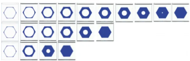
Figure 1 shows the filling of a hexagonal channel for various channel diameters over (computed) time.
Fig 1: filling of hexagonal shaped channel (top view) over computed time for various heights of the channel with hexagonal cross-section: 1mm (top), 0.7 mm (middle) and 0.5 mm (bottom).
Experimental results on channel filling are currently being analysed based on life/dead images taken at various time points (7 & 14 days) after seeding of skeletal progenitor cells onto the printed discs. Based on these results, the function vG will be determined.
4. Discussion and Conclusions
Calibration of the neotissue growth model with results from the basic canal shapes tested experimentally will generate a tool that subsequently can be used to design 3D printable geometries for dental applications.
Acknowledgements: We gratefully
acknow-ledge support from the Wallon Region through the BioWin project BIOPTOS.