IMAGE FOCUS
Ventricular septal defect and infective endocarditis
Anne-Catherine Bernarda,b, Stella Marchettaa,b, Raluca Dulgherua,b, Luc A. Pierarda,band
Patrizio Lancellottia,b
aGIGA Cardiovascular Sciences, University of Li!ege Hopsital, Li!ege, Belgium;bDepartment of Cardiology, Heart Valve Clinic, University Hospital Sart-Tilman, Li!ege, Belgium
ARTICLE HISTORYReceived 11 July 2019; Accepted 7 November 2019
KEYWORDS Ventricular septal defect; infective endocarditis; Streptococcus viridans; PET-CT scanner; management; complication
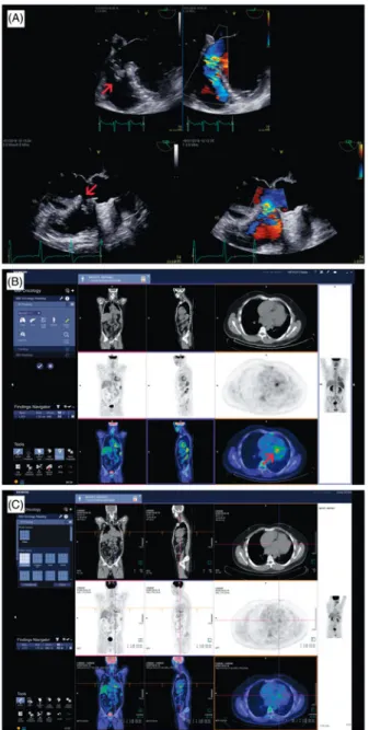
We report the case of a 42-year-old man known to have an asymptomatic small ventricular septal defect (VSD) and tricuspid dysplasia that presented in out-patient clinic with a prolonged state of dyspnoea, asthenia and night sweats. Biology showed an inflam-matory syndrome. Clinical examination revealed a pan-systolic murmur. Blood cultures were positive for Streptococcus viridans. Transoesophageal echocardiog-raphy (TOE) showed mobile vegetations in the peri-membranous VSD, predominantly on the right side but complex anatomy made echocardiographic assess-ment difficult (Figure 1, Panel A). A positron emission tomography-computed tomography (PET-CT) scanner was performed and confirmed the infection, which included the tricuspid valve and excluded peripheral embolisation (Figure 1, Panel B). The patient was treated during four weeks by Ampicillin intravenously, without an indication for surgery as the hemodynamic was stable. Oral hygiene and dental status were checked to lower the risk for IE recurrence and were normal. The patient’s healing was confirmed by the PET-CT control at the end of the treatment (Figure 1, Panel C). Although, not standard practice, PET-CT was considered necessary to ensure complete resolution of the inflammatory process within the interventricular
septum because follow-up TOE was non diagnostic. In patients with VSD, the incidence of infective endocar-ditis is 20–30 times higher than in the general popula-tion but the mortality seems lower. They are particularly sensitive to bacteria graft because of tur-bulent blood flow. In addition to systemic embolisa-tion, the specific risks are the increase of the shunt, with right ventricular overload, pulmonary embolism and involvement of the tricuspid valve. Prophylactic antibiotic therapy in congenital heart disease is limited to patients with untreated cyanotic congenital heart disease (CHD) and those with CHD who have postop-erative palliative shunts, conduits or prostheses. The non-specific prevention measures are essential as strict dental and cutaneous hygiene. Closure of the VSD, at distance from the IE episode, should be contemplated, given new data indicating the higher risk for IE in patients with uncorrected VSD. In this case, the PET-CT was precious for the confirmation of the diagnosis and the follow-up.
Disclosure statement
No potential conflict of interest was reported by the authors.
CONTACTPatrizio Lancellotti [email protected] Department of Cardiology, University Hospital Sart Tilman, Liege B–4000, Belgium ! 2019 Belgian Society of Cardiology
ACTA CARDIOLOGICA
Figure 1. Perimembraneous ventricular septal defect (VSD) presenting an aneurism and abnormal thickening of the defect mar-gins. Presence of a left to right intra-cardiac shunt at the level of the VSD seen with colour flow Doppler (Panel A). Whole body FDG PET/CT before antibiotic treatment showing abnormal hyper fixation of the tracer at the upper segment of the inter ventricu-lar septum suggesting an inflammatory/infectious process at this level (Panel B). Whole body FDG PET/CT showing decrease in tracer uptake intensity at the upper segment of the inter ventricular septum suggesting a regression of the inflammatory/infec-tious process after antibiotic treatment (Panel C).