part of
Fu
tu
re N
e
u
ro
log
y
A growing body of evidence indicates that
sleep promotes memory consolidation; the
protracted process by which fresh, labile
mem-ories are reorganized into stable memmem-ories
[1].
Although the first evidence for this effect was
collected more than a century ago
[2], the
potential underlying neural mechanisms are
only just beginning to be conceptualized and
characterized experimentally.
Memory consolidation is a hackneyed
expres-sion that may take different meanings. At the
cellular level, consolidation is often framed
in terms of long-term experience-dependent
changes in neural responsiveness, usually
result-ing from modifications in synaptic strength
(Figure 1)[3–19]
. At the systems level,
consolida-tion relates to the progressive reorganizaconsolida-tion of
memories within brain circuits
[20–21]. From a
behavioral perspective, consolidation refers to
an improvement of performance between
prac-tice sessions or a reduction of vulnerability of
a memory trace after the acquisition of a novel
skill
[22–23], although other phenomena such
as automatization
[24–25]or generalization
[26]might also be considered as behavioral markers
of memory consolidation
(Figure 1).
It is therefore not surprising that the effects of
sleep on memory consolidation were reported at
various levels of description. The early studies
assessed the effects of sleep on different memory
systems at the behavioral level. They contrasted
the influence of non-rapid eye movement sleep
(NREMS) with rapid eye movement sleep
(REMS) on declarative and procedural
memo-ries, respectively. As the understanding of the
effects of sleep on memory consolidation
pro-gressed, the hypotheses were increasingly framed
in terms of the neural processes occurring during
NREMS or REMS, and specifically, those
asso-ciated with phasic events such as slow waves,
spindles or phasic REMS.
This paper is organized into five sections.
The first section summarizes the key findings
of behavioral studies that suggest an effect of
sleep on memory. Earlier reviews have already
covered these results in detail
[27–31]. The
present article highlights recent data that call
for a reappraisal of these effects that, although
indisputable, seem quantitatively modest. The
article also emphasizes the higher sensitivity
of neuroimaging and neurophysiological
meas-ures in characterizing the effects of sleep on
memory. The following sections review the
two main hypotheses that are currently being
tested, namely the homeostatic synaptic
down-scaling and the sleep-dependent systems-level
consolidation. The final two sections
empha-size the intervention of post-training REMs
and wakefulness in memory processing.
Behavioral effects
The recognition of different memory systems
helped to characterize the effects of sleep on
memory consolidation
(Figure 2). On the
proce-dural side, spontaneous gains in performance in
a motor sequence learning task were observed
between initial training and later testing,
spe-cifically if the interval included sleep rather
than wakefulness
[32–33]. As these gains in
per-formance emerged without further practice, it
suggested that an ‘offline’ processing of
motor-memory was taking place during sleep. The
spontaneous enhancement of procedural abilities
appeared to depend primarily on REMS. Indeed,
the enhanced performance in procedural
learn-ing tasks was observed when sleep was allowed
Contribution of sleep to
memory consolidation
Anahita Shaffii-Le Bourdiec, Vincenzo Muto, Laura Mascetti, Ariane Foret,
Luca Matarazzo, Caroline Kussé & Pierre Maquet
††Author for correspondence: Cyclotron Research Centre (B30), University of Liège, Allée du 6 Août, 4000 Liège, Belgium n Tel.: +32 43 66 36 87 n Fax: +32 43 66 29 46 n [email protected]
The contribution of sleep to memory processing is being characterized at increasingly detailed levels. At a behavioral level, better performance at retrieval is usually observed after sleep, relative to a period of wakefulness. At a brain-systems level, functional neuroimaging techniques have demonstrated that the distribution of regional brain activity is influenced by previous waking experience. At present, the selective effects of sleep components, such as slow waves or spindles are being characterized. These effects are framed in terms of neural firing patterns and also in terms of the molecular mechanisms underpinning the effects of sleep on brain plasticity. Collectively, the available data indicate a positive influence of sleep on memory consolidation.
Keywords
n behavioral task n memory consolidation n neural processes n neuroimaging n sleep
Re
vie
w
Wake Sleep Wake Internal stimuli (ANS, hormones, chemical messengers) External stimuli Fiv e senses filte r - Accessibility of consolidated memories at a delayed retrieval Retrieval - Strengthening of fresh memory facts
- Transformation into more stable and persistent form - Integration into pre-existing knowledge networks - Plastic changes in brain circuitry, synaptic sensitivity - Editing, sorting, combining and pruning Consolidation Encoding - Acquisition of learning material - Newly built and labile representations
Future Neurol. © Future Science Group (2010)
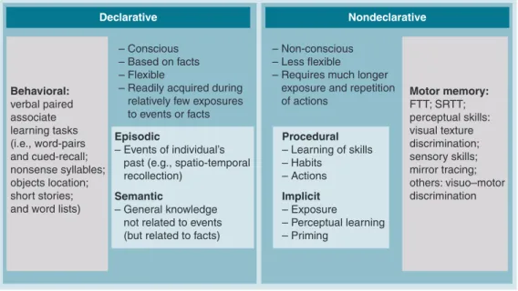
Declarative Nondeclarative
– Conscious – Based on facts – Flexible
– Readily acquired during relatively few exposures to events or facts
– Non-conscious – Less flexible
– Requires much longer exposure and repetition of actions Behavioral: verbal paired associate learning tasks (i.e., word-pairs and cued-recall; nonsense syllables; objects location; short stories; and word lists)
Motor memory: FTT; SRTT; perceptual skills: visual texture discrimination; sensory skills; mirror tracing; others: visuo–motor discrimination Episodic – Events of individual’s past (e.g., spatio-temporal recollection)
Semantic
– General knowledge not related to events (but related to facts)
Procedural – Learning of skills – Habits – Actions Implicit – Exposure – Perceptual learning – Priming
during the REMS-rich second part of the night
rather than during the deep NREMS-rich first
half
[34–35]. Likewise, perceptual visual
learn-ing (e.g., texture discrimination tasks, see
Box 1),
involving a different procedural learning
proc-ess, was demonstrated to be sensitive to REMS
deprivation
[36].
On the declarative side, despite early
con-troversial results
[37–40], declarative memory
was also shown to be better preserved when
sleep occurs immediately after encoding
[41],
and seems to benefit primarily from NREMS;
word-pair associates are better retained if sleep is
allowed during the first part of the night (rich in
deep NREMS) rather than in the second portion
of the night
[35]. If olfactory cues are presented
during a spatial source memory task and again
delivered during subsequent NREMS, memory
performance is enhanced during subsequent
retrieval
[42]. The same protocol has no effect
for a procedural task such as the finger-tapping
task (FTT)
[42].
These results were compatible with the ‘dual
process’ hypothesis, which claims that NREMS
and REMS are primarily involved in the
con-solidation of declarative and procedural
mem-ories, respectively. However, this theory
can-not easily account for data demonstrating, for
instance, that procedural learning also benefits
from NREMS-rich early night sleep
[43–45]. The
Figure 1. Memory is usually conceived to evolve in three distinct steps: encoding, consolidation and retrieval. Memoryconsolidation would occur covertly during both wakefulness and sleep. The objective now is to specify what are the conditions selectively present during sleep that promote memory consolidation.
ANS: Autonomic nervous system.
Figure 2. Memory is classically divided into two main groups involving different learning tasks.
alternative possibility, the ‘double-step’
hypoth-esis, assumes that the succession of NREMS
and REMS was required for optimal memory
consolidation
[46], whatever the memory
sys-tem considered. Some evidence has supported
this hypothesis in animal experiments
[47–51].
In humans, the evidence remains scarce. Motor
sequence learning is improved to a larger extent
by a complete night of sleep including
abun-dant amounts of both NREMS and REMS,
compared with late night sleep alone
[34].
Likewise, the positive effect of sleep on visual
perceptual learning is larger if it includes both
NREMS and REMS
[44,45].
Sleep is also believed to facilitate fluid
think-ing and problem solvthink-ing. Historically, anecdotal
evidence exists for the positive influence of sleep
on scientific and artistic creativity
[52]. Recently,
experimental evidence confirmed that sleep not
only enhances the verbatim retention of learned
material, but can also lead to behavioral
modi-fications during sleep that entail the generation
of novel representations based on
informa-tion extracted from learned exemplars
[53–54].
Similarly, sleep has been involved in selectively
enhancing false memories (relative to accurate
memories)
[55]. However, this effect is still
con-troversial. Another study did not report any
beneficial effect of sleep while sleep deprivation
enhanced false memories at retrieval
[56].
Despite all of this evidence, a critical review of
the effects of sleep suggests that the behavioral
effects of sleep on memory are smaller than
usu-ally believed. In the declarative domain, the
dif-ference in recall rates between sleep and waking
conditions, although significant, usually only
amounts to a marginal fraction of the learned
material (e.g.,
[41]). This suggests that most of
the learned material can still be retained and
consolidated during wakefulness, without the
necessary intervention of sleep (refer to the
lat-ter section of this article). However, one could
argue that the major behavioral effect of sleep
on memory consolidation is revealed under
con-ditions of interference. Indeed, sleep provides
active protection against potential interferences
resulting from encoding novel information on
the day following the initial learning
[57].
In the procedural domain, the effects of sleep
on memory consolidation were also not as
sim-ple as originally thought. Significant gains in
performance in motor sequence learning have
not been reported exclusively after sleep and can
occur over a waking period, depending on
vari-ous experimental factors: post-training interval
[33,58–59], circadian rhythms
[60]or subject’s
aware-ness of the sequential material
[23,34,58](TaBle 1).
In addition, gains in performance recorded with
the FTT are, in fact, overestimated, if the fatigue
accrued during initial training is not accounted
Box 1. Description of the most commonly used behavioral tasks for memory characterization.
n Word-pair associate– Subjects are asked to learn lists of visually presented pairs of words by usually forming a mental image of both objects.
– During the recall sessions, all previously learned words are tested. Subjects are presented with the first word of each pair. They are instructed to remember the second word with the help of the mental picture they imagined previously.
n Navigation
– In experimental conditions, navigation is usually conducted in a color 3D first-person view of the environment, which consists of various streets with distinctive walls, objects, sounds and background music and in which volunteers navigate using a joystick to control their moves.
n Finger-tapping task
– The finger-tapping task is a common motor sequence learning task in which participants have to produce an explicitly known five-element finger sequence on a keyboard, with their left non-dominant hand.
n Serial reaction time task
– In a serial reaction time task, participants face a computer screen where permanent position markers are displayed. A keyboard with spatially compatible response keys is within reach of the right hand. Subjects are asked to react as quickly and accurately as possible to the appearance of a stimulus below one of the markers by pressing the spatially corresponding key. Unknown to the subjects, the sequence of the stimulus positions is not random but either fixed (deterministic) or generated based on a probabilistic finite-state grammar that defines logical transitions between successive trials.
n Serial oculo–motor reaction time task
– Participants have to visually track a dot that, at any point in time, is displayed at one of several possible positions, and the color of which can briefly change. Participants are explicitly instructed to detect the changes in dot color. However, unbeknownst to them, the succession of dot positions follows a second-order eight-element sequence that is repeated during several practice blocks. A different but structurally equivalent second-order sequence is presented once at the end of training and during testing to allow for a direct measure of sequence learning. During both training and testing, performance is measured by the latencies of eye movements, defined as the time interval between the change in dot position and the first saccade initiated in the direction of the target. These latencies reflect the development of an implicit sequential knowledge, as they become progressively shorter with repetition of the learned sequence, and slow down when the untrained sequence is presented.
for
[61]. Finally, it should be stressed that a
benefi-cial effect of sleep over wakefulness in the
reten-tion of recently learned material has not been
reported for all experimental tasks. For example,
auditory perceptual learning does not seem to be
selectively enhanced by sleep
[62].
To tone down these reservations, it is also
possible that behavioral measures are
insensi-tive to sleep-related modifications in memory.
In other words, the absence of a behavioral
effect of sleep on memory does not necessarily
entail an absence of any effect. Owing to
redun-dancy in brain circuitry, similar performance
levels can be achieved by recruiting different
functional networks. For instance, the ability
to navigate a virtual maze learned 3 days earlier
is equivalent in volunteers who were either sleep
deprived (SD) or allowed to sleep on the first
postencoding night
(Figures 3 & 4)[25]. However,
during navigation, striatal activity is larger at
retrieval in the sleep group than in SD subjects.
These data suggest that brain activity is
restruc-tured during sleep in such a way that
naviga-tion in the virtual environment, initially related
to a hippocampus-dependent spatial strategy,
becomes progressively automatized and
medi-ated by the striatum. Likewise, the recollection
of emotional items 72 h after encoding does not
differ between sleeping or SD subjects during
the postencoding night
[63]. However, larger
responses were observed in the medial prefrontal
cortex and the hippocampus in the sleep group.
By contrast, after SD, subsequent recollection of
negative items is related to responses in an
amy-gdalo–occipital network. Again, this alternate
network suggests that recollection of emotional
stimuli relies on redundant neural systems.
In summary, there is currently a wealth of
evi-dence indicating that sleep enhances the retention
of newly learned material. Quantitatively, the
pos-itive influence of sleep on memory consolidation
appears smaller that initially estimated. However,
behavioral measures might be less sensitive than
direct measures of brain activity during sleep in
the assessment of sleep contribution to memory
consolidation. In this respect, measures of the
electromagnetic or hemodynamic activity of the
brain appear a necessary step in the evaluation of
the influence of sleep on memory processes.
Behavioral studies have also suggested that the
effects of sleep on memory consolidation could
not be easily framed merely in terms of sleep
states, suggesting that neural events associated
with specific sleep microstructure might be more
relevant for offline processing of recent
memo-ries. Accordingly, recent research that aimed to
characterize the mnemonic processes associated
with slow oscillations, D waves, spindles,
pha-sic REMS and sleep neuromodulatory context.
Recently, the emphasis was put on the impact
of NREMS oscillations on memory processes.
Two distinct, equally inspiring and not
neces-sarily exclusive hypotheses came under intense
experimental scrutiny: the homeostatic synaptic
downscaling and the systems-level consolidation
in hippocampo–neocortical circuits. They are
reviewed in the following sections.
Hypothesis of a homeostatic synaptic downscaling during NREM sleep
This hypothesis claims that wakefulness is
asso-ciated with a net increase in synaptic strength
in the brain, which would become energetically
unsustainable in the long term
[64–65]. During
deep NREMS a slow (<1 Hz) oscillation emerges
from cortical or thalamo–cortical populations.
Unit recordings have demonstrated that
neuro-nal activity during slow wave sleep (SWS) is
characterized by a fundamental oscillation of
membrane potential. This slow oscillation is
recorded in all major types of neocortical
neu-rons during NREMS and consists of a
depolar-izing phase, associated with important neuronal
firing (the ‘up state’), followed by a
hyperpolariz-ing phase durhyperpolariz-ing which cortical neurons remain
silent for a few hundred milliseconds (the ‘down
state’)
[66–67]. The slow oscillation occurs
syn-chronously in large neuronal populations, the
Table 1. Examples of factors modulating memory consolidation characteristics.
Type of
learning material
Type of retrieval test Type of learning Type of population Type of sleep surrounding learning Declarative/procedural Emotional /neutral New/old Difficult/easy Recall/recognition Explicit/implicit Fast/slow Motivational factors Attention/alertness/vigilance Genetics Gender Age Environment Health Delay, duration before/after learning Circadian Homeostatic Non-rapid eye movement sleep
Rapid eye movement sleep
Regular sleep group
Total sleep deprivation group
Immediate retrieval Delayed retrieval Day 1 Day 2 Day 3 Day 4
Night 1 Night 2 Night 3 Training
activity of which alternates between so-called
ON and OFF states
[65], in such a way that it
is reflected on EEG recordings as large
ampli-tude low frequency waves
[68]. D waves (1–4 Hz)
are faster waves of smaller amplitude that arise
when the synchronization in the network is less
marked
[65,69]. Slow wave activity (SWA), in
other words, the EEG power between 0.75 and
4 Hz, is a compound parameter that quantifies
the homeostatic sleep pressure accrued during
wakefulness
[70].
During NREM sleep, the hypothesis assumes
that the slow oscillation would be associated with
a downscaling of synaptic strength to a baseline
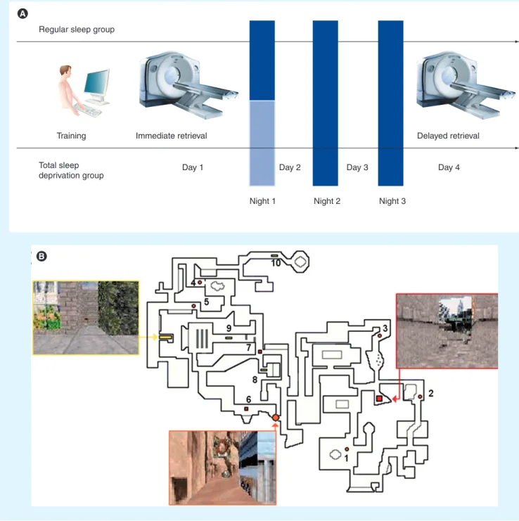
Figure 3. Experimental protocol and memory task. (A) Experimental design. After training on a desktop computer and subsequent
testing in the scanner on day 1 (immediate retrieval), subjects were either totally sleep deprived or allowed regular sleep on the first post-training night. They were all retested under identical conditions during a second functional MRI session on day 4 (delayed retrieval).
(B) The map depicts an aerial view of the color 3D virtual town in which subjects navigated at the ground level. Snapshots show the
three locations used as targets for testing during the fMRI sessions. The ten starting points are represented by numbers, with associated symbols indicating the target location to reach.
1 0.8 0.6 0.4 0.2 0 -0.2 -0.4 -0.6 -0.8 -1 -2 0 2 4 6 8 10 12 14 16 r = 0.41 r = 0.80 1 0.8 0.6 0.4 0.2 0 -0.2 -0.4 -0.6 -0.8 -1 -2 0 2 4 6 8 10 12 14 16 r = 0.41 r = 0.80
Mean distance score
B O L D s ig n al c h an g e E ff ec t si ze -4 -2 0 2 4
Figure 4. Sleep-dependent modulation of brain activity. Contrasts are displayed at p < 0.001 (uncorrected) superimposed on the
average T1-weighted MRI scan. Color bars code the value of the t-statistic associated with each voxel. (A) Higher activity elicited by place
finding for the regular sleep (RS) compared with the totally sleep-deprived (TSD) group at delayed retrieval bilaterally in the caudate nucleus. Blue crosshair, right caudate nucleus (14, 8 and 18 mm; Z = 3.73). (B) Between-group regression analyses of the average session
performance on cerebral activity at delayed retrieval (sagittal and coronal sections). Blue crosshair, right caudate nucleus (8, 22 and 4 mm; Z = 3.45). Scatter plot shows that brain response was correlated positively with performance in the RS group (blue; r = 0.41) but negatively in the TSD group (red; r = −0.80). (C) Psychophysiological interaction analysis using the right caudate nucleus (14, 8 and 18 mm; green
crosshair) as seed area. Coupling of activity between the caudate nucleus and the left hippocampus (coronal section) was negative in the RS group (blue), but positive in the TSD group (red). Blue crosshair, left hippocampus (22, 12 and 22 mm; Z = 3.15). Blue and red plots show the size of effect for each group. Error bars are standard deviations. Reproduced with permission from [25].
level, a process that is beneficial for learning and
memory. The synaptic downscaling would be
a general phenomenon occurring throughout
the brain, one that is modulated locally by the
amount of neural activity and synaptic strength
enhancement accumulated during the day.
In support of this hypothesis, an increase in
SWA is selectively observed after training to a
visuo–motor adaptation task, over scalp areas
that are deemed critical in this type of
learn-ing
[43]. By contrast, arm immobilization results
for a decrease in SWA over controlateral
senso-rimotor areas during subsequent NREMS
[71].
Local increases in SWA were also reported when
cortical activity was experimentally induced by a
vibratory stimulation of the hand
[72], transcranial
magnetic stimulation
[73]or when spike
timing-dependent activity was elicited during waking by
transcranial-paired-associative stimulation
[74].
Moreover, after learning lists of word-pair
asso-ciates, during the surface-positive half-waves of
NREMS slow waves, coherence is enhanced over
widespread scalp areas in the slow-oscillatory
and D bands
[75]. Similar increases in SWA were
reported in rats exposed to an enriched
environ-ment and appear to be associated with the release
of BDNF
[76]. At the cellular level, the slope and
amplitude of cortical evoked responses, taken as
markers of local synaptic strength, increase after
waking and decrease after sleep in proportion
with changes in SWA
[77]. At the cellular level,
multiunit recordings showed that firing rates and
synchrony decrease after sleep. Changes in firing
patterns in NREMS correlate with changes in
SWA
[65].
At the molecular level, in rat cortex and
hip-pocampus, GluR1-containing AMPA receptor
(AMPAR) levels are high during wakefulness
and low during sleep, and changes in the
phos-phorylation states of AMPARs, CamKII and
GSK3b are consistent with synaptic
potentia-tion during wakefulness and depression
dur-ing sleep
[77]. Similar findings were recently
reported in the fly, which provides evidence for
the generality of this phenomenon
[78].
However, for the sake of completeness, it
should be mentioned that not all forms of
sleep-dependent plasticity seem to follow this general
scheme. During neurodevelopment in kittens,
the plasticity induced by monocular
depriva-tion can be inhibited by the administradepriva-tion
dur-ing sleep of NMDA receptor (NMDAR) and
cAMP-dependent protein kinase antagonists
(PKA)
[79]. These findings suggest a synaptic
strengthening during sleep in contrast to the
downscaling hypothesis.
Sleep-dependent systems-level memory consolidation & hippocampo-neocortical dialog
The second hypothesis pertains primarily to
hippocampus-dependent memories. The
classi-cal theory of memory consolidation claims that
during consolidation, hippocampus-dependent
memories become progressively independent of
the limbic structures and are gradually stored
in cortical circuits
[21,80]. Sleep would
partici-pate in this systems-level memory consolidation
by promoting the functional interplay between
hippocampal neural ensembles and neocortical
areas
[81].
This hypothesis implies that new experiences
modify the activity of
hippocampo–neocorti-cal circuits during subsequent NREMS. At the
systems level, human brain activity during sleep,
as assessed by PET, is indeed significantly
modi-fied by previous waking experience. After the
exploration of a virtual 3D maze the activity
is enhanced during NREMS in occipital,
pari-etal and mesio-temporal areas
[82]. Moreover,
the increase in hippocampal activity is linearly
related to the individual gain in the ability to
navigate in the maze the next day,
suggest-ing that the changes in hippocampal activity
d uring NREMS relate to the offline processing
of t opographical memory.
By contrast, sleep deprivation should hinder
the offline processing of memory traces
dur-ing NREMS and alter the neural correlates of
subsequent retrieval. Accordingly, functional
neuroimaging studies showed that SD during
the first post-training night invariably modifies
responses subsequently associated with retrieval.
For instance, recall of word-pair associates 48 h
after encoding is associated with larger
hippo-campal responses in subjects allowed to sleep
on the post-encoding night compared with SD
subjects
[41]. Modifications of brain responses
during retrieval are also modified by
post-train-ing SD for other memory tasks: topographical
memory (see above)
[25], emotional memory (see
above)
[63]and motor sequence learning
[33–34].
After SD during the first post-training night,
changes in brain responses associated with
memory retrieval continue to be observed several
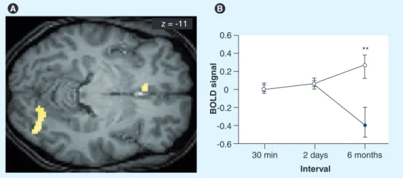
months after encoding (see
Figures 5 & 6)
[41,63,83],
suggesting that sleep immediately following
encoding modulates the early consolidation and
conditions the structure of long-term memory at
the systems level.
At the cellular level, the slow rhythm
organ-izes other NREMS oscillations in a coalescence
of rhythms so that spindles and g oscillations
z = -11 0.6 0.4 0.2 0 -0.2 -0.4 -0.6
30 min 2 days 6 months
BOLD signal
**
Interval
are more likely to occur during the up phase of
the slow oscillation
[84]. Likewise, hippo campal
activity during NREMS is characterized by sharp
waves and ripples, which are synchronous to the
cortical slow oscillation
[85–88], although it is not
yet clear which oscillation is driving the other
[86,89–91]. Finally, the slow oscillation organizes
the neural firing of arousing structures such as
the locus coeruleus (LC)
[92]. Collectively, these
data suggest that the up-state of the cortical slow
oscillation constitutes a remarkable period
dur-ing which the activity of various brain structures,
in the cortex (slow waves and spindles), the
hip-pocampus (sharp-wave ripples) and subcortical
structures (striatum and LC) is synchronized,
thereby fostering functional interactions
associ-ated with systems-level memory consolidation.
A key finding supporting this hypothesis is
that sequences of neural discharges recorded
in neural ensembles during wakefulness were
spontaneously repeated during sleep, especially
during hippocampal sharp waves and ripples,
which are coherent with cortical slow oscillation,
was observed in various brain structures such as
the hippocampus, neocortical areas
[87,93–95], the
thalamus
[96]or the striatum
[97]. Importantly,
reactivation of firing patterns in the neocortex is
synchronized to hippocampal sharp waves
[87,94],
a condition favorable to cortico–hippocampal
interactions. In addition, these reactivations
appear to depend on learning as the replayed
firing patterns appear only after the acquisition
of new rules
[94]. Simultaneous recordings in the
hippocampus and prefrontal cortex in the rat
demonstrated that cells in prefrontal cortex fire
consistently within 100 ms after hippocampal
cells during NREMS
[88]. This result provides
the first evidence at the single-cell-pair level
for highly consistent directional interactions
between these areas within the window of
plas-ticity. Moreover, these interactions were driven
by hippocampal sharp-wave/ripple bursts in
SWS and are sharply reduced during REMS
[88].
Collectively, these data support the
hypoth-esis that during sleep, hippocampo–neocortical
interactions may progressively transfer the
bur-den of memory from hippocampal ensembles to
long-term neocortical stores.
Sleep spindles are a hallmark of light NREMS
(mostly stage 2 sleep) and, in humans, consist of
waxing and waning 11–15 Hz oscillations, lasting
0.5–3 s
[19]. They are generated by the thalamus,
which acts as a pacemaker
[98–99], and result from
reciprocal rhythmic interactions between
reticu-lar and thalamo–cortical cells. Postinhibitory
rebound spike bursts in thalamo–cortical cells,
induced by a recurrent inhibition by reticular
cells, entrain cortical populations in spindle
oscil-lations
[100]. In turn, a cortico–thalamic feedback
synchronizes spindle oscillations in widespread
Figure 5. Differences in brain activity during a 6-month retest session for correctly recalled words learned before sleep versus before sleep deprivation in a paired-associate word task. (A) Correct word recall after 6 months activates the medial-prefrontal cortex (mPFC) and theoccipital cortex more strongly for words from the sleep condition than for words from the sleep-deprivation condition. Note that at the 2-day interval, no activity per se was found, but only a strong functional relation to hippocampal activity. At the 6-month interval, independent mPFC activity is found, but no more significant hippocampal activity. (B) The difference in brain activity in the mPFC
developed mainly during the interval between the 2-day and 6-month recall sessions. It is supported by a steady increase in mPFC activity for words from the sleep condition over the 6-month period (open circles) and a marked drop in mPFC activity for words from the sleep-deprived condition (filled circle) during the 6-month session ([−6 26 −10], Z = −3.87, P SVC = 0.004).
z = 22 z = -13
thalamic territories
[101]. As spindles entrain
syn-chronous firing in large thalamo-cortical neural
populations, they are in a good position to allow
for the modifications in the neural representations
of recent memories. In support of this
hypoth-esis, spindle activity increases after training on
declarative
[102–104]and procedural
[105–107]mem-ory tasks. Enhancing slow oscillations by direct
current transcranial stimulation increases the
power in the spindle frequency band and leads to
a better retention of declarative memory the next
morning
[108]. At the cellular level, it was shown
that repetitive spike bursts mimicking firing
pat-terns observed during spontaneous spindles
reli-ably induced short- and long-term potentiation
in cortical neurons of rat brain slices
[109]. It has
also been suggested that activity in the spindle
band would trigger molecular cascades involved
in brain plasticity by increasing intracellular
c alcium levels
[110].
Finally, the modifications in
neuromodula-tion associated with sleep might provide a
favo-rable context for memory offline processing.
NREMS is associated with a decrease in
cholin-ergic, adrenergic and serotonergic drive
[111]. For
example, acetylcholine inhibits feedback loops
within the hippocampus and between the
hip-pocampus and neocortex
[112]. A low cholinergic
activity during SWS would be required to
pro-mote the spontaneous replay of newly acquired
information in the hippocampus. In line with
this assumption, augmenting cholinergic tone
during NREMS by infusion of
physostig-mine in humans causes deterioration in later
p erformance on a declarative memory task
[113].
REMS & pontine waves
REMS represents a state of consciousness
dis-tinct from wakefulness and NREMS,
char-acterized by persistent (‘tonic’) features such
as relatively fast-frequency, low-amplitude
oscillations on EEG recordings and muscular
atonia, but also by phasic characteristics such
as bursts of REMS, muscle twitches and swift
modifications in autonomic functions
[114].
The implication of REMS in memory
process-ing is still a subject of debate
[115]and at the
very least appears dispensable in adult humans
since REMS suppression by
administra-tion of selective serotonin or norepinephrine
reuptake inhibitors has not demonstrated any
d etrimental effect on motor skill or word-pair
learning
[116].
On the other hand, there is also positive
evi-dence for the implication of REMS in memory
processing. At the systems level, training on
a procedural task (a motor sequence
learn-ing task based on probabilistic serial reaction
times,
Box 1) results in increased activity in
premotor and occipital cortices, thalamus and
upper brainstem during subsequent REMS. In
addition, the functional connectivity between
the premotor cortex and posterior parietal
cor-tex and presupplementary motor area is also
enhanced during post-training REMS
[117].
These changes in regional activity during
REMS are observed only if the learned
mate-rial is structured by hidden rules and not when
the material is random, suggesting that they
are related to the processing of the underlying
higher-order structure
[118].
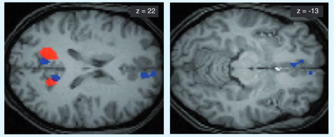
Figure 6. Obtained from the paired associate words task protocol, areas functionally related to the hippocampus during correct word recall on day 2 after sleep or sleep deprivation. At 2 days after learning, if subjects were allowed to sleep, the hippocampus was
functionally connected to the precuneus and the medial-prefrontal cortex during correct word recall (blue). However, if subjects were sleep-deprived the night after learning, the prefrontal sites did not relate to hippocampal activation (red).
Within REMS, it appears that
phasic-REMS-associated neural activity is involved in memory
consolidation. One of the main characteristics of
REMS in animals consists of prominent phasic
bioelectrical potentials that occur isolated or in
bursts during the transition from SWS to REMS
or during REMS itself (for a review see
119]).
These waves can be observed from many parts
of the animal brain
[120], but are most easily
recorded in the pons, the lateral geniculate
bod-ies and the occipital cortex
[121]and therefore
are termed ponto–geniculo–occipital (PGO)
waves in cats
[122]or pontine waves in rats
[122].
Typically, bursts of pontine waves are associated
with REMS. Several observations suggest that
PGO waves also occur during human sleep
[123– 128]. In rats, the density of pontine waves
dur-ing post-traindur-ing REMS is proportional to the
gain in performance to an avoidance task
[129].
Suppressing pontine waves, even without
modi-fying the amounts of REMS, annihilates
learn-ing. By contrast, the pharmacological induction
of pontine waves (by microinfusion of carbachol)
enhanced subsequent performance to the
avoid-ance task. These effects appear to be related to
the activation of glutamatergic
neurotransmis-sion, gene expression and protein synthesis
in the hippocampus and the amygdala
[130].
Accordingly, synaptic transmission and late-LTP
in the rat hippocampus are positively correlated
with REMS quantities
[131]. Reversely,
REMS-deprivation decreases synaptic transmission
and long-term potentiation selectively in
dor-sal hippo campus. Intriguingly, levels in GluR1,
NR1 and p-ERK1/2 decrease without changes
in GluR1 and NR1 mRNA expression,
suggest-ing sleep-induced modulation of translational,
rather than transcriptional mechanisms
[131].
Wakefulness
The data reviewed above does not mean that
mem-ory consolidation occurs exclusively during sleep.
Indeed, experimental evidence exists for
learning-related changes in spontaneous brain activity
dur-ing post-traindur-ing wakefulness. Replays of neural
firing sequences from a previous experience are
recorded in the rat hippo campus during brief
pauses in waking behavior, especially when the
rat has recently been in motion
[132]. Intriguingly,
neural firing sequences can be replayed in a
tem-porally reversed order during waking periods
relative to previous spatial experience
[133]. These
reverse replays would have a critical role in
sup-port of learning in hippocampus-dependent tasks
by propagating valuable information from the
rewarded location b ackwards along incoming
trajectories
[133].
Likewise, in non-human primates, the
cor-relation structure between somatosensory and
parietal cortex induced by reaching and
naviga-tion tasks was maintained during post-training
resting wakefulness
[134]. In humans, the brain
activity elicited during a new learning episode
(navigation or motor sequence learning)
modu-lates brain responses to an unrelated cognitive
task (auditory oddball task), during the
wak-ing period followwak-ing the end of trainwak-ing
[135].
Collectively, these data indicate that memory
is actively processed during post-training
wakefulness.
Executive summary
Behavioral effectsn Functional neuroimaging techniques show that the distribution of regional brain activity during sleep is influenced by previous
waking experience.
The hypothesis of a homeostatic synaptic downscaling during NREM sleep
n EEG and molecular data tend to support the hypothesis that NREM sleep, and more specifically slow-wave sleep, is the reflection of
both a global and local synaptic downscaling correlated with the amount of synaptic strengthening that has previously taken place during wakefulness.
The sleep-dependent systems-level memory consolidation & the hippocampo-neocortical dialogue
n Neuroimaging as well as neurophysiological data support the classical memory consolidation hypothesis stating that during sleep
hippocampo–neocortical interactions may progressively transfer the burden of memory from hippocampal ensembles to long-term neocortical stores.
REMS & pontine waves
n Often considered as dispensable, REMS is indeed implied in memory processing. Typical REMS phasic events such as the PGO waves are
implied in learning, and there is good evidence that REMS promotes synaptic potentiation.
Wakefulness
n Experimental evidence exists for learning-related changes in spontaneous brain activity during post-training wakefulness, showing that
memory is actively processed during wakefulness and not exclusively during sleep.
Conclusion
Financial & competing interests disclosure
Personal research reported in this review was sup-ported by the Belgian Fonds National de la Recherche Scientifique (FNRS), Fondation Médicale Reine Elisabeth (FMRE), Research Fund of the University of Liège and “Interuniversity Attraction Poles Programme – Belgian State – Belgian Science Policy”. Anahita Shaffii-Le Bourdiec, Laura Mascetti, Ariane Foret, Luca Matarazzo, Caroline Kussé and Pierre Maquet are supported by the FNRS. Vincenzo Muto is supported by the ULg. The authors have no other relevant affiliations or financial involvement with any organization or entity with a financial inter-est in or financial conflict with the subject matter or materials discussed in the manuscript apart from those disclosed.
No writing assistance was utilized in the p roduction of this manuscript.
Bibliography
Papers of special note have been highlighted as:
n of interest
nn of considerable interest
1. McGaugh JL: Memory – a century of consolidation. Science 287, 248–251 (2000).
2. Ebbinghaus H: Über das Gedächtnis,
Untersuchungen zur Experimentellen Psychologie. Duncker und Humblot, Leipzig,
Germany (1885).
3. Abel T, Lattal KM: Molecular mechanisms of memory acquisition, consolidation and retrieval. Curr. Opin Neurobiol. 11, 180–187 (2001).
4. Carew TJ, Sutton MA: Molecular stepping stones in memory consolidation. Nat.
Neurosci. 4, 769–771 (2001).
5. Izquierdo I, Bevilaqua LR, Rossato JI, Bonini JS, Medina JH, Cammarota M: Different molecular cascades in different sites of the brain control memory consolidation.
Trends Neurosci. 29, 496–505 (2006).
6. Wang H, Hu Y, Tsien JZ: Molecular and systems mechanisms of memory consolidation and storage. Prog. Neurobiol. 79, 123–135 (2006).
7. Bailey CH, Kandel ER, Si K: The persistence of long-term memory: a molecular approach to self-sustaining changes in learning-induced synaptic growth. Neuron 44, 49–57 (2004).
8. Bahar A, Dorfman N, Dudai Y: Amygdalar circuits required for either consolidation or extinction of taste aversion memory are not required for reconsolidation.
Eur. J. Neurosci. 19, 1115–1118 (2004).
9. Gould E, Beylin A, Tanapat P, Reeves A, Shors TJ: Learning enhances adult neurogenesis in the hippocampal
formation. Nat. Neurosci. 2, 260–265 (1999).
10. Guzman-Marin R, Bashir T, Suntsova N, Szymusiak R, McGinty D: Hippocampal neurogenesis is reduced by sleep
fragmentation in the adult rat. Neuroscience 148, 325–333 (2007).
11. Guzman-Marin R, Suntsova N, Bashir T, Nienhuis R, Szymusiak R, McGinty D: Rapid eye movement sleep deprivation contributes to reduction of neurogenesis in the hippocampal dentate gyrus of the adult rat. Sleep 31, 167–175 (2008).
12. Guzman-Marin R, Suntsova N, Methippara M, Greiffenstein R,
Szymusiak R, McGinty D: Sleep deprivation suppresses neurogenesis in the adult hippocampus of rats. Eur. J. Neurosci. 22, 2111–2116 (2005).
13. Shors TJ, Miesegaes G, Beylin A,
Zhao M, Rydel T, Gould E: Neurogenesis in the adult is involved in the formation of trace memories. Nature 410, 372–376 (2001).
14. Bliss TV, Collingridge GL: A synaptic model of memory: long-term potentiation in the hippocampus. Nature 361, 31–39 (1993).
nn Long-term potentiation is shown to be a molecular correlate of memory consolidation. 15. Benington JH, Frank MG: Cellular and
molecular connections between sleep and synaptic plasticity. Prog. Neurobiol. 69, 71–101 (2003).
16. Bergmann TO, Molle M, Marshall L, Kaya-Yildiz L, Born J, Siebner RH: A local signature of LTP- and LTD-like plasticity in human NREM sleep.
Eur. J. Neurosci. 27, 2241–2249 (2008).
17. McDermott CM, LaHoste GJ,
Chen C, Musto A, Bazan NG, Magee JC: Sleep deprivation causes behavioral, synaptic, and membrane excitability alterations in hippocampal neurons. J. Neurosci. 23, 9687–9695 (2003).
18. Tadavarty R, Kaan TK, Sastry BR: Long-term depression of excitatory synaptic transmission in rat hippocampal CA1 neurons following sleep-deprivation. Exp. Neurol. 216, 239–242 (2009).
19. Werk CM, Klein HS, Nesbitt CE, Chapman CA: Long-term depression in the sensorimotor cortex induced by repeated delivery of 10 Hz trains in vivo. Neuroscience 140, 13–20 (2006).
20. Doyon J, Benali H: Reorganization and plasticity in the adult brain during learning of motor skills. Curr. Opin Neurobiol. 15, 161–167 (2005).
21. Frankland PW, Bontempi B: The
organization of recent and remote memories.
Nat. Rev. Neurosci. 6, 119–130 (2005).
22. Maquet P: The role of sleep in learning and memory. Science 294, 1048–1052 (2001).
n Sleep reactivations of neuronal firing patterns triggered by new experiences during previous wakefulness may be linked to memory consolidation.
23. Robertson EM, Pascual-Leone A, Press DZ: Awareness modifies the skill-learning benefits of sleep. Curr. Biol. 14, 208–212 (2004).
24. Atienza M, Cantero JL, Stickgold R: Posttraining sleep enhances automaticity in perceptual discrimination. J. Cogn. Neurosci. 16, 53–64 (2004).
25. Orban P, Rauchs G, Balteau E et al.: Sleep after spatial learning promotes covert reorganization of brain activity.
Proc. Natl Acad. Sci. USA 103, 7124–7129
(2006).
26. Korman M, Raz N, Flash T, Karni A: Multiple shifts in the representation of a motor sequence during the acquisition of skilled performance. Proc. Natl Acad. Sci.
USA 100, 12492–12497 (2003).
27. Rasch B, Born J: Maintaining memories by reactivation. Curr. Opin Neurobiol. 17, 698–703 (2007).
Conclusions
Memory consolidation is among the few brain
functions that develop over time periods
rang-ing from a few hours to (potentially) several
months or years. It necessarily unfolds across
states of wakefulness and sleep (and also
vari-ous circadian phases). It is not surprising that
these factors influence memory consolidation.
The respective role of neural mechanisms
tak-ing place durtak-ing wakefulness and durtak-ing sleep
remains determined.
A comprehensive understanding of
mem-ory consolidation will eventually lead to its
improved characterization at various
lev-els of description, from the molecular to the
b ehavioral level.
28. Stickgold R, Walker MP: Sleep and memory: the ongoing debate. Sleep 28, 1225–1227 (2005).
29. Ellenbogen JM, Payne JD, Stickgold R: The role of sleep in declarative memory consolidation: passive, permissive, active or none? Curr. Opin Neurobiol. 16, 716–722 (2006).
30. Walker MP, Stickgold R: Sleep, memory, and plasticity. Annu. Rev. Psychol. 57, 139–166 (2006).
31. Born J, Rasch B, Gais S: Sleep to remember.
Neuroscientist 12, 410–424 (2006).
32. Walker MP, Brakefield T, Hobson JA, Stickgold R: Dissociable stages of human memory consolidation and reconsolidation.
Nature 425, 616–620 (2003).
33. Walker MP, Brakefield T, Morgan A, Hobson JA, Stickgold R: Practice with sleep makes perfect: sleep-dependent motor skill learning. Neuron 35, 205–211 (2002).
34. Fischer S, Hallschmid M, Elsner AL, Born J: Sleep forms memory for finger skills. Proc.
Natl Acad. Sci. USA 99, 11987–11991 (2002).
35. Plihal W, Born J: Effects of early and late nocturnal sleep on priming and spatial memory. Psychophysiology 36, 571–582 (1999).
36. Karni A, Tanne D, Rubenstein BS, Askenasy JJ, Sagi D: Dependence on REM sleep of overnight improvement of a perceptual skill. Science 265, 679–682 (1994).
37. Fowler MJ, Sullivan MJ, Ekstrand BR: Sleep and memory. Science 179, 302–304 (1973).
38. Tilley AJ, Empson JA: REM sleep and memory consolidation. Biol. Psychol. 6, 293–300 (1978).
39. Barrett TR, Ekstrand BR: Effect of sleep on memory: III. Controlling for time-of-day effects. J. Exp. Psychol. 96, 321–327 (1972).
40. Yaroush R, Sullivan MJ, Ekstrand BR: Effect of sleep on memory: II. Differential effect of the first and second half of the night. J. Exp.
Psychol. 88, 361–366 (1971).
41. Gais S, Lucas B, Born J: Sleep after learning aids memory recall. Learn Mem. 13, 259–262 (2006).
42. Rasch B, Buchel C, Gais S, Born J: Odor cues during slow-wave sleep prompt declarative memory consolidation.
Science 315, 1426–1429 (2007).
43. Huber R, Ghilardi MF, Massimini M, Tononi G: Local sleep and learning. Nature 430, 78–81 (2004).
nn The downscaling events taking place during sleep may be locally regulated. Hence, sleep may be a local phenomenon rather than a simple behavioral state.
44. Mednick S, Nakayama K, Stickgold R: Sleep-dependent learning: a nap is as good as a night. Nat. Neurosci. 6, 697–698 (2003).
45. Gais S, Plihal W, Wagner U, Born J: Early sleep triggers memory for early visual discrimination skills. Nat. Neurosci. 3, 1335–1339 (2000).
46. Giuditta A: A sequential hypothesis for the function of sleep. Sleep 222–224 (1984).
47. Ambrosini MV, Langella M, Carnevale GUA, Giuditta A: The sequential hypothesis of sleep function. III. The structure of postacquisition sleep in learning and nonlearning rats.
Physiol. Behav. 51, 217–226 (1992).
48. Ambrosini MV, Mariucci G, Bruschelli G, Colarieti L, Giuditta A: Sequential hypothesis of sleep function. V. Lengthening of post-trial SS episodes in reminiscent rats. Physiol.
Behav. 58, 1043–1049 (1995).
49. Ambrosini MV, Mariucci G, Colarieti L, Bruschelli G, Carobi C, Giuditta A: The structure of sleep is related to the learning ability of rats. Eur. J. Neurosci. 5, 269–275 (1993).
50. Ambrosini MV, Sadile AG, Gironi Carnevale UA, Mattiaccio A, Giuditta A: The sequential hypothesis on sleep function. II. A correlative study between sleep variables and newly synthesized brain DNA. Physiol. Behav. 43, 339–350 (1988).
51. Ambrosini MV, Sadile AG, Gironi Carnevale UA, Mattiaccio M, Giuditta A: The sequential hypothesis on sleep function. I. Evidence that the structure of sleep depends on the nature of the previous waking experience. Physiol. Behav. 43, 325–337 (1988).
52. Maquet P, Ruby P: Psychology: insight and the sleep committee. Nature 427, 304–305 (2004).
53. Ellenbogen JM, Hu PT, Payne JD, Titone D, Walker MP: Human relational memory requires time and sleep. Proc. Natl Acad. Sci.
USA 104, 7723–7728 (2007).
54. Wagner U, Gais S, Haider H, Verleger R, Born J: Sleep inspires insight. Nature 427, 352–355 (2004).
55. Payne JD, Schacter DL, Propper RE et al.: The role of sleep in false memory formation.
Neurobiol. Learn Mem. 92, 327–334 (2009).
56. Diekelmann S, Landolt HP, Lahl O, Born J, Wagner U: Sleep loss produces false memories. PLoS One 3, E3512 (2008).
57. Ellenbogen JM, Hulbert JC, Jiang Y, Stickgold R: The sleeping brain’s influence on verbal memory: boosting resistance to interference. PLoS One 4, E4117 (2009).
58. Press DZ, Casement MD, Pascual-Leone A, Robertson EM: The time course of off-line motor sequence learning. Brain Res. Cogn.
Brain Res. 25, 375–378 (2005).
59. Hauptmann B, Karni A: From primed to learn: the saturation of repetition priming and the induction of long-term memory. Brain
Res. Cogn. Brain Res. 13, 313–322 (2002).
60. Cajochen C, Knoblauch V, Wirz-Justice A, Krauchi K, Graw P, Wallach D: Circadian modulation of sequence learning under high and low sleep pressure conditions. Behav.
Brain Res. 151, 167–176 (2004).
61. Rickard TC, Cai DJ, Rieth CA: Sleep does not enhance motor sequence learning. J. Exp.
Psychol. Learn Mem. Cogn. 34, 834–842
(2008).
62. Gottselig JM, Hofer-Tinguely G, Borbely AA
et al.: Sleep and rest facilitate auditory
learning. Neuroscience 127, 557–561 (2004).
63. Sterpenich V, Albouy G, Boly M et al.: Sleep-related hippocampo-cortical interplay during emotional memory recollection. PLoS
Biol. 5, E282 (2007).
64. Tononi G, Cirelli C: Sleep function and synaptic homeostasis. Sleep Med. Rev. 10, 49–62 (2006).
n A new and promising approach to unravel the function of sleep – sleep as a
homeostatic process designed to downscale the neuronal assemblies potentiated during a previous waking period.
65. Vyazovskiy VV, Olcese U, Lazimy YM et al.: Cortical firing and sleep homeostasis. Neuron 63, 865–878 (2009).
66. Steriade M, Nunez A, Amzica F: Intracellular ana lysis of relations between the slow (<1 Hz) neocortical oscillation and other sleep rhythms of the electroencephalogram.
J. Neurosci. 13, 3266–3283 (1993).
67. Steriade M, Timofeev I, Grenier F: Natural waking and sleep states: a view from inside neocortical neurons. J. Neurophysiol. 85, 1969–1985 (2001).
68. Steriade M, Nunez A, Amzica F: A novel slow (<1 Hz) oscillation of neocortical neurons
in vivo: depolarizing and hyperpolarizing
components. J. Neurosci. 13, 3252–3265 (1993).
69. Esser SK, Hill SL, Tononi G: Sleep homeostasis and cortical synchronization: I. Modeling the effects of synaptic strength on sleep slow waves. Sleep 30, 1617–1630 (2007).
70. Achermann P, Borbely AA: Mathematical models of sleep regulation. Front Biosci. 8, S683–S693 (2003).
71. Huber R, Ghilardi MF, Massimini M et al.: Arm immobilization causes cortical plastic changes and locally decreases sleep slow wave activity. Nat. Neurosci. 9, 1169–1176 (2006).
72. Kattler H, Dijk DJ, Borbely AA: Effect of unilateral somatosensory stimulation prior to sleep on the sleep EEG in humans. J. Sleep
73. Huber R, Esser SK, Ferrarelli F, Massimini M, Peterson MJ, Tononi G: TMS-induced cortical potentiation during wakefulness locally increases slow wave activity during sleep. PLoS One 2, E276 (2007).
74. Huber R, Maatta S, Esser SK et al.: Measures of cortical plasticity after transcranial paired associative stimulation predict changes in electroencephalogram slow-wave activity during subsequent sleep. J. Neurosci. 28, 7911–7918 (2008).
75. Molle M, Marshall L, Gais S, Born J: Learning increases human
electroencephalographic coherence during subsequent slow sleep oscillations.
Proc. Natl Acad. Sci. USA 101, 13963–13968
(2004).
76. Huber R, Tononi G, Cirelli C: Exploratory behavior, cortical BDNF expression, and sleep homeostasis. Sleep 30, 129–139 (2007).
77. Vyazovskiy VV, Cirelli C, Pfister-Genskow M, Faraguna U, Tononi G: Molecular and electrophysiological evidence for net synaptic potentiation in wake and depression in sleep. Nat. Neurosci. 11, 200–208 (2008).
78. Gilestro GF, Tononi G, Cirelli C: Widespread changes in synaptic markers as a function of sleep and wakefulness in
Drosophila. Science 324, 109–112 (2009).
79. Aton SJ, Seibt J, Dumoulin MC, Coleman T, Shiraishi M, Frank MG: The sedating antidepressant trazodone impairs sleep-dependent cortical plasticity. PLoS One 4, E6078 (2009).
80. Alvarez P, Squire LR: Memory consolidation and the medial temporal lobe: a simple network model. Proc. Natl Acad. Sci. USA 91, 7041–7045 (1994).
81. Buzsaki G: Hippocampal sharp waves: their origin and significance. Brain Res. 398, 242–252 (1986).
82. Peigneux P, Laureys S, Fuchs S et al.: Are spatial memories strengthened in the human hippocampus during slow wave sleep? Neuron 44, 535–545 (2004).
83. Gais S, Albouy G, Boly M et al.: Sleep transforms the cerebral trace of declarative memories. Proc.
Natl Acad. Sci. USA 104, 18778–18783 (2007).
84. Steriade M, Amzica F: Coalescence of sleep rhythms and their chronology in corticothalamic networks. Sleep Res. Online 1, 1–10 (1998).
85. Clemens Z, Molle M, Eross L, Barsi P, Halasz P, Born J: Temporal coupling of parahippocampal ripples, sleep spindles and slow oscillations in humans. Brain 130, 2868–2878 (2007).
86. Isomura Y, Sirota A, Ozen S et al.: Integration and segregation of activity in entorhinal-hippocampal subregions by neocortical slow oscillations. Neuron 52, 871–882 (2006).
87. Ji D, Wilson MA: Coordinated memory replay in the visual cortex and hippocampus during sleep. Nat. Neurosci. 10, 100–107 (2007).
88. Wierzynski CM, Lubenov EV, Gu M, Siapas AG: State-dependent spike-timing relationships between hippocampal and prefrontal circuits during sleep. Neuron 61, 587–596 (2009).
89. Tononi G, Massimini M, Riedner BA: Sleepy dialogues between cortex and hippocampus: who talks to whom? Neuron 52, 748–749 (2006).
90. Molle M, Born J: Hippocampus whispering in deep sleep to prefrontal cortex – for good memories? Neuron 61, 496–498 (2009).
91. Diekelmann S, Wilhelm I, Born J: The whats and whens of sleep-dependent memory consolidation. Sleep Med. Rev. 13, 309–321 (2009).
92. Eschenko O, Sara SJ: Learning-dependent, transient increase of activity in noradrenergic neurons of locus coeruleus during slow wave sleep in the rat: brain stem–cortex interplay for memory consolidation? Cereb Cortex 18, 2596–2603 (2008).
93. Euston DR, Tatsuno M, McNaughton BL: Fast-forward playback of recent memory sequences in prefrontal cortex during sleep.
Science 318, 1147–1150 (2007).
94. Peyrache A, Khamassi M, Benchenane K, Wiener SI, Battaglia FP: Replay of rule-learning related neural patterns in the prefrontal cortex during sleep. Nat. Neurosci. 12, 919–926 (2009).
95. Qin YL, McNaughton BL, Skaggs WE, Barnes CA: Memory reprocessing in corticocortical and hippocampocortical neuronal ensembles. Philos. Trans. R Soc.
Lond. B Biol. Sci. 352, 1525–1533 (1997).
96. Ribeiro S, Gervasoni D, Soares ES et al.: Long-lasting novelty-induced neuronal reverberation during slow-wave sleep in multiple forebrain areas. PLoS Biol. 2, E24 (2004).
97. Pennartz CM, Lee E, Verheul J, Lipa P, Barnes CA, McNaughton BL: The ventral striatum in off-line processing: ensemble reactivation during sleep and modulation by hippocampal ripples. J. Neurosci. 24, 6446–6456 (2004).
98. Steriade M: Thalamic origin of sleep spindles: Morison and Bassett (1945). J. Neurophysiol. 73, 921–922 (1995).
99. Steriade M: The deafferented reticular thalamic nucleus generates spindle rhythmicity.
J. Neurophysiol. 57(1), 260–273 (1987).
100. Steriade M, Jones E, Llinas R: Thalamic
Oscillations and Signaling. Wiley-InterScience
NY, USA (1990).
101. Contreras D, Steriade M: Spindle oscillation in cats: the role of corticothalamic feedback in a thalamically generated rhythm. J. Physiol. 490 (Pt 1), 159–179 (1996).
102. Gais S, Molle M, Helms K, Born J: Learning-dependent increases in sleep spindle density. J. Neurosci. 22, 6830–6834 (2002).
n Sleep spindles as an EEG correlate of learning efficacy.
103. Schabus M, Gruber G, Parapatics S et al.: Sleep spindles and their significance for declarative memory consolidation. Sleep 27, 1479–1485 (2004).
104. Schmidt C, Peigneux P, Muto V et al.: Encoding difficulty promotes postlearning changes in sleep spindle activity during napping. J. Neurosci. 26, 8976–8982 (2006).
105. Fogel SM, Smith CT: Learning-dependent changes in sleep spindles and Stage 2 sleep.
J. Sleep Res. 15, 250–255 (2006).
106. Fogel SM, Smith CT, Cote KA: Dissociable learning-dependent changes in REM and non-REM sleep in declarative and procedural memory systems. Behav. Brain Res.. 180, 48–61 (2007).
107. Morin A, Doyon J, Dostie V et al.: Motor sequence learning increases sleep spindles and fast frequencies in post-training sleep. Sleep 31, 1149–1156 (2008).
108. Marshall L, Helgadottir H, Molle M, Born J: Boosting slow oscillations during sleep potentiates memory. Nature 444, 610–613 (2006).
109. Rosanova M, Ulrich D: Pattern-specific associative long-term potentiation induced by a sleep spindle-related spike train. J. Neurosci. 25, 9398–9405 (2005).
110. Sejnowski TJ, Destexhe A: Why do we sleep?
Brain Res. 886, 208–223 (2000).
111. Hobson JA: Current concepts. Sleep: biochemical aspects. N. Engl. J. Med. 281, 1468–1470 (1969).
112. Hasselmo ME: Neuromodulation: acetylcholine and memory consolidation.
Trends Cogn. Sci. 3, 351–359 (1999).
113. Gais S, Born J: Low acetylcholine during slow-wave sleep is critical for declarative memory consolidation. Proc. Natl Acad. Sci.
USA 101, 2140–2144 (2004).
114. Rechtschaffen A, Kales A (Eds). A Manual of
Standardized Terminology, Techniques and Scoring System for Sleep Stages of Human Subjects. B.I.S.B.R. Institute. University of
California, LA, USA, 1968.
n The bible of sleep EEG scoring methodology and patterns recognition.
115. Siegel JM: The REM sleep-memory consolidation hypothesis. Science 294, 1058–1063 (2001).
116. Rasch B, Pommer J, Diekelmann S, Born J: Pharmacological REM sleep suppression paradoxically improves rather than impairs skill memory. Nat. Neurosci. 12, 396–397 (2009).
117. Laureys S, Peigneux P, Phillips C et al.: Experience-dependent changes in cerebral functional connectivity during human rapid eye movement sleep. Neuroscience 105, 521–525 (2001).
118. Peigneux P, Laureys S, Fuchs S et al.: Learned material content and acquisition level modulate cerebral reactivation during posttraining rapid-eye-movements sleep.
Neuroimage 20, 125–134 (2003).
119. Callaway CW, Lydic R, Baghdoyan HA, Hobson JA: Pontogeniculooccipital waves: spontaneous visual system activity during rapid eye movement sleep. Cell Mol.
Neurobiol. 7, 105–149 (1987).
120. Hobson JA: The phasic electrical activity of the cortex and thalamus during desychonized sleep in cats. CR Seances Soc. Biol. Fil. 158, 2131–2135 (1964).
121. Mouret J, Jeannerod M, Jouvet M: Electrical activity of the visual system during the paradoxical phase of sleep in the cat.
J. Physiol. 55, 305–306 (1963).
122. Datta S: Cellular basis of pontine ponto–geniculo–occipital wave generation and modulation. Cell Mol. Neurobiol. 17, 341–365 (1997).
123. Peigneux P, Laureys S, Fuchs S et al.: Generation of rapid eye movements during paradoxical sleep in humans. Neuroimage 14, 701–708 (2001).
124. Inoué S, Saha UK, Musha T: Spatio–temporal distribution of neuronal activities and REM sleep. In: Rapid Eye Movement Sleep.
Mallick BN, Inoue S (Eds). Narosa Publishing, New Delhi, India 214–220 (1999).
125. Ioannides AA, Corsi-Cabrera M, Fenwick PB et al.: MEG tomography of human cortex and brainstem activity in waking and REM sleep saccades. Cereb
Cortex 14, 56–72 (2004).
126. Lim AS, Lozano AM, Moro E et al.: Characterization of REM-sleep associated ponto–geniculo–occipital waves in the human pons. Sleep 30, 823–827 (2007).
127. McCarley RW, Winkelman JW, Duffy FH: Human cerebral potentials associated with REM sleep rapid eye movements: links to PGO waves and waking potentials. Brain Res. 274, 359–364 (1983).
128. Salzarulo P: Direct depth recording of the striate cortex during REM sleep in man: are there PGO potentials in EEG?
Clin. Neurophysiol. 199–202 (1975).
129. Datta S: Avoidance task training potentiates phasic pontine-wave density in the rat: a mechanism for sleep-dependent plasticity. J. Neurosci. 20, 8607–8613 (2000).
130. Datta S, Li G, Auerbach S: Activation of phasic pontine-wave generator in the rat: a mechanism for expression of plasticity-related genes and proteins in the dorsal hippocampus and amygdala.
Eur. J. Neurosci. 27, 1876–1892 (2008).
131. Ravassard P, Pachoud B, Comte JC
et al.: Paradoxical (REM) sleep deprivation
causes a large and rapidly reversible decrease in long-term potentiation, synaptic transmission, glutamate receptor protein levels, and ERK/MAPK activation in the dorsal hippocampus. Sleep 32, 227–240 (2009).
132. Karlsson MP, Frank LM: Awake replay of remote experiences in the hippocampus.
Nat. Neurosci. 12, 913–918 (2009).
133. Foster DJ, Wilson MA: Reverse replay of behavioural sequences in hippocampal place cells during the awake state. Nature 440, 680–683 (2006).
134. Hoffman KL, McNaughton BL: Coordinated reactivation of distributed memory traces in primate neocortex. Science 297, 2070–2073 (2002).
135. Peigneux P, Orban P, Balteau E et al.: Offline persistence of memory-related cerebral activity during active wakefulness. PLoS Biol. 4, E100 (2006).
Affiliations
n Anahita Shaffii-Le Bourdiec
Cyclotron Research Centre, University of Liège, Belgium
Tel.: +32 4366 2306 Fax: +32 4366 2946 [email protected]
n Vincenzo Muto
Cyclotron Research Centre, University of Liège, Belgium
Tel.: +32 4366 2306 Fax: +32 4366 2946 [email protected]
n Laura Mascetti
Cyclotron Research Centre, University of Liège, Belgium
Tel.: +32 4366 2306 Fax: +32 4366 2946 [email protected]
n Ariane Foret
Cyclotron Research Centre, University of Liège, Belgium
Tel.: +32 4366 2306 Fax: +32 4366 2946 [email protected]
n Luca Matarazzo
Cyclotron Research Centre, University of Liège, Belgium
Tel.: +32 4366 2306 Fax: +32 4366 2946 [email protected]
n Caroline Kussé
Cyclotron Research Centre, University of Liège, Belgium
Tel.: +32 4366 2362 Fax: +32 4366 2946 [email protected]
n Pierre Maquet
Cyclotron Research Centre (B30), University of Liège, Allée du 6 Août, 4000 Liège, Belgium
Tel.: +32 4366 3687 Fax: +32 4366 2946 [email protected]