Université de Montréal
Mechanisms of translation regulation in long-term synaptic plasticity
Par Sarah Hébert-Seropian Département de neurosciences
Faculté de Médecine
Mémoire présenté à la Faculté des études supérieures en vue de l’obtention du grade de Maître en sciences en sciences neurologiques
Décembre 2014
© Sarah Hébert-Seropian, 2014 Université de Montréal Faculté des études supérieures
This thesis entitled:
Mechanisms of translation regulation in long-term synaptic plasticity
Presented by: Sarah Hébert-Seropian
Was evaluated by a committee composed of the following people: Dr Réjean Dubuc Chair Dr Richard Warren Committee member Dr Jean-Claude Lacaille Co-supervisor Dr Wayne Sossin Co-supervisor
RÉSUMÉ ET MOTS CLÉS
Les souvenirs sont encodés dans le cerveau grâce aux configurations uniques de vastes réseaux neuronaux. Chaque connexion dans ces circuits est apte à être modifiée. Ces changements durables s’opèrent au niveau des synapses grâce à une synthèse de protéines de novo et génèrent ce qu’on nomme des traces mnésiques. Plusieurs preuves indiquent que, dans certaines formes de plasticité synaptique à long terme, cette synthèse a lieu dans les dendrites près des synapses activées plutôt que dans le corps cellulaire. Cependant, les mécanismes qui régulent cette traduction de protéines demeurent encore nébuleux. La phase d’initiation de la traduction est une étape limitante et hautement régulée qui, selon plusieurs chercheurs, constitue la cible principale des mécanismes de régulation de la traduction dans la plasticité synaptique à long terme. Le présent projet de recherche infirme cette hypothèse dans une certaine forme de plasticité synaptique, la dépression à long terme dépendante des récepteurs métabotropiques du glutamate (mGluR-LTD). À l’aide d’enregistrements électrophysiologiques de neurones hippocampiques en culture couplés à des inhibiteurs pharmacologiques, nous montrons que la régulation de la traduction implique les étapes de l’élongation et de la terminaison et non celle de l’initiation. De plus, nous démontrons grâce à des stratégies de knockdown d’expression d’ARN que la protéine de liaison d’ARNm Staufen 2 joue un rôle déterminant dans la mGluR-LTD induite en cultures. Dans leur ensemble, les résultats de la présente étude viennent appuyer un modèle de régulation de la traduction locale de protéines qui est indépendante de l’initiation.
Mots clés : dépression à long terme dépendante des récepteurs métabotropiques du glutamate (mGluR-LTD), régulation de la traduction locale de protéines, protéines de liaison d’ARNm, répression de la traduction, Staufen 2 (Stau2).
ABSTRACT AND KEY WORDS
Memories are encoded in the unique configurations of the vast neuronal networks of the brain. Each of these connections possesses the ability to be modified. Such long-lasting changes at the synapse often require the synthesis of new proteins that create what we call memory traces. Evidence suggests that the signal-induced activation of translation in some forms of synaptic plasticity occurs locally, at the activated synapses, rather than in the soma. However, the mechanisms regulating local and rapid de novo protein synthesis are poorly understood. The initiation step of translation is a highly regulated step and is believed to be the main target of control. The present research project challenges this view for a certain form of long-term synaptic plasticity, metabotropic glutamate receptor-dependent long-term depression (mGluR-LTD). We show, using electrophysiological recordings of dissociated hippocampal neurons in cultures coupled to pharmacological inhibitors, that translation regulation depends on elongation and termination, rather than initiation. Moreover, by exploiting RNA knockdown strategies, we demonstrate that the RNA-binding protein Staufen 2 plays a crucial role in mGluR-LTD induced in cultures. Altogether, the findings of the present study support a model of translation regulation that is downstream of initiation.
Key words: metabotropic glutamate receptor-dependent long-term depression (mGluR-LTD), local translation regulation, RNA-binding proteins (RBPs), translation repression, Staufen 2 (Stau2).
TABLE OF CONTENTS
RÉSUMÉ ET MOTS CLÉS ... I ABSTRACT AND KEY WORDS ... II TABLE OF CONTENTS ... III LIST OF FIGURES ... V LIST OF TABLES ... VI LIST OF ABBREVIATIONS ... VII ACKNOWLEDGMENTS ... XII
CHAPTER I. INTRODUCTION ... 1
1.1 HIPPOCAMPAL-DEPENDENT LEARNING AND MEMORY ... 1
1.1.1 Role of hippocampus in learning and memory ... 1
1.1.2 Animal models of human amnesia ... 2
1.2 NEUROANATOMY OF THE HIPPOCAMPAL FORMATION ... 3
1.2.1 Anatomical organization ... 3
1.2.2 Cytoarchitectonic organization ... 4
1.2.3 The trisynaptic circuit ... 6
1.2.4 Field excitatory post-synaptic potentials ... 7
1.3 LONG-TERM SYNAPTIC PLASTICITY ... 7
1.3.1 Synaptic transmission at the Schaffer pathway ... 8
1.3.2 NMDA receptor-dependent LTP ... 10
1.3.2.1 From induction to expression ... 11
1.3.2.2 Maintenance of late phase of LTP ... 12
1.3.3 NMDA receptor-dependent LTD ... 14
1.3.3.1 From induction to expression ... 14
1.3.3.2 Maintenance of late phase NMDAR-LTD ... 16
1.3.4 mGlu receptor-dependent LTD ... 17
1.3.4.1 From induction to expression ... 17
1.3.4.2 Maintenance of late phase mGluR-LTD ... 19
1.4 LOCAL ACTIVITY-DEPENDENT PROTEIN SYNTHESIS ... 20
1.4.1 Translation regulation ... 21 1.4.2 mRNA localization ... 23 1.4.3 RNA-binding proteins ... 24 1.4.1.1 CPEB1 ... 24 1.4.1.2 ZBP1 ... 25 1.4.1.3 FMRP ... 25 1.4.1.4 Staufens ... 26
1.5 OBJECTIVES OF THIS MASTER’S THESIS ... 28
CHAPTER 2. RESULTS ... 30
2.1 CONTRIBUTION OF THE AUTHOR ... 30
2.2 ABSTRACT ... 32
2.3 INTRODUCTION ... 33
2.4.1 Dissociated hippocampal neurons ... 35
2.4.2 Lentivirus-mediated delivery of short hairpin RNA ... 35
2.4.3 Electrophysiology ... 36
2.5 RESULTS ... 38
2.5.1 mGluR-LTD in dissociated hippocampal neurons ... 38
2.5.2 mGluR-LTD is blocked by inhibitors of translation elongation but not of translation initiation ... 38
2.5.3 Knockdown of the RNA-binding protein Staufen 2 mGluR-LTD in cultured neurons ... 40
2.6 DISCUSSION ... 42
2.6.1 Expression mechanisms of mGluR-LTD ... 42
2.6.2 Translation regulation at the elongation step during mGluR-LTD ... 44
2.6.3 Translation regulation at the initiation step during mGluR-LTD ... 46
2.7 REFERENCES ... 48
2.8 TABLES ... 53
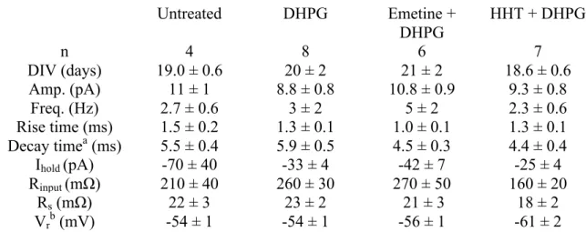
Table 1. Electrophysiological properties in neurons of each treatment group during baseline ... 53
Table 2. Electrophysiological properties in transduced neurons during baseline ... 53
2.9 FIGURES ... 54
2.9.1 Figure 5 ... 54
2.9.2 Figure 6 ... 55
2.10 LEGENDS ... 56
Figure 5. mGluR-LTD in cultured neurons and block by translation elongation inhibitor. ... 56
Figure 6. Knockdown of the RNA binding protein Staufen 2 impairs mGluR-LTD. ... 56
CHAPTER III. GENERAL DISCUSSION ... 57
3.1 REVIEW OF MAIN RESULTS ... 57
3.2 MECHANISMS OF LOCAL TRANSLATION REGULATION ... 57
3.2.1 Evidence for stalled polyribosomes ... 58
3.2.2 Role of Staufen 2 in mGluR-LTD ... 60
3.2.3 FMRP and Staufen 2 – an indispensable partnership ... 61
3.3 FUTURE PERSPECTIVES ... 62
3.3.1 Mechanism of stalling elongation ... 62
3.3.2 Distinct pathways of local translation regulation ... 63
3.3.3 Functional roles of mGluR-LTD ... 64
CHAPTER IV. GENERAL BIBLIOGRAPHY ... 68
LIST OF FIGURES
CHAPTER I 1
FIGURE 1 – BASIC ANATOMY AND CONNECTIVITY OF THE HIPPOCAMPUS 5 FIGURE 2 – SIGNALING PATHWAYS INVOLVED IN NMDAR-‐LTD 16
FIGURE 3 – SIGNALING PATHWAYS INVOLVED IN TRANSLATION REGULATION DURING MGLUR-‐LTD 20
FIGURE 4 – STEPS OF TRANSLATION INITIATION 23
CHAPTER II 30
FIGURE 5 – MGLUR-‐LTD IN CULTURED NEURONS AND BLOCK BY TRANSLATION ELONGATION INHIBITOR 55
FIGURE 6 – KNOCKDOWN OF THE RNA BINDING PROTEIN STAUFEN 2 IMPAIRS MGLUR-‐LTD 56
CHAPTER III 57
FIGURE 7 – MODEL OF TRANSLATION REGULATION IN MGLUR-‐LTD 66
LIST OF TABLES
CHAPTER II 30
TABLE 1 – ELECTROPHYSIOLOGICAL PROPERTIES IN NEURONS OF EACH TREATMENT GROUP DURING BASELINE
53
TABLE 2 – ELECTROPHYSIOLOGICAL PROPERTIES IN TRANSDUCED NEURONS DURING BASELINE 53
LIST OF ABBREVIATIONS
4E-BP Eukaryotic initiation factor 4E-binding protein
AMPAR α-amino-3-hydroxy-5-methyl-4-isoxazolepropionate receptor
AP2 Adaptor protein 2
Arc/Arg3.1 Activity regulated cytoskeletal-associated protein BDNF Brain-derived neurotrophic factor
Btz Barentsz
CA1 Cornu ammonis 1
CA2 Cornu ammonis 2
CA3 Cornu ammonis 3
CaMKII Calcium/calmodulin-dependent protein kinase II CBP80 Nuclear cap-binding protein 80
c-Fos FBJ osteosarcoma oncogene
CPEB1 Cytoplasmic polyadenylation element binding protein-1
CREB Cyclic adenosine monophosphate (cAMP) response element binding protein
DAG Diacylglycerol
DG Dentate gyrus
DHPG Dihydroxyphenylglycine
EC Entorhinal cortex
eEF2K Eukaryotic elongation factor 2 kinase EF1A Elongation factor 1A
eIF1A Eukaryotic initiation factor 1 A eIF2α Eukaryotic initiation factor 2 α eIF3 Eukaryotic initiation factor 3
eIF4F Eukaryotic initiation factor 4F complex eIF4G Eukaryotic initiation factor 4G
EJC Exon junction complex
e-LTP Early-LTP
EPSC Excitatory post-synaptic current ERK Extracellular signal-regulated kinases fEPSP Field excitatory post-synaptic potential FMRP Fragile X mental retardation protein
FXS Fragile X syndrome
GDP Guanosine diphosphate
GFP Green fluorescent protein
GRIP-ABP AMPAR-binding protein–glutamate receptor interacting protein GSK3β Serine/threonine kinase glycogen synthase kinase-3β
GTP Guanosine-5'-triphosphate
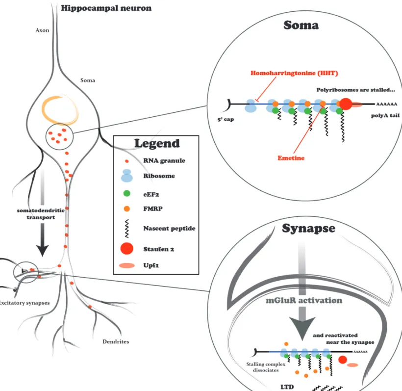
HHT Homoharringtonine
hnRNP Heterogeneous nuclear ribonucleoprotein
HPC Protein hippocalcin
IEG Immediate early gene
IP3 Inositol-1,4,5-trisphosphate
KH K-homology
l-LTP Late-LTP
LTD Long-term depression
LTP Long-term potentiation
MAP1B Microtubule associated protein 1B MAP2 Microtubule-associated protein 2 MAPK Mitogen associated protein kinases
mEPSC Miniature excitatory post-synaptic current Met-tRNAiMet Methionyl initiator transfer RNA
mGluR Metabotropic glutamate receptor
mGluR-LTD Metabotropic glutamate receptor-dependent long-term depression miRNP Micro RNA particles
MNK Mitogen-Activated Protein Kinase-Interacting Kinase MOI Multiplicity of infection
mRNA Messenger ribonucleic acid mTOR Mammalian target of rapamycin
NF-κB Nuclear factor kappa-light-chain-enhancer of activated B cell NSF N-ethylmaleimide-sensitive factor
NO Nitric oxide
NOS Nitric oxide synthase
NMD Non-sense mediated decay
NMDAR-LTD NMDAR-dependent long-term depression NMDAR-LTP NMDAR-dependent long-term potentiation
NMDAR N-methyl-D-aspartate receptor
Ophn1 Oligophrenin 1
P-bodies Processing bodies
PDK1 Phosphoinositide-dependent kinase 1 PERK Protein kinase-like ER kinase
PI3K Phosphoinositide 3-kinase
PIKE Phosphoinositide 3-kinase -enhancer PIC 43S pre-initiation complex
PICK1 Protein interacting with C-kinase 1
PKA Protein kinase A
PKC Protein kinase C
PKMζ Truncated form of protein kinase C, zeta
PLC Phospholipase C
PP1 Protein phosphatase 1
PP2B Protein phosphatase 2B PPF Paired-pulse facilitation
PP-LFS Paired pulse low frequency stimulation
PPR Paired pulse ratio
Pr Probability of transmitter release PRP Plasticity-related protein
PTK Protein tyrosine phosphatase Rap1 Repressor activator protein 1
RNP Ribonucleoprotein particle
RPM Ribopuromycilation
RSK1 Ribosomal S6 kinase-1
SC Schaffer collateral
shRNA Short hairpin RNA
SMD Staufen-mediated decay
SRF Serum response factor
Stau1 Staufen 1
Stau2 Staufen 2
siRNA Small interfering RNA
STEP Striatal-enriched protein tyrosine phosphatase STP Short-term potentiation
Upf1 Helicase up-frameshift 1 ZBP1 Zipcode-binding protein 1 Zif268 Early growth response 1
ACKNOWLEDGMENTS
I am deeply indebted to several people who have helped in bringing the best in me while writing this thesis. To all, I wish to convey my heartfelt gratitude, appreciation and acknowledgment. During the past two incredibly formative years, I have grown not only as a student and young scientist, but also as a more mature individual.
I want to thank both of my supervisors, Jean-Claude Lacaille and Wayne Sossin, for their guidance and advice. I benefited tremendously from their direction, technical support and above all encouragement. I am very appreciative of their many valuable suggestions and comments on this thesis. This work has been such a wonderful learning process for me and they have been major contributors to my development.
I am deeply grateful to have met fantastic colleagues, both in my lab and in the Department, without whom, this journey would not have been the same. I thank them for all their support and friendship. I will remember, most of all, the endless discussions, the laughter and the joy I felt of working surrounded by such remarkable individuals. I have learned from their depth of experience in science, but also in different aspects of life, and I hope to carry that with me as I begin a new adventure.
Finally, I would like to thank my family, especially my parents, for their unconditional support without which I would not be where I am now. Their faith in me has always
empowered me to achieve bigger and greater things, to be ambitious and strive for excellence. I cannot express in words how thankful I am for everything they have given me.
CHAPTER I. INTRODUCTION
1.1HIPPOCAMPAL-DEPENDENT LEARNING AND MEMORY
Although, today, the link between the hippocampus and memory is undisputed, neuroscientists were still searching for a clear hippocampal function until the mid-twentieth century (Andersen et al 2006). This impressive bulging brain structure of the temporal lobe attracted interest from the very start of brain investigations. It was implicated in a variety of functions ranging from olfaction (Brodal 1947) to harboring “the central emotive process of cortical origin” (Papez 1937) until the seminal and detailed report on the amnesic patient H.M. (Scoville & Milner 1957) provided direct evidence for a mnemonic function.
1.1.1 Role of hippocampus in learning and memory
H.M., who died in 2008, had been suffering from intractable epilepsy resistant to antiepileptic drug treatment for many years before he underwent bilateral resections of the medial portions of the temporal lobe. His seizures were successfully relieved, but he was left with a profound memory loss. Following the operation, H.M. was unable to retain any information about recent events or episodes of his life, people he had met, places he had visited or objects he had encountered (anterograde amnesia). His memories dating from some time prior to the intervention were also impaired (retrograde amnesia), although memories from early life appeared to be intact. In contrast, his general intellect, perceptual ability, working memory and some forms of long-term memory were unscathed (Milner et al 1968, Scoville & Milner 1957).
Since then numerous studies in both humans and animals have tried to elucidate how the hippocampus mediates memory processing. Learning and memory are extremely complex concepts involving a dynamic interplay between various brain regions that make different contributions to memory formation (Schacter & Tulving 1994, Squire 2004). To puzzle out the hippocampus’ role amidst the organization of memory systems in the brain is not a simple task (Kandel 2001). What does the hippocampus do? Does it perform tasks that other brain regions cannot accomplish? In which phase of memory processing is the hippocampus involved? The main processes include the encoding of the
information into memory, the consolidation process for stability over time and the retrieval and reactivation of the memory during recall (Morris 2006). To add a layer of difficulty in answering these questions, “memory traces” are encoded at different levels. They can be stored in synaptic weights as specific synapses modify their connection strength by undergoing biochemical modifications (Martin et al 2000), but such changes are embedded within a larger neural ensemble to which the memory has been allocated (Guzowski et al 1999, Hall et al 2001, Hebb 1949, Reijmers et al 2007, Sakaguchi & Hayashi 2012).
Although much is yet to be understood, great strides have been made and some conclusions have been reached about the function of the hippocampus in humans: (1) Amongst the different types of memory (Bruner 1969, Ryle 1949, Winograd 1975), the hippocampus is involved in one particular type termed declarative memory that refers to the recollection of facts and events (Tulving 1983) and can be associative, abstract and context-dependent (Manns & Eichenbaum 2006). Located at the confluence of highly processed multimodal sensory inputs, the hippocampus can bind information from the neocortex to form memories that are representational and model the external world (Squire & Alvarez 1995). (2) Its involvement in the storage and recovery of memories diminishes, or in any case, changes with time as consolidation proceeds and memory traces are reorganized. However, whether the hippocampus is a temporary memory system (Squire 1992, Squire & Alvarez 1995) or is engaged to some extent in the retrieval and storage of certain remote memories is still a subject of debate (Moscovitch et al 2006, Sutherland & Lehmann 2011). (3) The hippocampal region and the adjacent cortex are not involved in working memory in addition to a wide range of implicit or non-declarative tasks (e.g. motor skill memory) (Squire & Zola 1996). (4) The hippocampal region is not involved in non-mnemonic aspects of cognition (Craig 2006).
1.1.2 Animal models of human amnesia
Lastly, it is important to note that although there is great structural similarity and strong evolutionary conservation of circuitry of the hippocampus (in contrast to the diversity of the neocortex), there are some functional differences across the mammalian taxon (Manns & Eichenbaum 2006, Squire et al 2004). While it is unequivocally involved in
the initial acquisition and temporary storage of declarative memory, the rat hippocampus appears to be heavily involved in the formation of cognitive maps and their navigation through space compared to its human counterpart that is most engaged by episodic memory. Such differences could be accounted for by the different kinds of information the animal hippocampus is receiving in comparison to the humans. Just like the hippocampi of the two human hemispheres may be performing slightly different functions due to lateralization even though they are structurally symmetrical (Morris 2006), the rat hippocampus provides an output that is appropriate to the animal, not to humans. As Manns and Eichenbaum (2006) illustrate it ever so well, “it is perhaps not surprising that an animal such as the rat, whose survival normally depends on nighttime foraging, would place a premium of remembering spatial locations of important items. It is also not surprising that the human hippocampus, spoiled by the wonderful elaborative ability of its complex neocortex, would more likely reflect the paradoxes of episodic memory, being abstract yet structured, detailed yet imperfect.”
1.2NEUROANATOMY OF THE HIPPOCAMPAL FORMATION 1.2.1 Anatomical organization
The hippocampal formation is a discernible C-shaped tridimensional structure of the medial temporal lobe found in both hemispheres. In monkeys and humans, it lies horizontally on the floor of the temporal horn of the fourth ventricle. In rats, the structure curves (more vertically oriented than in primates) from the medial aspects, or septal pole (dorsal hippocampal), to the bottom tip of the temporal lobe, or the temporal pole (ventral hippocampus) (Amaral & Lavenex 2006). The course the hippocampal formation follows is termed the septotemporal axis and much of its internal structure remains relatively the same throughout its length. Orthogonal (perpendicular) to this axis is the transverse plane that clearly reveals the cytoarchitectonically distinct adjoining structures that make up the hippocampal formation: the hippocampus proper, dentate gyrus, subicular complex (subiculum, presubiculum, parasubiculum) and entorhinal cortex (Amaral & Lavenex 2006). The hippocampus proper is further subdivided into three subfields that bear the latin name cornu ammonis (CA) for Ammon’s Horn: CA1, CA2 and CA3. The general
term hippocampus is often used to refer to the hippocampus proper and dentate gyrus and will be used as such in this text. Moreover, we will focus on the rodent hippocampus since it is the animal model used in this study.
1.2.2 Cytoarchitectonic organization
The highly ordered organization of cells and terminating projections is one of the hallmarks of the hippocampus that has made it a model of choice for the study of the neurobiology of memory. The hippocampus consists of only one densely packed layer of principal neurons – it is an allocortical1 region unlike the surrounding neocortex – that form two interlocking C’s, reversed relative to each other, one of pyramidal cells of the hippocampus proper, and the other of granule cells of the dentate gyrus. Fibers originating from different cortical regions (including the hippocampus itself) make synaptic contact with distinct dendritic segments of their target principal neuron (Förster et al 2006). Thus, afferent fibers terminate in sharply segregated hippocampal layers to form a laminated network.
The principal neurons and zones of synaptic connections of the hippocampus proper are disposed in superimposed layers (Amaral & Lavenex 2006) (Fig. 1A):
(1) the lacunosum-moleculare layer is the deepest layer containing projections from the entorhinal cortex (perforant path) and extra-hippocampal inputs;
(2) the radiatum layer is the layer in which apical dendrites from the pyramidal cells extend and CA3-CA1 connections occur via the Schaffer collaterals from CA3 as well as associational connections2;
(3) the pyramidal layer contains the cell bodies of pyramidal cells;
(4) the oriens layer contains the basal dendrites of the pyramidal cells that make contact with some of the Schaffer collaterals and associational fibers;
(5) the alveus layer, on the other hand, contains the axons of the pyramidal cell. Much of these intrahippocampal projections travel along the transverse or oblique axis and exhibit a clear intrinsic connectivity (Andersen et al 1971) that we will discuss next.
1 Allocortical: a term applied to cortical regions having fewer than six layers. 2 Associational connections are ipsilateral CA3-CA3 connections.
Figure 1. Basic anatomy and connectivity of the hippocampus
(A) Layers of the hippocampus proper. Alveus layer (a), oriens layer (o), pyramidal cell layer (p), radiatum layer (r), lacunosum-moleculare layer (lm). Figure adapted from Freund and Buzsaki (1996). (B) Diagram of the trisynaptic circuit of the hippocampus. Figure adapted from Neves et al (2008). See text for details.
1.2.3 The trisynaptic circuit
The highly convergent-divergent internal connections of the hippocampus are more complex than the trisynaptic circuit, which was described early on (Ramón y Cajal 1911), might suggest. Although serial elements of the circuit lie within a transverse plane, axonal projections also diverge along the longitudinal axis (Amaral & Witter 1989). Moreover, it is now clear that it is rather a portion of the functional circuitry of the hippocampal formation than its major contributor (Amaral & Lavenex 2006). However, this unidirectional synaptic flow through the three important excitatory synapses depicted below remain of great significance for hippocampal research.
Enthorinal cortex (EC) è Dentate gyrus (DG) DG è CA3 CA3 è CA1
Synapse 1. The entorhinal cortex, via the perirhinal and parahippocampal cortices, receives a host of highly processed multimodal sensory inputs from various neocortical sources (Suzuki & Amaral 1994). This information is then relayed to the granule cells of the dentate gyrus via the perforant path. These entorhinal fibers perforate the subiculum before terminating into the outer molecular layer of the dentate gyrus (Fig. 1B). Of note, the entorhinal cortex also projects, to a lesser extent, to the hippocampus proper via the temporammonic pathway.
Synapse 2. Granule cells give rise to axons called mossy fibers (Fig. 1B) with unusually large boutons that form en passant synapses onto the CA3 pyramidal cells. This innervation stops at the border of CA3 with CA2, which is the main feature distinguishing these two regions. No other hippocampal projections are known to innervate CA3 (apart from CA3-CA3 commissural3 and associational connections).
Synapse 3. The CA1 subfield represents the last stage of this intrahippocampal loop and is densely innervated by CA3 pyramidal axons, the Schaffer collaterals (Fig. 1B). These fibers extend through both the stratum radiatum and stratum oriens layers. CA1 pyramidal cells send axons that travel parallel to the alveus in stratum oriens and mainly project back to the entorhinal cortex and subiculum (Amaral & Lavenex 2006).
Although pyramidal neurons largely outnumber any other cell type in the hippocampus, there is a great diversity of interneurons found in various layers of the hippocampus that play a crucial role in regulating and fine-tuning the activity of the network (Freund & Buzsaki 1996), ultimately modulating its output, thus adding to the complexity of the system.
1.2.4 Field excitatory post-synaptic potentials
A consequence of the laminar organization and connectivity of the hippocampus is the ability to generate large extracellular currents while stimulating Schaffer collaterals with an electrode in transverse slices. Such stimulation of parallel fibers causes the simultaneous synaptic activation of a population of cells and allows the observation of synaptic currents that would normally be too small to be detected in single unit recordings. When the recording electrode is placed parallel to the stimulating electrode, a negative change in potential occurs at the recording electrode relative to the reference electrode (ground) as positive depolarizing currents rush toward the current sink into the dendrites of activated cells. Synaptically generated current flows inside the cells and exit at the current source in the region of the soma where membrane area is greatest. A current loop is created and gives rise to field excitatory post-synaptic potentials (fEPSPs). With stronger stimulation, a population spike superimposed onto the rising phase of the fEPSP is observed and reflects the synchronous discharge of cells. The magnitude of the fEPSP is used as a measure of the efficacy of post-synaptic activation (Andersen et al 2006).
Hippocampal field potential studies were instrumental in further understanding synaptic function.
1.3LONG-TERM SYNAPTIC PLASTICITY
The idea that changes in the strength of the synapse serve as elementary components of memory storage was first brought forward by Ramón y Cajal (1894). However, tangible evidence was only provided years later. The first direct evidence to support the notion that neural circuits are modified by learning came from studies of simple forms of learning in invertebrate systems, including the gill-withdrawal reflex of Aplysia
(reviewed in Mayford et al 2012). During this same period, Bliss and Lomo (1973) found in vivo synaptic responses in the dentate gyrus of the hippocampus to display plasticity in the rabbit following stimulation of the perforant path. Since then, long-term synaptic plasticity, and particularly long-term potentiation (LTP), has been the subject of intense study and found to be present at a large number of excitatory synapses in the brain. Moreover, not only can neurons undergo bidirectional changes, such that synapses can be potentiated or depressed, but they can also express multiple forms of LTP and long-term depression (LTD) that differ in their molecular mechanisms and time domains thus, conferring several computational advantages (Malenka & Bear 2004); hence the widely held belief that modulation of synaptic transmission constitutes the physical substrate of information storage in the brain (Martin et al 2000). Given the wide variety and flavors of synaptic plasticity found in different brain regions, the following section will describe forms of plasticity limited to one of the better characterized synapses in the hippocampus: the Schaffer collateral (SC) synapse, a monosynaptic connection between CA3 and CA1 pyramidal neurons (CA3-CA1).
1.3.1 Synaptic transmission at the Schaffer pathway
Communication at chemical synapses involves the exocytotic release of the content of synaptic vesicles from the presynaptic terminal, diffusion across the synaptic cleft, and binding to postsynaptic receptors. At the SC CA3-CA1 synapse, synaptic transmission is excitatory and glutamate is released onto tiny protrusions called dendritic spines. Glutamate receptors located at this synapse are: α-amino-3-hydroxy-5-methyl-4-isoxazolepropionate receptors (AMPARs), N-methyl-D-aspartate receptors (NMDARs), kainate receptors and metabotropic glutamate receptors (mGluRs).
AMPARs are ligand-gated ionotropic receptors that respond rapidly to neurotransmitters released in the cleft. Their activation leads to large influx of sodium and smaller efflux of potassium, such that the postsynaptic membrane is depolarized and excitatory post-synaptic currents (EPSCs) are generated. AMPARs are composed of four subunits, which can be a homomeric or heteromeric mixture of GluA1 to GluA4 subunits. Most AMPARs contain at least one GluA2 subunit which renders them calcium-impermeable while GluA2-lacking AMPARs are permeable to calcium (Luscher &
Malenka 2012). Hippocampal principal cells mainly express GluA1 and GluA2 (Keinanen et al 1990).
NMDARs are also ligand-gated ionotropic receptors, but they are also voltage-dependent since magnesium blocks them at resting membrane potential and depolarization is needed to drive the divalent cation from the channel (Nowak et al 1984). NMDARs, when opened, are permeable to the monovalent cations sodium and potassium, but, unlike most AMPARs, they have a high permeability to calcium (Jahr & Stevens 1987). They also have slower kinetics, longer open time and higher affinity for glutamate (Dingledine et al 1999). Of note, NMDARs can possess subunits that contain a binding site for glycine and D-serine.
Kainate receptors are also ligand-gated ionotropic receptors that mediate fast excitatory neurotransmission. Although they contribute little to EPSCs, they supplement glutamate transmission by enhancing and extending the postsynaptic depolarization. They can also modulate transmission presynaptically (Pavel et al 2006).
mGluRs are G-protein coupled receptors that, when bound to glutamate, trigger various signaling cascades. There are eight receptor subtypes categorized in three groups based on their pharmacological and functional properties: Group I (mGluR1, mGluR5), Group II (mGluR2, mGluR3) and Group III (mGluR4, mGluR6, mGluR7, mGluR8) (Shigemoto et al 1997). mGluRs are generally located peri-synaptically, thus they are thought to require strong synaptic activation and glutamate spillover for the receptors to be activated. Group I mGluRs have a somatodendritic distribution and their activation leads to increased excitability of the neuron via the modulation of potassium, calcium, and nonselective cations channels as well as increased intracellular calcium postsynaptically. In contrast, group III mGluRs found near the pre-synaptic terminal act to inhibit excitatory transmission at the SC CA3-CA1 synapse (Pavel et al 2006). On the postsynaptic pyramidal neuron, there are high levels of mGluR5 and lower levels of mGluR1, while mGluR7 is abundant presynaptically (Bliss et al 2006).
EPSCs evoked at low-rates are mediated in great part by activation of AMPARs (Davies & Collingridge 1989). NMDARs contribute little to the postsynaptic response during basal synaptic activity but are critical for synaptic plasticity since they require the temporal coincidence of ligand release and depolarization for current to pass through.
1.3.2 NMDA receptor-dependent LTP
NMDAR-dependent LTP (NMDAR-LTP) is the predominant form of synaptic plasticity in the brain (Bliss & Collingridge 1993). As explained above, the properties of NMDA receptors are such that the coincidence of glutamate release and post-synaptic depolarization is required to open the channel and cause maximal post-synaptic influx of Ca2+. Calcium entry is an absolutely necessary trigger for NMDAR-LTP (Lynch et al 1983, Malenka et al 1992). Experimentally, activation of NMDARs is usually achieved by applying a high-frequency tetanic stimulation to the synapses or by directly depolarizing the postsynaptic neuron while applying a low-frequency synaptic stimulation (Citri & Malenka 2008). NMDAR-LTP exhibits a number of basic properties that relate to the properties of NMDARs. First, it is cooperative because a weak input, even if delivered at high frequency, does not induce LTP; a critical number of synapses must therefore be activated to reach threshold intensity. Second, it is associative because activity at one input can influence the ability of another active input to undergo plasticity. In other words, a weak input can be potentiated only if it coincides with a strong input. Finally, NMDAR-LTP is input specific since potentiation only occurs at synapses at which it is induced (Bliss et al 2006, Nicoll et al 1988).
As with other forms of synaptic plasticity, NMDAR-LTP involves phases of induction and expression. Following the appropriate pattern of stimulation of the Schaffer collaterals to activate NMDARs, an initial strong potentiation of the response is observed which decays over a period of 10 minutes to a stable, but persistently increased level compared to baseline when synaptic transmission is probed with a low stimulation rate (every 30 seconds). This initial phase is referred to as short-term potentiation (STP) and involves different mechanisms than those recruited in the long-term phase. The long-term phase, which can last from hours (in vitro) to days (in vivo), is in turn believed to be divided in two phases, early-LTP (e-LTP) and late-LTP (l-LTP), according to their respective sensitivity to protein synthesis inhibitors (Frey et al 1993), although this model has evolved (Reymann & Frey 2007). Different protocols of stimulation can also be used to isolate the early phase from the late phase (Frey & Morris 1997). It is important to keep in mind that LTP is not a unitary phenomenon and involves different molecular mechanisms that overlap in time. The multi-stage model of LTP is widely accepted and
these processes do seem to influence each other, but whether they consist of separate, parallel phases of expression or occur in series is still under investigation (Johnstone & Clark 2011, Park et al 2013, Reymann & Frey 2007).
1.3.2.1 From induction to expression
The expression of LTP can be achieved in several ways. Modifications to either pre- or postsynaptic terminals can lead to an enhanced synaptic transmission. Presynaptically, an increase in release probability would cause more glutamate to be released in the cleft and a larger postsynaptic response would be the consequence. On the other hand, this same observation can be made if there is an increase in the postsynaptic sensitivity to glutamate. This can be accomplished either through the modification of the receptor itself to enhance AMPAR conductance or the insertion of additional AMPARs into the postsynaptic density. The idea of ‘unsilencing synapses’ has also been suggested as a postsynaptic mechanism in which synapses previously lacking AMPARs are converted to functional synapses following LTP induction (Kerchner & Nicoll 2008). Extensive work has been done to clearly determine the locus of expression of NMDAR-LTP at the SC CA3-CA1 synapse (Bliss & Collingridge 2013). Although the controversy seems to have been resolved due to a large body of evidence pointing in the direction of new recruitment of AMPARs to silent synapses or to synapses already possessing some functional AMPARs (Kerchner & Nicoll 2008, Lynch 2004, Nicoll & Roche 2013), the subject is still a matter of debate (Bliss et al 2014, Johnstone & Clark 2011). These mechanisms of expression, however, are not mutually exclusive; the existence of one does not preclude the existence of another, even if their relative contribution is unequal. Moreover, the locus of expression may change overtime as STP progresses into the different stages of LTP (Johnstone & Clark 2011).
The induction of NMDAR-LTP is, by general consent, postsynaptic, but what are the biochemical cascades triggered by NMDAR activation that lead to the expression of LTP? Although there have been many proteins implicated in mediating LTP, it is generally agreed that calcium/calmodulin-dependent protein kinase II (CaMKII) activity is required for induction (Nicoll & Roche 2013). Calcium that entered through NMDARs binds to calmodulin, which then activates CaMKII. During this activation, CaMKII
undergoes autophosphorylation rendering it constitutively active and calcium independent. Calcium activated CaMKII translocates to the postsynaptic density where it can potentiate postsynaptic AMPA receptors in early phases of LTP. CaMKII enhances single channel conductance by phosphorylating specific sites on AMPAR subunits. Evidence also suggests that it is involved in the capture of AMPARs to the post-synaptic density, but its exact method of action remains unclear (Lisman et al 2012). Furthermore, the CaMKII/NMDAR complex is proposed to be a promising candidate for the maintenance of LTP and therefore the persistence of memory (Sanhueza & Lisman 2013).
Several other kinase cascades have also been found to be involved, some of them occurring in parallel. None of them seem to be obligatory since, depending on the conditions, LTP is not affected by inhibitors targeting their activity (Pavel et al 2006). Various factors are potentially at stake, but the induction protocol does appear to hold a determining role, which raises the possibility that there is more than one form of NMDAR-LTP at the SC CA3-CA1 synapse. Protein kinase A (PKA), protein kinase C (PKC), tyrosine kinases, the ERK/MAP kinase pathway (extracellular regulated kinases/mitogen associated protein kinases), phosphoinositide 3-kinase (PI3K), as well as the mammalian target of rapamycin (mTOR) and its downstream effectors, have all been implicated (Lynch 2004, Pavel et al 2006). Nitric oxide (NO) and brain-derived neurotrophic factor (BDNF) are just two substances amongst potential retrograde messengers to mediate the effects on presynaptic function (Regehr et al 2009). In the case of NO, calcium influx activates NO synthase (NOS) causing production of NO and diffusion from the postsynaptic to the presynaptic neuron to initiate presynaptic enhancement (Johnstone & Clark 2011).
1.3.2.2 Maintenance of late phase of LTP
We have seen that a plasticity-inducing event such as NMDAR-LTP activates distinct pathways that lead to post-translational modifications of pre-existing proteins and structural changes within the synapse. Specific alterations in protein synthesis are one of the consequences of the activation of such pathways and this is important for the induction of LTP. Protein synthesis is required for the persistence of synaptic change
during late phase LTP, whereas early phase LTP are unaffected by protein translation inhibitors (Frey et al 1988, Kelleher III et al 2004, Stanton & Sarvey 1984). When key signaling molecules and regulators of translation are disrupted, LTP is found to be abnormal in several of these instances (e.g. Alarcon et al 2004, Banko et al 2005, Costa-Mattioli et al 2005, reviewed in Costa-Costa-Mattioli et al 2009). But these studies do not provide information about whether protein synthesis initially derives in part from pre-existing mRNAs localized near the synapse and is independent of transcription or somatic translation or is general and somatic. In other words, where is protein translation occurring? Dendritic compartments are translationally competent since they contain all the necessary components of the protein synthesis apparatus (Tiedge & Brosius 1996). Moreover, some studies show that late phase LTP is maintained in synapses isolated from the soma by microsurgical cut (Vickers et al 2005) and destabilized microtubule networks (Vickers & Wyllie 2007). CaMKII protein levels are increased after tetanic stimulation (Ouyang et al 1999) and disrupting the dendritic localization of CaMKII mRNA transcript diminishes late-phase LTP (Miller et al 2002). Local application of protein synthesis inhibitors also impaired but did not completely block late phase LTP (Bradshaw et al 2003). While mRNA transcripts such as CaMKII, microtubule-associated protein 2 (MAP2), Shank and β-actin have been visualized in dendrites (Holt & Schuman 2013) and BDNF appears to mediate local protein synthesis that contributes to NMDAR-LTP (Leal et al 2014), it is still not clear how much protein is synthesized in the soma versus dendrites during LTP. Mechanisms of mRNA transport and regulation of local protein synthesis will be discussed in greater detail in section 1.4.
Following the induction of LTP, signaling pathways such as the ERK/MAPK and mTOR cascades upregulate the translation of ‘plasticity-related proteins’ (PRPs) (Costa-Mattioli et al 2009). However, the changes that are occasioned by the coordinated synthesis of PRPs are not inherently stable – there must be mechanisms in place to maintain them for periods that can last up to several hours in vivo (Klann & Sweatt 2008). One protein that has attracted a lot of interest in the last years is an atypical PKC isoform, PKMζ, that becomes, following its rapid synthesis and phosphorylation by phosphoinositide-dependent kinase 1 (PDK1), persistently active. It is thought to play a role in AMPAR trafficking to maintain enhancement of synaptic strength (Sacktor 2012).
Transcription can also become necessary in later phases to maintain the potentiation. A number of studies have shown the activation of specific patterns of gene expression following behavioral training (Guzowski 2002). Activity that induces LTP is linked to the nucleus by the activation of PKA, CaMKIV and MAPK and leads to the phosphorylation of the transcription factor CREB [cyclic adenosine monophosphate (cAMP) response element binding protein], which plays a central role in gating activity-regulated gene expression and the immediate early genes (IEGs) c-Fos (FBJ osteosarcoma oncogene) and Zif268 (early growth response 1). Arc/Arg3.1 (activity regulated cytoskeletal-associated protein) production, whilst it can depend on CREB activity, is also modulated by the transcription factors MEF2, SRF/Elk1 (serum response factor) and Zeste-like factor (Bramham et al 2010). IEGs subserve various cellular functions that are compatible with the structural and functional modifications that underlie synaptic plasticity (Abraham & Williams 2003, Guzowski 2002, Lynch 2004), thus they are believed to play a critical role in memory formation.
1.3.3 NMDA receptor-dependent LTD
Potentiation or depression of synaptic transmission can occur as a consequence of NMDAR activation, so what determines the direction of change? The degree and timing of calcium entry appears to be the determining factor in recruiting the intracellular molecules for the appropriate change in polarity (Artola & Singer 1993, Bliss et al 2006, Franks & Sejnowski 2002, Lisman 1989). In contrast to the induction of LTP, NMDAR-dependent LTD (NMDAR-LTD) is optimally induced by the repeated activation of the presynaptic neuron at low frequencies (Dudek & Bear 1992, Mulkey & Malenka 1992) to allow for a modest postsynaptic calcium entry (Malenka 1994). This suboptimal increase in calcium establishes a requirement for intracellular calcium store release, unlike during LTP induction (Nakano et al 2004).
1.3.3.1 From induction to expression
Mechanisms of expression in NMDAR-LTD appear to be largely postsynaptic with the removal of AMPARs from the synapse (Collingridge et al 2004), although there is
evidence that presynaptic alterations do occur and depend on several factors, notably the developmental stage (Collingridge et al 2010).
If LTP involves protein kinases, NMDAR-LTD activates protein phosphatases. The rise in postsynaptic calcium triggers the serine-threonine protein phosphatase cascade; calcium entering through NMDARs binds to calmodulin to activate calcineurin (protein phosphatase 2B, PP2B), which leads to the activation of protein phosphatase 1 (PP1) via dephosphorylation of inhibitor-1. PP1 acts to dephosphorylate several targets such as serine sites on AMPAR subunits. However, CaMKII, a protein kinase critical for potentiation of the synapse, is also a downstream target of calmodulin. It has been proposed that the different calcium dynamics are critical in determining which signaling pathway is activated (Lisman 1989).
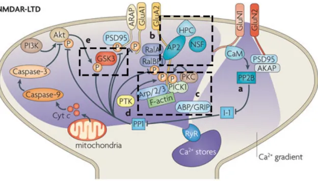
NMDAR-LTD involves regulation of AMPAR subunit cycling between the synaptic membrane and the cytoplasm. Upon NMDAR-LTD induction, the N-ethylmaleimide-sensitive factor (NSF) dissociates from GluA2-containing AMPARs and there is increased binding of the protein interacting with C-kinase 1 (PICK1) to the GluA2 subunit. This PICK1-GluA2 interaction is required for hippocampal NMDAR-LTD (reviewed in Anggono & Huganir 2012). The neuronal calcium sensor protein hippocalcin (HPC) is a high-affinity Ca2+ sensor that promotes the exchange of NSF with the adaptor protein 2 (AP2) to destabilize AMPARs and initiate clathrin-mediated endocytosis of AMPARs. Another mechanism involves the scaffolding proteins AMPAR-binding protein–glutamate receptor interacting protein (GRIP-ABP). Like NSF, GRIP-ABP dissociates from AMPARs when PICK1 is activated (PICK1 changes conformation when bound to GluA2). NMDAR-LTD is also associated with phosphorylation by protein tyrosine kinases of tyr876 of GluA2, which may also aid the exchange of PICK1 for ABP–GRIP, although this is more associated with PKC phosphorylation of GluA2. Another target of PP1 is the multifunctional serine/threonine kinase glycogen synthase kinase-3β (GSK3β) and is required for NMDAR-LTD. Dephosphorylation of a serine residue leads to further activation of GSK3β (reviewed in Anggono & Huganir 2012, Bliss et al 2006, Collingridge et al 2010, Kemp & Bashir 2001) (Fig. 2).
1.3.3.2 Maintenance of late phase NMDAR-LTD
NMDAR-LTD is also accompanied by a protein synthesis-dependent phase that becomes evident 4 hours after low frequency stimulation in freely moving rats (Manahan-Vaughan et al 2000). Protein synthesis dependence was also found in organotypic slices as translation inhibitors caused a rapid recovery to baseline levels of transmission after induction of LTD (Kauderer & Kandel 2000). However, the same translation inhibitor did not affect NMDAR-LTD in acute slices (Huber et al 2000), but the plasticity was only monitored 60 minutes after induction. In contrast, a study in acute slices by Sajikumar and Frey (2003) shows a protein synthesis-dependent phases appearing 3-4 hours after induction, thus NMDAR-LTD does appear to involve protein synthesis for the maintenance of its late phase.
Figure 2. Signaling pathways involved in NMDAR-LTD.
Calcium enters through NMDA receptors (consisting of GluN1 and GluN2 subunits) causing the upregulation of PP1 and the release of calcium from intracellular stores. AMPA receptor (GluA1 and GluA2 subunits) anchoring is destabilized and receptors are internalized. Figure adapted from Collingridge et al (2010).
1.3.4 mGlu receptor-dependent LTD
At least two different forms of LTD exist at the SC CA3-CA1 synapse. Depression can be elicited by the sole activation of metabotropic glutamate receptors (mGluRs) (Huber et al 2001, Palmer et al 1997), in contrast to the forms of plasticity we have discussed previously. NMDAR-LTD and mGluR-LTD are believe to be mechanistically independent since they do not occlude each other (Oliet et al 1997, Palmer et al 1997); in other words, when mGluR-LTD is saturated, NMDAR-LTD can achieve further depression. Interestingly, while evidence suggests that NMDAR-LTD reverses and erases LTP (also called depotentiation), mGluR-LTD is instead superimposed on LTP (Oliet et al 1997). Moreover, NMDAR-LTD appears to be more prominent in neonatal and juvenile rats, but a developmental shift favors mGluR-LTD in adults (Kemp et al 2000). mGluR-LTD can be induced synaptically or chemically. The application of a paired pulse low frequency stimulation (PP-LFS) to the presynaptic neuron generates an LTD that was found to be dependent upon the activation of group I mGluRs (mGluR1 and mGluR5) (Huber et al 2000). Chemical induction is often used in in vitro preparations. The group I mGluR agonist, dihydroxyphenylglycine (DHPG), is thought to elicit an LTD that relies preferentially on mGluR5 (Faas et al 2002). Although both induction protocols are thought to involve similar mechanisms because they occlude each other (Huber et al 2001), some differences have been found (Gladding et al 2009b). While DHPG induced-LTD is completely calcium independent (Fitzjohn et al 2001), synaptically induced-induced-LTD at the SC CA3-CA1 synapse is sensitive to intracellular calcium chelators (Bolshakov & Siegelbaum 1994, Oliet et al 1997). In these two studies, the calcium influx was identified to occur via T-type and L-type voltage gate calcium channels. However, a more recent study provided evidence that calcium is not an absolute requirement for synaptic induction of mGluR-LTD (small adjustments were made to the conventional PP-LFS protocol) (Kasten et al 2012).
1.3.4.1 From induction to expression
Whether the locus of expression is both presynaptic and postsynaptic or uniquely postsynaptic in mGluR-LTD is not clear, but it is evident that internalization of surface ionotropic GluRs occurs in response to mGluR activation (Gladding et al 2009a, Huang
et al 2004, Snyder et al 2001, Waung et al 2008, Xiao et al 2001, Zhang et al 2008). There is however conflicting evidence showing a lack of decreased postsynaptic sensitivity to uncaged glutamate (Rammes et al 2003) and enhanced responses to ionophoretic application of AMPA, kainic acid and NMDA (Tan et al 2003) following DHPG application. A presynaptic mechanism of expression is supported by findings indicating lasting increase in paired-pulse ratio, a reduction in miniature excitatory postsynaptic currents (mEPSCs) frequency and a decrease in neurotransmitter release following induction (Gladding et al 2009b).
Signaling pathways mediating mGluR-LTD are clearly distinct from those implicated in NMDAR-LTD. mGluRs function as G-protein-coupled receptors; receptor activity leads to G-protein activation promoting the exchange of GTP to GDP (guanosine-5'-triphosphate to guanosine diphosphate), which results in the modulation of protein-protein interactions and activation of second messenger cascades. Group I mGluR activation, is normally coupled to the activation of phospholipase C (PLC), the generation of diacylglycerol (DAG), inositol-1,4,5-trisphosphate (IP3) and the release of calcium
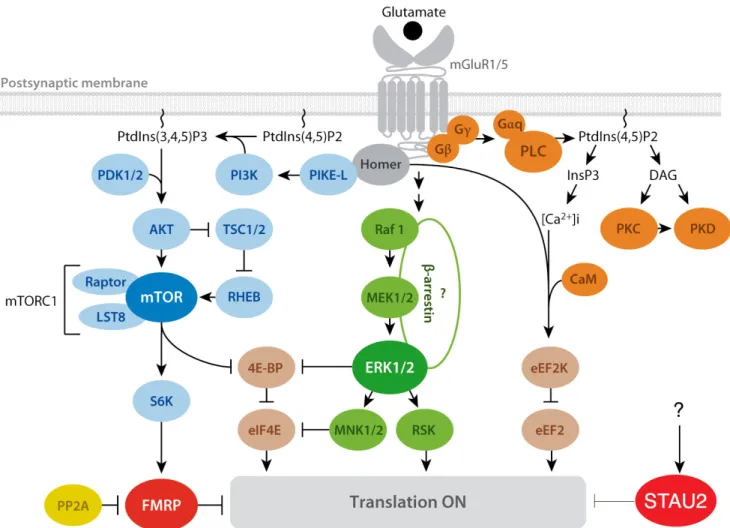
from intracellular stores, but, although this pathway is upregulated during mGluR-LTD (Mao et al 2005), it is not necessary (Fitzjohn et al 2001, Gallagher et al 2004, Schnabel et al 1999). The mitogen-activated protein kinase (MAPK) signaling cascades are activated as all three MAPK subclasses are involved: p38 MAPK, Jun N-terminal kinase (JNK), and ERK (Gallagher et al 2004, Moult et al 2008, Schmit et al 2013). MAPK cascades typically involve the sequential activation of a small GTPase (Ras), a MAPK kinase kinase (Raf), and a MAPK kinase (MEK). P38 MAPK is activated via the GTPase Rap1 (repressor activator protein 1), a pathway that is coupled to the endocytotic machineries and AMPAR internalization. ERK is also activated in this way and leads to the downstream activation of ribosomal S6 kinase-1 (RSK1), a key regulator of activity-dependent protein synthesis. Another important cascade coupled to translation regulation is the phosphoinositide 3-kinase-Akt-mammalian target of the rapamycin (PI3K-Akt-mTOR) pathway thought to mediate cap-dependent translation during mGluR-LTD in parallel with the MEK-ERK pathway. The mTOR pathway is activated through the coupling of mGluR5 with the postsynaptic-density scaffolding protein Homer that recruits the small GTPase that binds PI3K, PI3K-enhancer (PIKE), forming an
mGluR-Homer-PIKE complex. The formation of this complex turns on PI3K (Ronesi & Huber 2008) (Fig. 3). Protein tyrosine phosphatases (PTPs) are also implicated (reviewed in Anwyl 2006, Bliss et al 2006, Collingridge et al 2010, Gladding et al 2009b). An additional pathway activated by group I mGluR signaling alters the activity of the elongation factor 2 kinase (eEF2K) (Park et al 2008, Taha et al 2013, Verpelli et al 2010). Increased phosphorylation of eEF2 by eEF2K causes the arrest of general translation, whilst protein synthesis of specific transcripts such as Arc/Arg3.1 and the microtubule associated protein 1B (MAP1B) is increased (Davidkova & Carroll 2007, Park et al 2008).
1.3.4.2 Maintenance of late phase mGluR-LTD
mGluR-LTD exhibits a dependence on local protein synthesis within the first 10 minutes after induction (Huber et al 2000, Huber et al 2001, Park et al 2008, Waung et al 2008, Zhang et al 2008), unlike NMDAR-LTP/D. mGluR activation mediates the translation of “LTD proteins” such as striatal-enriched protein tyrosine phosphatase (STEP) (Zhang et al 2008), MAP1B (Davidkova & Carroll 2007), oligophrenin 1 (Ophn1) (Di Prisco et al 2014) and Arc/Arg3.1 (Park et al 2008, Waung et al 2008), which are involved in AMPAR endocytosis in some way. Other proteins synthesized during mGluR-LTD are the fragile X mental retardation protein (FMRP) (Weiler et al 1997), the ribosomal protein S6 and the elongation factor 1A (EF1A) (Antion et al 2008), which are proteins that themselves regulate translation.
mGluR-LTD is generally thought to involve translation rather than transcription, but there is evidence of the requirement for transcription factors 2 or 3 hours after induction. The modulation of factors such as NF-κB (nuclear factor kappa-light-chain-enhancer of activated B cells), ETS domain-containing protein and CREB is dependent on the ERK and PI3K pathways (Gladding et al 2009b).
Interestingly, when mGluR antagonists are applied after the induction, LTD is reversed, but is reestablished without any further induction when the drugs are washed out (Palmer et al 1997). It is therefore tempting to suggest that mGluR activation must be maintained for mGluR-LTD to persist.
1.4LOCAL ACTIVITY-DEPENDENT PROTEIN SYNTHESIS
A key mechanism for the persistence of synaptic changes at individual synapses is the requirement for local protein synthesis (Costa-Mattioli et al 2009, Pfeiffer & Huber 2006). Just as stress and growth signals can change the rate of synthesis of specific transcripts and bulk mRNAs, neurotransmission can control translation by regulating its machinery (Gal-Ben-Ari et al 2012).
Figure 3. Signaling pathways involved in translation regulation during mGluR-LTD.
PLC/calcium-calmodulin pathway (orange), the mTOR pathway (blue), and the ERK pathway (green). FMRP and Stau2 are both known RNA-binding proteins that normally repress translation, but upon mGluR activation, promote de-repression. Question marks indicate undetermined associations. Arrows indicate a positive consequence on downstream components; perpendicular lines indicate an inhibitory consequence.
Abbreviations: [Ca2+]i, calcium release from intracellular stores; CaM, calmodulin; ERK,
extracellular signal–regulated kinase; FMRP, fragile X mental retardation protein; Gαq, Gβ, Gγ, heterotrimeric G proteins; InsP3, inositol-1,4,5-triphosphate; mGluR, metabotropic glutamate receptor; mTOR, mammalian target of rapamycin; PtdIns, phosphoinositides; PLC, phospholipase C; PP2A, protein phosphatase 2A; Raptor, regulatory-associated protein of mTOR. Figure adapted from Bhakar et al 2012.
1.4.1 Translation regulation
Translation occurs in three steps: initiation, elongation and termination. Synthesis of most proteins is driven by cap-dependent translation; in other words, translation initiation is most often the rate-limiting step and target for regulation (Costa-Mattioli et al 2009).
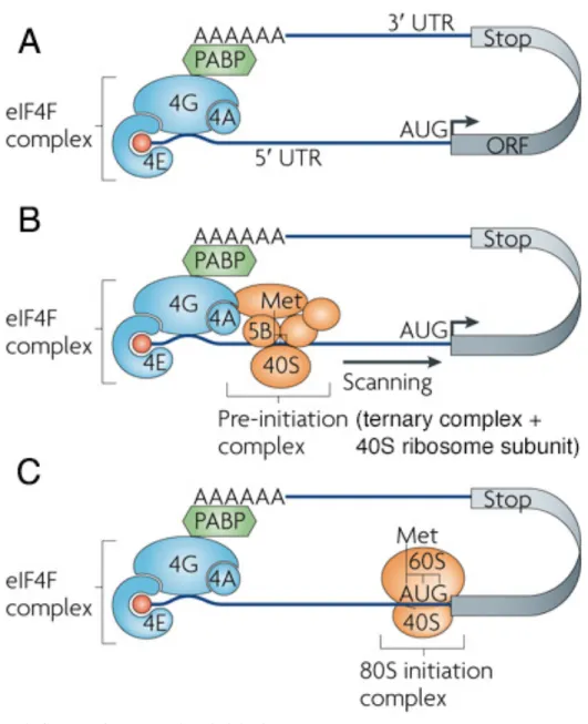
For initiation, the ribosome must be recruited to the mRNA. This is achieved through the interaction of the eukaryotic initiation factor (eIF) 4F complex with the 5’-m7G-cap (Fig. 4A), which then recruits the 43S pre-initiation complex (PIC). PIC is comprised of the 40S ribosomal subunit, eIF1, eIF1A, eIF3 and ternary complex (Jackson et al 2010). The ternary complex, for its part, brings together eIF2, GTP and the specific initiator methionyl initiator transfer RNA (Met-tRNAiMet) to form (eIF2)–GTP–Met-tRNAiMet (Sonenberg & Hinnebusch 2009). PIC begins scanning the 5’-UTR for the AUG start codon (Fig. 4B). eIFs are released and the 60S subunit joins the 40S subunit to form the 80S complex (Fig. 4C) (Sonenberg & Hinnebusch 2009). Elongation factors are recruited to regulate elongation. Upon recognition of the stop codon, termination factors promote the release of the polypeptide chain from the mRNA and ribosome.
Inhibition of the formation of the ternary complex and the eIF4F complex are the two major ways of regulating translation initiation (Gkogkas et al 2010). Phosphorylation of eIF2α, a component of the ternary complex, prevents the functional reconstitution of the complex. On the other hand, eIF4F complex assembly requires the interaction of the cap-binding protein eIF4E with eIF4G. This cap-binding interaction is disrupted and translation initiation is inhibited in the presence of the eIF4E-binding protein (4E-BP). Phosphorylation of 4E-BP by mTOR relieves inhibition. eIF4E can itself be phosphorylated by MNKs, a small family of protein kinases, some of which are regulated by MAPK signaling (Shveygert et al 2010).
The control of the level of peptide chain elongation, on the other hand, is mediated by the eukaryotic elongation factor 2 (eEF2). eEF2 is a GTP-binding protein that mediates the translocation of peptidyl-tRNAs from the A-site to the P-site on the ribosome as amino acids are added to the peptide chain. Phosphorylation of eEF2 inhibits eEF2–ribosome binding and arrests elongation. While general translation is slowed, eEF2 phosphorylation causes, by a mechanism that is unclear, the increased synthesis of specific transcripts (Bramham & Wells 2007).
Remarkably, these pathways that regulate the translation machinery have been shown to be upregulated by both NMDA and mGluR signaling (Gal-Ben-Ari et al 2012). Do these signaling pathways mediate control of general translation or a subset of mRNAS (Costa-Mattioli et al 2009)? Considerable evidence suggests that the coordinated translation of selective subsets of mRNAs, or “regulons” (Keene 2007), occurs in response to different patterns of synaptic transmission inducing plasticity (Costa-Mattioli et al 2009). So how is the concerted synthesis of specific plasticity-induced proteins achieved? One attractive mechanism involves RNA-binding proteins.
Figure 4. Steps of translation initiation
Figure adapted from Besse and Ephrussi (2008).
1.4.2 mRNA localization
For the local and rapid translation of specific proteins to occur on demand, mRNAs must first be transported to their final site of function. These subsets of transcripts are packaged into transport complexes called RNA granules. These higher-order assemblies contain important components of the translation machinery as well as RNA-binding proteins and their mRNA partners (Fritzsche et al 2013b, Sossin & DesGroseillers 2006) that render mRNAs translationally dormant (Besse & Ephrussi 2008). Synaptic activation
provokes their unmasking and release from their repressed state (Buxbaum et al 2014, Graber et al 2013). This dual function of mRNA localization and translation regulation confers several advantages (Martin & Ephrussi 2009). In addition to the ability to alter the synaptic input to one of its many dendrites without changing others, the neuron is provided with the opportunity to achieve this spatially restricted gene expression with a high temporal resolution.
RNA-binding proteins selectively bind transcripts for transport, hence their consideration as key candidates for differential regulation of plasticity-induced protein translation. Indeed, evidence suggests that different dendritic trafficking pathways exist which could allow for independent localization and distinct regulation of mRNAs involved in different forms of synaptic plasticity (Doyle & Kiebler 2011, Lebeau et al 2011, Mikl et al 2011).
1.4.3 RNA-binding proteins
Information about the destination of mRNA transcripts is encoded by cis-acting elements in the RNA most frequently found in the 3’ UTR, which are recognized by trans-acting RNA-binding proteins (RBPs) (Besse & Ephrussi 2008). RNA-binding proteins are not only incorporated into RNA granules but are also components of a variety of cytoplasmic RNA structures: translating polysomes, processing bodies (P bodies), stress granules, micro RNA particles (miRNPs) or the RNA interfering silencing complex, and RNA transport particles. Transport particles contain, similarly to RNA granules, mRNAs, RNA-binding proteins, adaptors that couple to the motor complex and motors, but they are devoid of ribosomes (Sossin & DesGroseillers 2006). Emerging evidence is suggesting that the composition and the pool of RNA granules are more heterogeneous then we previously thought (Bramham & Wells 2007, Fritzsche et al 2013b, Graber et al 2013). Here, we describe a few RBPs that have been identified to play an important role in localization and translation regulation of mRNA transcripts during synaptic plasticity.
1.4.1.1 CPEB1
Cytoplasmic polyadenylation element (CPE) binding protein-1 (CPEB1) is found in the postsynaptic density and controls mRNA translation by regulating the length of the
poly(A) tail. It forms a dual activity complex through its association with various factors that can both activate and repress translation of its target mRNAs by adding or removing the poly(A) tail. CPEB1 at the 3’UTR anchors a complex of proteins that includes an eIF4F binding protein. When bound together, CPEB1 competes with eIF4G for eIF4E binding, thus represses initiation. CPEB1 phosphorylation induces polyadenation and translation of CaMKIIα through NMDA receptor signaling. Deficits in LTP and LTD have been observed in CPEB1 KO mice (Richter 2007).
1.4.1.2 ZBP1
The mRNA of β-actin contains a localization element in the 3’UTR called zipcode that is specifically recognized by zipcode-binding protein 1 (ZBP1) and is involved in its localization (Tiruchinapalli et al 2003) and translation repression (Hüttelmaier et al 2005). Abolition of the function of the zipcode by mutation of the element itself, treatment with specific antisense oligonucleotides, or knockdown/out of ZBP1 protein leads to the mislocalization of β-actin mRNA and subsequent alterations of cell morphology, motility, and adhesion as well as failures in synaptic growth and deficiencies in dendritic spine number, maturation, and arborization (Eliscovich et al 2013). Buxbaum et al (2014), by using single-molecule in situ hybridization approaches, showed that β-actin transcripts are in a masked state, unavailable to probes for binding, but upon depolarization, become unmasked. ZBP1 has also been found to bind and regulate at least 116 other mRNAs (Eliscovich et al 2013).
1.4.1.3 FMRP
The fragile X mental retardation protein (FMRP) has generated a lot of interest due to its pathophysiology. Fragile X syndrome results from the complete absence of this protein due to transcriptional silencing of the gene FMR1. Loss of FMRP is caused by a trinucleotide (CGG) repeat expansion that leads to hypermethylation and transcriptional silencing. It is a protein that is normally highly expressed in neurons, thus its absence causes moderate to severe intellectual disability and autistic features. FMRP is also expressed in other tissues of the body causing a wide spectrum of abnormalities.