Investigation of Femtosecond Laser Technology for the
Fabrication of Drug Nanocrystals in Suspension
par Sukhdeep Kenth
Sciences pharmaceutiques Faculté de pharmacie
Mémoire présenté à la Faculté des études supérieures en vue de l’obtention du grade de Maitre en sciences (M.Sc.)
en sciences pharmaceutiques option technologie pharmaceutique
Décembre 2009
Faculté des études supérieures et postdoctorales
Ce mémoire intitulé:
Investigation of Femtosecond Laser Technology for the Fabrication of Drug Nanocrystals in Suspension
Présenté par : Sukhdeep Kenth
a été évaluée par un jury composé des personnes suivantes :
Dr Grégoire Leclair, président-rapporteur Dr Jean-Christophe Leroux, directeur de recherche
Dr Michel Meunier, co-directeur Dr Sophie Dorothée-Clas, membre du jury
Résumé
La technique du laser femtoseconde (fs) a été précédemment utilisée pour la production de nanoparticules d'or dans un environnement aqueux biologiquement compatible. Au cours de ce travail de maîtrise, cette méthode a été investiguée en vue d'une application pour la fabrication de nanocristaux de médicament en utilisant le paclitaxel comme modèle. Deux procédés distincts de cette technologie à savoir l'ablation et la fragmentation ont été étudiés. L'influence de la puissance du laser, de point de focalisation, et de la durée du traitement sur la distribution de taille des particules obtenues ainsi que leur intégrité chimique a été évaluée. Les paramètres ont ainsi été optimisés pour la fabrication des nanoparticules. L’évaluation morphologique et chimique a été réalisée par microscopie électronique et spectroscopie infrarouge respectivement. L'état cristallin des nanoparticules de paclitaxel a été caractérisé par calorimétrie differentielle et diffraction des rayons X. L'optimisation du procédé de production de nanoparticules par laser fs a permis d'obtenir des nanocristaux de taille moyenne (400 nm, polydispersité ≤ 0,3). Cependant une dégradation non négligeable a été observée. La cristallinité du médicament a été maintenue durant la procédure de réduction de taille, mais le
paclitaxel anhydre a été transformé en une forme hydratée. Les résultats de cette étude suggèrent que le laser fs peut générer des nanocristaux de
principe actif. Cependant cette technique peut se révéler problématique pour des médicaments sensibles à la dégradation. Grâce à sa facilité d'utilisation et la possibilité de travailler avec des quantités restreintes de produit, le laser fs pourrait représenter une alternative valable pour la production de nanoparticules de médicaments peu solubles lors des phases initiales de développement préclinique.
Abstract
Femtosecond (fs) laser ablation and fragmentation, a novel technique based upon the breakdown of material using laser energy was previously used for the production of fine gold nanoparticles in suspension. This technique has been newly investigated for the fabrication of paclitaxel nanocrystals in aqueous solution. In this work, we report the fabrication and characterization of paclitaxel nanocrystals generated by fs laser technology. Two distinct methods of this technology have been explored: ablation and fragmentation. The influence of the laser power, focusing position and treatment time on the particle size, size distribution and chemical integrity of the drug has been studied. Morphology and chemical composition of the finest paclitaxel nanocrystal formulation was studied by scanning electron microscopy and Fourier-transform infrared spectroscopy respectively. Differential scanning calorimetry and X-ray diffraction analyses were employed to evaluate the polymorphic state of the paclitaxel nanocrystals.
Optimal laser fabrication parameters have been established for the fabrication of uniformly small sized paclitaxel nanocrystals. Those optimal conditions generated finely-sized paclitaxel nanoparticles (400 nm, PDI ≤ 0.3) with a considerable degradation. The drug remained crystalline upon nanonization at high power, though the anhydrous crystals were converted to a partially hydrated form.
These findings suggest that drug nanocrystals could be produced using the fs laser technology; however, this technique may be inappropriate for drugs sensitive to degradation. Moreover, the simple fabrication of drug nanocrystals using the fs laser fragmentation presents a great asset for the intial phases of preclinical development of many poorly soluble drug candidates, which are not as sensitive as paclitaxel.
Table of Contents
RESUMÉ……….……. i
ABSTRACT……….... ii
LIST OF TABLES ………...…. vi
LIST OF FIGURES………... vii
ABBREVIATIONS……… x
ACKNOWLEDGEMENTS……….. xiii
CHAPTER 1 : STRATEGIES TO ENHANCE DRUG SOLUBILITY…….... 1
1.1. Introduction ... 2
1. 2. Salt formation ... 6
1.3. Complexation ... 7
1.4. Prodrugs ... 10
1.5. Cosolvents ... 13
1.6. Surfactants and Micelles ... 14
1.7. Emulsions, Microemulsions and Self-emulsifying delivery systems ... 16
1.8. Liposomes ... 17
1.9. Polymeric Solid dispersions ... 20
1.10 Size Reduction ... 21
1.11 References ... 24
CHAPTER 2: NANOCRYSTALS AND NANOSUSPENSIONS ... 28
2.1. Introduction ... 29
2.2. Methods of nanocrystal fabrication ... 31
2.2.1 Bottom-up strategies ... 31 2.2.2 Top-down strategies ... 33 2.3. Characterization of Nanocrystals ... 38 2.4. Administration routes ... 40 2.4.1 Oral ... 40 2.4.2 Parenteral ... 41
2.4.3 Ocular ... 41
2.4.4 Pulmonary ... 42
2.5 Marketed Nanocrystalline drugs ... 43
2.6 References ... 45
CHAPTER 3: LASER FABRICATION OF NANOCRYSTALS IN LIQUID ... 48
3.1 Introduction ... 49
3.2 Fabrication of inorganic nanosuspensions ... 52
3.3 Fabrication of organic nanosuspensions ... 56
3.4. Fabrication of drug nanosuspensions ... 57
3.5 Research proposal and Objectives ... 59
3.6 References ... 63
CHAPTER 4: PRESENTATION OF ARTICLE ... 67
4.1 Absract ... 68
4.2 Introduction ... 69
4.3. Experimental ... 72
4.3.1 Materials ... 72
4.3.2 Methods ... 72
4.3.2.1 Preparation of paclitaxel tablet and suspension ...72
4.3.2.2 Fs-laser treatment ... 72
4.3.2.3 Paclitaxel Assay... 73
4.3.2.4 Particle size and zeta potential measurements ... 74
4.3.2.5 Scanning electron microscopy... 74
4.3.2.6 Thermal Analyses... 75
4.3.2.7 Spectroscopic Analyses... 75
4.4. Results and Discussion ... 77
4.5. Conclusion ... 82
4.6. Acknowlegements ... 83
CHAPTER 5: DISCUSSION ... 100
5.1 Optimization of laser ablation procedure ... 102
5.2. Optimization of fragmentation (following ablation) ... 106
5.3. Optimization of single-step fragmentation strategy ... 107
5.4. Optimization of fragmentation in larger volume ... 109
5.5 Characterization of paclitaxel nanocrystals ... 110
5.6 References ... 112
CHAPTER 6: CONCLUSION ... 115
References ... 118
APPENDIX I: Mononuclear phagocyte system... i
APPENDIX II: Degradation products of paclitaxel... ii
List of tables
Table 1.1: Approved and marketed drug–cyclodextrin complexes in various world
markets ... 9
Table 2.1: Summary of nanocrystalline products currently on the market ………...43
Table 4.1: Degradation of the paclitaxel colloidal suspensions generated by ablation at three
focusing conditions. Mean ± SD (n=3) ... 91
Table 4.2 : Chemical degradation, size distribution and surface charge of paclitaxel
suspensions prepared by two-step fs-laser ablation and fragmentation. Mean ± SD (n=3)……….. 92
Table 4.3 : Chemical degradation and size distribution of paclitaxel particles obtained
by fragmentation (60 min) Mean ± SD (n=3)………...…… 93
Table 4.S1 : XRD peaks (angle 2θ) comparison of anhydrous paclitaxel, water-exposed paclitaxel, laser fragmented nanocrystals and dihydrate paclitaxel
...95
Table 5.1: Degradation and size analysis of paclitaxel particles by ablation and
fragmentation in two different surfactants at various concentrations n=1 ... 106
Table 5.2: Degradation and size analysis of fragmented paclitaxel suspension,
n=1... 107
List of figures
Figure 1.1: Drug administration and transport in the body………. . 3
Figure 1.2: The Biopharmaceutics Classification System (BCS)……….. 4
Figure 1.3: Chemical structure of a cyclodextrin molecule……….. 7
Figure 1.4: Complexation of drug and cyclodextrin………..… 8
Figure 1.5: An illustration of the prodrug concept………. 10
Figure 1.6: Paclitaxel prodrug strategies ……… 11
Figure 1.7: Representation of micelle formation……….……… 15
Figure 1.8: Structure of a liposome………..…… 17
Figure 1.9: Classification of liposomes……… 19
Figure. 1.10. Dissolution velocity dc/dt and saturation solubility Cs as a function of the size of drug powders ranging from coarse to nanonized drugs ………... 22
Figure 2.1: Comparison of plasma concentrations of active ingredient of EMEND® following oral administration in Beagle dogs of a conventional suspension and a NanoCrystal® dispersion formulation of MK-0869... 29
Figure 2.2: schematic representation of the media milling process... 34
Figure 2.3: High pressure homogenization technique: (right) forcing of drug suspension through narrow valve, (left) fabrication of drug nanocrystals due to generated forces... 36
Figure 3.1: (left) Typical experimental set-up for laser ablation at solid-liquid interface
and (right) laser fragmentation; irradiation of of suspension by laser ………... 50
Figure 3.2. (A) Illustration of the production of drug nanoparticles by femtosecond
laser ablation; B) Photography of a paclitaxel tablet immersed in a poloxamer 188 aqueous solution and irradiated by a femtosecond laser ………. 60
Figure 3.3. (A) Size distribution of drug nanoparticles produced by femtosecond laser
ablation before (■) and after (●) the second laser treatment step; (B) illustration of the fragmentation process during the second laser treatment step…………..………….... 60
Figure 4.1: Representation of fs-laser ablation (top) and fragmentation (bottom)
methods. ... 84
Figure 4.2: Chemical Structure of paclitaxel
(5β,20-epoxy-1,2α,4,7β,13α-hexahydroxytax-11-en-9-one 4,10- diacetate 2-benzoate 13-ester with (2R,3S)-N-benzoyl-3-phenyllisoserine) ………. 85
Figure 4.3 (top). Paclitaxel concentration of the suspensions prepared by fs-laser
ablation at focusing positions of z = 0 mm (squares), 1 (circles) and 1.5 (triangles), at power ranging from 25 to 400 mW. Mean ± SD (n=3). (bottom) Paclitaxel concentration of the colloidal suspensions prepared by fs-laser ablation at focusing positions z = 1, 150 mW, and time ranging from 10 to 60 min. Mean ± SD (n=3)………... 86
Figure 4.4: Scanning electron micrographs of (right) water-exposed non fragmented
paclitaxel and (left) laser fragmented paclitaxel nanocrystals (400 mW, 60 min). ………..… 87
Figure 4.5 :FTIR spectra of anhydrous paclitaxel (d), water exposed non-fragmented
paclitaxel (c), laser fragmented nanocrystals (b) and dihydrate paclitaxel
(a)………...…………88
Figure 4.6 : DSC thermograph of anhydrous paclitaxel (a), water exposed
non-fragmented paclitaxel b) laser non-fragmented (400 mW, 60 min) nanocrystals (c) and dihydrate paclitaxel (d). ………89
Figure 4.7: XRD analysis of anhydrous paclitaxel (d), water exposed non-fragmented
paclitaxel (c), laser fragmented (400 mW, 60 min) paclitaxel nanocrystals (b) and dihydrate paclitaxel (a). ………..……. 90
Figure 4.S1: TGA thermograph of paclitaxel anhydrous powder (a), water exposed
non-fragmented paclitaxel (b) laser fragmented (400 mW, 60 min) nanocrystals (c) and dihydrate paclitaxel (d). Weight percent is expressed in solid line and derivative weight percent is expressed as dashed line………... 94
Figure 4.S2: XRD analysis of milled paclitaxel (mill-PTX), dihydrate paclitaxel
(2H2OPTX) and anhydrous paclitaxel (PTX)... 97 Figure 5.1: SEM images of craters formed with ablation ( z = 1 mm, 20 min) on the
surface of paclitaxel tablet at 50 mW (Left) and 250 mw (Right). ... 102
Abbreviations
API Active pharmaceutical ingredient
BCS Biopharmaceutical Classification System
Bac Baccatin III
CD Cyclodextrin
CMC Critical micelle concentration DEPTX 10-Deacetyl-7-epipaclitaxel DLS Dynamic light scattering
DSC Differential scanning calorimetry
DPH Diphenylhydantoin
DPTX 10-Deacetylpaclitaxel EPTX 7-Epipaclitaxel
Fs Femtosecond
FT-IR Fourrier-tranform infrared spectroscopy HPMC Hydroxymethylcellulose
HPLC High performance liquid chromatography
GI Gastrointestinal
LD Laser diffractrometry
MPS Mononuclear phagocyte system
PCS Photon correlation spectroscopy PDI Polydispersity index
PEG Poly (ethylene glycol PLA Pulsed laser ablation PLD Pulsed laser deposition
PTX Paclitaxel
PTCDA 3,4,9,10-perylenetetracarboxylicdianhydride
PVP Polyvinylpyrrolidone
RESS Rapid expansion from supercritical solution
RESAS Rapid expansion from supercritical aqueous solution SAS Supercritical anti-solvent
SEDDS Self-emulsifying drug delivery systems SEM Scanning electron microscopy
TGA Thermal gravimetric analysis VOPc Vanadyl phthalocyanine
For my wonderful mother, my representation of strength, courage & patience
Acknowledgements
First and foremost, I would like to express my sincerest gratitude to my supervisor, Dr Jean-Christophe Leroux, for presenting me the opportunity to work in his group, with the responsibility of conducting such an exciting and innovative research project. I appreciate his guidance and support throughout my research studies.
Secondly, I would like to thank my co-director, Dr Michel Meunier, for the giving me the opportunity to work in the laser processing laboratory at École Polytechnique. I am grateful for his valuable opinions and guidance.
Next, I would thank Dr. Jean-Philippe Sylvestre, for providing valuable advice and assistance for my laser experiements. Furthermore, I am grateful to all my lab collegues for their guidance in the lab, and teaching me how to be a patient and viligant researcher. I enjoyed working with such intelligent and friendly people. Special thanks to Dr. Jeanne Leblond, Dr. Geneviève Gaucher and Maud Pinier for their support and friendship throughout my graduate studies.
I am also thankful to Fonds de la recherche en santé Québec (FRSQ) for financial support and to Dr Grégoire Leclair and Dr Sophie-Dorothee Clas for taking the time to review my thesis.
Lastly, I would like to thank my wonderful friends and family, especially my parents for their love, support and care.
CHAPTER 1:
1.1 Introduction
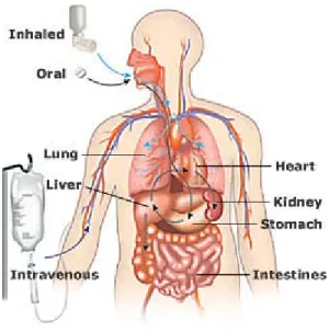
Pharmaceutical products may be administered through various routes, such as oral, nasal, dermal, intravenous, pulmonary, ocular, rectal, or vaginal delivery. However the oral route of administration is preferred, for its many advantages. When ingested orally, the drug is absorbed by the gastrointestinal (GI) tract and enters the hepatic system. Upon its passage through the GI tract, the drug encounters diverse environments with respect to pH, enzymes, electrolytes, fluidity, surface features, all of which may influence its absorption (Kwan 1997). Part of the orally administered dose may not be available for absorption as a consequence of chemical degradation in the GI tract, or it may be metabolized through the gut wall (Figure 1.1). Next, as the drug passes through the portal vein, it is carried to the liver before reaching systemic circulation, where it is distributed through the bloodstream to various tissues. The main problem here is that the liver metabolizes many drugs, and sometimes only a small fraction of active drug substance is available for distribution to the rest of the body. This phenomenon is known as the first pass effect, and greatly reduces bioavailability of some drugs. This effect is defined as the amount of ingested dose that reaches the systemic circulation (Mahota 2007). If administered intravenously the drug bypasses absorption and metabolism processes and is directly available in the bloodstream, and by definition bioavailability of a drug is 100%. The low oral bioavailability of drugs may be attributed to several factors, such as slow dissolution rate, poor solubility, metabolism by GI tract or liver due to first pass effect, chemical instability in the GI tract and/or poor permeability across intestinal membrane (Horter 2001). In addition, the absortption of the drug across the GI tract can highly depend upon membrane transport proteins (Rajendran 2010). Also known as drug transporters, these proteins allow for the active transport of many drug compounds across biological membranes (Lee 2009).
Figure 1.1: Drug administration and transport in the body. (nihseniorhealth.gov/images/drug_admin1.jpg)
Pharmacological activity of a drug partly depends on its bioavailability, which subsequently depends on its solubility, dissolution rate permeability. Therefore a drug must have an adequate aqueous solubility for systemic absorption and therapeutic response. Solubility, a thermodynamic property, refers to the concentration of saturated drug in solution, whereas dissolution, a kinetic parameter, is the rate at which the drug dissolves (Yvonne 2010). The aqueous solubility of a drug is a prime determinant of its dissolution rate. As a result of its poor solubility, the dissolution of the drug is so slow that dissolution takes longer than the transit time past its absorptive sites, resulting in incomplete bioavailability when administered orally (Horter 2001). In the case of parenteral administration (when the drug is dissolved in an organic solvent or micelles), it bears the high risk of precipitation in the circulatory system thereby becoming lodged in capillaries and obstructing their blood flow. Permeability, solubility and dissolution are major determinants of GI drug absorption. Permeability is defined as the rate at which the drug molecule is transported across a
membrane (Urban 2007). It is an important factor controlling the absorption and thus the pharmacological activity of a drug (Yvonne 2010). The Biopharmaceutics Classification System (BCS) categorizes drugs based upon their solubility and permeability (Figure 1.2). The solubility classification is based upon the dose strength of the drug. A highly soluble drug has a high strength such that it is soluble in a volume of 250 ml or less of aqueous media over pH range 1-7.5. If the drug does not exhibit high dose strength, it is considered as poorly soluble (Yu 2002). The pearmeability classification of BCS is based upon the extent of intestinal absorption of a drug in humans. Class I consists of drugs that demonstrate both high permeability and solubility. Such drugs are appropriate for the development of immediate release dosage forms. Class II drugs exhibit low solubility and high permeability, and may have limited bioavailability. However by formulating such drugs through physical means (eg. micronization or use of cosolvents), their dissolution may be improved. Class III (low permeability and high solubility) and class IV drugs (low solubility and low permeability) are much more difficult to formulate (Yvonne 2010). By altering the chemical structure, the permeability and thus the bioavailability of such drugs may be improved.
Over the years, progress in combinatorial chemistry, biology and genetics has lead to the steady increase in the number of drug candidates under development (Kesisoglou 2007). Drug design has attempted to explore new chemical species resulting in more complex molecules with higher hydrophobicities. This trend has often resulted in worsened aqueous solubility and intestinal permeability. Presently almost 40% of drugs in development pipelines and 60% of the drugs directly from synthesis are poorly water soluble (Lipinski 2002; Keck 2006). Such drugs typically display low bioavailability due to slow dissolution rate (Teeranachaideekul 2008).
Classic problems associated with poorly soluble drugs are low bioavailability and erratic absorption (Muller 2001). Innovative formulation approaches are continuously being investigated to increase bioavailability after oral administration or make available intravenously injectable forms (Keck 2006). Processes to improve solubility, dissolution and subsequent bioavailability of drugs include : (1) modification at the molecular level (eg. salt formation, prodrugs, co-solvents, complexation), (2) application of colloidal drug delivery systems (eg. micelles, emulsions, liposomes) and (3) modification of the drug properties at the particulate level (eg. nanosizing) (Yvonne 2010). These strategies are further discussed in detail in this chapter.
1.2 Salt
formation
Salt formation is a very common and effective strategy for increasing the solubility, dissolution rate and ultimately the bioavailability of poorly soluble ionizable drugs. Salts of acidic and basic drugs generally have higher water solubility than their corresponding acid or base (Serajuddin 2007). The salt formation takes place by varying the pH and using a counterion, with a significantly different pKa value than that of the drug (Yvonne 2010). The sufficient difference in pKa allows for the pH of the solution to be altered and thus generate a salt form of the acidic or basic drug of interest. Potential drug compounds are becoming more and more insoluble and sometimes salt formation is not enough to increase their solubility (Serajuddin 2007). For instance, for a compound with solubility 1 μg/mL, even a 1000 fold increase in solubility by salt formation will give a concentration of only 1 mg/mL, which may not be adequate for the purpose of dosage form development. Under such circumstances, salt formation may be combined with other solubilization technique to obtain an optimal aqueous drug solubility. Furthermore, salt formation is only applicable to weakly acidic or basic drugs and not for neutral ones. In many cases, this technique does not achieve better bioavailability because under physiological conditions, the salt converts back into the acidic or basic form of the drug. This latter approach only works for weak acids and bases that ionize between pH 2 and pH 9 and that are chemically resistant to degradation and hydrolysis. Furthermore, an extreme pH can have biocompatibility issues such as tissue irritation (Narazaki 2007).
1.3 Complexation
Complexation, another approach to dissolve hydrophobic drugs has gained popularity in recent years. Inclusion complexes obtained with cyclodextrins (CDs) are able to solubilise certain poorly soluble drugs. CDs are cyclic oligosaccharides derived from starch, composed of 6-8 glucopyranose units joined through α-(1,4) glucosidic bonds (Figure 1.3) (Brewster 2007).
Figure 1.3 : Chemical structure of a cyclodextrin molecule (Brewster 2007) The interior of the complex is relatively lipophilic whilst the exterior is hydrophilic, thus making it ideal for the inclusion of poorly water soluble drugs by non covalent interaction (Figure 1.4). There exist three natural CDs, α-, β-, and γ-CDs (with 6, 7, 8 units respectively), and they all differ in ring size and solubilisation properites. The use of CDs is often preferred for solubilisation of drugs (Stella 1997). For instance cosolvents such as alcohols and glycols will increase solubility of the poorly soluble drug in a non-linear way, with respect to the co-solvent concentration. For instance, a drug with solubility 0.1 mg/mL in water and 10 mg/mL in propylene glycol does not translate into 5 mg/mL solubility in a 50% mixture of water and propylene glycol. On
the other hand, CDs solubilize drugs as a linear function of their concentration, such that most drugs have a 1:1 complex with CD (Stella 1997). Hence the molecular weight of cyclodextrins becomes a limiting factor such that great amounts of cyclodextrin may be required for dissolve a small amount of drug.
Figure 1.4: Complexation of drug and cyclodextrin (Stella VJ 1997)
Presently, there are over 35 different drugs marketed as solid or solution based CD complex formulations (Thorsteinn 2004). Both synthetic and natural CDs have been employed to enhance drug solubility and are adminsitered via various routes (Table 1.1). In addition to enhancing solubility and dissolution rate of poorly water soluble drugs, CD complexes can reduce or prevent gastrointestinal and ocular irritation, prevent drug-drug or drug-additive interactions and convert oils and liquid drugs into microcrystalline or amorphous powders (Challa 2005).
Table 1.1 : Approved and marketed drug–cyclodextrin complexes in various world markets (Davis 2004)
Drug Administration route Trade name Market
-Cyclodextrin
Alprostadil (PGE1) Intravenous Prostavastin, Caverject,
Edex Europe, Japan, United States
Cefotiam hexetil HCl Oral Pansporin T Japan
Limaprost Oral Opalmon, Prorenal
-Cyclodextrin
Benexate Oral Ulgut, Lonmiel Japan
Dexamethasone Dermal Glymesason Japan
Iodine Topical Mena-Gargle Japan
Nicotine Sublingual Nicorette Europe
Nimesulide Oral Nimedex, Mesulid Europe
Nitroglycerin Sublingual Nitropen Japan
Omeprazole Oral Omebeta Europe
Dinoprostone (PGE2) Sublingual Prostarmon E Japan
Piroxicam Oral Brexin Europe
Tiaprofenic acid Oral Surgamyl Europe
2-Hydroxypropyl- -cyclodextrin
Cisapride Rectal Propulsid Europe
Hydrocortisone Buccal Dexocort Europe
Indomethacin Eye drops Indocid Europe
Itraconazole Oral, intravenous Sporanox Europe, United
States
Mitomycin Intravenous Mitozytrex United States
Randomly methylated -cyclodextrin
17 -Oestradiol Nasal spray Aerodiol Europe
Chloramphenicol Eye drops Clorocil Europe
Sulphobutylether -cyclodextrin
Voriconazole Intravenous Vfend Europe, United
States Ziprasidone maleate Intramuscular Geodon, Zeldox Europe, United
States 2-Hydroxypropyl- -cyclodextrin
1.4 Prodrugs
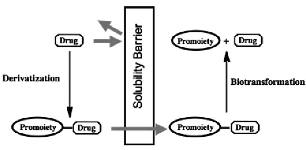
A common strategy to increase solubility of a drug is to redesign the molecule to a bioreversible, more water soluble derivative, called a prodrug. A prodrug is a pharmaceutical inactive compound that results from transient chemical modifications of a biologically active species and is designed to convert to biologically active species in vivo by a predictable mechanism (Xiaolong 2006). The drug is therapeutically inactive when administered, but becomes activated in the body when enzymatic or chemical processes break the promoiety linked to the drug (Figure 1.5) (Stella 2007).
Figure 1.5: An illustration of the prodrug concept (Stella 2007).
The successful design of the prodrug is based upon the efficient transformation of the promoiety into the active drug in vivo. There are a variety of chemical structures, but most prodrugs are classified based upon their chemical linkage (eg. ester, amides and salts) (Fleisher 1996). Other prodrug approaches include conversion of promoieties into amines, imides, hydroxyls, thiols carboxyls and carbonyls. A variety of
paclitaxel prodrugs have been synthesized in which the promoiety is attached at position C2 or C7 (Figure 1.6) (Skwarczynski 2006).
Figure 1.6 Paclitaxel prodrug strategies (Skwarczynski 2006).
Macromolecular prodrugs such as polymer-drug conjugates (polymeric prodrugs) are promising to enhance solubility of poorly water-soluble drugs (Duncan 2001). For instance, poly (ethylene glycol) (PEG), a water-soluble biocompatible polymer, dissolves in organic solvents quite readily. Its high solubility makes it a versatile candidate for polymer-drug conjugation (Peng 2008). Silybin, an antihepatotoxic agent used to treat liver and gall bladder has a solubility of 0.4 mg/mL. A soluble silybin prodrug conjugate with a linear PEG conjugated to the drug via a succinic
ester linkage enhanced the solubility to 800 mg/mL. Paclitaxel polymer prodrugs have also been reported. The first PEG prodrug of paclitaxel was reported by Greenwald et al. (Greenwald 1996). A water-soluble taxol 2’-PEG ester prodrug was designed and showed high solubility (660 mg/mL) (Skwarczynski 2006).
Prodrugs are not only used to enhance solubility of a drug, but offer other advantages as they increase drug stability, achieve sustained drug release, mask the taste of a drug and enable site specific drug delivery (Yvonne 2010). However, the use of the prodrug strategy is questionable due to the large costs and additional time needed to solve formulation and delivery problems (Stella 2007).
1.5 Cosolvents
The solubility of a drug may be enhanced by dissolving it in a cosolvent mixture, composed of water and a solvent (eg. ethanol, propylene glycol, glycerol and/or PEG) (Yvonne 2010). To achieve relatively high drug solubility, the concentration of the cosolvent mixture is usually high as well. Hence, upon dilution of the cosolvent mixture in the intravenous fluids or blood, the drug may easily precipitate (Singla 2002).
The use of cosolvent presents certain disadvantages such that an excess of solvent may be unsuitable and toxic for parenteral formulations. The intrinsic toxicity of cosolvent vehicles can be reduced by using a mixture of cosolvents or combining them with surfactants. Only few surfactants and organic cosolvents are regarded as safe for human use. Even fewer are compatible with injectable formulations possibly causing precipitation, pain, inflammation, and hemolysis. However, by raising the number of excipients in a given formulation, the risk of incompatibilities increases (Li 2007). Nonetheless, cosolvent strategies are explored to enhance solubility of many poorly water-soluble pharmaceutical agents. For instance, a cosolvent mixture of ethanol, polysorbate (Tween 80) and surfactant (Pluronic L64) in a 3:1:6 (v/v/v) ratio was used to formulate the poorly water-soluble drug paclitaxel (0.3 μg/mL).
The drug concentration in the cosolvent formulation was reported at 5 mg/mL, but the formulation became unstable upon dilution with water (Singla 2002).
1.6 Surfactants and Micelles
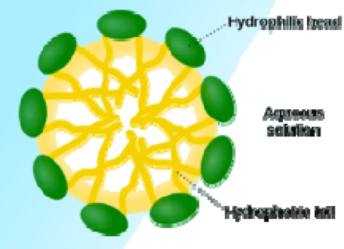
Surfactants are amphiphilic agents with a hydrophilic head and hydrophobic tail. This specific nature of surfactants gives them unique activity at interfaces, wherein they significantly reduce the surface tension of water. At low concentrations, these molecules are freely dispersed in an aqueous environment, however as the concentration is increased, the free energy of the system augments as a result of unfavourable interactions between the hydrophobic regions and the surrounding water molecules (Gaucher, in press). At a precise concentration, the surfactant molecules arrange themselves to form aggregates called micelles, thereby minimizing the energy of the system (Figure 1.7) (Torchilin 2007). The minimum surfactant concentration at which micelles assemble is referred to as the critical micellization concentration (CMC). Micelles are in dynamic equilibrium with free molecules in solution, and so they are constantly dissociating and reforming (Torchilin 2007). The size of micelles varies and depends on the size, shape and number of surfactant monomers used. The formation of micelles is driven by the decrease of free energy in the system because of the removal of hydrophobic fragments from the aqueous environment and the re-establishing of the hydrogen bond network in water. Consequently, hydrophobic fragments of the amphiphilic molecules form the core and hydrophilic fragments form the shell (Torchilin 2007). Due to such a structure, micelles solubilise molecules of poorly soluble nonpolar drugs within its core and polar drug molecules may be adsorbed on the micelle surface (Jones 1999). Such solubilisation using micelle-forming surfactants results in an increased water solubility of poorly soluble drug and its improved bioavailability, and enhanced permeability across biological barriers (Torchilin 2001; Torchilin 2007).
Figure 1.7: Representation of micelle formation at CMC (Wikipedia)
Polymeric micelles have attracted much attention over the years. Polymeric micelles are formed by block co-polymers consisting of hydrophilic and hydrophobic monomer units with the length of a hydrophilic block exceeding to some extent that of a hydrophobic one (Gaucher 2005). Similar to micelles formed by the conventional surfactants, block copolymer micelles assume a core-shell structure where the core is comprised of hydrophobic blocks, and is stabilized by the corona composed of hydrophilic chains (Gaucher, in press). In comparison to the conventional micelles, polymeric micelles form at lower CMC and are more stable upon dilution accordingly protecting the incorporated drug from fast degradation (Devalapally 2007). Genexol – PM is a methoxy-PEG-poly (D-L-lactide) micelle formulation of paclitaxel that obtained a pre-market approval in Korea in 2006. In
vivo studies demonstrated that the antitumor efficacy of this formulation is better than
1.7 Emulsions, microemulsions and self-
emulsifying delivery systems
An emulsion is composed of a minimum of two immiscible liquid phases (eg. oil and water), where a dispersed phase (dispersed globules) is in a continuous phase (liquid phase) and the whole system is stabilized by an emulsifying agent (Mahato 2007). Depending on which phase is dispersed into the other, either an oil-in-water (o/w) or water-in-oil (w/o) emulsion is obtained. Micron or submicron sized droplets are formed upon applying high energy to the system (Yvonne 2010). Microemulsions; much smaller one-phase systems are more stable colloidal solutions. Microemulsions contain appropriate mixture of oil (polar lipids) water and surfactants, and the oil component is used to deliver the poorly soluble drug. In addition, whereas emulsions are composed of spherical droplets, microemulsions range from droplet-like micellular structures to bicontinous structures. Self-emulsifying drug delivery systems (SEDDS), composed of an isotropic mixture of drug, lipid and surfactants, are used to deliver hydrophobic drugs. SEDDS form fine emulsion droplets (50-100 nm) upon dilution in physiological fluids (Tang 2008). Thus, the hydrophobic drug remains in solution and is delivered to the gut. SEDDS exist as solutions or are made into solid dosage forms. A few of the marketed o/w emulsion products include Diazepam®, Etomidate® and Propofol® (Muller 2005).
1.8 Liposomes
Liposomes are spherical vesicles composed of phospholipids self-assembled in aqueous medium (Rawat 2006). Due to their amphiphilic nature, lipids in excess of water spontaneously self-associate above a concentration called the critical aggregation concentration, which is in the order of 10-10 M. The self-assembly process is spontaneous and cooperative (Mahato 2007). The lipid moieties spontaneously orient in water, and the lipid chains orient inward, giving rise to bilayer structures (Figure 1.8). The aggregation phenomenon is a result of both the polar interactions between the hydrophilic heads of the phospholipids, and the hydrophobic effect of the aliphatic chains (Simard 2007).
Figure 1.8: Structure of a liposome (Devalapally 2007)
Liposomes range from 50 nm to a few micrometers in diameter (Haley 2008). They consist of one or more concentric lipid bilayers separated by water compartments. The rationale of using liposomes as drug delivery systems to the cells was originally based upon the possibility of fusion of the liposome to the target cell membrane.
Hence, the preparation of liposomes had to be prepared from natural lipids found in cell membrane, such as glycerolipids, sphingolipids and sterols (Simard 2007). Synthetic lipids that mimic the structure and physicochemical properties of natural lipids may also be used to fabricate liposomes. Furthermore, these liposomes offer greater advantage such that they may be altered for improved properties. In comparison to other colloidal drug delivery systems, liposomes have the ability to encapsulate both hydrophilic (inside the aqueous region) and lipophilic drugs within the bilayer regions.
Liposomes are classified and described according to their size, the number of bilayers, or their preparation method. Multilamellar vesicles (MLV) are usually 1-5 μm in diameter, whereas unilamellar vesicles (UV) range from 40 nm to 1 μm. There exist four major types of liposomes: conventional, sterically stabilized, targeted and cationic (Simard 2007). Conventional liposomes are either neutral or negatively charged, and are composed of phospholipids, glycolipids, and/or cholesterol. Such liposomes have a limited circulation lifetime because they are removed by cells from the mononuclear phagocyte system (MPS) (Appendix I) (Janoff 1999). These liposomes are mainly used for passive targeting to the phagocytic cells of the MPS, localizing predominantly in the liver and spleen.
Sterically stabilized liposomes have a polymer coating to obtain prolonged circulation times. PEGylated (polyethylene glycol) lipids reduce the uptake of the liposomes by the MPS and subsequently circulation time of the vesicle is greatly increased. By residing in the bloodstream for a longer time, PEGylated liposomes (also known as STEALTH® liposomes) could localize into tumours and most inflammation sites (Mahato 2007). Targeted liposomes usually have a biological moiety attached to enable recognition by a specific receptor at the site of interest. Targeting ligands include monoclonal antibodies (generating an immunoliposome), vitamins or specific antigens. Most of the targeting liposomes are also PEGylated, and so are able to target nearly any cell type in the body and deliver drugs. Finally, cationic liposomes
are positively charged and are mainly used for the delivery of nucleic acids (Papahadjopoulos 1991). The positively charged liposomes interact with the negatively charged backbone of DNA or RNA, and lead to neutralization.
Figure 1.9: Classification of liposomes (www.imcr.uzh.ch/static/onkwww/images/lipos4.gif)
Liposomes were one of the first drug delivery system to be approved for clinical use, and today there exists many liposomal formulations. One very popular formulation is Doxil®, which encapsulates the poorly soluble drug Doxorubicin in 80-100 nm-sized liposomes of PEGylated distearoyl phosphatidyl ethanolamine, hydrogenated soy phophatidylcholine and cholesterol (Yvonne 2010).
1.9 Polymer Solid dispersions
As discussed earlier, polymers play a vital role in solubilising poorly soluble drugs. Alternative applications of polymers include their use in solid dispersions as a strategy to improve solubility of poorly soluble drugs (Leuner 2000).
Solid dispersions consist of molecularly or physically dispersing drug in a water-soluble carrier (eg. PEG or PVP) or a water inwater-soluble carrier (Sivert 2009). Drug dissolution can be improved by molecularly dispersing a drug in a hydrophilic polymer matrix via preparation processes such as solvent evaporation or hot-melt extrusion. Polymers have been used quite extensively to generate solid dispersions, because they make amorphous solid dispersions. Solubility of a given solid is a sum of crystal packing energy, cavitation and solvation energy. Different solid-state forms of a material have different crystal packing energy. The amorphous form, due to absence of an ordered crystal lattice requires minimal energy provides the maximal dissolution advantage as compared to the crystalline and hydrated forms of a drug (Vasconcelos 2007). Polymers used include both synthetic (PEG, polymethacrylates), and natural polymers such as hydroxymethylcellulose (HPMC), ethylcellulose and CDs (Vasconcelos 2007). When the solid dispersion is exposed to an aqueous milieu, the carrier dissolves allowing the release of the incorporated drug.
Due to the intensive work involved in the optimization of solid dispersions, the latter are rarely considered as a viable option to formulating poorly-water soluble drugs. In addition, the amorphous form of the drug within the solid dispersion may undergo crystallization over time.
1.10 Size reduction
A much more classical method to increase drug dissolution rate is through size reduction (Keck 2006). The Noyes-Whitney equation (equation 1), where dW/dt is
Equation 1
the rate of dissolution, A is the surface area of the solid, C is the concentration of the solid in the bulk dissolution medium, Cs is the concentration of the solid in the diffusion layer surrounding the solid, D is the diffusion coefficient, and L is the diffusion layer thickness explains that dissolution rate may be readily improved by reducing the size of the drug particle, and thereby increasing surface area available for dissolution (Figure 1.10) (Merisko-Liversidge 2003; Muller 2004). The size reduction also decreases the thickness of the diffusion layer around each particle, thus further promoting the dissolution. The surface area may be adjusted by altering the particle size (eg. micronization). Micronization, which involves coarse drug powder milled into an ultrafine powder of particle size distributions in the range of 1 - 25 μm, is a common method used to improve bioavailability of the poorly soluble drug (Merisko-Liversidge 2003).
Fig. 1.10. Dissolution velocity dc/dt (or dW/dt) and saturation solubility Cs as a function of the size of drug powders ranging from coarse to nanonized drugs (Muller 2004)
Many of the new drugs exhibit such a low solubility that micronization is insufficient to increase the surface area of the drug to a large extent. At very low solubility, the achieved increase in dissolution rate is not significant enough to lead to high enough bioavailability. Though micronization increases dissolution rate, it does not change the saturation solubility of the poorly soluble drug and consequently desired therapeutic blood levels are not attained. Hence, the next approach involves further reduction of the drug particles, down to the nanoscale. Moreover, according to the Ostwald-Freundlich equation (equation 2),
Equation 2
whereR is the gas constant, T is the absolute temperature, M themolecular weight of the solid in solution, σ is the interfacialtension between the solid and liquid, ρ is the density of thesolid, and S1 and S2 are the solubilities of particle of radiusr1 and r2,
respectively, the equilibrium solubility of particles increases withdecreasing particle size. Particularly size reduction in the range below ca. 1 µm is accompanied by an increase in saturation solubility (Borm 2006).
Though many textbooks and articles follow this theory, the validity of the Oswald-Freundlich equation to describe the relationship between the size of a particle and its solubility is controversial (Godec 2009, Nancollas 2009). Experimental observations have shown that Oswald ripening is not due to the increased solubility of smaller crystals but rather to a negative tension between a solid and a solution (Wu 1998). Oswald stated that the driving force for ripening is the variation in solubility between large and small particles. However, the alternative mechanism concludes that the ripening is due to a thermodynamically unstable interface between the solid and solution. Wollaston and Curie assumed the hypothesis that finely divided solids should have larger solubilities than coarser ones in the late 19th century. Later Oswald and Freundlich used similar arguments to establish the equation above and prove that solubility of finely sized crystals in a function of particle size. However, when the first experimental investigations were made to test the supersaturation, particle size and solid-liquid interfacial tensions, limitations were encountered and the Oswald-Freundlich equation was subjected to criticism (Wu 1998). Despite the controversy, many believe that particle size reduction does indeed lead to enhanced dissolution rate.
Nanosizing is the reduction of the active pharmaceutical ingredient (API) particle size down to the sub-micrometer range (Kesisoglou 2007). By definition, drug nanocrystals are nanoparticles of pure crystalline drug without any matrix material typically ranging from 200 – 500 nm in size (Muller 2001). The following chapter comprehensively describes the advantages, methods of fabrication and applications of drug nanocrystals.
1.11 References
Borm P, Klaessig FC, et al, Wood S 2006. Research Strategies for Safety Evaluation of Nanomaterials, Part V: Role of Dissolution in Biological Fate and Effects of Nanoscale Particles. Toxicol Sci 90:23-32.
Breitenbach, J. (2002). Melt extrusion: from process to drug delivery technology. Eur
J Pharm Biopharm, 54: 107-117.
Brewster, M., Loftsson T. (2007). Cyclodextrins as pharmaceutical solubilizers. Adv
Drug Deliver Rev, 59: 645-666.
Challa, R., Ahuja A., et al. (2005). Cyclodextrins in drug delivery: An updated review. AAPS PharmSciTech, 6: E329-E357.
Davis ME, Brewster ME 2004. Cyclodextrin-based pharmaceutics: past, present and future. Nat Rev Drug Discov 3(12):1023-1035
Devalapally, H., Chakilam A. et al. (2007). Role of nanotechnology in pharmaceutical product development. J Pharm Sci, 96: 2547-65.
Duncan, R., Gac-Breton S. et al. (2001). Polymer-drug conjugates, PDEPT and PELT: basic principles for design and transfer from the laboratory to clinic. J Control
Release, 74: 135-146.
Gaucher G., Dufresne M.-H, et al. (2005). Block copolymer micelles: preparation, characterization and application in drug delivery. J Control Release, 109: 169-188. Godec A, Gaberšček M, Jamnik J 2009. Comment on the Article “A New
Understanding of the Relationship between Solubility and Particle Size” by W. Wu and G.H. Nancollas. J Sol Chem 38:135-146.
Haley B., Frenkel E. (2008). Nanoparticles for drug delivery in cancer treatment.
Urol Oncol-Semin Ori, 26 :57-64.
Horter, D., Dressman J. B. (2001). Influence of physicochemical properties on dissolution of drugs in the gastrointestinal tract. Adv Drug Deliv Rev, 46 :75-87. Janoff, A. S. (1999). Liposomes: Rational Design. New York, Marcel Dekker Inc.
Jones, M.-C., Leroux J.-C. (1999). Polymeric micelles - a new generation of colloidal drug carriers. Eur J Pharm Biopharm, 48: 101-111.
Keck, C. M., Muller R. H. (2006). Drug nanocrystals of poorly soluble drugs produced by high pressure homogenisation. Eur J Pharm Biopharm, 62: 3-16.
Kesisoglou, F., Panmai S., et al. (2007). Nanosizing--oral formulation development and biopharmaceutical evaluation. Adv Drug Deliv Rev,59: 631-44.
Kwan, K. C. (1997). Oral Bioavailability and First-Pass Effects. Drug Metab Dispos, 25: 1329-1336.
Lee EJD, Lean CB, Limenta LMG 2009. Role of membrane transporters in the safety profile of drugs. Expert Opinion on Drug Metabolism & Toxicology 5(11):1369-1383.
Leuner, C., Dressman J. (2000). Improving drug solubility for oral delivery using solid dispersions. Eur J Pharm Biopharm, 50: 47-60.
Li P,. (2007). Developing early formulations: practice and perspective. Int J Pharm, 34: 1-19.
Lipinski, C. (2002). Poor Aqueous Solubility – an Industry Wide Problem in Drug Discovery. Am Pharm Rev: 5: 82
Mahato, R. I. (2007). Pharmaceutical Dosage Forms and Drug Delivery. Boca Raton, Florida, CRC Press, Taylor & Francis Group, LLC.
Merisko-Liversidge, E., Liversidge G. G., et al. (2003). Nanosizing: a formulation approach for poorly-water-soluble compounds. Eur J Pharm Sci, 18: 113-20.
Muller, R. H., Peters K, (1998). Nanosuspensions for the formulation of poorly soluble drugs: I. Preparation by a size-reduction technique. Int J Pharm, 160: 229-237.
Muller, R. H., Jacobs, C. et al. (2001). Nanosuspensions as particulate drug formulations in therapy. Rationale for development and what we can expect for the future.Adv Drug Deliv Rev, 47: 3-19.
Nancollas G, Wu W 2009. Response to “Comment on “A New Understanding of the Relationship between Solubility and Particle Size” by Godec, A., Jamnik, J., and Gaderšček, M. J Sol Chem 38:147-148.
Narazaki, R., Sanghvi, R., et al. (2007). Estimation of Drug Precipitation upon Dilution of pH & Cosolvent Solubilized Formulations. Chem Pharm Bull, 55:1203-1206.
Papahadjopoulos D., Gabizon A. T., et al (1991). Sterically stabilized liposomes: improvements in pharmacokinetics and antitumor therapeutic efficacy. Proc Natl
Acad Sci U SA. 84:11460-4.
Peng, Z., Y. Hai, et al. (2008). Water soluble poly(ethylene glycol) prodrug of silybin: Design, synthesis, and characterization. J Appl Polym Sci, 107: 3230-3235. Rajendran L, Knolker H-J, Simons K (2010). Subcellular targeting strategies for drug design and delivery. Nat Rev Drug Discov 9:29-42.
Simard P., Leroux J.-C., et al (2007). Nanoparticles for Pharmaceutical Applications,. Valencia, American Scientic Publishers
Sivert A., Bérard, V. et al. (2009). New binary solid dispersion of indomethacin with surfactant polymer: From physical characterization to in vitro dissolution
enhancement. J Pharm Sci, 99: 1399-1413.
Rasenack, N., Muller B. W. (2004). Micron Size Drug Particles: Common and Novel Micronization Techniques. Pharm Dev Technol, 9: 1-13.
Rawat, M., Singh D., et al. (2006). Nanocarriers: promising vehicle for bioactive drugs. Biol Pharm Bull 29: 1790-8.
Serajuddin, A. (2007). Salt formation to improve drug solubility. Adv Drug Deliv
Rev , 59: 603-616.
Singla, A. K., Garg A., et al. (2002). Paclitaxel and its formulations. Int J Pharm 235: 179-92.
Stella, V. J., Nti-Addae K. W. (2007). Prodrug strategies to overcome poor water solubility. Adv Drug Deliv Rev 59: 677-694.
Stella V.J, (1997). Cyclodextrins: their future in drug formulation and delivery.
Pharm Res, 5: 556-567.
Stevens, P., Sekido M., et al. (2004). A Folate Receptor–Targeted Lipid Nanoparticle Formulation for a Lipophilic Paclitaxel Prodrug. Pharm Res, 21 2153-2157.
Teeranachaideekul V., Junyaprasert V. B, et al. (2008). Development of ascorbyl palmitate nanocrystals applying the nanosuspension technology. Int J Pharm, 354: 227-234.
Thorsteinn, L., Marcus B., et al. (2004). Role of Cyclodextrins in Improving Oral Drug Delivery. Am J Drug Del 2: 261-275.
Torchilin, V. (2007). Micellar Nanocarriers: Pharmaceutical Perspectives. Pharm
Res, 24 : 1-16.
Torchilin, V. P. (2001). Structure and design of polymeric surfactant-based drug delivery systems. J Control Release 73: 137-172.
Vasconcelos, T., Sarmento B., et al. (2007). Solid dispersions as strategy to improve oral bioavailability of poor water soluble drugs. Drug Discov Today 12 :1068-1075. Wu W, Nancollas GH 1998. A New Understanding of the Relationship Between Solubility and Particle Size. J Sol Chem 27:521-531.
Xiaolong Li (2006). Design of Controlled Release Drug Delivery Systems. New York, McGraw Hill.
Yu LX, Amidon GL, Polli JE, Zhao H, Mehta MU, Conner DP, Shah VP, Lesko LJ, Chen M-L, Lee VHL, Hussain AS 2002. Biopharmaceutics Classification System: The Scientific Basis for Biowaiver Extensions. Pharmaceutical Research 19(7):921-925.
Yvonne P., T. R. (2010). Pharmaceutics- Drug Delivery and Targetting. London, UK, Pharmaceutical Press
nihseniorhealth.gov/images/drug_admin1.jpg (retrieved December 1st 2009)
CHAPTER 2:
Nanocrystals and Nanosuspensions
2.1 Introduction
Whilst reduction in particle size has been applied in the pharmaceutical field for several decades, newer technologies and the deeper understanding of colloidal drug systems has allowed for the production of nanoscale drug particles. Drug nanocrystals are composed of pure crystalline drug devoid of polymeric or lipidic carriers and may be sized from few nanometers up to 400 nm. Nanosuspensions are dispersions of the nanocrystals in a liquid media, which are stabilized by surfactants or polymeric stabilizers (Junghanns 2008). Nanosuspensions overcome the delivery issues of poorly soluble compounds by obviating the use of potentially harsh vehicles, such as pH-buffers or cosolvents, thereby reducing toxicity and increasing efficacy (Rabinow 2004).
As presented section 1.10, a significant feature of drug nanocrystals is the increase in dissolution velocity (subsequently in bioavailability). Another exceptional feature of nanosuspensions is the increased adhesiveness compared to microparticles which is an additional factor for improving the oral absorption of poorly soluble drugs (Junghanns 2008). As a result of this phenomenon, nanocrystals remain in contact with the gut wall for longer time, thereby enhancing bioavailability of the drug upon oral administration (Gao 2008).
Being physically stable and displaying increased dissolution rate, a nanocrystal formulation greatly enhances biological performance of poorly soluble drugs (Merisko-Liversidge 2003).
By increasing the dissolution rate, nanonization enhances the rate and extent of absorption of drugs administered orally so that the bioavailability is less affected in the fed/fasted state. For instance, by nanosizing the active ingredient (MK-0869) of the drug EMEND®, previously FDA approved for prevention of chemotherapy-induced nausea, the advantages of a nanocrystalline formulation over a conventional dosage form were confirmed (Figure 2.1) (Wu 2004). Additionally, owing to their small size, stable drug nanocrystals may be injected by the intravenous (i.v.) route
without the risk of embolic complications. More importantly, the total drug amount in the formulation may be adjusted to high levels, allowing usage of smaller administration volumes.
In this chapter, various methods of nanocrystal production, their characterization and applications are discussed in detail.
Figure 2.1: Comparison of mean (±S.E.) plasma concentrations of MK-0869 (active ingredient of EMEND®) following oral administration in Beagle dogs (N = 5) of a conventional suspension (●, fasted; ○, fed) and a NanoCrystal® dispersion formulation ( , fasted; , fed) of MK-0869 at a dose of 2 mg/kg (Wu 2004).
2.2
Methods of nanocrystal fabrication
Methodologies for the generation of drug nanocrystals are mainly categorized as “bottom-up” or “top-down” methods (Van Eerdenbrugh 2008). The first is based upon the strategy of growing drug nanocrystals, whereas the latter breaks down coarse drug powder into nanoparticles.
2.2.1 Bottom-up strategies
Precipitation techniques and supercritical fluid technology are the commonly used bottom-up approaches to fabricate drug nanocrystals.
The precipitation technique consists of dissolving the poorly soluble drug in a solvent (usually organic) and adding this solution to an anti-solvent (eg. water) under agitation (Kesisoglou 2007). This leads to a supersaturation which results in rapid nucleation of the drug, thereby forming nanoparticles (Gao 2008). Several parameters such as stirring rate, volume ratio of anti-solvent/ solvent and drug content require optimization for the production of uniformly sized nanoparticles. A faster stirring rate generates smaller particles and a greater anti-solvent/solvent ratio brings about a faster nucleation. The drug content should be adjusted to a moderate level, because very large drug quantities may cause uncontrolled agglomeration of the nanocrystals. The drug nanocrystals are further processed for removal of organic solvent.
The principle advantage of the precipitation technique is that it is relatively simple and inexpensive, however this technique does have its disadvantages. It is known that the particle size is not always controlled using this method because growth of the crystals is not hindered, and microcrystals are very often generated (Keck 2006). In order to avoid spontaneous agglomeration and subsequent formation of microcrystals, stabilizers are used. Another limitation of this technique is that the drug must be soluble in a solvent which is miscible with the anti-solvent. This poses difficulties
because most poorly soluble drugs needing to be nanosized, are poorly soluble in both aqueous and organic solvent, thus making them unsuitable candidates for the precipitation technique (Grau 2000). Amorphous drug content often is generated during the precipitation process. Though this state of a drug is more soluble and exhibits a higher dissolution than the crystalline state, it is not as stable and converts to crystalline over time (Gao 2008). Despite its limitations, liquid precipitation is low cost method offering convenient processing of nanocrystals (Zhang 2009). This bottom-up technology is marketed by DowPharma (Midland, MI, USA) and by BASF Pharma Solutions (Florham Park, NJ, USA) (Kesisoglou 2007).
Supercritical fluid based technologies, such as rapid expansion from supercritical solutions (RESS), rapid expansion from supercritical aqueous solutions (RESAS), and supercritical anti-solvent methods (SAS), have been investigated as an alternative bottom-up strategy for the fabrication of drug nanocrystals (Date 2004). This approach requires complex operating conditions and induces high costs (Zhang 2009).
The RESS method consists of dissolving the drug in a supercritical fluid (eg. CO2) at a programmed temperature and pressure. The drug solution is introduced into a precipitation unit and allowed to expand rapidly though a capillary into a region of lower pressure and temperature (Date 2004). This phenomenon results in the dissolved drug precipitating rapidly as a finely sized powder, thus generating nanocrystals. The shortcoming of this method is that only drugs exhibiting solubility in supercritical fluids could be used. Similar to the liquid precipitation technique, generation of microcrystals are also obtained, resulting in large size distribution. The RESAS is an evolution of the RESS method, such that an aqueous solution containing stabilisers is used in addition to the supercritical fluid. Though size is better controlled, this technique displays similar concerns because of the use of solvents. The SAS method consists of dissolving the drug in an appropriate organic solvent or co-solvent, and then atomizing it into an excess flowing continuum of supercritical
CO2, thus forming nanoparticles in a dried state. Hydrophobic drugs with poor solubility in supercritical fluids may be processed using the SAS procedure, however broad particle size distribution is obtained.
2.2.3 Top-down strategies
Top-down methods are more widely employed for the production of drug nanocrystals. This specific strategy relies on mechanical attrition to break down large crystalline particles into nanoparticles (Kesisoglou 2007). Such technologies include high-pressure homogenization and media milling.
Media milling (or wet milling) is based upon an attrition procedure in which micrometer sized drug crystals are milled in the presence of grinding media and stabilizers (Rabinow 2004). This patent protected technology was developed in 1990 by Liversidge et al. (Merisko-Liversidge 2008). In this process, a nanocrystal suspension is generated by high shear milling equipment composed of a milling chamber, milling beads (or grinding media), a rotor and a recirculation chamber (Figure 2.2).
Figure 2.2: Schematic representation of the media milling process (Date 2004).
The poorly soluble drug is dispersed in an aqueous surfactant solution, and the resulting suspension is wet-milled with the grinding media (Gao 2008). The grinding material spherical in form, is typically composed of zirconium oxide, zirconium silicate or other media such as glass, titania, alumina (Hu 2004). The stabilizers (or surface modifiers) used comprise of various polymers, low molecular weight oligomers, natural products and surfactants. During the milling process, the generated shear forces and those created during impaction of the milling media with the solid drug provide high energy to break the drug crystals into nanoparticles (Merisko-Liversidge 2003). Particle size is determined by stress intensity (which is a function of kinetic energy of the grinding beads), number of contact points (which
may be increased by using smaller grinding media), and the time of milling (Rabinow 2004). The optimization of the following parameter: drug content, amount of grinding or milling pearls, milling speed and time are important for the generation of desirable nanosuspensions. Liversidge et al. reported that particles of Naproxen, a poorly soluble drug were reduced to 270 nm, from an average initial size of 20-30 μm over a period of 5 days of wet milling. PVP K-15 was used as the stabilizer (5:3 drug to stabilizer ratio) and the nanosuspension remained physically and chemically stable for up to 4 weeks at 4°C (Hu 2004).
Though the wet-milling procedure enables the production of stable drug nanosuspensions, there are a few drawbacks to this technique. Not only is it time consuming, but there have been reports on the contamination of the product by grinding material (Muller 2001). Erosion of the grinding material may contaminate the final drug product. In addition, wet-milling is a batch process; therefore batch-to-batch variations may be detected in the quality of the dispersion, processing time, degree of drug crystallinity and particle size distribution. Such variations affect the drug particle size and the efficiency of the delivery system. Moreover milling over several days may also bring risks of microbiological problems, particularly when performing the process at 30°C or using a dispersion media providing nutrition to bacteria (Gao 2008). Regardless of these limitations, this mechanical method of producing nanocrystals is successfully marketed as the NanoCrystal® technology (Élan) (Merisko-Liversidge 2008). Four oral products generated via the NanoCrystal® technology are currently marketed in the US and other countries: Rapamune®, Emend®, TriCor 145® and MegaceES® (section 2.5).
Another top-down method, known as high pressure homogenization, was invented by Muller et al. in the mid 1990s (Muller 1998). Homogenization involves the forcing of a drug suspension under pressure through a valve with a narrow aperture (Figure 2.3). Therefore, the formation of the nanosuspension is based on the cavitation energy generated in the high pressure homogeniser (eg. the piston-gap homogenizer).
In this process, the poorly soluble drug is dispersed in an aqueous surfactant solution
via high speed stirring, and the resultant suspension is passed though a high pressure
homogeniser (gap width of approximately 25 μm) at a pressure of 1500 bar over 3- 20 homogenization cycles (Hu 2004; Rabinow 2004). Due to the narrowness of the gap, the streaming velocity of the drug suspensions and the dynamic fluid pressure increase. Furthermore the static pressure of the fluid decreases below the boiling point of water (at room temperature), and as a consequence the water boils and gas bubbles form which implode when the fluid leaves the homogenization gap (Muller 2001). The generated cavitation forces are very strong and break the drug particles, forming nanocrystals of approximately 40 – 500 nm. The size is dependent upon the hardness of the drug substance and may be controlled by adjusting the processing pressure and the number of cycles applied (Hu 2004).
Figure 2.3: High pressure homogenization technique: (right) forcing of drug suspension through narrow valve, (left) fabrication of drug nanocrystals due to generated forces (Junghanns 2008).
This process also is time-consuming as it takes several cycles to reduce particle size which highly depends on the pressure in the homogenizer. Moreover, the drug
powder requires micronization prior to homogenization in order to avoid blockage of the narrow valve. Changes in drug crystallinity have been reported with this process. A portion of drug nanoparticles was reported as amorphous, and in some cases, complete transformation to amorphous state was observed (Muller 2001). In comparison to the milling procedure, high pressure homogenization displays much less contamination due to erosion of the equipment. Muller et al. investigated the contamination of nanosuspensions produced under harsh conditions (eg. 20 cycles and pressure of 1500 bar), and found iron contamination to be under 1 ppm, low enough to not pose health issues (Krause 2000).
DissoCube® was the first high pressure homogenization technology to be marketed. It involved the fabrication of nanocrystals in water. Shortly after, Nanopure® technologies was developed which involved homogenization in non-aqueous media or mixtures of water and immiscible substances (eg. PEG). The company Baxter developed a combined technology known as NANOEDGE® system in which homogenization procedure is combined with the precipitation technique discussed earlier. The homogenization step is used to avoid growth of the drug particles after precipitation takes place (Gao 2008).
2.3
Characterization of Nanocrystals
The essential characterization parameters for nanosuspensions include evaluation of size and size distribution, zeta potential, crystalline state, dissolution velocity and saturation solubility, surface hydrophilicity/hydrophobicity (Patravale 2004). Other tests include chemical investigation such as evaluation of possible degradation products and evaluation of moisture of nanocrystals (for lyophilized API) (Rabinow 2004).
The mean size and size distribution (polydispersity index) are important parameters because they govern properties such as saturation solubility, dissolution velocity, physical stability, and certain biological performances. Photon correlation spectroscopy (PCS) or dynamic light scattering technique (DLS) are employed, but limited to measuring sizes of 3 nm to 3 μm, therefore laser diffractrometry (LD) is used to detect aggregates of drug nanocrystals. LD is able to measure particles of 0.05 μm to 2000 μm (Muller 2001). Scanning (or transmission) electron microscopy (SEM, TEM) may also be used for size evaluation. The polydispersity index (PDI) is an important index of physical stability of the nanocrystal. PDI values vary between 0 (monodisperse particles) to 1 (broad distribution), however lower values (≤0.3) are usually more appreciable for long-term stability of the nanosuspension.
Surface charge is an important parameter also governing the stability of the nanosuspensions. It is measured by means of electrophoresis and is expressed as electrophoretic mobility or converted to zeta potential. This measurement allows for the prediction of storage stability of the nano-dispersions. Usually the particles with sufficient zeta potential are less likely to aggregate. Literature states that a zeta potential of at least -30 mV for electrostatic and -20 mV for sterically stabilized nanoparticles is desirable for physically stable suspensions (Gao 2008).
Evaluation of crystalline character is required to ensure that crystallinity of the drug has been retained upon nanonization because fabrication procedures may alter the polymorphic state of the drug. For instance high pressure homogenization may generate nanocrystals with amorphous fraction (Muller 2001). Differential scanning