Université de Montréal
A comparison between DNA-DNA checkerboard
hybridization and culture techniques for the detection of
Candida species in denture stomatitis
Par
Muhammad Faheem Khiyani
Département de dentisterie de restauration Faculté de médecine dentaire
Université de Montréal
Mémoire présenté à la Faculté des études supérieures et postdoctorales en vue de l’obtention du grade de
Maîtrise ès Sciences (M.Sc.) En sciences buccodentaires
Octobre 2017
Université de Montréal
Faculté des études supérieures et postdoctorales
Ce mémoire intitulé:
A comparison between DNA-DNA checkerboard hybridization and culture techniques for the detection of Candida species in denture stomatitis
Présenté par:
Muhammad Faheem Khiyani
A été évalué par un jury composé des personnes suivantes:
Dre Gisèle Mainville, présidente-rapporteur Dre Elham Emami, directrice de recherche Dr Jean Barbeau, co-directeur de recherche Dr Robert Durand, membre du jury
iii
RÉSUMÉ
Introduction
Selon la littérature, les évidences sur l'utilisation et l'application potentielles de la technique d'hybridation à damier d'ADN-ADN dans le diagnostic de la stomatite prothétique associée à la Candida (DS) sont limitées. En outre, la littérature suggère que les biomarqueurs inflammatoires de la salive pourraient offrir une nouvelle avenue pour le diagnostic précoce de cette maladie.
Objectifs
Les objectifs de ce projet de recherche de maîtrise étaient les suivants: 1) Fournir des informations sur la précision diagnostique de la culture conventionnelle et de la technique d'hybridation à damier d'ADN-ADN pour la détection d'espèces de Candida dans DS et d'étudier son impact sur le diagnostic clinique de cette maladie, et 2) Examiner systématiquement les données disponibles sur les biomarqueurs salivaires présents dans DS.
Méthodes
Objectif 1): Le biofilm palatin de 26 participants diagnostiqués avec DS a été analysé pour détecter et quantifier les espèces de Candida en utilisant des techniques d’hybridation à damier d’ADN et d’ADN-ADN. En utilisant chaque technique comme référence standard pour l'autre, la précision diagnostique des deux techniques a été examinée et comparée à l'aide des tests Kappa et McNemar. Le test de Spearman a été utilisé pour examiner l'association entre la quantité totale de Candida et les scores d'inflammation totale.
Objectif 2): La revue systématique a suivi les lignes directrices relatives aux rapports systématiques et aux méta-analyses (PRISMA). Le niveau de preuve a été évalué à l'aide de
iv
l'échelle 2011 du centre d'Oxford pour la médecine fondée sur des preuves (OCEBM). La qualité méthodologique a été évaluée à l'aide de la déclaration du renforcement des rapports d’études observationnelles en épidémiologie (STROBE) et classée selon l'échelle d'Olmos.
Résultats
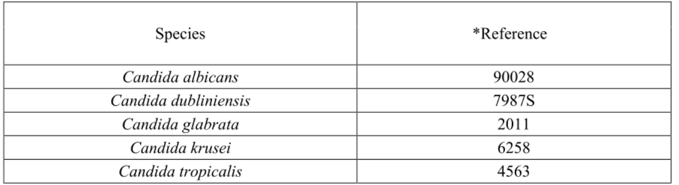
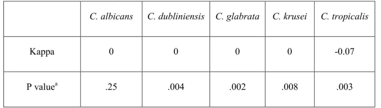
Objectif 1): Pour toutes les espèces de Candida, la spécificité de la technique de culture variait entre 52% et 88,5% et entre 92,9% à 100% pour le damier. Il y avait un désaccord entre les deux techniques. La sensitivité pour les deux techniques a été observée comme nulle pour toutes les espèces. La corrélation entre Candida et les scores d'inflammation n'a pas été statistiquement significative pour la culture, mais une corrélation statistiquement significative a été observée avec la technique du damier (p = 0,05).
Objectif 2): La majorité des études incluses dans la revue systématique ont montré que les niveaux d'IL-6, CCL3 et TGF-β, GM-CSF et TNF-α étaient plus élevés chez les personnes âgées atteintes de DS, comparativement aux plus jeunes ou individus sains (p <0,05). Quelques études ont toutefois observé une différence non statistiquement significative dans les niveaux de la plupart des cytokines salivaires (IL2, IL12, IFN-Ƴ, IL-4, IL-8, IL-10, IL-17, TNF-α et ICAM -1) entre DS et les porteurs sains de prothèses dentaires.
Conclusion
Les résultats des études menées dans le cadre de ce projet de recherche de maîtrise suggèrent que l'hybridation à damier d'ADN-ADN a une meilleure précision diagnostique par rapport à la culture pour la détection d'espèces de Candida dans la DS. En outre, les taux de certaines cytokines salivaires spécifiques peuvent être associés à l'inflammation palatine observée dans la DS. Une recherche plus poussée est nécessaire pour confirmer ces résultats.
v
Mots-clés
Stomatite prothétique, hybridation à damier ADN-ADN, Candida, biofilm oral, biomarqueurs, revue systématique.
vi
ABSTRACT
Introduction
According to the literature, evidence on the potential use and application of DNA-DNA checkerboard hybridization technique in the diagnosis of Candida-associated Denture Stomatitis (DS) is scarce. Furthermore, the literature suggests that the inflammatory biomarkers in saliva could offer a new venue for the early diagnosis of this disease.
Objectives
The objectives of this master's research projects were to: 1) Provide evidence on the diagnostic accuracy of conventional culture and DNA-DNA checkerboard hybridization techniques for the detection of Candida species in DS, and to investigate its impact on the clinical diagnosis of this disease, and 2) To systematically examine the available evidence on the salivary biomarkers present in DS.
Methods
Objective 1): Palatal biofilm of 26 participants diagnosed with DS was analyzed to detect and quantify Candida species using culture and DNA-DNA checkerboard hybridization techniques. Using each technique as the standard reference for the other, the diagnostic accuracy of both techniques was examined, and compared using Kappa and McNemar tests. Spearman's rank test was used to examine the association between total Candida and total inflammation scores.
Objective 2): The systematic review followed the Preferred Reporting Items for Systematic Reviews and Meta-Analyses (PRISMA) guidelines. The level of evidence of the included studies was graded using the Oxford Center for Evidence-Based Medicine (OCEBM) 2011
vii
scale. The methodological quality was assessed using Strengthening the Reporting of Observational Studies in Epidemiology (STROBE) statement, and graded according to the Olmos scale.
Results
Objective 1): For all Candida species, the specificity of the culture technique ranged from 52% to 88.5%, and between 92.9% to 100% for the DNA-DNA checkerboard hybridization technique. There was a lack of agreement between the two techniques. The sensitivity for both the techniques was observed to be zero for all species. The correlation between Candida and inflammation scores was not statistically significant for the culture method, however a statistically significant and positive correlation was observed for the DNA-DNA checkerboard hybridization technique (p=0.05).
Objective 2): The majority of studies included in the systematic review, showed that the levels of IL-6, CCL-3, TGF-β, GM-CSF, and TNF-α were higher in older individuals with DS, as compared to younger individuals with DS, or healthy individuals (p<0.05). In contrast, a few studies also observed a non-statistically significant difference in the levels of most salivary cytokines (IL-2, IL-12, IFN-Ƴ, IL-4, IL-8, IL-10, IL-17, TNF-α, and ICAM-1) between DS and healthy denture wearers.
Conclusion
The results of the studies undertaken during this master's research project suggest that DNA-DNA checkerboard hybridization shows greater diagnostic accuracy for the detection of
Candida species in DS, as compared to the culture technique. Furthermore, the levels of some
specific salivary cytokines may be associated with the palatal inflammation observed in DS. Further research is needed to confirm these results.
viii
Keywords
Denture stomatitis, DNA-DNA checkerboard hybridization, Candida, oral biofilm, biomarkers, systematic review.
ix
Table of Contents
RÉSUMÉ ... iii
ABSTRACT ... vi
LIST OF TABLES ... xii
LIST OF FIGURES ... xiii
LIST OF SYMBOLS AND ABBREVIATIONS ... xiv
DEDICATION ... xviii ACKNOWLEDGMENTS ... xix CHAPTER 1 ... 20 LITERATURE REVIEW ... 20 1.1 INTRODUCTION ... 20 1.2 DENTURE STOMATITIS ... 21 1.2.1 Epidemiology ... 21 1.2.2 Classification... 23
1.3 ETIOLOGY AND RISK FACTORS FOR DENTURE STOMATITIS ... 25
1.3.1 Mucosal trauma ... 27
1.3.2 Oral biofilm and bacterial species... 28
1.3.3 Candida species and denture stomatitis ... 29
1.3.3.1 Candida virulence factors ... 30
1.4 HOST IMMUNE RESPONSE OBSERVED IN DENTURE STOMATITIS ... 33
1.5 METHODS FOR THE ISOLATION AND DETECTION OF CANDIDA SPECIES ... 35
1.5.1 Candida isolation techniques ... 35
1.5.2 Candida detection and differentiation techniques ... 38
CHAPTER 2 ... 46
METHODOLOGY ... 46
2.1 PROBLEMATIC AND OBJECTIVES ... 46
2.1.1 Checkerboard vs. culture ... 47
2.1.2 Systematic review ... 47
2.2 RESEARCH METHODOLOGY... 48
x
2.2.1.1 Study design and study participants ... 48
2.2.1.2 Data collection and measurement instruments ... 48
2.2.1.3 Data analysis ... 50
2.2.1.4 Ethical considerations ... 51
2.2.2 Systematic review ... 51
2.3 Research Significance ... 52
2.4 STUDENT'S ROLE IN THE PROJECT ... 52
CHAPTER 3 ... 54
RESULTS ... 54
3.1 MANUSCRIPT 1 ... 54
Comparison between DNA-DNA checkerboard hybridization and culture for identification of Candida species in Candida-associated denture stomatitis ... 54
3.2 MANUSCRIPT 2 ... 78
Salivary biomarkers in denture stomatitis: A systematic review ... 78
CHAPTER 4 ... 108
DISCUSSION ... 108
4.1 COMPARISON BETWEEN DNA-DNA CHECKERBOARD HYBRIDIZATION AND CULTURE FOR THE DETECTION OF CANDIDA SPECIES ... 109
4.1.1 Application for Candida detection in denture stomatitis ... 109
4.1.2 Methodological issues ... 113
4.2 SALIVARY BIOMARKERS IN DENTURE STOMATITIS ... 115
4.3 STUDY LIMITATIONS AND RECOMMENDATIONS FOR FUTURE STUDIES ... 117
CHAPTER 5 ... 119
CONCLUSION ... 119
BIBLIOGRAPHY ... 120
APPENDICES ... 148
APPENDIX I: COMPARISON BETWEEN CULTURE AND DNA-DNA CHECKERBOARD HYBRIDIZATION TECHNIQUES FOR THE DETECTION OF CANDIDA SPECIES ... 148
xi
APPENDIX II: DIAGNOSTIC ACCURACY OF CULTURE FOR THE DETECTION OF
CANDIDA SPECIES USING DNA-DNA CHECKERBOARD HYBRIDIZATION
TECHNIQUE AS REFERENCE ... 150 APPENDIX III: DIAGNOSTIC ACCURACY OF DNA-DNA CHECKERBOARD HYBRIDIZATION TECHNIQUE FOR THE DETECTION OF CANDIDA SPECIES USING CULTURE AS REFERENCE ... 151
xii
LIST OF TABLES
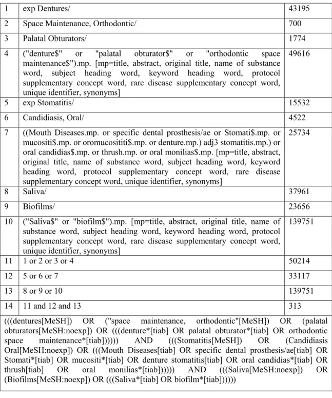
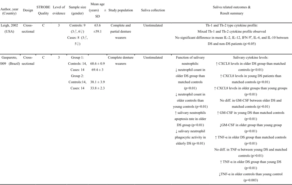
Table 1: Microbial species assessed by DNA-DNA checkerboard hybridization ... 73 Table 2: Coding index for signals generated by DNA-DNA checkerboard hybridization ... 73 Table 3: Conversion index of culture based CFU count to DNA-DNA checkerboard hybridization scores ... 74 Table 4: Percentage of total samples positive for each Candida species using DNA-DNA checkerboard hybridization and culture techniques ... 75 Table 5: Comparison between DNA-DNA checkerboard hybridization and culture techniques for the detection of Candida species ... 75 Table 6: Medline and PubMed search strategy ... 102 Table 7: Summary of characteristics of included studies and their findings ... 103
xiii
LIST OF FIGURES
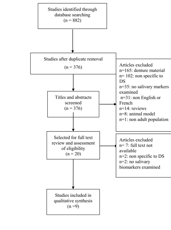
Figure 1: Distribution of inflammation and total Candida score using culture technique ... 76 Figure 2: Distribution of inflammation and total Candida scores using checkerboard techniques ... 77 Figure 3: PRISMA flowchart of the systematic review ... 106 Figure 4: Risk of bias assessment ... 107
xiv
LIST OF SYMBOLS AND ABBREVIATIONS
%: PercentageAIDS: Acquired immune deficiency syndrome ALS: Agglutinin-like sequence
ATCC: American type culture technique BCR: Biofilm and cell wall regulator
BHIYE: Brain heart infusion broth supplemented with yeast extract CAH: Carbonic anhydrase
CCH: Calcium channel homolog CFU: Colony forming units CMA: Corn meal agar
CRC: Concentrated rinse cultures CYTN: Cystatin
DES: Denture-associated erythematous stomatitis DNA: Deoxyribonucleic acid
DS: Denture stomatitis
EFG: Enhanced filamentous growth protein ELA: Elastase activity
ELISA: Enzyme-linked immunoabsorbent assay FIG: Factor-induced gene
GM-CSF: Granulocyte-macrophage colony-stimulating factor GPI: Glycosylphosphatidylinositol
xv GTT: Germ tube test
HAE: Haemophilus aegyptius endonuclease HWP: Hyphal wall protein
ICAM: Intercellular adhesion molecule IFN: Interferon
Ig: Immunoglobulins IL: Interleukin
LC-MS/MS: Liquid chromatography-mass spectrometry/liquid chromatography-tandem mass spectrometry
LIP: Lipase precursor
m/z: Mass-to-charge ratio
MID: Mating pheromone-induced death MeSH: Medical subject heading
MSP: Mitochondrial sorting of proteins NaCl: Sodium chloride
NaOH: Sodium hydroxide NLA: Neisseria lactamica NO: Nitric oxide
NRC: Neat rinse cultures
OCEBM: Oxford centre for evidence-based medicine PBS: Phosphate buffered saline
PCR: Polymerase chain reaction PDA: Potato dextrose agar
xvi PFGE: Pulsed field gel electrophoresis
PFU: Pyrococcus Furiosus PM: Percent method
PPIA: Peptidyl-prolyl cis-trans isomerase
PRISMA: preferred reporting items for systematic reviews and meta-analyses PWB: Primary wash buffer
RFLP: Restriction fragment length polymorphism rRNA: Ribosomal ribonucleic acid
SAP: Secreted aspartic proteases SCM: Standard curve method SDA: Sabouraud dextrose agar SDB: Sabouraud dextrose broth
SELDI-TOF/MS: Surface enhanced laser desorption/ionization time of flight mass spectrometry
sIg: Secretory immunoglobulins SPO: Salivary peroxidase spp: Species
SSA: Stress-seventy subfamily A STAT: Statherin
SWB: Secondary wash buffer TAQ: Thermus aquaticus
TE: Tris-Ethylenediaminetetraacetic acid TEC: Transposon enhancement control
xvii TGF: Transforming growth factor
TNF: Tumor necrosis factor
VEGF: Vascular endothelial growth factor VSM: Visual scoring method
xviii
DEDICATION
Dedicated to my parents, wife, and my three beautiful children
This was only possible with your prayers, encouragement, support, and positivity all of you bring to my life
xix
ACKNOWLEDGMENTS
First and foremost, I would like to thank Dr. Elham Emami for her exceptional support and guidance through the course of this academic program. Without her passion, innovative thinking, and critical feedback, this thesis would not have been possible. Most importantly, I would like to thank her for being a role model and showing me what empathy truly means.
I would like to thank; Dr. Jean Barbeau for co-supervising this thesis. His subject expertise was of tremendous value in giving the final shape to my project; Dr. Raphael F de Souza for sharing his experience, and his constructive comments during my master's degree. I would also like to acknowledge the effort of Mr. Pierre Rompre for helping in the design of the statistical analyses and assisting me throughout the process. I thank Mrs. Natalie Clairoux for her assistance in the design of the search strategy for this systematic review and Dr. Jocelyne Feine for revising the manuscript of this review.
Lastly, I am thankful to all my friends at the research unit especially Issam Kersheh, Aminah Alesawy and Amal Idrissi Janati for keeping my spirits high.
20
CHAPTER 1
LITERATURE REVIEW
1.1 INTRODUCTION
Denture Stomatitis (DS) or Denture-associated Erythematous Stomatitis (DES) is an oral biofilm-associated chronic inflammatory disease, which affects the oral mucosa covered by a removable prosthesis [1-7]. It is the most prevalent oral disease among edentate individuals [7-10], remaining asymptomatic or often presenting with symptoms like mucosal tenderness, bleeding, halitosis, burning sensation, xerostomia and dysphagia [4, 11-14]. Additionally, it has been linked to various systemic diseases, especially among hospitalized patients, individuals with a compromised immune system and elders with cognitive impairments and dementia [15-18].
While multiple risk factors have been investigated for their role in the etiology of this disease, the role of Candida continues to be frequently highlighted in the literature [5, 19, 20]. Consequently, current practice primarily focuses on antifungal prescriptions in addition to oral hygiene improvement [2, 21-23]. Following laboratory culture, a diagnosis may be made if the
Candida count is found to be >400 CFU/ml of saliva, in an otherwise healthy individual
[24-26]. However, in the current literature, there continues to be a lack of good quality reports to determine a direct cause-and-effect relationship between Candida and DS. We argue that the association of any microorganism to a disease is limited to the ability of the technology or method used to detect the microorganism. Therefore, clearly, the presence of Candida and its potential association with DS is dependent on the diagnostic test applied and its accuracy to
21
detect Candida spp. within the samples. The relationship between these two variables of interest should be investigated utilizing recently developed molecular methods such as the DNA-DNA checkerboard hybridization technique [27]. Since such techniques drastically differ from previous non-genetic tests such as the conventional laboratory culture, it is therefore imperative to determine the diagnostic accuracy of such methods in comparison to culture, to improve our understanding of the role of Candida in DS.
In the following sections of this chapter, we focus on the literature encompassing the risk factors of DS, the observed salivary immune response, and the methods available for the identification and quantification of Candida.
1.2 DENTURE STOMATITIS
1.2.1 Epidemiology
DS, DES or Candida-associated denture stomatitis is a chronic, erythematous oral inflammatory condition, observed on the oral mucosa covered by a dental prosthesis [1, 3-7, 10]. It affects both, complete and partial denture wearers and is most commonly observed on the denture bearing palatal mucosa, with a lower incidence in the mandibular mucosa [1, 9, 13, 21, 28]. DS is the most prevalent form of oral disease reported among completely edentate individuals and serves as the main indicator of poor oral health in this population [29, 30]. Despite the fact that people now tend to retain their natural teeth well into old age, the increase in the average life expectancy coupled with poorer socioeconomic status results in tooth loss and hence, the use of complete or partial prostheses becomes inevitable [7, 30-32]. Since complete tooth loss and denture use are most prevalent among disadvantaged individuals with
22
a low socioeconomic status, it is expected that this disease is more prevalent in this population group [29, 33].
DS affects a significant number of denture-wearing individuals. It is reported that 1 in every 3 [7-9], or 2 in every 3 individuals wearing dentures [34, 35], may present with some severity of this disease. The global prevalence of DS is reported to range between 15% and 77% [7, 30, 36-39]. University-based studies in the province of Quebec in Canada, have reported a prevalence up to 77.5% in a sample of complete denture wearers who visited university dental clinics [7, 40, 41]. While wearing complete dentures has been frequently reported to have a statistically significant correlation between DS [7, 9, 38, 42-44], some studies have also observed DS within study samples wearing partial dentures [8, 26, 33, 45].
Several studies have also shown that children and adults wearing acrylic partial dentures, obturators and ortho-appliances can be affected by DS [13, 33, 46, 47]. Furthermore, a systematic review by Emami et al. [21] reported that up to 36% individuals wearing chrome-cobalt or acrylic partial dentures may also present varying degrees of DS. However, it must be considered that the wide variations in the reported global prevalence of denture stomatitis may be attributed to the differences in the diagnosis, methods of data collection, choice of the study population; and associated geographic, socio-demographic and lifestyle characteristics [7, 21, 40, 41].
A higher prevalence of DS is observed in elders due to long-term denture use, lack of dexterity in performing oral hygiene, polymedication and decreased host immunity [38, 48-52]. It has also been reported to affect female denture wearers more often than males [12, 13, 38]. A possible explanation of a higher incidence among women observed by other authors may be due to the possibility that more women may sleep with their dentures due to aesthetic concerns
23
as previously reported by Coelho et al. [53]. In contrast, a recent study by Iosif et al. [54] in 2016 with a small sample size of 56 participants concluded that there were no statistically significant age and sex differences between subjects with and without Candida-associated DS. However, such difference in studies’ results could be related to type II error and underpowered studies.
1.2.2 Classification
Various classifications have been presented in the literature over the last few decades, aiding in the clinical diagnosis and staging of DS. The most commonly used classification continues to be the one presented by Newton [55], which is as follows:
Type I: Pinpoint hyperaemic lesions, particularly around the orifices of the ducts of the palatal
mucous glands (localized inflammation).
Type II: Diffuse erythema observed on the denture bearing mucosa (generalized
inflammation).
Type III: Inflammatory papillary hyperplasia (granular appearance).
A modified version of Newton's classification was presented by Barbeau et al. [28]. This modified version considers not only the type or intensity of inflammation but also identifies the extent or the spread of inflammation, by dividing the denture bearing mucosa into quadrants.
Type I: Pinpoint hyperaemic lesions, particularly around the orifices of the ducts of the palatal
mucous glands (localized inflammation).
Type II: Diffuse erythema observed on the denture bearing mucosa (generalized
24
Subclass A: Inflammation limited to 1 or 2 quadrants Subclass B: Inflammation extending to 3 or 4 quadrants
Type III: Inflammatory papillary hyperplasia (granular appearance).
Subclass A: Inflammation limited to 1 or 2 quadrants Subclass B: Inflammation extending to 3 or 4 quadrants
While both, the original and the modified Newton classification are regularly used, a more comprehensive classification presented by Schwartz et al. [56] provides a better representation of the severity (intensity) and area (extent) of the disease, making it easier to apply in a clinical setting:
Severity index:
0: Normal pink mucosa
1: Slight erythematous or mildly inflamed mucosa 2: Moderately inflamed mucosa
3: Severe or very pronounced inflamed mucosa
Area index:
0: No inflammation
1: Inflammation extending up to 25% of denture-bearing tissue
2: Inflammation extending between 25% and 50% of denture-bearing tissue 3: Inflammation extending over 50% of the denture-bearing tissue
The score obtained on the severity and area index are then summed up to obtain a final inflammation score which may vary between 0 and 6 [56].
25
1.3 ETIOLOGY AND RISK FACTORS FOR DENTURE
STOMATITIS
The etiology of DS is considered to be multifactorial in nature and continues to be poorly understood [7, 25, 54, 57, 58]. As the name of this pathological condition suggests, the presence of mucosal inflammation associated with DS is dependent upon the introduction of a denture into the oral cavity [59]. The mere presence of a partial or complete denture will initiate contact and promote microbial adhesion to the oral mucosa [45, 60]. Several underlying factors may predispose individuals and increase their susceptibility to DS [61, 62]. These risk factors may, therefore, be divided into "local" or prosthesis-associated factors, and "general" or systemic risk factors [9, 58, 63, 64].
Local or prosthesis-associated modifiable risk factors of DS are trauma from unadjusted or ill-fitting dentures, the age of the prosthesis, denture hygiene related factors which include denture cleaning/brushing and denture wearing habits i.e., interrupted, continuous and/or nocturnal wear [2, 5, 7, 9, 25, 28, 38, 41, 43, 65, 66]. As the dentures age, they lose retention and stability due to the pathological changes in the edentulous oral cavity such as the development of mobile ridges, and reduction in the vertical dimension of occlusion, thus inducing trauma to the oral mucosa [25, 41, 44, 67-71]. It has also been suggested that an important cause of denture instability is an improper inter-occlusal relationship altering the patterns of occlusal load transmission to the tissues under the denture bases, resulting in DS [67, 72, 73].
The continuous and nocturnal denture wear is considered to inflict uninterrupted pressure on the denture bearing tissues, inhibit the oxygenation of the oral mucosa, and impede the
26
cleaning effect of the tongue and saliva, thus making the oral mucosa more sensitive to cell injury and prone to inflammation [9, 28, 41, 74-76]. Furthermore, mucosal coverage by the denture base creates an acidic and an anaerobic local microenvironment that promotes pathogens like Candida spp. and other microorganisms to proliferate within the biofilm, producing toxins and metabolic waste responsible for cell injury and resultant inflammation, the main clinical feature of DS [66, 77-80].
General or systemic risk factors for DS reported in the literature include old age, smoking, obesity, sugar consumption [7, 9, 10, 14, 28, 38, 45, 63, 81, 82], xerostomia, diabetes mellitus and immunosuppressive conditions such as AIDS [14, 45, 63, 83, 84], the use of antibiotics, corticosteroids, hormones and other xerogenic agents [45, 82, 85-88], and lastly, malnutrition including deficiencies in proteins, iron, vitamin A and B [9]. Martori et al. [80] conducted a cross-sectional study involving 84 geriatric denture wearers and examined the correlation between various local and systemic risk factors and DS. Using multiple logistic regression models with the observed inflammation as the dependent variable, an association was observed between DS and low salivary pH (OR 0.057; 95% CI 0.01-0.48), smoking (OR 152.8; 95% CI 2.28 to >999) and sugar consumption (OR 6.917; 95% CI 1.17-40.9).
In general, it is difficult to ascertain a direct cause-and-effect relationship between the factors nominated as etiological factors in the literature because of the studies’ design and their cross-sectional nature [21, 28, 40, 41]. However, from the available evidence, three factors may play an important role in the occurrence of DS [7, 89, 90]. These include mucosal trauma [28, 36, 41, 91], oral biofilm and specific bacteria [76, 92], as well as pathogens such as Candida spp., and more specifically, Candida albicans [2, 36, 40, 76, 78, 93].
27
1.3.1 Mucosal trauma
The role of denture-induced trauma has been frequently reported as a risk factor for DS [25, 36, 41, 80, 91]. Historical studies conducted by Budtz-Jorgensen research group involving 58 DS patients using complete dentures for a very long period of time (mean age of denture 26.8 years), suggested that the inflammation observed in DS was increasingly linked with poor denture hygiene and continuous mucosal irritation resulting in mechanical trauma caused by ill-fitting dentures and unbalanced occlusion [4, 70].
The susceptibility of the palatal mucosa to trauma induced by a denture may also be dependent upon the presence or absence of natural teeth in the opposing jaw, as well as the type of prosthesis [67]. This is further supported by a study conducted by Emami et al. [41] which concluded that the risk of DS was 4.5 times greater in patients wearing mandibular conventional dentures than in those who were rehabilitated with more stable implant-assisted overdentures.
An animal model study on mucosal biomechanics showed a high correlation between histopathological changes in the palatal mucosa and occlusal forces transmitted due to instability and poor retention of the prosthesis [94]. These continuous forces compromise the circulation under the mucosa, thus resulting in swelling, edema, mucosal inflammation and eventually bone resorption [95-99]. It is therefore suggested that the mucosa covered by a denture base may not exhibit signs of inflammation in the absence of mechanical pressure or trauma caused by dentures [7, 28, 73, 94, 97].
28
1.3.2 Oral biofilm and bacterial species
The commensal microbiota of the oral cavity comprises a wide variety of microorganisms, including viruses, protozoa, fungi, and bacteria [100]. These microorganisms colonize different parts of the oral cavity like the teeth, gingiva, tongue, mucosa, throat and the palate [101], by adhering to a glycoprotein pellicle, and proliferating to form the dental plaque [102], and in the presence of a denture, forming the denture biofilm. This biofilm is a “microbially
derived sessile community characterized by cells that are irreversibly attached to a substratum or interface or to each other, embedded in a matrix of extracellular polymeric substances that they have produced” [103-108].
The oral biofilm covering the mucosa and denture surfaces provides ideal conditions for the microorganisms within the biofilm to proliferate, and cause mucosal inflammation [109, 110]. These microorganisms continue to co-aggregate, further utilizing habitat-specific nutrients [111], thus forming a heterogeneous and highly diverse ecological environment in the oral cavity, upon which the health and disease status of the host is dependent [10, 112].
Comparing the microbiome of denture wearers, those with and without DS, it has been suggested that bacterial species such as α-hemolytic Streptococci and Neisseria may play an important role in the inflammation associated with DS [113]. It has also been observed that while Candida spp. were in higher quantities in DS patients, bacterial species such as
Streptococci, Lactobacilli, and Actinomyces were also present in the denture biofilm of
patients with DS [114]. Budtz-Jorgensen et al. [115] observed similar findings in their study utilizing 1239 isolates of denture biofilm samples taken from DS patients. They concluded that DS was associated with a high bacterial count, mainly gram-positive rods (median 45%) comprising of Lactobacillus spp. (median 19%) and Actinomyces spp. (median 9%).
29
Van Reenen [116] examined the changes in the counts of Streptococci and Candida spp. isolated from the denture biofilm of denture wearers with DS. They observed a reduction in inflammation after prescribing antibiotics, suggesting that bacteria may play an important role in the inflammation observed. This may be explained by the synergistic nature of Streptococci, which create a favorable environment for yeasts by producing lactate, providing carbon for the yeast to feed and thrive upon [117]. Investigating the oral microbiome associated with DS using a high-throughput 16S rRNA sequencing technology, O'Donnell et al. [10] showed that the denture biofilm in DS subjects had a higher proportion of Bacteroidia attributed to
Prevotella and Veillonella (p<0.05). Additionally, the inflamed mucosa also had a high
prevalence of Actinobacteria and Bacteroidia, suggesting similarities between the denture and mucosal biofilm [10].
The microbial diversity of the oral biofilm may consequently point towards a possible role of non-candidal microorganisms in addition to Candida spp. in the etiology of DS [93, 118].
1.3.3 Candida species and denture stomatitis
Candida spp. have received the most attention as the primary etiology of DS, as studies report
a high prevalence of these microorganisms in DS patients [5, 19, 20]. Candida spp. exist as commensal but opportunistic microorganisms on the epithelial surfaces of the human body, including the oral cavity [109, 119-121]. It has been reported that about 75% to 100% of the population may demonstrate Candida specific immunity [121-123], suggesting previous exposure to the microorganism.
The most common site considered to harbor Candida is the mucosal surface of the denture base [4, 124]. Budtz-Jorgensen et al. [125] conducted an epidemiological study involving 560
30
individuals above the age of 65 years and compared mucosal and denture biofilm samples of those with and without DS. Their results showed that in yeast form, Candida albicans were the most common species grown in pure culture among both study groups. However, there was a statistically significant difference in the concentration of Candida hyphae between the two groups; 77% in individuals with DS and 47% in individuals without DS (x2 test, p <0.001) [125]. Additionally, the presence of inflammatory cells along with hyphae was higher in DS patients (65%), as compared to healthy participants (14%) [125]. The presence of yeast has also been reported in the unstimulated saliva in 90% of study subjects with DS [126].
Candida albicans have also been reported to be the most prevalent species isolated from
healthy and immunocompromised patients suffering from DS [127-129]. Budtz-Jorgensen et al. [110] showed that Candida spp. count in DS subjects was 100 times higher than in healthy denture wearers, with Candida albicans, Candida tropicalis and Candida glabarata as the most commonly isolated species. Furthermore, MacFarlene et al. [1] reported that the most prevalent Candida spp. in DS lesions were the albicans, followed by glabrata and tropicalis.
1.3.3.1 Candida virulence factors
The ability of Candida to trigger a host immune response and cause inflammation is due to various virulence factors. These factors include:
I. Dimorphism
An important property of Candida, that plays a role in its virulence and pathogenicity is dimorphism, which is the ability that Candida exhibits to transition between yeast and hyphal forms, frequently observed in diseased conditions [130].
31
Dimorphism plays an important role in the formation of the biofilm and also aids in tissue invasion [131]. The biofilm formation involves two steps: Following the initial role of adherins, dimorphism ensures candidal adherence to the substrate in the hyphal state and later, dispersion from the biofilm in the yeast state following hyphal replication and extracellular matrix formation [132, 133]. While Candida in the hyphal form is observed to show a higher level of invasiveness, the yeast form exhibits increased virulence [134, 135]. Several Candida transcriptional factors, namely Bcr1, Tec1, and Efg1 are also considered to play an important role in the formation of the oral biofilm on mucosal and prosthetic surfaces [136].
Dimorphism also regulates the contact sensing ability of Candida through which it senses contact surfaces and switches from yeast form to hyphal growth, resulting in tissue invasion [131]. Furthermore, certain extracellular calcium channels; Cch1, Mid1, and Fig1, as well as the polarisome module; Ras-like GTPase Rsr1/Bud1, have shown to regulate the ability of hyphae to grow in a particular directional pattern, depending on the topology and surface characteristics of the substrate [137, 138].
II. Host recognition and cellular attachment
Of particular importance in the oral cavity, is the ability that Candida possesses to recognize and attach to host cells through various surface mannoproteins called adhesins [139]. Most notable of these are Agglutinin-like sequence (ALS) proteins Als1–7 and Als9, particularly Als3 which has been shown to be up-regulated in oral epithelial cells [140, 141]. Another protein, Hwp1 aids in forming covalent links between candidal hyphae and host cells [142]. Both ALS and Hwp1 proteins are glycosylphosphatidylinositol (GPI)-linked proteins and aid in candidal adhesion to host cells as well as biofilm formation [142, 143]. In addition to host cell adhesion, Als3 and Ssa1 have also shown to play a role in the cellular invasion by acting
32
as invasins that bind with host cell ligands resulting in induced endocytosis, whereby the fungal cell is engulfed into the host cell [144, 145]. In contrast to a passive endocytosis mechanism triggered by invasins, the viable Candida hyphae penetrate the host cells through an active invasion mechanism [146].
III. Tissue hydrolysis
Another important virulent characteristic of Candida is its ability to release hydrolytic enzymes such as proteases, phospholipases and lipases which aid in tissue penetration following cellular adhesion [147]. The proteases (Sap1-10) comprise the largest hydrolase family in Candida albicans and are considered to play a significantly virulent role in epithelium invasion [148, 149]. Phospholipases (A, B, C, and D), and lipases (LIP 1-10) are also considered to play an important role in the disruption of host cell membrane and pathogenicity of Candida albicans [150, 151].
IV. Withstanding pH
Candida have also shown great adaptability to the surrounding pH of the host environment,
which can vary greatly depending upon the location; from very acidic (pH 2) in the stomach to slightly alkaline on the palatal mucosa (pH 7.34) [152, 153]. In addition, its ability to transform into hyphal form helps it to withstand an acidic environment [130]. Furthermore, it also inherits processes to regulate extracellular pH by uptaking amino acids and cleaving them intracellularly, thus producing ammonia to alkalinize the surrounding pH [154, 155].
More recently, literature has raised questions regarding the association between Candida
albicans and DS. For instance, Emami et al. [40] investigated the relationship between
33
in the counts of Candida albicans, among patients with DS and healthy subjects. Similarly, another study observed that the high Candida count in DS was associated with the area of inflammation (mucosal area coverage), and not the intensity or severity of inflammation [28]. This coupled with evidence of a high recurrence rate of DS after the cessation of antifungal therapy [156-158], may suggest a more complex role of other factors such as the host immune response to microbial insult.
1.4 HOST IMMUNE RESPONSE OBSERVED IN DENTURE
STOMATITIS
Saliva plays a critical role in maintaining the integrity of the hard and soft tissues in the oral cavity by regulating the local immune response observed in oral inflammatory diseases, like DS [159]. The inflammatory cascade is triggered as a protective response to cell injury, exhibiting an interaction between cells and inflammatory mediators such as vasoactive amines (histamine, serotonin), phospholipids (platelet-activating factor), arachidonic acids (prostaglandins, leukotrienes) and cytokines (tumor necrosis factor, interleukins, interferons, and colony stimulating factors) [160-162]. This physiological process leads to vasodilatation, increased microvascular permeability, cellular activation, cellular adhesion and coagulation, which increases the available oxygen and nutrients at the site of injury, thus generating heat and provoking tissue edema [160, 162].
A notable immune response is observed within saliva in response to the pathogenic microorganisms causing DS [50, 163-165], thereby playing a protective role in the host defense mechanism [166]. Salivary and blood neutrophils serving as biomarkers mediate cytokine liberation through diapedesis, chemo-attraction, phagocytosis and activation events,
34
therefore responding in an acute manner to protect against the establishment of oral diseases [167].
Saliva also plays a role in the humoral and cell-mediated adaptive immunity, displayed by the high prevalence of immunoglobulins [168] and distinct cytokine profiles [163]. In fact, the predisposition towards Candida infections among the elderly denture-wearing population may be explained by salivary immunosenescence or the deterioration of immunity due to the advancement of age [169]. Furthermore, defense mechanisms such as phagocytosis (or uptake) have been shown to be impaired among the elderly [50], while induced neutropenia through IL-17 pathway blockade in a mice model also increased disease susceptibility [170-172]. Other components such as salivary proteins like secretory immunoglobulin A (IgA), lactoferrin, lysozyme, and histatins, also function as biomarkers and have antifungal effects [173, 174]. Among these, IgA acts as a barrier protecting against antigen invasion and is seen in a higher concentration in DS [175]. Most importantly, lactoferrin inhibits bacterial growth by sequestering essential iron and also exhibits non-iron-dependent antibacterial, antifungal, antiviral, antitumor, anti-inflammatory, and immunoregulatory activities [174]. Differential susceptibility to DS may be attributed to differential concentrations of the aforementioned salivary proteins, which are measured by proteomic profiling of saliva in DS patients [176]. For instance, levels of Vascular endothelial growth factor (VEGF), which are considered to be the underlying markers of chronic inflammatory or autoimmune conditions in the oral cavity, were observed to be decreased in type I DS and increased in type II DS, when comparing DS subjects with and without type 2 diabetes mellitus [71, 177, 178].
35
1.5 METHODS FOR THE ISOLATION AND DETECTION OF
CANDIDA SPECIES
1.5.1 Candida isolation techniques
A variety of methods have been used for the isolation of microorganisms including Candida from the site of inflammation in the oral cavity. The decision to select a particular method is dependent on the objective of the research, the nature of the lesion observed and the kind of technique planned to be used for the quantification and identification of various Candida spp. The following methods are validated sampling methods used for Candida isolation from the oral cavity:
I. Smear
This process involves the collection of a superficial sample from the site of inflammation in the oral cavity by firmly wiping the area using a sterile wooden stick or blade [25, 179], or gently using a cytology brush [180]. Obtained smears are observed on a glass slide either in moist form fixed with ether/alcohol (1:1) [179], Cytofix/Cytoperm (Becton, Dickinson and Company, Franklin Lakes, NJ) [25], or as a dry smear following staining using either Gram-stain or Periodic acid-Schiff (PAS) techniques [179, 181]. Microscopic examination of an oral smear obtained from a suspected Candida-associated denture stomatitis lesion will reveal the presence of Candida spp., visible either as hyphae or blastophores [181, 182].
II. Swab
In addition to smears, mucosal swabs are also one the most widely used methods to screen and diagnose oral and systemic diseases [183]. Additionally, they are also frequently used to
36
isolate DNA for genomic and forensic research [183, 184]. In order to isolate microorganisms such as Candida spp. found in DS, a cotton swab sterilized in the laboratory, or a pre-packaged sterile swab may be used [185]. Similar to a smear collection, the sample is collected by gently running the cotton swab over the site of the inflammatory lesion on the palate, rugae area and the denture fitting surface [2, 25, 186]. In order to ensure microbial viability, the sample is stored on ice while being transported to the laboratory [186].
III. Imprint culture
Benefiting from the adherence properties of Candida, another technique termed as imprint culture is also routinely used for isolating and quantifying Candida from the oral cavity [20, 181, 186, 187]. This involves using a sterile foam-like pad dipped in saline or a liquid medium like Sabouraud dextrose broth (SDB), placing it at the specific site to be investigated i.e., inflamed oral mucosa or the denture fitting surface, and leaving it in place for 10 to 60 seconds [24, 188-190]. The imprint pad is then placed on Sabouraud dextrose agar (SDA) and left for 60 minutes to ensure that the sample has been adequately transferred onto the agar before incubation [189]. A variation of this method allows the foam to be left in-situ for the first eight hours of the 48-hour incubation cycle [179].
IV. Oral rinse
A swab or an imprint culture is often followed by an oral rinse for further analysis. This is done by asking the patient to rinse with 10ml of sterile phosphate buffered saline (PBS: 0.01 M, pH 7.2) for 60 seconds [179, 191]. The rinse is collected in a sterile container and transferred to the laboratory for analysis.
37
In the laboratory, the collected oral rinse may either be cultured on SDA as neat rinse cultures (NRC) or as concentrated rinse cultures (CRC) [191]. In order to concentrate the obtained rinse, the neat rinse is centrifuged at 1700g for 10 minutes [186, 191]. More recently, centrifuging the neat rinse at 2000g for 10 minutes [192] and at 2300g for 20 minutes [185] has also been reported. Following the removal of the supernatant, the pellet obtained from the centrifugation process is mixed in a predefined amount of the original solution (500 μL) and inoculated onto agar media in 100 μL aliquots using a spiral plating system [185, 188].
V. Saliva
Biomarkers within human saliva provide extensive information about the etiology, pathophysiology, and prognosis of various diseases [193-195]. Additionally, a collection of biofluids such as human saliva serves as a non-invasive method for the screening and diagnosis of oral and systemic diseases [183, 195]. Salivary culture serves as a reliable method for quantifying Candida spp. isolated from the oral cavity and assists in differentiating between the carrier and infectious states [196]. As a reference, Epstein et al. [197] demonstrated that a salivary Candida count of >400 CFU/ml is considered as an infected state, while <400 CFU/ml of saliva is considered as a carrier state [25, 26, 198].
Depending on the study objective, stimulated or whole unstimulated saliva may be collected utilizing various commercially available collection kits. Stimulated saliva can be collected using Salivette® with cotton swabs (Sarstedt, Nümbrecht, Germany) or using paraffin gum (Ivoclar Vivadent, Schaan, Lichtenstein) [199]. The patient is asked to swallow the saliva already present in the mouth, then chew on a paraffin gum or strip for 2 minutes, followed by spitting the saliva in a sterile container [25]. Provided that the patient has normal salivary production, unstimulated whole saliva may be used for quantifying Candida using culture
38
techniques [163, 169, 200, 201]. The standard protocol for the collection of unstimulated saliva requires the patient to sit upright with their head tilted slightly forward, and passively drool into a sterile 50ml falcon tube for 5 minutes [168, 202]. The total quantity of saliva produced over the predefined duration of time is recorded in order to calculate the average salivary flow (ml/min) [168, 202].
VI. Tissue biopsy
Candida-associated DS in its severe form may present with hyperplastic tissue or papillary
hyperplasia on the palatal tissue region covered by the denture [55]. A tissue biopsy taken from the affected site is indicated for histopathological examination. The procedure involves anesthetizing the palatal mucosa, followed by a 2-4 mm full thickness punch biopsy from the keratinized epithelium to the periosteum [25]. The histopathological examination involves observing the biopsies for epithelial and connective tissue inflammatory reactions [203].
1.5.2 Candida detection and differentiation techniques
In a laboratory setting, the samples obtained from the above-mentioned methods may be processed using a variety of techniques. While some of these techniques are simply limited to the detection (absence or presence) and quantification of Candida using culture media, others may be used in the identification and differentiation of particular Candida spp. based on their morphology and genetics.
I. Laboratory culture
Culture media have been used for isolating and detecting microorganisms in a controlled laboratory environment for well over a century. The initial attempts to use solid media for growing microorganisms outside the human body can be traced back to the early 1830s when
39
Italian scientist Bartolomio Bizio successfully cultured a chromomeric bacterial species,
Serratia marcescens [204]. However, it was not until 1881, when Robert Koch upon receiving
advice from Fanny Hesse used agar to make the first stable solid culture media [205]. The French dermatologist Sabouraud later formulated a standardized method of developing agar media to culture fungi and bacteria [206].
Sabouraud dextrose agar (SDA) and Sabouroud dextrose broth (SDB) continue to be the most commonly used media for the isolation of Candida spp. [207]. The slightly acidic nature (5.6 pH) and high content of dextrose (4%) of this medium ensure rapid fermentation and acid production, thus inhibiting bacterial growth [207, 208]. The clinical samples are inoculated on the medium and incubated at 37oC for 24-48 hours [25, 209] or 48-72 hours [210, 211]; following which convex, smooth, creamy colored Candida colonies can be observed [210, 211]. Certain antifungals like azoles may also be added to the culture medium for the selective growth of Candida, or to test drug susceptibility [212-215]. In addition to SDA and SDB, numerous other commercially available non-selective agar and broth media may also be used for growing Candida, including Potato dextrose agar, Nutrient agar, Brain heart infusion broth supplemented with yeast extract (BHIYE) and Corn meal agar (CMA) [216]. However none of these culture media permit the differentiation between various Candida spp., and therefore the use of differential media is employed [207, 208, 217].
Differential media such as Pagano-Levin agar, CHROMagar Candida (CHROMagar, Paris, France), Albicans ID (bioMerieux, Marcy l'Etoile, France) and Fluoroplate (Merck, Darmstadt, Germany) allow for the differentiation between various Candida spp. [188, 218, 219]. Based on the color of individual colonies observed on CHROMagar Candida following the incubation cycle, Candida spp. can be differentiated as C. albicans (green), C. tropicalis
40
(blue) and C. krusei (light pink / pale rose), with a sensitivity and specificity between 95% [220] and 99% [218]. Albicans ID media (bioMerieux, Marcy l'Etoile, France) and CHROMagar Candida, both have shown comparable sensitivity and specificity close to 100% for the differentiation between albicans and non-albicans Candida spp. [221], which appear as blue and green colored, smooth colonies on each medium, respectively [218, 219].
While the above-mentioned media rely on the chromogenic substrate in their composition, Fluoroplate (Merck, Darmstadt, Germany) contains a fluorogenic substrate which differentiates between albicans and non-albicans Candida spp. based on their fluorescence observed under a 365nm UV light, whereas all non-albican species appear pale white [219, 222, 223].
II. Morphologic test
The use of microbiological cultures is often followed by a morphologic test such as the Germ tube test (GTT), for the presumptive identification and differentiation between albicans and non-albicans Candida spp. [188, 210, 211]. The test utilizes the dimorphic nature of Candida
albicans recognized as a virulence factor, which gives it the ability to switch between yeast
and mycelial forms, and the formation of chlamydospores [224-229]. Candidal dimorphism or morphological switching can be induced under conditions such as the presence of an inducing substrate like human serum, optimal temperatures (>33oC), an approximately neutral pH and starvation [227, 230].
Using a straight wire, a colony of yeast or a small inoculum grown on solid media is transferred into a tube containing either human or animal serum and incubated at 37oC for 2-3 hours [231-233]. Other media used may include serum substitutes [229], 1% bactopeptone in 2% agar [234, 235], 0.1% glucose in 2% New Zealand agar [235], rice extract and
41
carbohydrate media [236], and bovine albumin [232, 233]. Following incubation, a small amount of the suspension is placed on a glass slide, covered with a coverslip and observed under a microscope for the formation of filaments or hyphae [211, 231, 232]. Microscopic examination reveals the formation of true hyphae which appear as cylindrical tubes extending from the body of the yeast with no constriction at their base [237], an appearance typical of C.
albicans and C. dubliniensis, differentiating them from C. glabrata and C. krusei [210, 211].
Furthermore, when inoculated on solid agar containing Tween 80 and incubated for 72 hours at 22oC, C. albicans and C. dubliniensis exhibit chlamydospore formation [188, 238].
Apart from C. albicans and C. dubliniensis other Candida spp. like C. stellatoidea and C.
tropicalis may also exhibit germ tube formation [188, 239]. It is therefore essential to adhere
to the 2-3 hour time limit for the incubation cycle, as species other than albicans and C.
dubliniensis may also start to develop germ tubes as the incubation period increases [225,
240]. Since C. albicans and C. dubliniensis share morphological similarities, that is, both species develop germ tube and chlamydospores [224-226, 241]; further differentiation between the two is often required. Incubation of the inoculated media at 42oC can be used as a confirmation of the presence of C. albicans, as C. dubliniensis do not form germ tube at an elevated temperature and test negative for germ tube formation [225].
III. Genetic tests
While the above mentioned conventional techniques are valid methods for Candida detection and differentiation, they are nonetheless limited to the presumptive identification of the microorganisms, based on either the color and appearance on culture media or their morphology following a GTT [237]. The following methods, however, are more sensitive and
42
specific, providing a definitive identification of Candida spp. based on the genetic variability between various strains [242].
i. Electrophoretic karyotyping & Restriction fragment length polymorphism
Electrophoretic karyotype analysis involves the separation of the Candida chromosomal DNA or other macromolecules on an electrophoretic gel such as the agarose gel matrix [243, 244]. The separation is done based on the size of the DNA molecule and involves two steps; preparing the DNA while ensuring minimal to no degradation, and separating the DNA molecules by applying an electrical current through a process called gel electrophoresis [243, 245, 246].
However, conventional electrophoresis techniques are often limited due to their inability to separate molecules over 25-50 kilobases (kb) [243, 244, 247]. Yeast DNA molecules, which may range over several hundred kilobases can, therefore, be separated using mechanisms such as Pulsed field gel electrophoresis (PFGE) [248, 249], thus enabling the detection and identification of fungi, including Candida spp. [182, 246, 247]. The original technique as described by Schwartz et al. separates the DNA molecules in agarose matrices by the alternate activation of electrical fields placed perpendicular to one another [243, 247]. A distinct pattern can be observed due to the relative number and size of the chromosomes of various microbial species using PFGE, therefore making electrophoretic karyotyping useful in differentiating between closely related microbial species [247].
Restriction fragment length polymorphism (RFLP) provides an alternate to karyotyping. This process involves the isolated DNA to be digested and cleaved, or fragmented using DNA restriction enzymes (MspI, NlaIII, HaeIII, DdeI, EcoRI and BfaI), prior to being subjected to electrophoresis in an agarose gel matrix [250-253]. DNA fragments of varying length then
43
hybridize with the specific DNA sequence used as an RFLP probe, following gel electrophoresis [250]. The resultant bands can be observed due to the luminescent dye used in the gel [188]. However the use of electrophoretic karyotyping and RFLP have several limitations, as they are expensive, require specialised equipment, and need 48 hours for DNA extraction and 72 hours for PFGE (which depending on the size of the molecules can take up to weeks) [247].
ii. Polymerase Chain Reaction (PCR)
Polymerase chain reaction (PCR) based techniques provide a rapid and cost-effective alternative to electrophoretic karyotyping and RFLP [254, 255]. In addition, they are highly sensitive and specific [256] for the detection of pathogenic microorganisms, including oral pathogens [249, 257, 258], as compared to conventional microbiological techniques [249]. They are based on the production of a large quantity of any specified DNA for analysis, by repeating the DNA extension reaction, bounded by primers [258, 259].
The original technique described by Saiki et al. [256, 260], was first employed for the enzymatic amplification of beta globulin genomic sequences for the prenatal diagnosis of sickle cell anemia. Since then, various PCR techniques developed over the course of years have been widely used for the detection and identification of yeast and Candida spp. [252, 261-267]. Furthermore, PCR is also regularly used for the definitive differentiation between various Candida spp., especially C. albicans and C. dubliniensis [253, 268-270].
The components of a conventional PCR typically include a DNA template which contains the target sequence, a DNA polymerase enzyme such as the Taq DNA polymerase or Pfu DNA polymerase [254, 270], which are short pieces of single stranded DNA responsible for producing DNA sequences complementary to the target DNA, and primers which are short
44
strands of complementary DNA that enable the DNA polymerase enzyme to add nucleotides to the primer DNA strand [271].
Each PCR cycle involves three stages: Denaturation of the template DNA by heating at 94oC for 1 minute in order to break the hydrogen bonds between the strands, cooling down of the reaction to approximately 57oC for 1 minute resulting in the primers forming bonds with the template DNA in a process termed as annealing; and the final process called extension where the reaction is reheated to 72oC for 1 minute, allowing the polymerase enzymes to add nucleotides to the primers thus completing a single DNA replication cycle [126, 260, 267].
iii. DNA-DNA checkerboard hybridization
DNA-DNA checkerboard hybridization technique is a culture independent, molecular technique used for the identification and quantification of microorganisms, including those that are non-cultivable [188, 272, 273]. The technique was initially introduced by Socransky et al. [27] for the study of microorganisms isolated from periodontal lesions based on their genetic variability and has since been widely used in dentistry for studying microbiota in a variety of oral conditions [272-279]. Instead of focusing on a few microorganisms, this technique analyzes the samples for a large number of microorganisms, allowing for a more exploratory perspective and thus, may successfully highlight a microorganism in quantities that may be out of its normal range. That is to say, a microorganism previously neglected by researchers may show a pattern which may play an important role in the pathogenesis of the disease [280].
The DNA-DNA checkerboard hybridization technique allows 28 samples to be simultaneously analyzed for 40 microbial species on a single membrane, using whole genomic DNA probes as controls [27, 272, 273, 281]. As described by Socransky et.al [27], and modified by
45
Nascimento et al. [282], samples collected from the oral cavity or the site of the lesion are stored in tubes containing 0.15ml TE buffer, into which 0.15ml of 0.5M NaOH is added and boiled for 5 minutes. Following this denaturation process, the denatured DNA samples are deposited onto a 15x15 nylon membrane (Boehringer Mannheim® or Hybond N+®, GE Healthcare Life Sciences do Brazil, São Paulo-SP, Brazil) using a Minislot 30 TM (Immunetics, Cambridge, MA, U.S.A) and affixed using a UV light (Stratalinker 1800, Stratagene, La Jolla, CA, U.S.A), followed by baking at 120oC for 20 minutes [27] or at 80oC for 2 hours [282]. The membrane with the fixed sample DNA and control DNA (105 and 106 microbial cells of each species), is prehybridized at 42oC for 1 hour [27] or 60oC for 2 hours (0.5 M NaCl; 0.4% w/v blocking reagent) [274, 282], and then placed in a Miniblotter 45TM (Immunetics; Cambridge, MA, U.S.A) in a perpendicular or cross-ways pattern and hybridized overnight at 42oC [27] or 60oC [274]. The membrane is then washed and visualized for hybridization signals using Storm FluorimagerTM (Molecular Dynamics, Sunnyvale, CA, U.S.A) which are then converted to absolute counts [27, 280].
46
CHAPTER 2
METHODOLOGY
2.1 PROBLEMATIC AND OBJECTIVES
Candida species have been considered the most important factor predisposing denture wearers
to DS [2, 40, 118, 283]. The diagnosis of Candida-associated DS can only be made by analyzing the biological samples of patients with DS using a variety of microbiological techniques. Microbial culture is the most commonly employed laboratory technique, providing a semi-quantitative or quantitative estimate of the Candida count, expressed as Colony forming units (CFU) [207, 284]. A count of >400 CFU/ml is indicative of a moderate to high
Candida load and may be considered for antifungal treatment [25]. In addition to non-specific
media like SDA, selective media such as CHROMagar is also required to specifically differentiate between various Candida spp. [284].
Performing multiple procedures for the accurate detection of Candida spp. can be a laborious and cumbersome task that requires a significant amount of time and resources. DNA-DNA checkerboard hybridization technique developed by Socransky et al. [27] is a molecular diagnostic method, that provides an alternate method for Candida detection, and has been used in numerous studies to identify and quantify multiple microbial species including Candida in clinical samples [242, 272, 285, 286].
According to the literature, evidence on the potential uses and application of DNA-DNA checkerboard hybridization technique in the diagnosis of Candida-associated DS is scarce.
47
Furthermore, since the inflammatory biomarkers in saliva could offer a new venue for the early diagnosis of this disease, this two-part master's research project aims at providing new evidence and shed light on these topics.
Specific objectives
2.1.1 Checkerboard vs. culture
1. Primary objective: To compare the diagnostic accuracy of microbial culture and DNA-DNA checkerboard hybridization techniques for the detection of Candida spp. in the palatal biofilm of denture wearers with DS, using each technique as the reference for the other.
2. Secondary objective: To compare the relationship between Candida counts using the two methods, and the extent and severity of palatal inflammation, in denture wearers diagnosed with DS.
We hypothesize that there is no statistically significant difference between microbial culture and DNA-DNA checkerboard hybridization techniques in their diagnostic accuracy for the detection of Candida spp., and there is no association between palatal inflammation in denture wearers diagnosed with DS and Candida counts measured by the two methods.
2.1.2 Systematic review
1. To identify and evaluate the quality of literature examining the differences in salivary biomarker profiles of healthy denture wearers, and those with DS.
48
2.2 RESEARCH METHODOLOGY
2.2.1 Checkerboard vs. culture
2.2.1.1 Study design and study participants
The first part of this master's research project is a secondary analysis of the data, which was collected in our previous two-center (Canada, Brazil) trial entitled: "The effect of palatal brushing on denture stomatitis" (registered as NCT01643876 on Clinicaltrials.gov) [2].
The data analysis was conducted only on the data obtained from Brazil (University of São Paulo, Ribeirão Preto) since checkerboard hybridization was only conducted at this center. Therefore, this study included twenty-six participants (male, n = 4, female, n = 22) as previously detailed in the published article [2].
2.2.1.2 Data collection and measurement instruments
Data collection included a clinical examination for the diagnosis of DS and a microbiological investigation using microbial culture and DNA-DNA checkerboard hybridization.
Clinical investigation
Clinical diagnosis of DS was carried out by two trained dentists and was defined according to the Schwartz's area and severity index [56].
I. Schwartz index: Inflammation area index: 0: No inflammation