AVIS
Ce document a été numérisé par la Division de la gestion des documents et des archives de l’Université de Montréal.
L’auteur a autorisé l’Université de Montréal à reproduire et diffuser, en totalité ou en partie, par quelque moyen que ce soit et sur quelque support que ce soit, et exclusivement à des fins non lucratives d’enseignement et de recherche, des copies de ce mémoire ou de cette thèse.
L’auteur et les coauteurs le cas échéant conservent la propriété du droit d’auteur et des droits moraux qui protègent ce document. Ni la thèse ou le mémoire, ni des extraits substantiels de ce document, ne doivent être imprimés ou autrement reproduits sans l’autorisation de l’auteur.
Afin de se conformer à la Loi canadienne sur la protection des renseignements personnels, quelques formulaires secondaires, coordonnées ou signatures intégrées au texte ont pu être enlevés de ce document. Bien que cela ait pu affecter la pagination, il n’y a aucun contenu manquant.
NOTICE
This document was digitized by the Records Management & Archives Division of Université de Montréal.
The author of this thesis or dissertation has granted a nonexclusive license allowing Université de Montréal to reproduce and publish the document, in part or in whole, and in any format, solely for noncommercial educational and research purposes.
The author and co-authors if applicable retain copyright ownership and moral rights in this document. Neither the whole thesis or dissertation, nor substantial extracts from it, may be printed or otherwise reproduced without the author’s permission.
In compliance with the Canadian Privacy Act some supporting forms, contact information or signatures may have been removed from the document. While this may affect the document page count, it does not represent any loss of content from the document.
Modulators and Effectors of Inositol Hexakisphosphate Activity in Prostate Cancer CeUs: From Clinical Prognosis to Enhanced Therapeutics
par
Jean-Simon Diallo
Programme de Biologie Moléculaire Faculté des études supérieures
Thèse présentée à la Faculté des études supérieures En vue de l'obtention du grade de
Ph.D en biologie moléculaire
Juin 2008
Cette thèse intitulée :
Modulators and Effectors of Inositol Hexakisphosphate Activity in Prostate Cancer Cells: From Clinical Prognosis to Enbanced Tberapeutics
Présentée par: Jean-Simon Diallo
à été évaluée par un jury composé des personnes suivantes:
Réjean Lapointe président rapporteur
Ore. Anne-Marie Mes-Masson directrice de recherche
Dr.
Fred Saad co-directeur de recherche Gérardo Ferbeyre membre du jury William Muller examinateur externe Jean-François CôtéChez l'homme, le cancer de la prostate (CaP) est le cancer le plus fréquemment diagnostiqué en Amérique du Nord. Lorsque localisé à la prostate, la chirurgie et la radiothérapie sont souvent efficaces bien qu'une proportion significative des patients subissent une rechute de la maladie (aussi connue comme récurrence biochimique ou RCB). Le traitement principal pour le CaP avancé ou récurent est la thérapie par privation d'androgènes (TPA), qui vise à diminuer l'activité transcriptionelle du récepteur aux androgènes (AR). Bien que la TP A puisse prolonger la vie des patients, un CaP dit androgéno-indépendant (AI) se développe éventuellement, pour lequel les traitements courants sont essentiellement de nature palliative. Plusieurs études indiquent qu'il existe plusieurs mécanismes AR-dépendants et AR-indépendants permettant aux cellules du CaP à résister à la TP A qui pourraient aussi influencer la réponse des cellules du CaP AI envers d'autres modalités de traitement.
Les objectifs de ce projet de doctorat étaient d'innover dans le traitement du CaP et d'approfondir nos connaissances vis-à-vis les facteurs impliqués dans la progression clinique du CaP et la réponse thérapeutique. Comme tremplin pour atteindre ces objectifs, nous avons utilisé l'inositol hexakisphosphate (IP6), un agent phytochimique oralement non-toxique. Dans le chapitre 2, nous avons découvert que bien que l'IP6 présentait un activité anticancérese contre plusieu lignées du CaP, son effet était maximal contre les cellules du CaP AI n'exprimant pas le AR. Dans le contexte de la lignée cellulaire du CaPAI PC3 (AR-négative), l'efficacité de l'IP6 pouvait être modulée par l'expression stable du AR, indépendamment de la présence d'androgènes. En explorant ce phénomène, nous avons découvert que l'expression du AR empêchait l'apparition des manifestations de l'apoptose induites par l'IP6. De plus, l'expression du AR diminuait l'effet de l'IP6 sur l'expression d'un sous-groupe de gènes, impliquant potentiellement l'action de NF-KB et de protéines pro-apoptotiques de la famille BCL-2 tels NOXA et PUMA.
Suites à ces découvertes initiales, dans les capitres 3 et 4, nous avons utilisé des micro-étalages de tissus en combinaison avec l'immunohistochime, des méthodes d'analyse innovatrices, en plus d'algorithmes de modélisation multivariés émergeants (arbres de survie à base de partition récursive), pour évaluer l'omnipotence de NOXA, PUMA et du AR dans les tissus prostatiques. Pour la première fois, ces études ont identifié NOXA et PUMA en tant que cibles potentielles pour le traitement du CaP et ont établi NOXA comme étant un indicateur pronostique de RCB lorsque qu'évalué en combinaison avec certaines caractéristiques c1inico-pathologiques. Ces études ont également souligné l'implication potentielle du AR localisé au noyau dans les étapes initiales de la . progression du CaP. De plus, les résultats obtenus supportent l'étude future des fonctions cytoplasmiques et androgéno-indépendantes du AR, en vue de la prévalence du AR cytoplasmique dans le CaP AI et en vue de son utilité pronostique potentielle dans certàins contextes.
Dans le chapitre 5, nous avons poursuivi l'exploration des mécanismes de l'IP6 dans le contexte des cellules du CaPAI PC3. Nous avons établi que, du moins dans cette lignée cellulaire, il est peu probable que l'action de l'IP6 implique NF-KB. Par contre, nous avons présenté des évidences supportant le rôle des protéines de la famille BCL-2 dans le mécanisme d'action de l'IP6. D'autant plus important, les informations générées au sujet des mécanismes de l'IP6 nous ont permis de rationaliser une stratégie pour augmenter l'efficacité de l'IP6 à l'aide d'inhibiteurs du protéasome, une option thérapeutique émergente pour le traitement du CaP AI. En effet, nous avons découvert qu'un traitement combiné à l'IP6 et aux inhibiteurs du protéasome augmente significativement la cytotoxicité observée, qui impliquerait la dépolarisation mitochondriale et potentiellement les membres pro-apoptotiques de la famille BCL-2.
Dans leur ensemble, les résultats présentés dans cette thèse de doctorat ont contribué au domaine du traitement du cancer de la prostate par l'entremise de découvertes de nouveaux marqueurs pronostiques et d'options thérapeutiques. Pris avec les recherches futures, nous croyons que les innovations avancés durant
de projet de doctorat pourraient éventuellement aider à mieux gérer le CaP et le CaPAI
Mots clés:
Cancer de la prostate Inositol hexakisphosphate Récepteur aux androgènes Protéines de la famille BCL-2 Marqueurs pronostiques Arbres de survie
SUMMARY
Prostate cancer (PCa) is the most frequently diagnosed cancer in North American men. When localized to the prostate, surgery and radiotherapy are often used successfully although a significant proportion of patients experience disease relapse (also termed biochemical recurrence or BCR). The mainstay for treatment of advanced or recurrent PCa is androgen-deprivation therapy (ADT), which aims to reduce the transcriptional activity of the androgen receptor (AR). Although ADT can prolong the life of PCa patients, androgen-independent (AI) PCa eventually arises, for which treatment is essentially palliative. Several studies indicate that there are both AR-dependent and AR-independent mechanisms involved in the resistance to ADT, which may also influence the efficacy of other treatment modalities. As such, there is a pressing need to improve our ability to match patients with the appropriate treatment and to increase the number and potency of clinically available therapeutic options for PCa and AIPCa patients.
The objectives of this doctoral thesis were to innovate in the treatment of PCa and to further our understanding of the factors involved in both clinical outcome and therapeutic response. Throughout the doctoral thesis, we have used inositol hexakisphosphate (IP6), an orally non-toxic phytochemical, as a stepping stone towards achieving these objectives. In chapter 2, we discovered that although IP6 presented anti-cancer activity against several PCa cell lines, its effect was greatest in AR-negative AIPCa cells. In the context of AR-negative PC3 AIPCa cells, the efficacy of IP6 could be modulated by stable expression of the AR, independently of the presence of androgens. In exploring this phenomenon, we discovered that the expression of the AR prevented the manifestations of apoptosis induced by IP6 as weIl as the up-regulation of a subset of genes, which indicated the potential implication of NF-KB and pro-apoptotic members of the BCL-2 family such as PUMA and ~OXA.
Following these initial discoveries, in chapters 3 and 4 we employed tissue microarray technology combined with immunohistochemistry, innovative analysis methods, as well as emerging multivariate modeling algorithms (recursive
partitioning-based survival trees) to assess the omnipotence of NOXA, PUMA and the AR in prostate tissues. For the first time, these studies identified NOXA and PUMA as potential targets for PCa treatment and established NOXA as a promising prognostic indicator of BCR when inc1uded within multivariate survival tree models inc1uding key c1inico-pathological features. These studies also highlighted the potential involvement of nuc1ear AR in the initial stages of PCa. In addition, given the observed prevalence of cytoplasmic AR in AIPCa patients and the finding that cytoplasmic AR may ho Id prognostic information in certain contexts; these studies provided further rationale for studying cytoplasmic androgen-independent functions of the AR.
In chapter 5, we continued to explore the mechanisms of IP6 in the context of PC3 AIPCA cells. We established that, at least in these cells, the action ofIP6 does not likely involve NF-KB. However, we provided further evidence supporting a role of BCL-2 family members in mediating the effects of IP6. Most importantly, the information collected allowed us to rationalize a strategy to enhance the activity of IP6 using proteasome inhibitors, an emerging therapeutic option for AIPCa. Indeed, we discovered that combined treatment of AIPCa cells with proteasome inhibitors and IP6 leads to a significantly enhanced cytotoxicity involving mitochondrial depolarization and likely implicating pro-apoptotic BCL-2 family members.
Overall, the results presented in this doctoral thesis have contributed to the field of prostate cancer therapy through the discovery of both novel prognostic markers and therapeutic options. Altogether with future research, we believe that the innovations brought forth during the course of this doctoral project could eventually lead to the better management of PCa and AIPCa.
Keywords: Prostate cancer Inositol hexakisphosphate Androgen receptor BCL-2 family proteins Prognostic markers Survival trees Proteasome inhibitors
TABLE OF CONTENTS
Résumé
Mots clés Hi
Summary iv
Key words vi
List of tables xix
~~~~ ~
List of abbreviations xix
Remerciements/ Acknowledgments xxvii
CHAPTERI 1
1. Introduction 1
1.1 Cellular Homeostasis 1
1.1.1 DNA Homeostasis
1.1.1.1 DNA Replication and DNA Degradation 1 1.1.1.2 Genome Integrity and the Impact of DNA Mutation 2
1.1.2. RNA Homeostasis 2
1. 1.2. 1. RNA Transcription and its Regulation 3 1.1.2.2. RNA Processing and Degradation 4
1.1.3. Prote in Homeostasis. 4
1.1.3.2. Prote in Degradation and the Ubiquitin/Proteasome System 5 l.2.Disruption of Cellular Homeostasis: Cell cycle, Apoptosis, and Cancer 6
1.2. 1. The Cell Cycle 6
1.2.1.1 Cyclin-Dependent Kinases 7
1.2.1.2. Cyclins 7
1.2.1.3 Cell Cycle Checkpoints and Cell Cycle Inhibitors 7
1.2.2. Regulation of the Cell Cycle. 8
1.2.2.1 Transcriptiona1 Control of Cell Cycle Components 9 1.2.2.2. mTOR and the Trans1ationa1 Control of Cell Cycle
Figure L 10 1.2.2.3. Proteasome-Mediated Degradation ofCell Cycle
Components Il
1.2.3. Apoptosis. 11
1.2.3.1 The Mitochondria 12
1.2.3.1.1. Mitochondrial Energy Production 12 1.2.3.1.2. Mitochondrial and Cellular Inter-Dependence 13 1.2.3.1.3. VDAC/ANT Complexes and the Inter-Membrane
Space 13
1.2.3.2. The Regulation of Mitochondrial Outer Membrane Permeability .
1.2.3.2.1 Anti-Apoptotic BCL-2 Family Members 1.2.3.2.2. Multi-Domain Pro-Apoptotic BCL-2 Family Members
1.2.3.2.3. BH3-0nly Pro-Apoptotic Proteins
14 14
15 15 1.2.3.2.4. Control of outer membrane permeability by
BCL-2-family proteins 15
1.2.3.2.5. Transcriptional Control of BCL-2 Fami1y Members 17 1.2.3.2.6. Post-Translational Control ofBCL-2 Family
Members 18 1.2.3.3. Effectors of Apoptosis 19 1.2.3.3.1 Caspases 19 1.2.3.3.2 Caspase Targets 21 1.2.3.3.3. Inhibitors of Apoptosis 21 Figure 2. 22
1.2.3.4. Non-Apoptotic Cell Death 23
1.2.4. Deregu1ation of the Cel! Cycle and Apoptosis in Cancer 23 1.2.4.1 Important Conditions for the Development of Cancer 23 1.2.4.1 Se1f-Sufficiency in Growth Signaling. 24 1.2.4.1.1. Receptor Tyrosine Kinases and the Ras Pathway 24
Figure 3.
1.2.4.1.3. The NF-KB Pathway 25
27 1.2.4.1.4. Altemate Pathways: from Wnt to Nuclear Receptors
28 1.2.4.2. Insensitivity to Growth Inhibitory SignaIs 28 1.2.4.2.1 Pocket Proteins and Cell Cycle Inhibitors 28 1.2.4.2.2. p53 and the Response to DNA Damage 29
1.2.4.3. Resistance to Apoptosis 29
1.2.4.4 Other Important Characteristics. 31
1.2.4.4.1. Unlimited Replication 31
1.2.4.4.2. Sustained Angiogenesis 32
1.2.4.4.3. Enhanced Invasiveness 32
1.3. Prostate Cancer Deve10pment and Pathology 32
1.3.1 Anatomy of the prostate 33
1.3.1.1 Cellular composition of the secretory glands 33 1.3.2. Factors Important for Prostate Development 34 1.3.3. Prostate Cancer: Origins, Prevalence, and Risk Factors 34 1.3.4. Genetic and Molecular Events Involved in Prostate Cancer
Progression 36
1.3.4.1. Chromosomal Losses and the Role of Tumor Supressors 36 1.3.4.2. Chromosomal Gains and the Role ofOncogenes 37 1.3.4.3. Other Important Genetic Events 37
1.3.5. The Androgen Receptor 38
1.3.5.1. The AF-l Domain 38
1.3.5.2. The Hinge Region and the DNA-binding Domain 39 1.3.5.3. The AF-2 and Androgen-Binding Domains 39
1.3.5.4. AR Phosphorylation 40
1.3.5.5. Effects of Androgen Binding 40 1.3.5.6. Cytoplasmic Androgen Receptor Functions 41 1.3.6. Commonly used Prostate Cancer Celllines. 41
1.3.6.2. The PC3 Cell Line 1.3.6.3. The 22Rv1 Cell Line 1.3.6.4. The LNCaP Cell Line 1.3.6.5. RWPE-1 Cells
42 42 43 43 1.3.7. Detection, Pathology, and CUITent Treatment of Prostate Cancer.
43 1.3.7.1. Detection of Prostate Cancer
1.3.7.2. Diagnosis and Histopathology
1.3.7.3. Prostate Cancer Staging and Treatment 1.3.8. Androgen-Independent Prostate Cancer
1.3.8.1. Androgen Receptor-Dependent Pathways 1.3.8.2. Androgen Receptor-Independent Pathways
1.3.8.3. CUITent Treatment of Androgen-Independent Prostate 44 44 45 46 46 47 Cancer 48
1.4. Emerging Therapeutic Strategies in the Management of Prostate Cancer 48
1.4.1. Chemoprevention 48
1.4.2. Molecular markers 49
1.4.2.1 Methods for the evaluation of molecular markers 49 1.4.2.2. Prognostic Biomarkers for Predicting Prostate Cancer
Relapse 51
1.4.2.2.1. Cell Cycle Markers 51
1.4.2.2.2. Apoptosis Markers 51
1.4.2.2.3. The Androgen Receptor 52
1.4.3. Emerging Therapies in Prostate Cancer Treatment 52
1.4.3.1. Single-Target Strategies 52
1.4.3.2. Multi-Target Strategies 53
1.4.3.2.1. Proteasome Inhibitors 53
1.4.4. Inositol Hexakisphosphate 54
1.4.4.1. The Synthesis of Inositol Hexakisphosphate 55 1.4.4.2. Inositol Hexakisphosphate and Cell Signaling 55
1.4.4.3. Inositol Hexakisphosphate in DNA repair and mRNA export 56 1.4.4.5. The Many Potentia1 Anti-Cancer Mechanisms ofIP6. 56 1.4.4.5.1. Metal Chelation and Anti-Oxidant Properties 56 1.4.4.5.2. Effects on Cell Membrane and Signaling 57 1.4.4.5.3. Effects Relating to the Cell Cycle and Apoptosis 57
1.4.4.5.4. In Vivo Effects 58
2. Doctoral thesis objectives 59
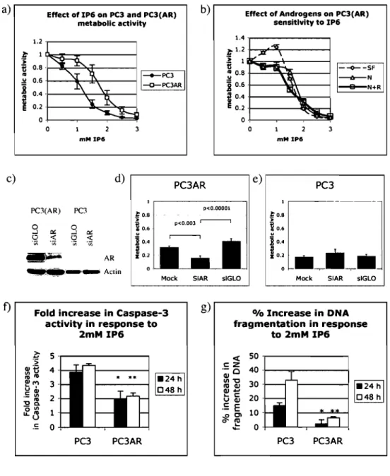
CHAPTER II: An Androgen-Independent Androgen Receptor Function Protects from Inositol Hexakisphosphate Toxicity in the PC3/PC3(AR) Prostate Cancer Cell Lines
Abstract Introduction
Materials and Methods Results Discussion Conclusions: Acknowledgements: References Figure 1. Figure 2. Figure 3. Figure 4. Figure 5. 62 63 64 65 70 74 78 78 79 85 87 89 92 94
CHAPTER III: NOXA and PUMA Expression Add to Clinical Markers in Predicting Biochemical Recurrence of Prostate Cancer Patients in a Survival Tree Model
Abstract Introduction
Materials and Methods Results Discussion Conclusion References Acknowledgements Table 1. Table 2. Figure 1 Figure 2. Figure 3. Figure 4. Figure 5.
CHAPTER IV: Co-Assessment ofCytoplasmic and Nuclear Androgen Receptor Localization in Prostate Specimens: Potential Implications for Prostate Cancer Development and Prognosis
131
Abstract Introduction
Materials and Methods Results Discussion Conclusion 96 97 98 100 104 107 110 112 118 119 120 121 123 125 127 129 132 133 135 138 141 146
Acknowledgements 146 References 147 Table 1. 153 Table 2. 154 Figure 1. 156 Figure 2. 158 Figure 3. 160 Figure 4. 162
CHAPTER V: Inositol hexakisphosphate and Proteasome Inhibitors Elicit Enhanced Mitochondrial Depolarization in Prostate Cancer Cells:
Implication of BCL-2 family proteins 164
Abstract Introduction
Materials and methods Results Discussion Conclusion Acknowledgements References Figure 1. Figure 2. Figure 3. Figure 4. Figure 5. Figure 6. CHAPTERVI 6. Discussion 165 166 168 174 179 184 184 186 192 194 196 198 200 202 204 204
6.1. AR expression and the sensitivity to IP6 204 6.2. NOXA and PUMA as molecular targets and prognostic markers for prostate
cancer 209
6.3. The implications of androgen receptor expression and sub-cellular
localization in the development of prostate cancer. 213 6.4. IP6 in combination with proteasome inhibitors: mechanistic and therapeutic
implications 218
6.5. Conclusion 225
REFERENCES
APPENDIX: Co-author signatures
226 22681
LIST OF TABLES CHAPTERIII Table 1. Table 2. CHAPTERIV Table 1. Table 2.
Patient cohort characteristics Brier scores and associated 95% CI for selected RPART models
Patient cohort characteristics AR expression in correlation with clinico-pathological features
119
120
153
LIST OF FIGURES CHAPTERI Figure 1. Figure 2. Figure 3. CHAPTER II Figure 1. Figure 2. Figure 3. Figure 4. Figure 5. CHAPTER III Figure 1 Figure 2. Figure 3.
Overview of the cell cycle and its regulators Overview of mitochondrial functions
and intrinsic/extrinsic pathways of apoptosis The Receptor Tyrosine Kinase, Ras, P13K1Akt, and NF-KB pathways.
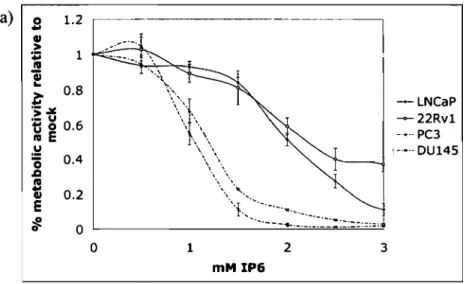
Effect of IP6 on prosate cancer celllines Androgens do not modulate
the efficacy of IP6 in prostate cancer cells Effect of AR expression on
the efficacy of IP6 in PC3 cells
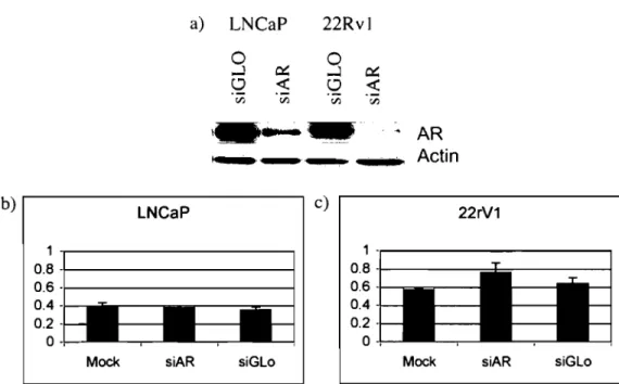
AR down-regulation does not modulate the efficacy of IP6 in prostate cancer cells endogenously expressing the AR
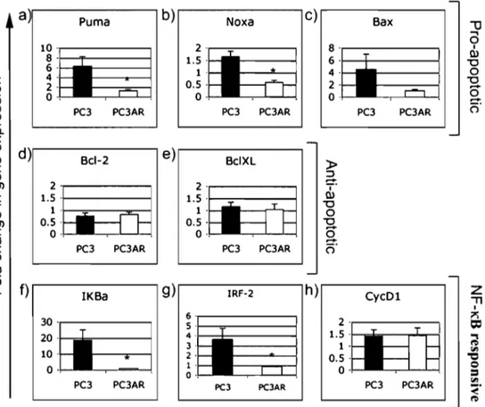
Effect of IP6 on the expression of NF-KB-responsive genes and of genes involved in apoptosis
NOXA and PUMA expression in PCa celllines
NOXA and PUMA expression in prostate tissues
Average NOXA and PUMA
expression in prostate tissue subtypes
10 22 27 85 87 89 92 94 121 123 125
Figure 4. Figure 5. CHAPTERIV Figure 1. Figure 2. Figure 3. Figure 4. CHAPTER V Figure 1. Figure 2. Figure 3. Figure 4. Figure 5.
NOXA and PUMA expression
in relation to the onset ofbiochemical recurrence following radical prostatectomy Top-ranking multivariate survival
tree models
AR expression and sub-cellular localization in prostate tissues Average cytoplasmic, nuclear
and nuclear / cytoplasmic AR staining intensity ratio in prostate specimen subtypes AR expression and sub-cellular
localization in relation to biochemical recurrence following radical prostatectomy Two highest ranking survival tree models
NF -KB in the response to IP6
IP6 induces temporal changes in the levels of BCL-2 family proteins
The effect of IP6 requires protein translation but not rnRNA transcription
A proteasome inhibitor sensitizes AIPCa cells to the effect of IP6
IP6 and proteasome inhibitors present
an enhanced effect on mitochondrial depolarization
127 129 156 158 160 162 1922 1944 1966 1988
Figure 6.
that requires protein translation IP6 and MG-132 skew pro-apoptotic to anti-apoptotic protein ratios by altering the levels of BCL-2 family proteins
200
LIST OF ABBREVIATIONS 17-AAG 19S 26S 40S 4E-BPI 60S Al ABD ActD ADT AF-l AF-2 AI AIF AIPCa Akt ALLN ALT Aly ANT AP-2 AP-3/API80 Apaf-l APC APC/C Ape-l
AR
ARAARE
ARF-BPI ATF2 ATM ATP ATPaseATR
BAD BAKBAX
BBC3 BCL-2 BCL-2L-I0 17 -Allylamino-l 7-Demethoxygeldanamycin sedimentation coefficient of 19 sedimentation coefficient of 26 sedimentation coefficient of 40eukaryotic elongation initiation factor 4E binding protein 1
sedimentation coefficient of 60 GM-CSF-inducible gene, Al androgen-binding domain Actinomycin D
androgen deprivation therapy activating function 1
activating function 2 androgen-independent apoptosis inducing factor
androgen-independent prostate cancer
thymoma viral proto-oncogene / prote in kinase B N-acetyl-Ieucinyl-Ieucinyl-norleucinal
alternative lengthening of telomeres mutated in alymphoplasia
adenine nuc1eotide translocator Adaptor protein 2
Adaptor prote in 3/ 180
apoptotic protease activating factor-l adenomatous polyposis coli
Anaphase promoting complex or cyc1osome apurinic endonuc1ease-l
Adrogen receptor
Andorgen receptor associated protein androgen response element
ADP-ribosylation factor binding prote in 1 activating transcription factor-2
Ataxia telangiectasia mutated Adenosine tri-phosphate
Adenosine tri-phosphate phosphatase Ataxia telangiectasia related
BCL-XLlBCL-2-associated death promoter homolog BCL-2 antagonistlkiller
BCL-2 associated prote in X BCL-2 binding component 3 B-celllymphoma protein 2 BCL-2 like gene 10
BCL-B B-celllymphoma protein B BCL-GL B-celllymphoma prote in G long BCL-GS B-celllymphoma protein G short BCL-RAMBO BCL-2 homolog rambo
BCL-W B-celllymphoma prote in W
BCL-XES B-celllymphoma prote in X extra short BCL-XL B-celllymphoma prote in X long BCL-XS B-celllymphoma protein X short BCR biochemical recurrence
bFGF basic fibroblast growth factor
BFL-l BCL-2 homolog isolated from fetalliver 1 BHI BCL-homology domain 1
BH2 BCL-homology domain 2 BH3 BCL-homology domain 3 BH4 BCL-homology domain 4
BID BH3 interacting domain death agonist BIK BCL-2 homolog induced killer
BIM BCL-2 interacting mediator of cell death BMF BCL-2-modifying factor
BMP7 bone morphogenic prote in 7 BNIPI BCL-2 homolog previously Nip-l BNIP3 BCL-2 homolog previously Nip-3 BOD BCL-2-related ovarian death gene BOK BCL-2-related ovarian killer Boo BCL-2 homologue of ovary BPH benign prostate hyperplasia BRAG-I brain-related apoptosis gene 1 BRCAI breast cancer 1
CAD caspase activated DNAse caspase cysteine aspartate protease CBP CREB-binding protein cdc37 cell-division-cycle 37 CDK cyclin dependent kinase CDKI cyclin dependent kinase 1 CDK2 cyclin dependent kinase 2 CDK4 cyclin dependent kinase 4 CDK6 cyclin dependent kinase 6 CDK7 cyclin dependent kinase 7
cDNA complementary DNA
CHEK2 chekpoint kinase 2
CHOP-C CIEBP homologous protein
CHX cycloheximide
clAPI cellular inhibitor of apoptosis 1 clAP2 cellular inhibitor of apoptosis 2
Cip Cipl Cip2 CKII COXYb CREB c-Rel DAG DBD DBP5 DD DHT Diablo dicer DISC Diva DNA DNA-PK DNAse DR3 DR-4 DRE ~'I'm El E2 E2F E2FI E2F4 E3 EBP-a eEF eEF2 EGF eIF eIF4E eIF4G ELAC2 EMEA EnR ER ErbB ErbB-3 cdk inhibtor prote in cdk inhibtor protein 1 cdk inhibtor protein 2 casein kinase II Cytochrome oxidoreductase Yb
cAMP response element binding protein
cellular homo log of reticuloendotheliosis virus protein diacylglycerol
DNA-binding domain DEAD-box prote in 5 death domain
dihydroxytestosterone
direct inhibitor of apoptosis binding prote in with low pl bidentate RNaseIII family double-strand RNA
endonuclease
Death-induced signaling complex
death inducer binding to vBCL-2 and Apaf-l Deoxyribonucleic acid
Deoxyribonucleic acid-dependent protein kinase Deoxyribonucleic acid nuclease
death receptor 3 death receptor 4 digital rectal exam
mitochondrial electrochemical gradient ubiquitin activating enzyme
ubiquitin conjugating enzyme
adenovirus E2 promoter binding factor adenovirus E2 promo ter binding factor 1
adenovirus E2 promo ter binding factor 4 ubiquitin ligase enzyme
Enhancer binding protein alpha eukaryotic elongation factor eukaryotic elongation factor 2 epidermal growth factor
eukaryotic elongation initiation factor eukaryotic elongation initiation factor 4E eukaryotic elongation initiation factor 4G elaC homo log 2
European Agency for the Evaluation of Medicinal Products
endoplasmic reticulum estrogen receptor
erythroblastosis erbB-related prote in erythroblastosis erbB-related prote in 3
ERK
Ets FADD Fas Fbw7 FDA FGF FGFRiiib FHRLI GI G2 GIel GSK3 HARAKIRI/DP5 HAT HDAC Hect HER-2IN euJErbB2 HIF-la Hoxb13 HR HRPCa Hsp90 IAP lBS ICAD IGF IGFBP3 IKB IKK IL-3 IL-6 INK4 INK4a INK4b INK4c INK4d Ins5P2K IP3 IP3R IP6 IRF-2 ITM2B (S)extracellular response kinase E26 transformation-specific
Fas associated prote in with a death domain Fas antigen
F-box protein with WD40 domains 7 Food and Dug Administration fibroblast growth factor FGF-receptor iiib
forkhead transcription factor like 1 Gap 1
Gap 2
glucine-leucine-phenylanine-leucine lethal mutant Glycogen synthase kinase 3
Harakiri or death prote in 5 Histone acetyl-tranferase histone de-acetylase
homologous to the E6-AP carboxyl terminus
EGFR homo log 2INeuregulinierythroblastosis erbB-related protein 2
Hipoxia induced factor 1 alpha Homeobox prote in b 13
hormone refractory
hormone-refractory prostate cancer Heatshock protein 90
inhibitor of apoptosis integrated brier score
Inhibitor of caspase activated DNAse insulin growth factor
IGF-binding prote in 3 Inhibitor of kappaB
inhibitor of kappa B kinase interleukin 3
interleukin 6
inhibitor of cyc1in-dependent kinase 4
inhibitor of cyc1in-dependent kinase 4 family member a inhibitor of cyc1in-dependent kinase 4 family member b inhibitor of cyc1in-dependent kinase 4 family member c inhibitor of cyc1in-dependent kinase 4 family member d 1,3,4,5,6-pentakisphosphate 2-kinase
inositol-I,4,5 tri-phosphate
inositol-I,4,5 tri-phosphate receptor inositol hexakisphosphate
interferon regulatory factor 2 Integral membrane prote in 2B short
I-TRAQ JAK JC-I JNK Ki67 Kip2 Ku Ku70/80 LH LHRH LPS M MAP-I MAPK MCL-I MCLI-S mdm2 MEK MG-132 ML-IAP MMP MOMP mRNA MSH MTD mTOR MudPIT Mule NAD NADH NBK NBSI NCIC NF-KB NFXI NK NKX3.1 NOXA Nup OCT1I2 Omi/HtrA2
isobaric tags for relative and absolute quantification Janus kinase
5,5' ,6,6' -tetrachloro-l, l' ,3,3' -tetraethyl-benzimidazo 1carbocyanide iodide Jun Kinase
Ki-67 antigen
kinase inhibitor prote in 2 Ku antigen
Ku antigens 70/80 lutheinizing hormone
lutheinizing hormone releasing hormone lipopolysaccharide
Mitosis
Modulator of Apoptosis 1
mitogen activated protein kinase Myeloid cellieukemia 1
Myeloid cellieukemia lshort mini double-minute 2
Mitogen effector kinase Z-Leu-Leu-Leu-CHO
Me1anoma linked ihibitor of apoptosis matrix metaloproteinase
mitochondrial outer-membrane permeation messenger ribonuc1eic acid
MutS homo log
matador, a Spanish word for killer mammalian target of rapamycin
multidimensionl prote in identification technology MCL-I ubiquitin ligase E3
nicotinamide adenine dinuc1eotide Hydrogenated nicotinamide dinuc1eotide natural born killer
Nijmegen breakage syndrome 1
National Cancer Institute of Canada Nuc1ear factor tha binds kappa light chain immunoglobulin enhancer in B cells
Nuc1ear factor that binds to Xl region of X box natural killer
re1ated to mouse NK-3 homeobox prote in stands for damage
Nuc1eoporin
octamer specifie transcription factor 1/2
mammalian homolog ofbacterial heatshock serine protease HtrA
P/CAF p300/CBP -associated factor plOO 100 kilo Dalton protein pl07 107 kilo Dalton protein p130 130 kilo Dalton protein pl5 15 kilo Dalton protein pl6 16 kilo Dalton protein p18 18 kilo Dalton protein p19 19 kilo Dalton protein p21 19 kilo Dalton prote in p27 27 kilo Dalton prote in p50 50 kilo Dalton protein p52 52 kilo Dalton protein p53 53 kilo Dalton protein p57 57 kilo Dalton protein p65 65 kilo Dalton protein p73 73 kilo Dalton protein
PABP Poly adenosine binding protein 1
PARP Poly(Adenine Dinuc1eotide Phosphate-ribose) po1ymerase
PCa prostate cancer
PCNA proliferating cell antigen PCR polymerase chain reaction
PDEF prostate-derived Ets transcription factor PDGF platelet-derived growth factor
PDGFR platelet derived growth factor receptor PDK phosphatidylinositol-dependent kinase PDKI phosphatidylinositol-dependent kinase 1 PEST pro line-apartate-serine-threonine
PI3K Phosphatidyl-inositol-3-kinase
PIDD p53-induced protein with a death domain PIN prostatic intra-epithelial neoplasia
PIP2 phosphatidyl-inostol (4,5) bi-phosphate PIP3 phosphatidyl-inosoitol (3,4,5) tri-phosphate PKA protein kinase A
PKC protein kinase C
PLC phospholipase C
PolyA polyadenosine PP1 prote in phosphatase 1
PP2A protein serine/threonine phosphatase 2A PP3 protein phosphata se 3
pRb retinoblastoma protein PSA prostate speeifie antigen
PSMA prostate specifie membrane antigen
PTEN PUMA pVHL RAIDD RAN REF RelA Re lB RING-H2 RISC RNA RNAse RNAseL ROC ROCK-l RP RPART rRNA RTK S SCF SCFpTRCP SELECT shRNA siRNA Skpl Skp2 Smac SMAD SPIKE SRC-l STAT Succ-LLVY-AMC SUV39Hl SUV39H2 TAP tBID TFIIH TGF-a
phosphatase and tensin homo log p53-up-regulated mediator of apoptosis Voh Hippel-Lindau prote in
RIP-associated ICH-lICED-3-hmologous prote in with a death domain
Ras-related nuclear GTPase Ribonucleic and export factor
reticuloendotheliosis virus prote in homo log A reticuloendotheliosis virus prote in homolog B Zinc finger family protein motif
Ribonucleic acid induced silencing complex Ribonucleic acid
Ribonucleic acid nuclease Ribonucleaic acid nuclease L Receiver operating characteristic Rho kinase 1
radical prostatectomy
recursive partitioning regression trees ribosomal ribonucleic acid
receptor tyrosine kinase Synthesis
Skp-Cul-F-box complex
Skp-Cul-F-box complex containing Beta-tranducin repeat containing protein
selenium and vitamin E chemoprevention trial short hairpin ribonucleic acid
short interfering ribonucleic acid Shaggy kinase in Schizosaccharomyces pombe 1
Shaggy kinase in Schizosaccharomyces pombe 2
Second mitochondrial activator of caspase related to Sma and mothers against dpp small prote in with inherent killing effect steroid receptor coactivator-l
signal transducer and activator of transcription
Succinate-Leu-Leu-Val-Trp-amino-4-methy 1coumarin
Su(var) or supressor ofvariability 3-9 homo log 1
Su(var) or supressor ofvariability 3-9 homo log 2 Tip ofherpesvirus saimiri-associated protein truncated BID
Transcription factor II H
TGF-~ TMRPSS2 TNF TNF-Rl TNM TOM TPA TRADD TRAIL Trail-RI TRAMP tRNA TURP Ubal UBC UVB VDAC VEGF VSV Wafl Wnt WST-l xIAP Z-ARR-AMC Z-LLE-AMC ZVAD-fmk
Transforrning growth factor beta transmembrane protease serine 2 Tumor necrosis factor
Tumor necrosis factor receptor 1 tumor, lymph nodes, metastasis translocase of the outer membrane tetradecanoylphorbol 13-acetate
TNF-receptor associated protein with a death domain
TNF -receptor associated apoptosis inducing ligand
Trail receptor 1
transgenic adenocarcinoma of the mouse protate transfer ribonuc1eic acid
trans-urethral resection of the prostate
Ubiquitin activating enzyme 1
Ubiquitin conjugating enzyme Ultraviolet radiation B
voltage-dependent anion channel vascular endothelial growth factor vesicular stomatitis virus
Wid-type p53-activated fragment 1
Drosophila melanogaster segment polarity gene wingless/ int-l
4-[3-( 4-iodophenyl)-2-( 4-nitrophenyl)-2H-5-tetrazolio]-I,3-benzene disfonate
x-linked inhibitor of apoptosis
z-Ala-Arg-Arg-amino-4-methy1coumarin z-Leu-Leu-Leu-Asp-amino-4-methy1coumarin z-valine-alanine-asparagine-fluoro- methy l-ketone
REMERCIEMENTS/ACKNOWLEDGEMENTS
It is quite certain that without the efforts and help of many, l would not have had the possibility to accomplish the monumental task of presenting a doctoral thesis. Since aIl cannot be named, l will begin by presenting my apologies to those people who l may not have specifically mentioned in this brief acknowledgement, and by thanking them sincerely.
This being said, l would like to thank Dr. Lee Wall, my masters' thesis supervisor who passed away in May 2002, from whom l leamed a sense of organization and critical thinking, and who l will always remember as an eccentric but brilliant scientist. l would also like to offer my most heartfelt thanks to my director and co-director Dr. Anne-Marie Mes-Masson and Dr. Fred Saad. l am sincerely grateful to them for accepting me in their research te am after the tragic death of Dr. Wall, and for having supported me throughout the years. They are in my opinion exemplary models of leadership, integrity, brilliance, and compassion. l will always value them as both mentors and friends.
l'aimerais également remercier tous mes collègues de l'Institut du Cancer de Montréal, passés et présents, qui m'ont toujours aidé par l'entremise de leurs bons conseils, leur support moral et leur sens critique. l'aimerais particulièrement remercier Philippe Gannon, Hervé Koumakpayi, Laurent Lessard et Benjamin Péant, avec qui j'ai voyagé et travaillé côte à côte depuis longtemps et que j'estimerai toujours comme de bons amis. l'aimerais également souligner à cet effet, le support de mes collègues et ami(e)s du laboratoire Mes-Masson /Saad / Provancher, notamment Abdulhadi Aldejmah, Mona Alam Fahmy, Blandine Betton, Ali Filali Mouhim, Julie Lafontaine, Cécile Le Page, Jason Madore, Véronique Ouellet, et Magda Zietarska, avec qui j'ai collaboré de près et de loin. Je tiens aussi à remercier Chantale Auger, Louise Champoux, Manon de Ladurataye, Nathalie Delvoye, Louise Portelance, Nathalie Tapp et Maral
Tersakian pour leur aide technique et administratif, sans lequel je n'aurais pu réussir.
Je dois aussi faire part de remerciements sincères envers les autres laboratoires membres de l'institut du Cancer de Montréal. Particulièrement, j'aimerais remerCIer le Dr. Richard Bertrand ainsi que les membres de son laboratoire. Spécifiquement, j'aimerais souligner l'aide et l'expertise de Myriam Beauchemin, Claudie Paquet, et finalement Nicolas Parent, avec qui j'ai partagé de longues soirées de travail mais aussi de célébration. De plus, j'aimerais remercier les membres des laboratoires passés et présents des Dr. Belmaaza, Bradley, Chartrand, Lapointe, Langelier, Maugard, Perrotte, Prentki, Royal, Santos et Wu, qui m'ont souvent dépanné en temps critiques et qui m'ont également conseillé et supporté moralement. J'aimerais aussi remercier Dr. Bégin, Dr. Ferbeyre, Dr. Mukhopadhyay, Dr. Sircar and Dr. Aprikian pour leur aide et collaboration précieuse.
Finalement, je voudrais remercier mes parents ainsi que ma famille. Bien que certains aient été loin de moi, ils ont toujours été près de mon cœur et c'est cette présence qui ma permis de continuer durant les moments difficiles. D'ailleurs, il est certain que ces moments auraient été d'autant plus difficiles si ce n'était pas pour le doux sourire de ma fiancée Erin, qui m'a donné le courage de persévérer.
Over the last century, cancer has emerged as one of the most problematic diseases of the western world. With the advent of globalization and the increasing life expectancy worldwide [1], cancer is likely to become one of the most significant challenges to human medicine for centuries to come. In this chapter, we will begin by briefly reviewing the maintenance of cellular homeostasis and explore how this homeostasis is fundamentally altered in cancer cells. We will subsequently focus on the development of prostate cancer and its CUITent treatment. Emerging therapeutic concepts in prostate cancer will then be introduced and the potential of inositol hexakisphosphate will be showcased within this context.
1.1 Cellular Homeostasis
Cellular homeostasis can be defined as the equilibrium between production and destruction of cellular components. Although the cell processes a wide range of molecules, we will focus on the homeostasis deoxyribonucleic acids (DNA), of ribonucleic acids (RNA), and of proteins, as they are intimately linked and as they are of central importance for the development of cancer.
1.1.1 DNA Homeostasis
Chromosomal DNA is considered to be the primary holder of the genetic information within the cell and acts as a template for the production of RNA and proteins. The specific sequence of DNA within chromosomes constitutes the template for RNA synthesis and subsequent protein synthesis. This intimate relationship is of utmost importance for the cell, as changes in the DNA sequence may have a severe impact on cellular functions and consequently on the overall cellular homeostasis.
1.1.1.1 DNA Replication and DNA Degradation
In the normal ce Il , production of DNA through DNA replication is a tightly regulated process, allowed to occur only during the synthesis or S phase of the cell cycle (see section 1.2.1) [2]. This process is mediated by the action of various proteins including DNA primases, DNA ligases, DNA helicases,
topoisomerase, and finally DNA polymerases that play a most direct role by synthesizing a complementary DNA strand from a single strand template [3]. Counter to DNA polymerase activity, DNAses act to break down the DNA polymer into smaller sub-components. Exonucleases effectively digest DNA from one end to the other (either 5' to 3' or 3' to 5 ') while endonucleases cut within a DNA strand. The action of endo and exo-nucleases is important for diverse processes su ch as DNA repair (eg. Ape-l [4], see below) and apoptosis (eg. CAD [5], see section 1.2.3.3.2).
1.1.1.2 Genome Integrity and the Impact of DNA Mutation
It is ofutmost importance for the cell to maintain the integrity of the DNA sequence as any unwarranted change can potentially lead to the disruption of cellular homeostasis. In addition, this disruption may be transmitted to daughter cells following cell division. As such, various proteins (that are synthesized based on the DNA sequence, see section 1.1.3.1) are normally responsible for maintaining chromosomal integrity. These include enzymes involved in the repair ofDNA single-strand breaks [ego Ape-1, [4], PARP [6]], or double-strand breaks [e.g ATM, [7], DNA-PK and Ku proteins [8]], DNA mismatches [e.g, MSH, PCNA [7]], or that maintain the stability of DNA ends or te10meres (e.g Telomerase [9]). Other proteins su ch as p53 act as genome guardians and coordinate cellular response to DNA damage [10]. The proper regulation of these proteins is therefore also critical for maintaining a stable genome (see section
1.2.4.2.2).
1.1.2. RNA Homeostasis
RNA is to DNA as paper is to a paper press and therefore any change that is made to the DNA template is automatically transferred unto the corresponding RNA message. The process of making RNA from DNA is referred to as transcription and occurs mainly in the nucleus and within the Gap 1 (G 1), S, and Gap 2 (G2) phases of the cell cycle [11] (see section 1.2.1.). As RNA is subsequently translated into proteins that play major structural and functional roles within the cell (see section 1.1.3.1), the tight regulation of transcription is extremely important for maintaining cellular homeostasis. As such, many
cell-signaling pathways are devoted to the transcriptional control of genes (see sections 1.2.2.1, 1.2.3.2.5, 1.2.4.1, and 1.3.5.5).
1.1.2.1. RNA Transcription and its Regulation
Transcription is regulated both by DNA and prote in. Specific sequences in DNA (e.g gene promoters and enhancers) act as binding sites for transcription factors that initiate or activate the process of RNA synthesis through the action of RNA polymerases [e.g RNA Polymerase I [12], II [13], III [14]]. As such, DNA mutations that modify either the specificity of the DNA sequences recruiting transcription factors or that modify the specificity of transcription factors themselves can have a major impact on RNA homeostasis.
The condensation state of chroma tin is one of the most fundamental regulators of gene expression. In its most basic form, chromatin is composed of negatively charged DNA wrapped around positively charged histone proteins (altogether called a nucleosome). Condensed chromatin (heterochromatin) is much less accessible to transcription factors, RNA polymerases, and their associated co-factors. Consequently, gene transcription generally occurs within
100 se chromatin domains (euchromatin) [15, 16].
Whereas the DNA sequence regulates which transcription factors (including RNA polymerases) bind where, histone proteins are the structural regulators of the chromatin condensation state. Histone proteins can be post-translationally modified by covalent addition of various moieties including acetyl, methyl, phosphoryl, and ev en ubiquitin groups. Post-translational modification of histones can lead to the alteration of chromatin structure. These modifications occur on specific histone residues and altogether constitute a histone code that acts in parallel to the genetic code provided by the DNA sequence [16].
Although researchers are finding the histone code to be increasingly complex [16, 17], it is generally considered that the acetylated state of histones is associated to transcription permissive euchromatin whereas de-acetylated histones are rather associated to repressive heterochromatin. Importantly, histone acetyl tranferases (HATs) mediate the addition of acetyl groups whereas histone
de-acetylases (HDACs) mediate the removal of these moieties. In effect, activator transcription factors often act to recruit HA Ts while repressor transcription factors recruit HDACs [18].
1.1.2.2. RNA Processing and Degradation
Once RNA is produced, it is subsequently processed and spliced into its mature form (rnRNA), void of intron sequences and equipped with a 5' methy1guanosine cap and a 3' poly-adenosine (PolyA) tail. While the 5' methylguanosine cap plays a role in the initiation of protein translation (see section 1.1.3.1) both the 5' methyl cap and the PolyA tail play a ro1e in protecting the RNA from degradation by RNAses, which oppose the action of RNA polymerases and promote RNA de-polymerization [11]. Recently, it has been found that the cell possesses a highly specific system of RNAses that permits the degradation of mRNA in sequence-specific manner. The RNA-induced si1encing complex (RISC) in concert with the RNA endonuclease dicer induces the degradation of specific mRNAs. N otably, exploitation of this endogenous cellular machinery has spawned the emergence small interfering RNA (siRNAs) and short hairpin RNA (shRNAs) technology that allows for the specific down-regulation of genes and their associated proteins using short complementary RNA sequences [19,20] (see chapter II).
1.1.3. Protein Homeostasis.
The main purpose of mRNAs is to act as an intermediate template for the production of protein through a process termed translation. This process is mediated by the ribosome, a large ribonucleoprotein complex composed of two sm aller subunits (40S and 60S). Much like RNA production, prote in translation occurs mainly in the G1, S, and G2 phases of the cell cycle [11]; however, unlike RNA transcription, this process occurs mainly in the cytoplasm and endoplasmic reticulum, although more restricted translation also occurs in other organelles such as the mitochondria [21] (see section 1.2.3.1) and the nucleolus [14, 22]. Notably, the nucleolus is the primary site for ribosomal RNA transcription and ribosome maturation [22].
One of the first control points for prote in production is mRNA export from the nucleus, which proceeds through the TAPINFXl pathway and involves a large number of proteins including nuclear export receptors (eg. TAPINFXI), nuclear export adaptors (Aly/REF), A TPases/RNA helicases (eg. DBP5) and nucleoporins (eg. GIel and Nup family proteins) [23]. The second major control point for prote in production from mRNA involves translation initiation and recruitment of the 40S ribosome. This involves numerous elongation initiation factors (eIF) such as eIF4E that binds the 5' methylguanosine cap, eIF4E binding proteins (e.g 4E-BPI), and eIF4G that also causes circularization of mRNA indirectly by its interaction with poly(A) binding protein (PABPI). Although regulation at the translation initiation step is better described, prote in translation can also be regulated at the elongation and termination steps through the action of elongation factors (eEFs) such as eEF2 [24]. As prote in production can have rather immediate functional consequences for cellular homeostasis, prote in translation is also tightly regulated through the concerted action of many signaling pathways, one ofwhich is the mTOR pathway ([25], see section 1.2.2.2),
1.1.3.2. Protein Degradation and the Ubiquitin/Proteasome System
Although lysosomal proteases account for 10-20% of cellular protein degradation [26], the 26S proteasome is the principle mediator of prote in degradation responsible for the ATP-dependent processing of 80-90% of cellular proteins [27]. The 26S proteasome is a large 2000 kDa multiprotein complex composed of a barrel-shaped 20S subunit and two "lid" 19S subunits. The 20S subunit contains several proteolytic sites within its two central p-rings and possesses trypsin-like, chymotrypsin-like and caspase-like protease activity. Notably, the chymotryptic-site is thought to be rate limiting in proteasome-mediated degradation [27]. The 19S subunits are mainly regulatory in nature and direct proteasomal degradation by binding poly-ubiquitinylated proteins that, as such, have been targeted for degradation. Thus, the targeting of proteins for proteasome-mediated degradation by the process of poly-ubiquitination is a key regulatory step of protein homeostasis and can profoundly impact cellular homeostasis.
The ubiquitination of cellular proteins is mediated by a series of three types of enzymes, namely El activating enzymes, E2 ubiquitin-conjugating proteins, and E3 ubiquitin ligases [28, 29]. While mono-ubiquitination can play a role in altering protein localization [30] and is increasingly thought to play a role in chromatin organization [31], poly-ubiquitinylation (4 ubiquitin subunits or more) targets proteins for proteasomal degradation [28]. While only one El enzyme (Ubal) is required to conjugate ubiquitin to a small number of cellular E2 enzymes (UBCs) that have limited specificity, there are several E3 ubiquitin ligases (eg. Skp1l2, APC/C, mdm2, SCF~ TRCP) that confer most of the substrate specificity as weIl as temporal and spatial control to the ubiquitinlproteasome system [28, 29, 32]. The E3 ubiquitin ligases contain either Hect or RING-H2 domains and are notably involved in a variety of cellular processes including genomic surveillance (e.g mdm2 [33] see section 1.2.2.2 and 1.2.4.2), cell signaling (e.g SCF~TRCP[34], see section 1.2.4.1.3), cell cycle (eg. SCF (skp1l2) [35, 36], APC/C [37], see section 1.2.2.3) and cell death (e.g. Mule/ARF-BP1[38] see section 1.2.3.2.6).
1.2.Disruption of Cellular Homeostasis: Cell cycle, Apoptosis, and Cancer By definition, the homeostatic state is a stagnant balance between import, export, production, and degradation of each component of the cell. In reality, this balance is in continuaI reassessment. As the cell reacts to various stresses and stimuli, the homeostatic balance is perturbed and the state of the cell is altered. As we will soon discuss, the outcome of these perturbations depends on both the stimulus and the state of the cell.
1.2.1.The Cell Cycle
The cell cycle can be viewed as a programmed and controlled alteration of cellular homeostasis that leads to cytokinesis and cellular proliferation. The cell cycle is divisible in four stages, Gl, S, G2, and M (mitosis) [11]. These stages are associated to distinct cellular activities (see sections 1.1.1-1.1.3) and are periodically regulated at "checkpoints" that play a crucial role in overseeing the repli cation of the genome and its proper distribution within the resulting daughter
cells. The cell cycle checkpoints also play a crucial role in the control of cellular proliferation within the organism, as they can be regulated by growth factors and various extracellular signaIs [39](Refer to Chapter 1 Figure. l for an overview).
1.2.1.1 Cyclin-Dependent Kinases
The cell cycle is controlled by a complex network ofproteins ofwhich the mam constituents are cyclins and cyclin-dependent kinases (CDK). Although there are at least ni ne known CDKs, five of them play an active role throughout the cell cycle. Specifically, CDK 4, 6, and 2 are active during the G l phase, CDK2 is active in S phase while CDKI is active in both G2 and M phases. CDK7 is active at aIl cell cycle phases. Although the kinase activity of CDK6, 4, 2 and 1 changes throughout the cell cycle, their protein levels generally remain stable. CDK activity is regulated by cyclins, whose levels rise and fall periodically throughout the cell cycle [40-42].
1.2.1.2. Cyclins
Much like CDKs there are over sixte en identified cyclins although many of them are not related to the cell cycle. cyclins DI, D2 and D3 are expressed maximally during the G 1 phase and associate to CDK4 and CDK6. cyclin E is expressed during the G liS phase transition and associates with CDK2. The activity of CDK2 following the G liS transition is maintained by its association with cyclin A, which is expressed throughout the S phase, progressively decreasing until the G2/M phase transition where it interacts instead with CDKl. During the initial stages of M phase, cyclin B levels are at their highest, leading to sustained activation of CDKI until the cycle repeats itself [41]. Notably, in contrast with cyclins E, A and B, D-type cyclin expression is regulated by growth factor signaling [42]. Importantly, CDKlcyclin complexes have varying specificities and can phosphorylate a wide range of proteins that promote changes in cellular organization, cell cycle progression, and eventually cell division.
Acting as natural "brakes" for the cell cycle and playing important roles at the cell cycle checkpoints, CDK inhibitors block the activity of CDKlcyclin complexes. The better-known CDK inhibitors faU into two major families. The INK4 family comprises p15 (INK4b), p16 (INK4a), p18 (INK4c) and p19 (INK4d) and present ankyrin repeat domains. INK4 family CDK inhibitors are generaUy thought to compete with D-type cyclins to block the cell cycle in G 1. The Cip/Kip fami1y comprises p21(Wafl/Cipl), p27(Cip2), p57(Kip2) and a1so share a comm6n inhibitory domain. Although Cip/Kip family CDK inhibitors act on cyclin D/CDK complexes in vitro, they preferentially act on CDK2 complexes in vivo [41,43].
Although INK4 and Cip/Kip family proteins are more or less direct CDK inhibitors, a third category of cell cycle inhibitors, the pocket proteins, mediate their effect through E2F transcription factors. E2F family transcription factors are key regulators of the G liS cell cycle transition but are also involved in cell differentiation and apoptosis [44, 45] (see section l.2.3.2.5). This family of proteins consists of at least seven members, of which E2F l, 2 and 3 are considered transcriptional activators that act upon release from their interaction with pRb: the prototypical pocket prote in family member. While the function of the other E2F family members is more obscure, E2F4 and E2F5 are thought to act as transcriptional repressors when bound to pocket proteins (including pRb, pl 07 and p130) that also recruit HDACs. Among others, Rb proteins can be phosphorylated by CDK(4/6)/cyclin D or CDK2/cyclin E complexes during cell cycle progression. Phosphorylation of pRb family members results in their dissociation from E2F binding partners. The coordinated activity of E2F members results in the modulation of E2F-regulated gene expression and cell cycle progression [44, 46-48].
1.2.2. Regulation of the Cell Cycle.
Net cyclin and CDK inhibitor expression at the protein level depends on mRNA availability, prote in production, and prote in degradation. As such this offers severallayers of regulation for cell cycle control.
1.2.2.1 Transcriptional Control of Cell Cycle Components
The first level of control occurs at the level of mRNA transcription. For example, cyclin Dl mRNA expression is regulated by various transcription factors including NF-KB [49] and ~-catenin [50]. Upstream signaling pathways in tum regulate the activity of these transcription factors, such as that of PI3K/ AktlIKK (NF-KB) and WntlGSK3 (~-catenin) (see sections 1.2.4.1.1 to
1.2.4.1.4). Moving forward in the cell cycle, cyclin E can be up regulated by E2F 1 following hyper-phosphorylation of pRb [51]. In contrast, to haIt cell cycle progression, the expression of the pI5 (INK4b) CDK inhibitor can be up regulated at the mRNA level following cell stimulation by TGF-~ [52]. Similarly, the p53 transcription factor can induce p21 (Wafl/Cip1) mRNA up-regulation in response to DNA damage [53].
1.2.2.2. mTOR and the Translational Control of Cell Cycle Components One of the better-described pathways that regulate cell cycle and other proteins at the translation level is the mTOR (mammalian target of rapamycin) pathway. As its name implies, mTOR was first identified as a target ofrapamycin,
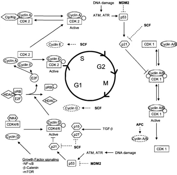
DNA damage MDM2 1 : . . ..1.-Active .. --- SCF 1 CDK 1 1 €clin~ 1 CDK21 (QI~n3> ~
----.
,...::s--"-{~~
@
~§P
'\~
~ APC ---.~ ~.. TGF-p : ~ Active ~ t~
T
~
i2~~I~~~~'
'\.
@
~--- ~CF
~
'\. ATM, ATR +-- DNA damage Growth Factor signaling,,~(
-NF-KB Ô ~---- MDM2 -p-Catenin -mTOR
rI
Activel
1 CDK 1 1Figure 1. Overview of the cell cycle and its regulators. Refer to sections 1.2.2 through 1.2.2.3. Grey circles indicate a phosphorylation event whereas dashed lines indicate proteasome-mediated degradation. Lines ending with bars indicate an inhibitory effect. Ubiqutin ligases are highlighted in boldo Adapted from [41].
a drug that inhibits cell proliferation [54]. In normal cells, the mTOR pathway is typically initiated by an increased availability in amino acids or by upstream activation of PI3K and Akt. mTOR activation has been shown to lead to the inhibition of 4E-BPl that normally represses translation by binding eIF4E. Hence, activation of mTOR promotes translation of various proteins including cyclin Dl whereas inhibition of mTOR leads to decreased protein production and cell cycle arrest [25].
1.2.2.3. Proteasorne-Mediated Degradation of Cell Cycle Cornponents
Another important cell cycle regulation mechanism is proteasome-mediated degradation of cyclins and other proteins involved in cell cycle control. As mentioned previously, this involves several E3 ubiquitin ligases. SCF (skp/ cul / F-box protein) E3 ligase complexes play an important role to this effect, mediating the degradation of CDK inhibitors such as p2l Cipl, p27Kipl and p57Kip2
[32, 55] as well as of cyclin Dl [56] and cyclin E [57]. Notably, the composition of the SCF complex defines its target specificity. For example SCFI (skpl/cull/F-box protein) complexes alter specificity through the changing of the F-(skpl/cull/F-box protein. Skp2, Fbw7 and ~-TrCP are the best known F-box proteins and while skp2 and fbw7 play direct role in the cell cycle, SCF complexes containing ~
TrCP play a more indirect role by controlling cell signaling through the NF-KB and ~-catenin pathway [32, 34, 50] (see sections 1.2.4.1.3 and 1.2.4.1.4). In a similar fashion to SCF complexes, the APC (Anaphase Promoting Complex) ubiquitin ligase mediates the degradation of cyclin A and B [58]. Other important E3 ubiquitin ligases include MDM2, which controls the expression of the genome surveillance factor p53 [59].
1.2.3. Apoptosis.
If the cell cycle can be viewed as a controlled perturbation of cellular homeostasis leading to cell division, then apoptosis may be described as a controlled response to the disruption of cellular homeostasis leading to cell death.
Likely almmg at containing cellular toxins that may be damageable towards surrounding cells, the apoptotic cell undergoes distinct morphological and biochemical changes. These typically inc1ude cell shrinkage, cell membrane blebbing and presentation of phosphatidyl-serine at the cell surface, loss of nuc1ear envelope integrity, chromatin condensation, and DNA fragmentation [60, 61]. The apoptotic program can be initiated by both intracellular (intrinsic) and extracellular (extrinsic) signaling pathway [62]. Apoptosis is a highly regulated process and its progression involves a cascade of molecular events, implicating a myriad of proteins inc1uding pro- and anti-apoptotic proteins (see sections 1.2 .. 3.2.1 to 1.2.3.2.3), inhibitors of apoptosis (lAPs, see section 1.2.3.3.3), proteases (see section 1.2.2.3.1) and DNAses (see section 1.2.3.3.2) [63,64] (see Chapter I Figure. 2 for an overall schematization). In the context of tissues and organs, the process of apoptosis is extreme1y important as it counterbalances the effects of cell division, thereby maintaining tissue homeostasis or active1y sculpting organ shape [65,66].
1.2.3.1 The Mitochondria
Before we begin to review the molecular details of apoptosis and the processes of cell death, it is important to describe one of its central regulatory organelles: the mitochondrion. The mitochondrion possesses its own DNA, leading sorne to suggest that it may have evolved separately from the cell and was later incorporated in a symbiotic relationship [67]. Importantly, mitochondria not only provide eukaryotic cells with energy by acting as the main sites of ATP (adenosine tri-phosphate) production, but also act as key regulators of apoptosis and cell death in general [64,67].
1.2.3.1.1. Mitochondrial Energy Production
The mitochondrion is composed of an outer membrane and a larger ruffled inner membrane encirc1ing the mitochondrial matrix. While the outer membrane acts as agate between the cytosol and the mitochondria, the inner membrane plays a major role in the production of ATP, holding all the components of the
e1ectron transfer chain. The electron transfer chain mega-complex is responsible for the establishment of a proton gradient between the inter-membrane space and the mitochondrial matrix. It is composed of several sub-complexes inc1uding the nicotinamide adenine dinuc1eotide (NAD) dehydrogenase complex, iron-suIf ur proteins, Co-enzyme Q (ubiquinone), cytochrome B complex (BIC 1), cytchrome C and also cytochrome oxydase (cytochrome NA3 complex) [68]. Notably, whereas most of these complexes reside within the inner-mitochondrial membrane, cytochrome C sits in the inter-membrane space. The process of ATP production by ATP synthase, which sits on the matrix side of the mitochondrial inner membrane, is driven by the proton motility force that results from the pH gradient between the outer/inner membrane space and the mitochondrial matrix. [64, 67, 68]. As the inter-membrane space is more acidic than the mitochondrial matrix, this yields an electrochemical gradient referred to as ~\{'m[64].
1.2.3.1.2. Mitochondrial and Cellular Inter-Dependence
Within the mitochondrial matrix lays the mitochondrial genome, which is circular and much smaller than the nuc1ear genome (roughly 16 000 bp) and encodes for two ribosomal RNAs (rRNAs), 22 transfer RNAs (tRNAs) and 13 polypeptides. The mitochondrial matrix also harbors its own translation-competent ribosomes, although these are of significantly different composition than cytosolic ribosomes [21, 69] (see section 1.1.3.1 ). However, the mitochondrion is not auto-sufficient in that it requires the import of proteins encoded by nuc1ear genes, which are likely imported both post and co-translationally (e.g as they traverse the outer-membrane) [70]. Mitochondrial import of RNA has also been described, particularly for transfer RNAs [71], although the mechanisms involved are much more obscure than those involved in protein import, that are known to proceed through the action of the Translocase of the Outer Membrane (TOM) pore complex [72].
1.2.3.1.3. VDAC/ANT Complexes and the Inter-Membrane Space
Another key mitochondrial membrane pore complex involved III the transport of diverse molecules is VDACI ANT. This complex is formed of VDAC
(Voltage-dependent anion channel) which lies within the outer membrane, ANT (adenine nucleotide translocator) located in the inner membrane, and cyclophilin D that associates to the matrix portion of ANT. In addition, benzodiazepine receptor and hexokinase, an enzyme involved in glucose phosphorylation, are associated to VDAC on the cytosolic side of the channel. [73-80]. Although this remains debated, sorne studies suggest that this highly regulated inner and outer membrane spanning channel plays a role in apoptosis because it allows for the dispersion of known pro-apoptotic factors that are present in the inter-membrane space. As will be discussed subsequently, these include cytochrome C that binds Apaf-l and activates the apoptosome [81, 82], Apoptosis inducing factor (AIF) that plays several roles including chromatin condensation and DNA fragmentation [83], Diablo/Smac and Omi/HtrA2, both antagonizers of lAPs [84-86], EndoG that activates caspase-activated DNAse (CAD) [87, 88], as weIl as caspases 2 and 9 [89-92] (see section).
1.2.3.2. The Regulation of Mitochondrial Outer Membrane Permeability.
Given the presence of pro-apoptotic factors within the inter-membrane space and given the importance of the maintenance of the proton gradient and
~ \ } ' f i for ATP production by ATP synthase in the inner membrane, outer
mitochondrial membrane permeability is an extremely weIl regulated process. In fact, a whole family of proteins is dedicated to its control. The BCL-2-family of proteins is characterized by the presence ofBCL homology domains (BHI, BH2, BH3 or BH4) and presently contains over 30 known members. These are generally categorized according to whether they promote or prevent apoptosis and how many (and which) BH domains the proteins contain [93].
1.2.3.2.1 Anti-Apoptotic BCL-2 Family Members
Anti-apoptotic proteins of the BCL-2 family ultimately prevent the permeation of the mitochondrial outer membrane and generaIly contain at least two different BH domains. These currently include the prototypical BCL-2 [94], BCL-XL [95], BCL-XES [96], BCL-W [97], MCL-l [98], AlIBFL-1 [99], BRAG-I [100], BOO/DIVA [101], BCL-B [102] and BCL-2L-1O [103].
1.2.3.2.2. Multi-Domain Pro-Apoptotic BCL-2 Family Members
In contrast to anti-apoptotic BCL-2 proteins, pro-apoptotic proteins of the BCL-2 family lead to the leakage of apoptosis effectors residing within the inter-membrane space into the cytoplasm (as described in section 1.2.3.1). These fall into two sub-categories based on whether they contain multiple different BH domains (multi-domain) or only one or sometimes two BH3 domains (termed BH3-only). Known multi-domain BCL-2-family members inc1ude BAX [104], BAK [105], BCL-XS [95], BOKlMTD [106], BCL-RAMBO [107] and BCL-GL [108]. Importantly, sorne of these proteins such as BAX, BAK and BID (see below) can form pore structures and permeate synthetic liposomes in vitro and may permit direct mitochondrial outer membrane permeation [109].
1.2.3.2.3. BH3-0nly Pro-Apoptotic Proteins
Currently, known BH3-on1y pro teins incIude BAD [110], BID [111], BIK/NBK [112], BIM/BOD [113], HARAKIRI/DP5 [114], BNIP1 [115], BNIP3 [116], PUMNBCB3 [117], NOXA [118], BCL-GS [108], MCL1-S [119], BMF [120], MAP-1 [121], ITM2B(S) [122] and SPlKE [123]. It is generally thought that the major ro1e of these proteins is to antagonize anti-apoptotic members of the BCL-2 family, although sorne such as BID and PUMA, may more directly promote pore formation mediated by multi-domain pro-apoptotic proteins such as BAX and BAK [124].
1.2.3.2.4. Control of outer membrane permeability by BCL-2-family proteins
This long list of pro and anti-apoptotic factors of the BCL-2-family gives us a general sense of the importance and complexity of mitochondrial membrane permeability control. Overall, it is thought that the balance of active pro- and anti-apoptotic proteins within the cell dictates the release of apoptogenic factors from the inter-membrane space of the mitochondria. Hence, we can invoke the notion of homeostasis to explain the action of BCL-2 family members on the mitochondria. For example, if the cell shifts its production towards accumulation of pro-apoptotic proteins, cells can survive in conditions where they would