S54 ESTRO 33, 2014 EP-1196
Accelerated partial breast irradiation: Arctherapy versus 3D-CRT G. Farha1, M. Cheve2, G. Auzac2, S. Lopes Da Silva2, S. Rivera1 1Gustave Roussy Cancer Campus, Radiation Oncology Department,
Villejuif, France
2Gustave Roussy Cancer Campus, Medical Physics Department, Villejuif,
France
Purpose/Objective: To dosimetrically compare two techniques of accelerated partial breast irradiation (APBI): Arc therapy by RapidArc (RA) and 3D conformal external beam irradiation (3D-CRT) by two mini-tangents and an 'en face'electron beam.
Materials and Methods: A retrospective dosimetric comparison of RA and 3D-CRT was performed. Eight left-sided breast cancer patients treated by 3D-CRT APBI in a prospective phase II multicentric trial were included for a dosimetric comparison of the dose received to the ipsilateral breast, heart, Non-Target Breast Tissue Volume (NTBTV), ipsilateral lung and PTV. All patients were treated with 42 Gy in 10 fractions twice daily using two mini-tangents and an 'en face' electron beam; the dosimetric constraints of this APBI protocol were respected. The lumpectomy cavities (CTV) were imported from the original scans. The PTV was constructed as a uniform expansion of 1.8 cm for all patients and was limited to 5 mm below the skin. Normal structures including ipsilateral lung, breast and heart were also imported from original scans. The same contoured simulation CT was used to calculate treatment plan and dosimetry with both techniques (RA and3D-CRT) for each patient. To evaluate dose to the ipsilateral breast, heart, ipsilateral lung, NTBTV and PTV, dose-volume histogram (DVH) analysis was performed. Results: The average percentage of the breast volume receiving 30 and 20 Gywas higher in the 3D-CRT group (22.4% and 24.2 % respectively) compared with RA (21% and 22,8% respectively) . Improved coverage of the PTV was noted in the 3D-CRT plans compared with RA plans. With 3D-CRT technique, 97.03% of the PTV received 40 Gy compared with 95,73% with RA technique. The average of the mean and maximal doses to PTV was higher by 1.9% and 5.3% respectively in RA compared with 3D-CRT (p=0.002). Homogeneity index was lower with 3D-CRT (0.087) than RA (0.104). V5 Gy and mean dose to the heart were not significantly improved in RA (0.49 % and 0.54 Gy respectively compared to 0.94 % and 0.79 Gy respectively; p=0.762 for V5 Gy;p=0.935 for mean dose). V5 Gy and mean dose to the ipsilateral lung were not significantly higher in RA (5.91 % and 1.25 Gy respectively compared to 5.31 % and 1.10 Gy respectively; p=0.347 for V5 Gy; p=0.258 for mean dose). V10 Gy and mean dose to the NTBTV were not significantly improved in RA (35.83 % and 10.96 Gy respectively compared to 37.91 % and 15.77 Gy respectively; p=0.604 for V10Gy; p= 0.995 for mean dose).
Conclusions: In patients treated with 3D-CRT, coverage of the PTV was not significantly better and mean dose to ipsilateral lung was not significantly lower. As we did observe a trend in favor of RA we think it would be useful to do the dosimetric comparison for all patients in that trial because a better PTV coverage with 3D-CRT might come at the cost of a higher integral dose to the remaining normal breast. RA might give a better sparing of the heart with lower doses to NTBTV but higher maximal dose to PTV. The dosimetric comparison of the rest of the patients in the protocol is ongoing in order to improve the statistical power of this study and better define patients who will benefit more from one technique or another.
EP-1197
Patterns of difficult cases for breast irradiation: where multi-beam IMRT and SIB should be the primary choice
S. Ben Mustapha1, D. Dechambre1, C. Mievis1, P. Coucke1, S. Cucchiaro1,
A. Gulyban1, F. Lakosi1
1Liege University Hospital, Department of Radiation Oncology, Liege,
Belgium
Purpose/Objective: To present the dosimetric advantages of multi-beam(MB) IMRT combined with SIB over tangential field (TG) treatment in breast cancer patients with unusual clinical situations.
Materials and Methods: Four difficult cases out of last 40 patients were identified and planned with inverse optimized MB-IMRT. 2/4 patient with early stage invasive lobular carcinoma were treated by mastectomy+reconstruction (TRAM flap, prosthesis) in the past. Both patients have developed infiltrating ductal carcinoma foci(s) in the axillary scar at the level of the superior and middle axillary region. Local excision was performed in each case. One of these cases had limited pulmonary function as well due to existing chronic obstructive pulmonary disease. The target volume comprised the reconstructed chest wall, 'tumor bed'with regional lymph nodes. In the other 2/4 patient breast conserving surgery was performed in the left para-sternal region
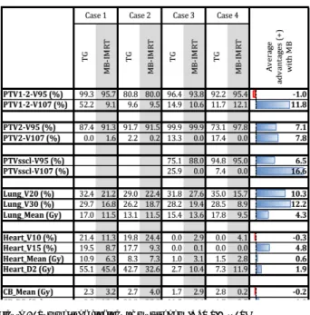
challenging tangential field treatment in terms of heart and contralateral breast dose delivery. In two cases the surgical margin was positive demanding elevated tumor bed doses. Due to the challenging anatomy and respiration function we have decided to treat these patients with MB-IMRT and to evaluate if simultaneously integrated boost could even further improve the plan. Comparison against the classical TG field-in-field approach was made by matching the median doses of the corresponding targets. As the cases had different prescription levels for the breast/chest-wall and the boost volume, therefore relative DVH parameters were used for comparison: V95%and V107% for the breast/chest wall target excluding the boost volume (PTV1-2), boost volume (PTV2) and the regional lymph nodes (PTV_sscl). For the ipsilateral lung V20,V30,for the heart V10, Dmean, D2 and for the contralateral breast Dmean and D2 were documented.
Results: The main dosimetric findings of the TG vs. MB-IMRT presented at Figure 1. For the planning target volumes the homogeneity parameters showed an average 8.1% improvement. For patient with regional lymph node irradiation, these advantages were even higher. For OARs the relevant volumetric and dose parameters showed a reduction of 6.7% and 1.5 Gy by using MB-IMRT. The reduction of lung V20 from the average of 32% to 21% would likely make a clinically significant risk reduction of pneumonitis, especially in the presence of existing pulmonary problems and systemic therapy. For the leftsided patients the decreased heart mean dose (4.7 and 1 Gy) could lead into a5-20% reduction of risk for major coronary events (Darby et al., NEJM 2013).
Figure 1. Summarized findings of multi-beam IMRT SIB vs. TG
Conclusions: Multi-beam IMRT with SIB has a clear advantage over classic tangential treatment arrangements in the presence of unusual clinical challenges. These relevant improvements should encourage centers to elaborate multi-beam approach at least for selected breast cancer patients.
EP-1198
Cavity Boost (CB) following Fractionated External Beam Radiotherapy (EBRT): Time to move on from clinical mark-ups?
S. Chatterjee1, S. Chakraborty1, S. Das1, S. Tamil Selvan1, R.K. Shrimali1,
R. Achari1, I. Mallick1, R. Ahmed2, A. Manke2, A. Mahata1 1Tata Medical Center, Radiation Oncology, Kolkata West Bengal, India 2Tata Medical Center, Surgical Oncology, Kolkata West Bengal, India
Purpose/Objective: Tumour cavity boost (CB) is essential post External Beam Radiotherapy (EBRT). Surgical clips (SC) marking of the tumour cavity volume (TCV) has enabled radiation oncologists to better visualise the tumour bed. Whilst technological advancement in forward planned breast radiotherapy has enabled clinicians to achieve acceptable homogeneity,cavity boost methodology varies significantly between centres. Clinical Electron Mark up (CEM) is the commonest method of tumour bed boost but CT based conformal Photon(COP) and conformal electron (COE) boost could also be done.
Materials and Methods: In 50 patients with invasive carcinoma, cavity demarcation was done by the surgeons during breast conservation surgery according to a set institutional protocol using SC. CEM to boost