HAL Id: hal-02879680
https://hal.sorbonne-universite.fr/hal-02879680
Submitted on 24 Jun 2020
HAL is a multi-disciplinary open access
archive for the deposit and dissemination of
sci-entific research documents, whether they are
pub-lished or not. The documents may come from
teaching and research institutions in France or
abroad, or from public or private research centers.
L’archive ouverte pluridisciplinaire HAL, est
destinée au dépôt et à la diffusion de documents
scientifiques de niveau recherche, publiés ou non,
émanant des établissements d’enseignement et de
recherche français ou étrangers, des laboratoires
publics ou privés.
Cataract and glaucoma combined surgery: XEN® gel
stent versus nonpenetrating deep sclerectomy, a pilot
study.
Vincent Theillac, Esther Blumen-Ohana, Jad Akesbi, Pascale Hamard,
Alexandre Sellam, Emmanuelle Brasnu, Christophe Baudouin, Antoine Labbé,
Jean-Philippe Nordmann
To cite this version:
Vincent Theillac, Esther Blumen-Ohana, Jad Akesbi, Pascale Hamard, Alexandre Sellam, et al..
Cataract and glaucoma combined surgery: XEN® gel stent versus nonpenetrating deep sclerectomy, a
pilot study.. BMC Ophthalmology, BioMed Central, 2020, 20, pp.231. �10.1186/s12886-020-01492-z�.
�hal-02879680�
R E S E A R C H A R T I C L E
Open Access
Cataract and glaucoma combined surgery:
XEN® gel stent versus nonpenetrating deep
sclerectomy, a pilot study
Vincent Theillac
1*, Esther Blumen-Ohana
1, Jad Akesbi
1, Pascale Hamard
2, Alexandre Sellam
1,
Emmanuelle Brasnu
2, Christophe Baudouin
2,3, Antoine Labbe
2,3and Jean-Philippe Nordmann
1Abstract
Background: To compare the efficacy of phacoemulsification (PKE) combined with nonpenetrating deep sclerectomy (NPDS) with mitomycin C (MMC) versus XEN® gel stent with MMC.
Methods: In this nonrandomized, retrospective, comparative, single-center pilot study, 105 consecutive eyes of 75 patients with uncontrolled primary open-angle glaucoma (POAG) and cataract who underwent PKE combined with either XEN implantation (n = 47) or NPDS (n = 58) between May 2013 and November 2018 were included. The primary outcome was complete success at 9 months, which was defined as intraocular pressure (IOP)≤18, 15 or 12 mmHg without treatment; qualified success was IOP≤18, 15 or 12 mmHg with antiglaucoma medications. Secondary outcome measures included the number of antiglaucoma medications, visual acuity (VA), and postoperative adverse events.
Results: Using the 18 mmHg threshold, complete or qualified success was achieved in 69.6 and 89.1% in the PKE + XEN group, and 63.8 and 89.7% in the PKE + NPDS group (p = .54 and p = .93), respectively, at 9 months. The mean IOP decreased from 20.8 ± 6.8 mmHg to 16.2 ± 2.8 mmHg in the PKE + XEN group (p < .001, 18.9% mean drop), and from 21.5 ± 8.9 mmHg to 14.9 ± 3.9 mmHg in the PKE + NPDS group (p < .001, 25.6% mean drop). Best-corrected VA significantly improved (p < .001) in both groups. The mean number of antiglaucoma medications was significantly reduced from 2.66 ± 1.1 to 0.49 ± 1.0 in the PKE + XEN group (p < .001) and from 2.93 ± 0.9 to 0.69 ± 1.2 in the PKE + NPDS group (p < .001). Conclusions: The XEN stent combined with PKE seemed to be as effective and safe as PKE + NPDS at 9 months in this pilot study.
Keywords: XEN, Nonpenetrating deep sclerectomy, Primary open angle glaucoma, Microinvasive glaucoma surgery, Cataract, Combined surgery
Background
Glaucoma is one of the world’s leading causes of irrevers-ible blindness and should affect 76 million people world-wide in 2020 [1]. Although trabeculectomy remains the gold standard glaucoma surgery, nonpenetrating deep
sclerectomy (NPDS) is an alternative for the surgical treat-ment of primary open-angle glaucoma (POAG). NPDS has been demonstrated to be effective with fewer surgical com-plications than trabeculectomy [2]. In the elderly popula-tion, cataract is frequently associated with glaucoma when a surgical intervention is needed for glaucoma [3]. Com-bined phacoemulsification-NPDS (PKE + NPDS) is a safe and effective procedure when cataract is associated with evolutive glaucoma [4]. Its efficacy is comparable to an NPDS performed as a single procedure [5], or a PKE
© The Author(s). 2020 Open Access This article is licensed under a Creative Commons Attribution 4.0 International License, which permits use, sharing, adaptation, distribution and reproduction in any medium or format, as long as you give appropriate credit to the original author(s) and the source, provide a link to the Creative Commons licence, and indicate if changes were made. The images or other third party material in this article are included in the article's Creative Commons licence, unless indicated otherwise in a credit line to the material. If material is not included in the article's Creative Commons licence and your intended use is not permitted by statutory regulation or exceeds the permitted use, you will need to obtain permission directly from the copyright holder. To view a copy of this licence, visithttp://creativecommons.org/licenses/by/4.0/. The Creative Commons Public Domain Dedication waiver (http://creativecommons.org/publicdomain/zero/1.0/) applies to the data made available in this article, unless otherwise stated in a credit line to the data.
* Correspondence:v.theillac@gmail.com
1Department of Ophthalmology 2, Quinze-Vingts National Ophthalmology
Hospital, IHU FOReSIGHT, University Paris Descartes, 28 rue de Charenton, 75012 Paris, France
combined with trabeculectomy [6]. Nevertheless, combined PKE + trabeculectomy seemed to have more complications than combined PKE-NPDS [7].
Recently, the development of microinvasive glaucoma surgery (MIGS) with an optimal safety profile promotes combined cataract and glaucoma surgery [8]. Among these techniques, XEN® Gel Stent (Allergan Inc., Irvine, CA, USA), a glutaraldehyde cross-linked porcine colla-gen tube 6 mm in length and 45μm in diameter is the only MIGS with ab interno implantation [9] that uses the subconjunctival drainage pathway [10]. Cataract sur-gery combined with XEN has been described and low-ered IOP by 25–42% [11, 12]. To the best of our knowledge, no studies have compared PKE + XEN and PKE + NPDS.
The purpose of this study was to compare the efficacy and safety of PKE combined with NPDS (PKE + NPDS) and mitomycin C (MMC) versus PKE combined with the XEN® gel stent (PKE + XEN) and MMC in OAG.
Methods
This retrospective pilot study was conducted at the Quinze-Vingts National Ophthalmology Hospital, Paris, France, in accordance with the tenets of the Declaration of Helsinki. The medical records of all consecutive pa-tients with no previous glaucoma surgery who underwent PKE + XEN® between June 2017 to November 2018 were reviewed. This group was compared to a matched group (age, sex, surgeon) of patients who underwent PKE + NPDS as first glaucoma surgery between May 2013 and March 2018. Surgical procedures were performed by six glaucoma surgery experts (JA, CB, EBO, EB, PH, and AL).
Preoperative data comprised best-corrected visual acuity (BCVA), subjective refraction, anterior segment and fundus examination, gonioscopy, pachymetry, IOP measurement using a Goldman applanation tonometer, 24–2 ± 10–2 Humphrey visual field (SITA-standard 24–2 and SITA-fast 10–2 programs, with stimulus III-White, of the Humphrey visual field analyzer (Carl Zeiss Meditec, Dublin, CA, USA) with mean deviation (MD), pattern standard deviation (PSD), visual field index (VFI); ganglion cell complex (GCC), and retinal nerve fiber layer (RNFL) optical coher-ence tomography (OCT Cirrus HD-OCT (Carl Zeiss Medi-tec, Dublin, CA, USA), and the number of treatments. The BCVA was measured on a Snellen chart and then con-verted to LogMAR according to the Ferris table [13]. The refractive results were assessed according to the calculation of the change in the spherical component by subtraction of spherical equivalents as previously described [14].
The PKE + XEN® procedure was performed as previ-ously described [11]: topical anesthesia with oxybupro-caine and xylooxybupro-caine gel, standard phacoemulsification, intraocular injection of a myotic agent, subconjunctival in-jection of 0.1 mL of 0.2 mg/mL MMC with a 27-G needle
beneath the superonasal Tenon and spread away from the limbus, introduction of the preloaded injector through a temporal incision, injector placement in the superonasal quadrant and then 45° rotation towards 12 o’clock when the needle was visible subconjunctivally, injection of the implant ideally through the scleral spur with passage from the sclera to the subconjunctival space 3.0 mm from the limbus, gonioscopic examination to verify the intraocular part of the XEN (about 2 mm) and then visibility and mo-bility assessment of the subconjunctival part, cautious washing of the viscoelastic, intraocular injection of 0.1 mL of 1 mg/0.1 mL cefuroxime, and hydro-sutures of the cor-neal edges.
The PKE + NPDS procedure was also performed as pre-viously described [15]: topical anesthesia with oxybupro-caine and xylooxybupro-caine gel, limbus-based conjunctival flap, application of sponges soaked with 0.2 mg/mL MMC in the sub-Tenon space for 2 min, abundant rinsing and then completion of the first scleral flap until achieving clear cornea, standard PKE, cautious washing of the viscoelastic, intraocular injection of myotic agent, hydro-sutures of the corneal edges, intraocular injection of 0.1 mL of 1 mg/0.1 mL cefuroxime, completion of a deep scleral rectangular flap until careful peeling of the external trabecular mem-brane up to satisfactory filtration, and conjunctival sutures with resorbable 8/0 Vicryl®.
All patients received topical antibiotics for 1–4 weeks and anti-inflammatory therapy for 6–8 weeks, adjusted according to the situation.
For each patient, the postoperative follow-up consisted of a visit at 1 day, 1 week, and 1, 3, 6, 9 and 12 months after surgery with BCVA, IOP measurement, number of antiglaucoma medications, and notification of a potential complication. The primary outcome was successful sur-gery at 9 months defined as complete success, correspond-ing to a postoperative IOP ≤18, 15 or 12 mmHg in the absence of antiglaucoma treatment, and qualified success defined as a postoperative IOP≤18, 15 or 12 mmHg under one or more antiglaucoma medications; failure was an IOP > 18 mmHg, with or without treatment [16]. Second-ary outcomes were IOP decrease, number of antiglaucoma medications, surgery duration, complications, number of needling procedures, visual acuity, refraction, and visit number. We conducted a subgroup analysis in order to observe the effect of postoperative IOP at Day 1 < 9 mmHg [17]. The effective operating times of both tech-niques were calculated using Hospital Manager Bloc soft-ware (Softway Medical®, Meyreuil, France).
Quantitative variables were compared using a Student t-test. The chi2
test was used for the analysis of qualita-tive data. The Fisher exact test was used for the analysis of qualitative data when at least one calculated theoret-ical value was less than 3. Survival curves were generated using the Kaplan-Meier method. All statistical analyses
were performed using GraphPad Prism 7® for Windows® (GraphPad Software, La Jolla, CA, USA). Double-sided p-values <.05 were considered statistically significant.
Results
Forty seven eyes of 36 patients and 58 eyes of 39 patients were respectively included in the PKE + XEN® group and in the PKE + NPDS group. There was no difference for age, sex, ethnicity, diabetes, first-degree relatives with POAG, duration of glaucoma, preoperative BVCA, glau-coma type, mean preoperative IOP, and preoperative number of antiglaucoma medications. The mean follow-up period was 8.7 ± 4.2 months for the PKE + XEN grofollow-up and 20.4 ± 15.6 months for the PKE + NPDS group. The mean preoperative IOP was 20.8 ± 6.8 mmHg in the
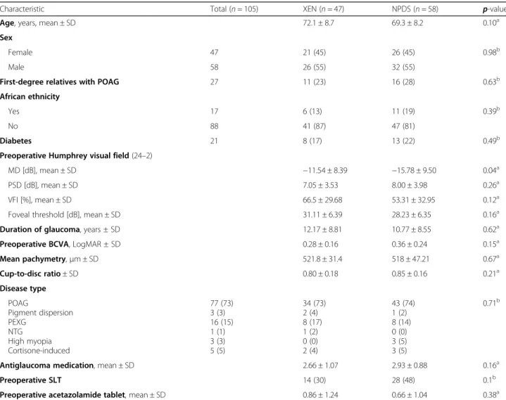
PKE + XEN group and 21.5 ± 8.9 mmHg in the PKE + NPDS group (p = .66). The mean number of preoperative treatments was 2.66 ± 1.1 in the PKE + XEN group and 2.93 ± 0.9 in the PKE + NPDS group. The MD was sig-nificantly lower in the PKE + NPDS group than in the PKE + XEN group (− 15.78 ± 9.50 dB versus − 11.54 ± 8.39 dB, p = .04) but there was no difference for PSD, VFI, and foveal threshold. The patients’ baseline clinical characteristics are shown in Table1.
Complete and qualified success at 9 months, using 18 mmHg threshold, was achieved, respectively, in 69.6 and 89.1% in the PKE + XEN group and 63.8 and 89.7% in the PKE + NPDS group (p = .54 and p = .93) (Table2).
The mean IOP at 9 months was 16.2 ± 2.8 in the PKE + XEN group (p < .001 compared to preoperative IOP) and
Table 1 Baseline characteristics
Characteristic Total (n = 105) XEN (n = 47) NPDS (n = 58) p-value
Age, years, mean ± SD 72.1 ± 8.7 69.3 ± 8.2 0.10a
Sex
Female 47 21 (45) 26 (45) 0.98b
Male 58 26 (55) 32 (55)
First-degree relatives with POAG 27 11 (23) 16 (28) 0.63b
African ethnicity
Yes 17 6 (13) 11 (19) 0.39b
No 88 41 (87) 47 (81)
Diabetes 21 8 (17) 13 (22) 0.49b
Preoperative Humphrey visual field (24–2)
MD [dB], mean ± SD −11.54 ± 8.39 −15.78 ± 9.50 0.04a
PSD [dB], mean ± SD 7.05 ± 3.53 8.00 ± 3.98 0.26a
VFI [%], mean ± SD 66.5 ± 29.68 53.31 ± 32.95 0.12a
Foveal threshold [dB], mean ± SD 31.11 ± 6.39 28.23 ± 6.35 0.16a
Duration of glaucoma, years ± SD 12.17 ± 8.81 10.77 ± 8.55 0.62a
Preoperative BCVA, LogMAR ± SD 0.28 ± 0.16 0.36 ± 0.24 0.15a
Mean pachymetry,μm ± SD 521.8 ± 31.4 518 ± 47.21 0.67a Cup-to-disc ratio ± SD 0.80 ± 0.18 0.85 ± 0.16 0.21a Disease type POAG Pigment dispersion PEXG NTG High myopia Cortisone-induced 77 (73) 3 (3) 16 (15) 1 (1) 3 (3) 5 (5) 34 (73) 2 (4) 8 (17) 1 (2) 0 (0) 2 (4) 43 (74) 1 (2) 8 (14) 0 (0) 3 (5) 3 (5) 0.71b
Antiglaucoma medication, mean ± SD 2.66 ± 1.07 2.93 ± 0.88 0.16a
Preoperative SLT 14 (30) 28 (48) 0.1b
Preoperative acetazolamide tablet, mean ± SD 0.86 ± 1.24 0.66 ± 1.04 0.38a
The results are displayed as n (%) for categorical variables
SD standard deviation, NPDS nonpenetrating deep sclerectomy, HFA Humphrey visual field analyzer, MD mean deviation, PSD pattern standard deviation, VFI visual field index, BCVA best corrected visual acuity, POAG primary open-angle glaucoma, PEXG pseudoexfoliative glaucoma, NTG normal-tension glaucoma, SLT selective laser trabeculoplasty
a
Student t-test
b
14.9 ± 3.9 mmHg in the PKE + NPDS group (p < .001 com-pared to preoperative IOP). There was no mean IOP differ-ence between the two groups at 9 months (p = .25). The mean IOP lowering between the preoperative and the 9-month evaluation was − 18.9 ± 5.2% in the PKE + XEN group and− 25.6 ± 4.3% in the PKE + NPDS group (p = .39) (Fig.1). The mean number of preoperative treatments de-creased from 2.66 ± 1.1 to 0.49 ± 1.0 in the PKE + XEN group (p < .001) and from 2.93 ± 0.9 to 0.69 ± 1.2 in the PKE + NPDS group at 9 months (p < .001). The treatment-free survival curves (Fig. 2) did not differ significantly be-tween the two groups (p = .39).
BCVA was significantly improved by surgery (p < .001 in both groups) with no postoperative difference be-tween the two groups (Table3).
The preoperative and final postoperative spherical equivalents were similar in both groups (Table4).
The mean operating time was 52.97 ± 14.37 min in the PKE + NPDS group versus 39.09 ± 12.75 min in the PKE + XEN group (p < .001). As detailed in Table5, the number of immediate complications was similar in both groups. No cases of loss of light perception, nor IOP < 5 mmHg leading to hypotony related maculopathy were recorded.
There was no significant difference in the number of needling procedures between the two groups: 14 patients required ≥1 needlings (30%) in the PKE + XEN group and 11 (19%) in the PKE + NPDS group (p = .19). The survival curves (Fig.3) of the number of needling proce-dures did not differ between the two groups (p = .17). Twenty-four goniopunctures (41.4% of the patients) were performed in the PKE + NPDS group, with a mean completion time of 90.7 ± 31.9 days.
IOP < 9 mmHg on Day 1 after PKE + NPDS surgery was associated with complete success (p = .02). This was not the case in the PKE + XEN group (p = .37).
Discussion
In POAG patients, cataract is frequently associated with glaucoma when a glaucoma surgical intervention is
needed. The development of microinvasive glaucoma surgery (MIGS) with an optimal safety profile and rapid recovery has recently renewed interest in combined cata-ract and glaucoma surgery. In the present study, there was no statistically significant difference in success rate between PKE combined with XEN or PKE combined with NPDS after a 9-month follow-up period.
The NPDS procedure supports the goal of a safer pro-file than trabeculectomy [2], as does MIGS. Despite sur-gical difficulties and the need for sursur-gical experience, NPDS is a common procedure in France: it accounted for 64.7% of the number of trabeculectomies in 2014 [18]. Eldaly et al. [2] indicated that success in deep scler-ectomy did not differ from that in trabeculscler-ectomy.
The IOP results of combined PKE + XEN were consist-ent with those described in the literature [11, 12]. Complete and qualified success rates ranged, respect-ively, from 50% [19] to 90% [20] and from 50% [19] to 97.5% [21], in accordance with our results.
Publications on combined PKE + NPDS surgery have re-ported IOP results consistent with our findings [22]. Pre-vious studies have found an IOP-lowering effect around 38% [22] for combined PKE + NPDS surgery. Our results were more modest with IOP lowered by 25.6%. This could be explained by the especially low preoperative IOP in the PKE + NPDS group (21.5 mmHg) compared to the pre-operative IOP reported in the literature [22].
Day 1 IOP appears to be a significant prognostic factor in combined PKE + NPDS surgery, as shown by Garcia-Pérez et al. Day 1 IOP < 9 mmHg was associated with a higher success rate and fewer treatments and gonio-punctures [17]. In the present study we had similar re-sults with a complete success rate significantly higher in the subgroup of patients undergoing PKE + NPDS with an IOP < 9 mmHg at Day 1 (p = .02). However, this was not observed for the PKE + XEN procedure in our study. In NPDS, IOP at Day 1 is an indicator of adequate inner scleral flap dissection and juxtacanalicular trabecular meshwork removal [17], which provides a better result. Although XEN implant provides constant resistance with a steady-state pressure of approximately 6–8 mmHg following an uncomplicated implantation under normal physiologic conditions and without conjunctival resist-ance to the outflow [9], Day 1 IOP does not seem to be a predictor of long-term outcomes for the XEN, whether it was a standalone or a combined procedure [23].
Combined PKE + trabeculectomy remains the gold standard surgical treatment for cataract and glaucoma surgery [24], but it has potentially severe surgical com-plications [7]. In the present study, we found no major complications in both groups during the 9-month study period. The short follow-up duration and the small number of patients who underwent PKE + XEN did not allow concluding on long-term complications such as
Table 2 Combined surgery success at 9 months
XEN (n = 46 eyes) NPDS (n = 58 eyes) p-value Complete success[without treatment]
≤ 18 mmHg 32 (69.6) 37 (63.8) 0.54
≤ 15 mmHg 25 (54.4) 29 (50.0) 0.67
≤ 12 mmHg 13 (28.3) 17 (29.3) 0.91
Qualified success [with treatment]
≤ 18 mmHg 41 (89.1) 52 (89.7) 0.93
≤ 15 mmHg 32 (69.6) 38 (65.5) 0.66
≤ 12 mmHg 18 (39.1) 21 (36.2) 0.76
Failure 5 (10.9) 6 (10.3) 0.93
The results are displayed as n (%)
extrusion, avascular bleb, blebitis, or endophthalmitis [25, 26]. NPDS postoperative management allows fine adjustments with goniopuncture, although XEN en-ables only bleb management with needling and sur-gical revision. The needling rate in the PKE + XEN group was within the low range of those found in the XEN literature (between 30.7% [27] and 47% [16]). The needling rates were not significantly dif-ferent between the two groups while one surgical re-vision in each group was observed among the patients included in this pilot study. Seven patients (14.9%) had a use of aqueous suppressant for sus-pected bleb encapsulation with Dorzolamide 2%-Timolol 0.5% twice a day during 2 weeks as a post-operative management. This particular treatment has been previously recommended by an expert review as a first step treatment if a bleb encapsulation de-velopment was suspected while needling was
suggested if aqueous suppressants failed [28]. This procedure might explain the low needling rate in our study.
Patients who underwent combined PKE + NPDS sur-gery had more severe MD than those who underwent PKE + XEN. Nevertheless, no significant difference in the other parameters of glaucoma severity was found in this study (PSD, visual field index, C/D). Patients referred for their first glaucoma surgery in Europe had a mean MD of − 13.8 dB, consistent with that found in our two groups [29]. The difference in MD could be explained by the surgeon’s caution regarding the new PKE + XEN technique and its unfamiliar out-comes, which led to the selection of patients with a less impaired visual field to prevent the potential risk of sudden visual loss (“wipe-out”).
There is, to date, no study assessing the cost of XEN [30]. The economic rationale proposed is based on com-bined surgery: the rapid implantation of XEN would save operating time with regard to filtering surgery compared to conventional filtering surgery. This time savings, which could be used to perform other surgical proce-dures, would offset the extra cost related to the XEN im-plant. In the present study, there was indeed a clinically significant 14.82-min difference in surgery duration, which could make it possible to perform more proce-dures if XEN is used. This finding needs to be confirmed in a complete operating program and at least on a monthly operating volume, but XEN shows a fast learn-ing curve by both experienced surgeons and novice resi-dents [31], unlike the NPDS, which implies delicate
Fig. 1 Mean intraocular pressure (IOP) and mean IOP lowering from baseline through 9 months of follow-up, by surgery group. Error bars represent 95% confidence intervals
Fig. 2 Kaplan-Meier survival analyses of the probability of avoiding glaucoma medication after surgery
Table 3 Postoperative visual acuity outcome
XEN (n = 45 eyes)
NPDS (n = 53
eyes) p-value
Postoperative VA, LogMAR mean ± SD
0.07 ± 0.13 0.12 ± 0.17 0.12 VA evolution, LogMAR ± SD −0.21 ± 0.17 −0.24 ± 0.31 0.65
VA visual acuity, NPDS nonpenetrating deep sclerectomy, MAR minimal angle of resolution, SD standard deviation
Table 4 Comparison of spherical equivalent evolution in PKE + XEN and PKE + NPDS
XEN (n = 45 eyes) NPDS (n = 53 eyes) p-value Preoperative spherical equivalent, mean ± SD −1.23 ± 4.33 −1.22 ± 3.33 0.99 Postoperative spherical equivalent, mean ± SD − 0.55 ± 1.03 − 0.55 ± 1.16 0.99 Spherical component variation, mean ± SD 0.67 ± 4.18 0.71 ± 3.18 0.97
dissection. However, the quality-of-life indicators remain to be determined.
We acknowledge limitations to this study. First, we conducted a retrospective study, and the conclusions would be strengthened by a randomized controlled trial. However, in the PKE + XEN group, patients were con-secutively and exhaustively included. The PKE + XEN group was matched with PKE + NPDS on age, sex, and surgeon. Although, for a limited number of patients, we included both eyes in the statistical analysis, the large number of subjects included in the present study might have limited this bias. In our pilot study, the mean follow-up was of only 8.7 ± 4.2 months so it was not pos-sible to conclude on the long-term efficacy of combined PKE + XEN surgery. XEN must necessarily be assessed in the long term: it has been developed to reduce the risk of hypotony without considering conjunctival resist-ance [32], but the resistance of XEN to aqueous humor flow is increased by that of conjunctival healing [9]: the
long-term efficacy of XEN could only be assessed with mature blebs [32].
Conclusions
In this pilot study, the XEN implant combined with PKE confirmed a favorable risk/benefit ratio, comparable to that of PKE combined with NPDS. This new combined technique is safe and effective, and significantly lowers IOP and the number of anti-glaucoma treatments in the medium term. The operating time of PKE + XEN surgery is significantly shorter than that of PKE + NPDS surgery.
Thus, combined PKE + XEN surgery appears to be a promising procedure, although its place in the surgical arsenal of glaucoma treatment remains to be clarified.
Abbreviations
PKE:Phacoemulsification; NPDS: Nonpenetrating deep sclerectomy; MMC: Mitomycin C; POAG: Primary open-angle glaucoma; IOP: Intraocular pressure; VA: Visual acuity; MIGS: Minimally/Micro-invasive glaucoma surgery; BCVA: Best-corrected visual acuity; MD: Mean deviation; PSD: Pattern standard deviation; VFI: Visual field index; GCC: Ganglion cell complex; RNFL: Retinal nerve fiber layer
Acknowledgements Not applicable.
Authors’ contributions
All authors (VT, EBO, JA, PH, AS, EB, CB, AL, JPN) contributed to conception and design of the study. VT performed the acquisition, analysis,
interpretation of data and drafting the manuscript. All authors (VT, EBO, JA, PH, AS, EB, CB, AL, JPN) have been involved in revising the manuscript critically for important intellectual content and giving the final approval of the version to be published. All authors (VT, EBO, JA, PH, AS, EB, CB, AL, JPN) agreed to be accountable for all aspects of the work in ensuring that questions related to the accuracy or integrity of any part of the work are appropriately investigated and resolved. All authors read and approved the final manuscript.
Funding
No funding was received for this research.
Availability of data and materials
The datasets used and/or analyzed during the current study are available from the corresponding author on reasonable request.
Table 5 Postoperative complications in XEN and NPDS eyes
Complication XEN (n = 47 eyes) NPDS (n = 58 eyes) p-value
Choroidal detachment 0 1 (1.7) 0.91 Leak/dehiscence 1 (2.1) 2 (3.5) 0.69 Conjunctival suture 0 2 (3.5) 0.20 Goniopuncture 24 (41.4) 2nd surgery 2 (4.3) 2 (3.5) 0.82 NPDS 2 (4.3) 0 Trabeculectomy 0 1 (1.7)
Trans-scleral cyclodiode laser 0 1 (1.7)
Use of aqueous suppressant for suspected bleb encapsulation 7 (14.9)
Iris incarceration 1 (2.1) 3 (5.2) 0.42
Bleb revision surgery 1 (2.1) 1 (1.7) 0.88
The results are displayed as n (%)
Fig. 3 Kaplan-Meier survival analyses of the probability of avoiding needling after surgery
Ethics approval and consent to participate
The data documentation and retrospective analysis was approved by the Ethics Committee of the French Society of Ophthalmology (IRB 00008855 Société Française d’Ophtalmologie IRB#1) and adhered to the tenets of the Declaration of Helsinki. According to the approval of the Ethics Committee, the data for this retrospective analysis were evaluated pseudonymized and no patient informed consent was required. No administrative permission, nor license were acquired to access the data.
Consent for publication Not applicable.
Competing interests
The authors declare that they have no competing interests.
Author details
1Department of Ophthalmology 2, Quinze-Vingts National Ophthalmology
Hospital, IHU FOReSIGHT, University Paris Descartes, 28 rue de Charenton, 75012 Paris, France.2Department of Ophthalmology 3, Quinze-Vingts
National Ophthalmology Hospital, IHU FOReSIGHT, Paris and Versailles Saint-Quentin-en-Yvelines University, Versailles, France.3INSERM U968; UPMC
Univ Paris 06, UMR_S968, Institut de la Vision; CNRS, UMR 7210; CHNO des Quinze-Vingts, INSERM-DHOS CIC 503, Paris, France.
Received: 1 October 2019 Accepted: 1 June 2020
References
1. Tham Y-C, Li X, Wong TY, Quigley HA, Aung T, Cheng C-Y. Global prevalence of glaucoma and projections of glaucoma burden through 2040: a systematic review and meta-analysis. Ophthalmology. 2014;121:2081–90. 2. Eldaly MA, Bruce C, El Sheikha OZ, Wormald R. Non-penetrating filtration
surgery versus trabeculectomy for open-angle glaucoma (Review). Cochrane Database Syst Rev. 2014;(2):CD007059.
3. Tseng VL, Yu F, Lum F, Coleman AL. Risk of fractures following cataract surgery in Medicare beneficiaries. JAMA. 2012;308:493–501.
4. Yuen NSY, Chan OCC, Hui SP, Ching RHY. Combined phacoemulsification and nonpenetrating deep sclerectomy in the treatment of chronic angle-closure glaucoma with cataract. Eur J Ophthalmol. 2007;17:208–15. 5. Jiang N, Zhao G-Q, Lin J, Hu L-T, Che C-Y, Wang Q, et al. Meta-analysis of
the efficacy and safety of combined surgery in the management of eyes with coexisting cataract and open angle glaucoma. Int J Ophthalmol. 2018; 11:279–86.
6. Funnell CL, Clowes M, Anand N. Combined cataract and glaucoma surgery with mitomycin C: phacoemulsification-trabeculectomy compared to phacoemulsification-deep sclerectomy. Br J Ophthalmol. 2005;89:694–8. 7. Gianoli F, Schnyder CC, Bovey E, Mermoud A. Combined surgery for cataract
and glaucoma: phacoemulsification and deep sclerectomy compared with phacoemulsification and trabeculectomy. J Cataract Refract Surg. 1999;25: 340–6.
8. Lavia C, Dallorto L, Maule M, Ceccarelli M, Fea AM. Minimally-invasive glaucoma surgeries (MIGS) for open angle glaucoma: a systematic review and meta-analysis. PLoS One. 2017;12:e0183142.
9. Sheybani A, Reitsamer H, Ahmed IIK. Fluid dynamics of a novel micro-fistula implant for the surgical treatment of Glaucoma. Invest Ophthalmol Vis Sci. 2015;56:4789–95.
10. Kerr NM, Wang J, Barton K. Minimally invasive glaucoma surgery as primary stand-alone surgery for glaucoma. Clin Exp Ophthalmol. 2017;45:393–400. 11. Mansouri K, Guidotti J, Rao HL, Ouabas A, D’Alessandro E, Roy S, et al.
Prospective evaluation of standalone XEN gel implant and combined phacoemulsification-XEN gel implant surgery: 1-year results. J Glaucoma. 2018;27:140–7.
12. Widder RA, Dietlein TS, Dinslage S, Kühnrich P, Rennings C, Rössler G. The XEN45 gel stent as a minimally invasive procedure in glaucoma surgery: success rates, risk profile, and rates of re-surgery after 261 surgeries. Graefes Arch Clin Exp Ophthalmol. 2018;256:765–71.
13. Ferris FL, Kassoff A, Bresnick GH, Bailey I. New visual acuity charts for clinical research. Am J Ophthalmol. 1982;94:91–6.
14. Touzeau O, Costantini E, Gaujoux T, Borderie V, Laroche L. Calculations of mean refraction and variation of refraction using a dioptric space. J Fr Ophtalmol. 2010;33:659–69.
15. Leleu I, Penaud B, Blumen-Ohana E, Rodallec T, Adam R, Laplace O, et al. Central 10-degree visual field change following non-penetrating deep sclerectomy in severe and end-stage glaucoma: preliminary results. Graefes Arch Clin Exp Ophthalmol. 2018;256:1489–98.
16. Sheybani A, Dick HB, Ahmed IIK. Early clinical results of a novel Ab Interno gel stent for the surgical treatment of open-angle Glaucoma. J Glaucoma. 2016;25:e691–6.
17. García-Pérez JL, Rebolleda G, Muñoz-Negrete FJ. Intraocular pressure on the first postoperative day as a prognostic indicator in phacoemulsification combined with deep sclerectomy. J Cataract Refract Surg. 2008;34:1374–8. 18. Bron AM, Mariet A-S, Benzenine E, Arnould L, Daien V, Korobelnik JF, et al. Trends in operating room-based glaucoma procedures in France from 2005 to 2014: a nationwide study. Br J Ophthalmol. 2017;101:1500–4.
19. Fea AM, Spinetta R, Cannizzo PML, Consolandi G, Lavia C, Aragno V, et al. Evaluation of Bleb Morphology and Reduction in IOP and Glaucoma Medication following Implantation of a Novel Gel Stent. J Ophthalmol. 2017;2017.
20. Pérez-Torregrosa VT, Olate-Pérez Á, Cerdà-Ibáñez M, Gargallo-Benedicto A, Osorio-Alayo V, Barreiro-Rego A, et al. Combined phacoemulsification and XEN45 surgery from a temporal approach and 2 incisions. Arch Soc Esp Oftalmol. 2016;91:415–21.
21. De Gregorio A, Pedrotti E, Russo L, Morselli S. Minimally invasive combined glaucoma and cataract surgery: clinical results of the smallest ab interno gel stent. Int Ophthalmol. 2018;38:1129–34.
22. Bilgin G, Karakurt A, Saricaoglu MS. Combined non-penetrating deep sclerectomy with phacoemulsification versus non-penetrating deep sclerectomy alone. Semin Ophthalmol. 2014;29:146–50.
23. Karimi A, Lindfield D. Is a day 1 postoperative review following ab interno Xen gel stent surgery for glaucoma needed? Clin Ophthalmol. 2018;12: 2331–5.
24. American Academy of Ophthalmology. Primary Open-Angle Glaucoma. 2015. https://www.aao.org/preferred-practice-pattern/primary-open-angle-glaucoma-ppp-2015. Accessed 17 Mar 2019.
25. Lapira M, Cronbach N, Shaikh A. Extrusion and breakage of XEN gel stent resulting in Endophthalmitis. J Glaucoma. 2018;27:934–5.
26. Arnould L, Theillac V, Moran S, Gatinel D, Grise-Dulac A. Recurrent exposure of XEN gel stent implant and Conjunctival erosion. J Glaucoma. 2019;28: e37–40.
27. Galal A, Bilgic A, Eltanamly R, Osman A. XEN Glaucoma implant with Mitomycin C 1-year follow-up: result and complications. J Ophthalmol. 2017; 2017:5457246.
28. Vera V, Ahmed IIK, Stalmans I, Reitsamer H. Gel stent implantation -recommendations for preoperative assessment, surgical technique, and postoperative management. US Ophthalmic Rev. 2018;11(1):38–46. 29. Holló G, Schmidl D, Hommer A. Referral for first glaucoma surgery in
Europe, the ReF-GS study. Eur J Ophthalmol. 2019;29:406–16. 30. Agrawal P, Bradshaw SE. Systematic literature review of clinical and
economic outcomes of micro-invasive Glaucoma surgery (MIGS) in primary open-angle Glaucoma. Ophthalmol Ther. 2018;7:49–73.
31. Marques RE, Ferreira NP, Sousa DC, Pinto J, Barata A, Sens P, et al. Glaucoma gel implant learning curve in a teaching tertiary hospital. J Glaucoma. 2019; 28:56–60.
32. Lim KS. Control and optimisation of fluid flow in glaucoma drainage device surgery. Eye (Lond). 2018;32:230–4.
Publisher’s Note
Springer Nature remains neutral with regard to jurisdictional claims in published maps and institutional affiliations.