T
HÈSE DE DOCTORAT DE
L’UNIVERSITÉ DE NANTES
COMUEUNIVERSITÉ BRETAGNE LOIRE ÉCOLEDOCTORALEN°601
Mathématiques et Sciences et Technologies de l’Information et de la Communication
Spécialité : Génie informatique, automatique et traitement du signal, section CNU61 Par
Konstantin AKHMADEEV
Modèles probabilistes fondés sur la décomposition d’EMG
pour la commande de prothèses
Thèse présentée et soutenue à Laboratoire des Sciences du Numérique de Nantes, le 20/11/2019 Unité de recherche : Laboratoire des Sciences du Numérique de Nantes (LS2N)
Thèse N°:
Rapporteurs avant soutenance :
Christine Servière Chargé de Recherche CNRS, GIPSA-lab, Grenoble Frédéric Marin Professeur, Université de Technologie de Compiègne
Composition du Jury :
Président : Yann Péréon Professeur, Université de Nantes Examinateurs : Dario Farina Professeur, Imperial College London
Yann Péréon Professeur, Université de Nantes Yannick Aoustin Professeur, Université de Nantes
Éric Le Carpentier Maître de Conférences, École Centrale de Nantes Dir. de thèse : Yannick Aoustin Professeur, Université de Nantes
Co-encadrant : Éric Le Carpentier Maître de Conférences, École Centrale de Nantes
Invité(s) :
Acknowledgement
Firstly, I would like to express my sincere gratitude to my supervisors Yannick Aoustin and Eric Le Carpentier. It was thanks to them that I could come to Nantes and start working here in the first place. Their support during these four years helped me a lot when I felt overwhelmed by all the questions that arise in a scientific study. I would like to thank Yannick Aoustin for his kindness, patience, and for those multiple moments when he convinced me to do things that I did not believe in at first but that turned out to be extremely helpful and interesting later. I would like to thank Eric Le Carpentier for being always so well tempered, as well as for his ability to process my fuzzy ideas and translate them into clear problem statements. I learned a lot from both of you.
My colleague and teammate, Tianyi Yu, thank you for your kindness and readi-ness to work. We have done very nice job together and I hope we will be able to continue that way.
I would like to thank Yann P´ereon for his professional advice and the example of an infinite devotion to the work. The half day I spent observing your practice in Hˆopital Nord La¨ennec is one of the highlights of my thesis. Your Myostorming days are the great example of how strong professional communities are built.
I’m very grateful to Dario Farina, who was the first to introduce me to my subject, five years ago in Gottingen, Germany. Your attention and support during these years have always encouraged me to try new ideas, some of which have finally become the most important parts of my thesis.
For their professional advice and support, I would like to thank Rapha¨el Gross, Antoine Nordez, Philippe Jouandeau and Pierre-Antoine Gourraud. My very special gratitude is to Mathieu Courcelle for his immense help setting up the experiments. I am very grateful to my family for their support during these four years. My mom, dad, brother, grandparents - everyone - have never questioned my choices and always cheered me. I couldn’t do it without you. I also thank my dear friends Gleb Degtyaryov, Anatole Zaprudnov, Jurii Panyov and Bogdan Khomutenko for their unceasing support. You’re a great part of my life, even across such distance. My best wishes to all my friends in Russia with whom we managed to stay in touch during these years, as well as to those with whom we, unfortunately, didn’t.
Working in IRCCyN and then in LS2N, I have met plenty of doctorants, master and bachelor students with whom I had and, I hope, will have the greatest experi-ences in my life. I cannot name all of you here, guys, but if you are reading this, know that I’m talking about you and please receive my warmest greetings.
Abstract
Nowadays, robotic prostheses of lower and upper extremities draw more and more of academic and commercial interests. Such devices have the potential of significantly improving the everyday live of the amputees. This is due to the growing versatility and robustness of the robotic arms, forearms, and legs, including numerous low-cost 3d-printable designs that emerged in the last few years.
However, the wide functionality of the robotic part of a prosthesis requires an adequate interface with the user. That is, a device that replaces a missing limb cannot be controlled in the same way as a computer or a smartphone, i.e. using a keyboard or voice commands. In order for the control to be comfortable and easy, it should be perceived by the user as natural, require a small mental e↵ort and provide fast responses to the commands with a possibility of an immediate adjustment. Thus, prosthetic control should be based on a di↵erent source of information about the command desired by the user.
The primary solution for the aforementioned problem is interfacing with the neural system of the user. The desired command, or intent, originates in the motor cortex and, via several intermediate levels, descends to the spinal cord, where it is transformed into excitation-inhibition patterns applied to the muscles by means of the activity spinal motor neurons. Thus, information about the intent can be extracted from the three levels: supraspinal (motor cortex), spinal and muscular. The first two, being actively studied in academia and, lately, by private companies, remain impractical since they require complex invasive procedures. Muscular level, in contrary, is much easier to interface, due to such tool as electromyography (EMG). EMG is a recording of the electrical activity of muscles that accompanies their contraction. It permits to assess the contraction patterns of residual muscles of the stump, which can be then associated with the corresponding intent. There exist numerous EMG-based approaches to prosthetic control and new ones are being continuously proposed in academia. The majority of these approaches are based on the so-called surface EMG (sEMG), i.e. the EMG acquired by electrodes applied to the skin. In the first part of this work, we have conducted an experimental study of a classic sEMG-based gesture classification approach in order to better understand its advantages and limitations. The general relationship between the movement and sEMG has also permitted us to apply a similar technique to detection of multiple sclerosis in humans.
However, applicability of sEMG in the prosthetic control is limited by such fac-tors as loose electrical contact between the electrode and the skin, skin and muscle
sition relative to the muscle. These factors can be partly alleviated using EMG decomposition, a technique that decodes the activity of spinal motor neurons. This, being the most direct representation of the neural command, is of interest in modern prosthetic control.
EMG decomposition is widely used outside of prosthetic control, e.g. in studies of the motor system and neurology. Usually, it is applied to pre-acquired signals in an a posteriori setup. Although, during the last few years, a number of real-time decomposition methods were presented in the literature. The real-time aspect makes this technique applicable to prosthetic control. However, due to the computational complexity of decomposition task, these methods propose simultaneous decoding of only a few motor neurons (up to ten), which may be considered insufficient for establishing a precise prosthetic control.
The main interest of the present thesis is exploring the ways to extract maximum possible information from this limited decomposition, in order to infer the underlying intent. As we will show later in the text, a model-based approach that uses the known relation between the intent and the firing behavior of motor neurons can provide promising results for decomposition-based prosthetic control.
To provide fully controlled conditions for the tests of the proposed approach, we have developed a simulation model of EMG and muscle contraction process. This model yields a number of additional features that may be of interest in other studies of motor control, such as a detailed simulation of multichannel recordings by electrodes that change their position in the muscle.
Tests carried out on the base of both simulated and experimental data show that the model-based approach to the intent inference from decomposition can outper-form an existing commonly used one, especially when the number of decoded motor neurons is small. These results strengthen the potential of the decomposition-based approaches being applied in prosthetic control.
R´
esum´
e
Actuellement, des proth`eses robotis´ees de bras suscitent de plus en plus d’int´erˆet acad´emique et commercial. Ces syst`emes ont le potentiel d’am´eliorer consid´erablement la vie quotidienne des amput´es. Cela est dˆu aux capacit´es croissantes des bras et jambes robotiques, y compris les nombreux mod`eles d’impression 3D bon march´e, qui sont apparus lors des derni`eres ann´ees.
Ces proth`eses m´ecaniquement tr`es sophistiqu´ees n´ecessitent une interface ap-propri´ee avec l’utilisateur. C’est-`a-dire que la proth`ese qui remplace un membre manquant ne peut pas ˆetre pilot´ee de la mˆeme fa¸con qu’un ordinateur ou smart-phone : par l’interm´ediaire de clavier ou commande vocale. Pour l’utilisateur, la commande doit ˆetre naturelle, ne pas exiger de forte concentration mentale et fournir la possibilit´e d’ajustement imm´ediat.
La solution principale pour le probl`eme mentionn´e ci-dessus est l’interface avec le syst`eme nerveux de l’utilisateur de la proth`ese. La commande d´esir´ee, ou inten-tion, provient de cortex moteur du cerveau, descend `a travers de multiples couches interm´ediaires vers la moelle ´epini`ere, o`u elle est transform´ee en excitations et inhi-bitions appliqu´ees aux muscles. Ainsi, l’information sur l’intention peut ˆetre extraite `a partir de trois niveaux di↵´erents : supra-vert´ebrale, vert´ebrale et musculaire. Les deux premiers, ´etant un des int´erˆets de la recherche actuelle, restent toujours diffi-cilement r´ealisables en pratique. Le niveau musculaire, au contraire, est plus facile `a connecter grˆace `a l’´electomyographie (EMG).
EMG est un enregistrement de l’activit´e ´electrique des muscles qui accompagne leur contraction. Elle caract´erise la contraction des muscles r´esiduels du moignon et peut ˆetre associ´ee `a l’intention correspondante. Il existe de nombreuses m´ethodes de pilotage de proth`ese fond´ees sur les signaux EMG, ainsi que des nouvelles m´ethodes apparaissent r´eguli`erement. La plupart de ces approches sont fond´ees sur l’EMG de surface (sEMG), l’EMG enregistr´e par une ´electrode plac´ee sur la surface de peau. Dans la premi`ere partie de cette th`ese, nous avons r´ealis´e une ´etude exp´erimentale sur la reconnaissance des gestes classique fond´ee sur la sEMG, pour mieux compren-dre ses avantages et inconv´enients. Le lien g´en´eral entre l’EMG et le mouvement nous a permis d’appliquer une m´ethode similaire `a la d´etection de la scl´erose en plaques chez les patients.
Par contre, le potentiel du sEMG dans le pilotage des proth`eses est limit´e par des facteurs sp´ecifiques comme l’instabilit´e de contact entre l’´electrode et la peau, la variation de la conductivit´e de la peau entraˆın´ee par la transpiration et la fatigue de l’utilisateur, ainsi que par le d´eplacement des ´electrodes par rapport aux muscles.
technique qui d´ecode les activit´es des moto-neurones de la moelle ´epini`ere. Ces activit´es, ´etant les repr´esentations les plus directes de la commande neuronale, sont int´eressantes pour les approches modernes au pilotage des proth`eses.
La d´ecomposition d’EMG est largement utilis´ee hors du domaine de pilotage des proth`eses, par exemple pour l’´etude du syst`eme moteur et en neurologie. Normale-ment, elle est appliqu´ee aux signaux d´ej`a acquis, pourtant, lors des derni`eres ann´ees, plusieurs m´ethodes de d´ecomposition en temps r´eel ont ´et´e propos´ees. L’aspect temps r´eel rend cette technique applicable au pilotage des proth`eses. Cependant, du fait de la complexit´e des calculs engendr´es, ces m´ethodes ne fournissent que la d´ecomposition d’un nombre limit´e de motoneurones simultan´ement actifs. Cette in-formation peut ˆetre consid´er´ee insuffisante pour repr´esenter la commande neuronale et, par cons´equent, pour ˆetre utilis´ee dans le pilotage des proth`eses.
L’int´erˆet principal de cette th`ese est l’´etude des m´ethodes d’extraction du maxi-mum d’informations possible sur la commande neuronale `a partir de la d´ecomposition limit´ee. Nous allons montrer qu’une m´ethode bas´ee sur la mod´elisation de la relation entre l’activit´e d’un motoneurone et l’intention, peut fournir des r´esultats promet-teurs, qui soutiennent l’id´ee du pilotage fond´e sur la d´ecomposition.
Afin d’´etudier la m´ethode propos´ee dans un environnement contrˆol´e, nous avons d´evelopp´e un mod`ele de simulation d’EMG et de contraction de muscle. Ce mod`ele poss`ede plusieurs caract´eristiques suppl´ementaires qui peuvent ˆetre utiles dans autres ´etudes du syst`eme moteur, telles que la simulation des signaux EMG multi-canaux, ainsi que l’EMG dans des ´electrodes qui changent leur position dans le muscle avec le temps.
Les tests ex´ecut´es avec les donn´ees simul´ees et exp´erimentales montrent que la m´ethode propos´ee d’inf´erence d’intention `a partir de la d´ecomposition peut sur-passer les m´ethodes qui existent, en particulier quand le nombre des motoneurones d´ecompos´es est tr`es bas. Ces r´esultats renforcent le potentiel des m´ethodes fond´ees sur d´ecomposition dans les applications de pilotage des proth`eses.
Contents
1 Introduction 11
1.1 Basic notions of EMG, motor control and EMG decomposition . . . . 13
1.1.1 EMG signal generation . . . 13
1.1.2 EMG electrodes . . . 14
1.1.3 Motor control . . . 15
1.1.4 EMG decomposition . . . 17
1.2 Existing myoelectric control strategies. . . 19
1.2.1 Myoelectric control classification and terminology . . . 19
1.2.2 Dual-channel amplitude-based myoelectric control strategy . . 20
1.2.3 sEMG pattern recognition myoelectric control strategy . . . . 21
1.2.4 Other sEMG-based myoelectric control strategies . . . 22
1.2.5 iEMG-based myoelectric control strategies . . . 23
1.2.6 Existing decomposition-based control strategies . . . 24
1.3 Problematic of decomposition-based myoelectric control . . . 25
1.4 Problematic of this research . . . 27
1.4.1 Potential advantages of decomposition-based myoelectric control 27 1.4.2 Challenges of decomposition-based myoelectric control . . . . 27
1.4.3 Physiological basis for approaches that use small number of decomposed MUs . . . 28
1.5 Conclusion . . . 29
2 Surface EMG studies. Gesture classification and multiple sclerosis diagnosis. 39 2.1 Introduction . . . 39
2.2 Real-time gesture classification using commercial EMG armband . . . 40
2.2.1 Introduction . . . 40
2.2.2 Experimental equipment and protocol. . . 42
2.2.3 Feature extraction . . . 43
2.2.4 Gesture classification . . . 46
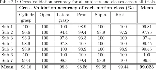
2.2.5 Results . . . 47
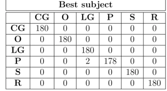
2.2.6 Conclusion . . . 48
2.3 Aid in multiple sclerosis diagnosis using commercial EMG armband . 50 2.3.1 Introduction . . . 50
2.3.2 Experimental equipment and protocol. . . 51
2.3.3 Classification . . . 53 1
2.3.4 Results. . . 54
2.3.5 Conclusion and future work . . . 56
2.4 General conclusions on the MYO armband . . . 56
3 Models for intent estimation from EMG decomposition 63 3.1 Introduction and main definitions . . . 63
3.1.1 Organization of motor control . . . 63
3.1.2 Model that relates the e↵ect to the MN firing behavior . . . . 64
3.1.3 Definition of the intended e↵ect estimation problem and main assumptions . . . 64
3.1.4 General approach to the decomposition-based e↵ect estima-tion problem. . . 65
3.2 Inference of the intended e↵ect from the set of recruited motor neurons 67 3.2.1 Probabilistic model of MN recruitment . . . 67
3.2.2 Recruitment model for multiple MNs . . . 68
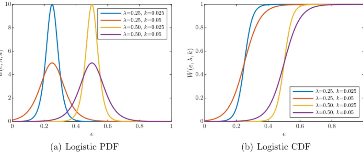
3.2.3 Choice of PDF for RT distribution . . . 70
3.2.4 Cost function for ML-estimation of RT distribution parameters 71 3.2.5 Cost function for ML-estimation of e↵ect from available MU activation data . . . 71
3.2.6 Example on simulation data . . . 73
3.3 E↵ect inference from firings rates of motor neurons . . . 75
3.3.1 Hidden Markov model of a spike train . . . 76
3.3.2 Inter-pulse interval distribution . . . 77
3.3.3 Criteria for IPI-based estimation model fitting and inference . 78 3.3.4 Iterative inference procedure . . . 79
3.3.5 Example on simulation data . . . 80
3.3.6 Joint e↵ect estimation model based on recruitment and IPI distribution . . . 81
3.3.7 Example on simulation data . . . 82
3.4 Conclusion . . . 83
4 Simulation of iEMG 87 4.1 Introduction . . . 88
4.2 Modeling of motor unit territories and muscle fiber innervation . . . . 90
4.2.1 Recruitment thresholds and sizes of motor neurons . . . 90
4.2.2 Distribution of the innervation centers in the muscle cross-section . . . 91
4.2.3 Muscle fibers geometry and distribution in the muscle cross section . . . 92
4.2.4 Assignment of muscle fibers to the motor neurons . . . 94
4.2.5 Results of the MFs innervation modeling . . . 95
4.2.6 MF diameters and conduction velocities modeling . . . 96
4.3 Neuromuscular junction modeling . . . 96
4.3.1 Structure of the axon branching model . . . 97
4.3.3 Coordinates of neuromuscular junctions. . . 100
4.3.4 Delay of MNAP propagation . . . 101
4.4 MUAP and EMG modeling for multichannel and shifting electrodes . 102 4.4.1 Transmembrane potential and current density . . . 103
4.4.2 Single fiber action potential modelling . . . 104
4.4.3 Motor unit action potential modelling. . . 104
4.4.4 EMG in a single observation point . . . 106
4.4.5 EMG in a single-channel electrode. . . 106
4.4.6 EMG in a multichannel electrode . . . 107
4.4.7 EMG in a shifting electrode . . . 108
4.4.8 Results and application example for multichannel MUAP sim-ulation . . . 109
4.4.9 Results and application example for scanning acquisition sim-ulation . . . 110
4.4.10 Results and application example for MUs territory assessment 112 4.5 Annotation and MUAP dictionary generation . . . 112
4.5.1 Instrumentation noise level setting for simulation . . . 113
4.5.2 Selection of detectable MUs . . . 114
4.5.3 Annotation and dictionary generation for a simulated signal . 114 4.5.4 Results and application example for multichannel iEMG de-composition . . . 115
4.6 Conclusion . . . 116
5 Simulation of muscle contraction 121 5.1 Introduction . . . 121
5.2 Contraction force simulation model . . . 122
5.2.1 Excitation-rate curves . . . 122
5.2.2 Firing instants modelling . . . 122
5.2.3 Twitch model . . . 124
5.2.4 Total force produced by a muscle . . . 125
5.3 Target profile . . . 125
5.3.1 Compensation of excitation-force nonlinearity . . . 126
5.3.2 Identification of excitation-force model . . . 126
5.3.3 Tuning of a PI controller . . . 130
5.4 Implementation details . . . 131
5.4.1 Global simulation model structure. . . 131
5.4.2 Class structure . . . 132
5.4.3 Sub-sampling of the force . . . 133
5.4.4 Incremental computation . . . 133
5.4.5 Data-set generation . . . 135
6 Simulation and experimental results 139
6.1 Simulated data set generation . . . 140
6.2 Experimental data set . . . 141
6.3 Evaluation methods . . . 142
6.3.1 Criterion of estimation quality . . . 142
6.3.2 Reference e↵ect estimation model . . . 142
6.4 O✏ine e↵ect estimation for simulated data . . . 143
6.5 O✏ine e↵ect estimation for experimental data . . . 145
6.5.1 Ramp contractions . . . 145
6.5.2 Trapezoidal and constant contractions . . . 145
6.6 Online simulated e↵ect estimation . . . 146
6.6.1 Virtual subject . . . 146
6.6.2 Results. . . 148
6.7 Online experimental e↵ect estimation: current state and perspectives 151 6.7.1 Description of the real-time decomposition system . . . 151
6.7.2 Pilot setup for experimental real-time decomposition . . . 152
6.8 Discussion and conclusion . . . 153
Conclusion and perspectives 157 Results. . . 157
List of Figures
1.1 Illustration of mathematical model of EMG signal. . . 14
1.2 Schematic illustration of spatial selectivity of intramuscular and sur-face EMG electrodes. . . 16
1.3 Illustration of EMG decomposition process. . . 17
1.4 Illustration of prosthetic control taxonomy on the example of wrist extension/flexion and pronation/supination. . . 20
1.5 Graph representing the classic dual-channel myoelectric control strat-egy. . . 21
2.1 MYO armband by Thalmic Labs Inc. . . 40
2.2 Grasp types or classes used in the gesture classification study. . . 43
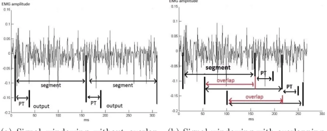
2.3 Two types of signal windowing: without and with overlapping. . . 44
2.4 Accuracies of training, online validation and online validation after taking o↵ and putting the armband on. . . 49
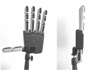
2.5 Front and side views of the robotic hand used in the tests. . . 49
2.6 Instruction for armband positioning on the forearm. . . 52
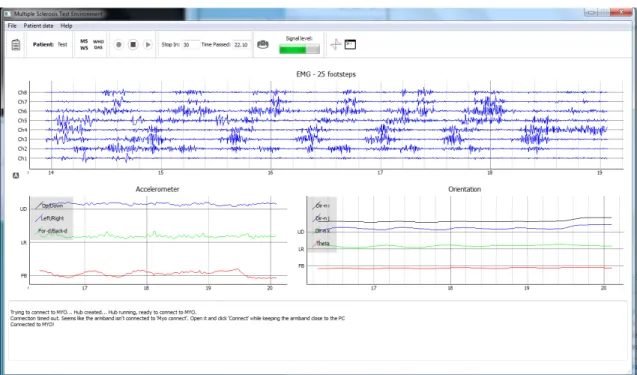
2.7 Graphical user interface developed for multiple sclerosis data acqui-sition. . . 52
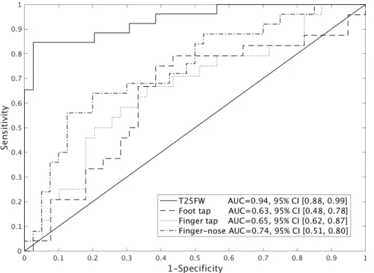
2.8 ROC curves for MS-healthy classification on four motor function tests. 55 3.1 Estimates of the e↵ect given by individual e↵ect-rate models. . . 66
3.2 Bayesian graph representing the probabilistic model of recruitment of a MN. . . 68
3.3 Bayesian graph representing the probabilistic model of recruitment of multiple MNs. . . 68
3.4 Examples of logistic probability and cumulative distribution functions. 70 3.5 Example of ML-estimation of RT distribution parameters on a simu-lated data set containing e↵ect samples close to the RT.. . . 72
3.6 Cost function for di↵erent e↵ect values as function of the set of active MNs. . . 73
3.7 Simulated contraction force and its estimate obtained by minimiza-tion of RT-based inference cost funcminimiza-tion. . . 74
3.8 Values of cost function for RT-based inference during the contraction in Figure 3.7. . . 75
3.9 Relation between sawtooth sequence, spike train and activation of a MN. . . 77 3.10 Bayesian graph representing the hidden Markov model of MN neuron
firing activity. . . 78 3.11 Example of IPI-based estimation of e↵ect, on simulated spike trains. . 81 3.12 xample of joint e↵ect estimation, on simulated spike trains. . . 82 4.1 Geometrical model of the muscle. . . 92 4.2 Muscle fibers distribution in a 1-mm2 area, generated by drawing
from constant distribution and by Farthest Point Sampling.. . . 93 4.3 Simulated territory centers of N =100 motor neurons in the
cross-sectional area of muscle. . . 93 4.4 The number of fibers assigned to each motor neuron for a simulated
muscle. . . 97 4.5 Simulated innervation territories of motor neurons . . . 98 4.6 Histogram of simulated fiber diameters. . . 99 4.7 Structure of motor neuron axon branching, modeled as a tree with a
single bifurcation. . . 99 4.8 Neuromuscular junction z-coordinates distribution model. . . 100 4.9 Scheme of the SFAP generation model adopted in the simulation. . . 103 4.10 E↵ect of propagation delays of MNAPs and SFAPs on resulting motor
unit action potential . . . 105 4.11 E↵ect of the neuromuscular jitter on MUAP waveform. . . 106 4.12 Simulation of multichannel MUAP acquired by an array of electrodes
inserted to a muscle at an angle to the muslce fibers. . . 110 4.13 A simulation of scanned MUAP within the innervation territory of a
MN. . . 111 4.14 Results of territory assessment simulation. . . 112 5.1 Excitation-rate relationships of motor neurons modeled as linear
sat-urated functions. . . 123 5.2 Schematic representation of the motor control system. . . 127 5.3 Excitation-force data from a simulated slow contraction with linear
excitation, and its polynomial fit. . . 128 5.4 PID controller for force simulation arrangement. . . 129 5.5 Identification and validation of the linear OE model of
excitation-force relation dynamic. . . 130 5.6 An example of simulated contraction with trapezoidal target profile. . 131 5.7 Global outline of the simulation model. . . 132 5.8 Schematic representation of the parts of the simulation model that
run at di↵erent sampling frequencies. . . 134 6.1 Simulated innervation numbers and innervation territories areas,
com-pared to their values imposed by the model. . . 141 6.2 Estimates provided by CST-based and the proposed estimation
6.3 Average values and standard deviations of R2 measure for the esti-mations performed by the proposed and CST-based models. . . 144 6.4 Estimation of abduction angle during experimental ramp contraction
reaching 50% of maximum angle. . . 145 6.5 Average values and standard deviations of R2 measure for the
esti-mations performed by the proposed and CST-based models on exper-imental ramp data set. . . 146 6.6 Results of angle estimation for experimental trapezoidal contraction.. 147 6.7 Results of angle estimation for experimental constant contraction
pro-file. . . 147 6.8 Schematic representation of the simulation model in cases of training
and testing of the e↵ect estimation model in online setup.. . . 149 6.9 Results of estimation for a simulated trapezoidal contraction in online
setup. . . 150 6.10 Average value and variance of R2 measures as functions of resulting
number of MNs included in the decomposition, online simulated setup.150 6.11 Structure of the real-time decomposition system.. . . 152
List of Tables
2.1 Cross-Validation accuracy for all subjects and classes across all trials. 47
2.2 Confusion matrix for user with best performance. . . 48
2.3 Confusion matrix for user with worst performance . . . 48
2.4 Demographics of participants. . . 53
2.5 Clinical evaluation of patients. . . 53
2.6 Features most represented in first principal components. . . 54
2.7 Confusion matrix for combined classification approach. . . 56
4.1 Main acronyms and notations . . . 87
6.1 Simulation parameters. . . 155
Chapter 1
Introduction
Application of the electromyography (EMG) to upper limb prosthetics is widely studied nowadays. This is due to the fact that control of a modern prosthetic device is required to be intuitive, robust and provide wide information flow between the user and the prosthesis. A way to ensure that is to establish a system that would autonomously infer user’s intent and pass it to the mechanical part of the prosthesis to execute it. EMG possesses some of the important features that can help meet these requirements.
The requirements of intuitiveness and wide information flow are dictated by increasing capabilities of the mechanical part of the prosthesis, which now provides larger functionality than existing prosthetic control approaches can cover. As an example, the individual finger manipulation in robotic hands was achieved a long time ago [1,2,3], while the inference of corresponding intent from the user still stays a problem that is not solved.
The EMG-based intent inference may be also considered in a more general con-text of human-machine interfaces (HMI). There is a growing interest in versatile HMIs, which brings in larger commercial interest to the field. Private companies, such as Emotiv1, Neuralink2, and open-source projects, such as OpenBCI3, engage
more and more in the development of HMIs oriented to both prosthetic and non-prosthetic use. The potential of EMG in such systems is supported by the commer-cial success of a wearable gesture recognition device Thalmic MYO4.
Physiological manifestations of user intent may be captured on three di↵erent levels: first is motor cortex, where the planning and onset of the motion is performed; the second level is the spinal cord, which transforms the high-level input command to low-level excitation-inhibition patterns; and the third level are the muscles, where the incoming motor command is transformed into the mechanical action.
Methods of interfacing the first level are being actively developed in the domains of neurorehabilitation and neuroscience. As an example, electroencephalography
1Emotiv official website 2Neuralink official website 3OpenBCI website
4Thalmic labs has ceased the production of MYO armbands and closed the corresponding website. We provide alink to MYO programming packages instead.
(EEG) is a technique of recording the electrical activity of the brain using a grid of non-invasive electrodes attached to the skull skin. A significant number of studies on EEG-based BCIs were made, proposing a cursor control [4], motion intent classifica-tion [5], and others [6]. The reasons why EEG-based interfaces are not being used in commercial prostheses are, first of all, a requirement for the user to be highly con-centrated on the performed task, as well as a low number of recognizable patterns, and the fact that constant wearing of an array of electrodes is generally impractical. Invasive brain and spinal cord interfaces that could overcome these limitations are yet far from being widely used in humans [7]. However, a recent white paper by Neuralink [8] suggests that basic invasive brain interfaces can be accessible on the market in a few years.
At the moment, among the three mentioned interfacing approaches, muscle level provides the most promising results. This is due to the fact that the user’s intent ul-timately transforms into the muscle contraction process, which can be characterized by the accompanying EMG. Techniques of EMG acquisition are far less demanding than brain or spinal cord interfacing, which makes EMG a promising information source in prosthetic control.
EMG-based prosthetic control is being studied in academia and industry for al-ready 60 years, with an extensive growth of popularity within the last three decades. However, there is a number of problems and limitations that were not yet overcome. First, the control strategy that practically reaches the amputees is still very similar to that used in the first myoelectric prostheses created in 1960s. Known disad-vantages of this approach coupled with the imperfections and expensiveness of the robotic parts lead to a high rejection rate among the amputees [9, 10]. A complex development of new control strategies, more comfortable stump sockets and reliable end-e↵ectors is still needed.
Another limitation of the EMG-based prosthetic control is the fact that in all variety of amputation cases, muscles of interest may be missing. For example, in the case of transhumeral amputation, none of the forearm muscles, which actuate the hand, are present, and therefore a source of information about the intent of hand motion is missing. One of the ways to overcome this limitation is to remap the patterns used in intent inference, to other muscles, such as those of the lower limb [11]. Another solution is targeted muscle reinnervation [12], a surgical method involving the relocation of amputated limb’s nerves into a healthy muscle in order to produce intent-related EMG.
In this chapter, we will cover the physiological and technical aspects of EMG, as well as the organization of motor control. Then, we will discuss existing EMG-based strategies of prosthetic control and their classification. Further, the potential roles of intramuscular EMG and EMG decomposition will be analyzed. That will bring us to the problematic of this thesis, which is a decomposition-based inference of user intent.
1.1
Basic notions of EMG, motor control and EMG
decomposition
EMG is a recording of the electrical activity of muscle fibers that naturally accom-panies their contraction during the execution of motor tasks. The contraction of muscle fibers is regulated by alpha motor neurons (MN) of the spinal cord, which, in their turn, are defined by supra-spinal commands originating in the motor cor-tex, and are regulated by numerous inter-spinal mechanisms of excitation, inhibition, and feedback. EMG is, therefore, a function of the total neural input to the muscle. In prosthetic applications, this makes EMG a great source of information about the user’s intent. In this section we will shortly cover the main point about the physiology and modelling of EMG.
1.1.1
EMG signal generation
A motor unit (MU) is an elementary entity of the motor system. It consists of a MN located in the spinal cord and of a set of muscle fibers innervated by its axon [13]. The last originates in the soma of MN and reaches the specific muscle or muscle group as a part of an e↵erent nerve. The axon then separates from the nerve in the vicinity of the muscle and forms a so-called terminal arborization to innervate a certain number of muscle fibers, usually referred to as muscle unit (MsU).
As a nerve cell, MN responds to excitatory input by a series of its cell membrane depolarizations. The last propagate through the axon in form of action potentials (MNAP) up to the junction with the muscle fibers. Due to the specific interfacing mechanism between an axon and a muscle fiber, called neuromuscular junction, MNAP excites an action potential in the fiber (SFAP). SFAP travels along the fiber, causing its contraction until it reaches the tendon and fades.
All fibers belonging to a single MU receive the MNAP almost synchronously with specific delays determined by the di↵erences in their axon branch lengths. This creates a unique spatial configuration of SFAPs, which, propagating along fibers with di↵erent velocities, further de-synchronize with each other.
Each SFAP causes the variation of the electric potential field around the muscle fiber, which can be detected by a metal electrode placed in its vicinity. Group of SFAPs evoked by the same MNAP produce a potential variation that can have unique temporal form when acquired by the electrode. The last also depends on the mutual positioning of the electrode and muscle fibers, their shapes and sizes. These shapes, as they are recorded by the electrode and the following acquisition system, are called motor unit action potentials, MUAPs.
While receiving excitation, MN generates MNAPs in a rhythmic fashion, with intervals between depolarizations of about 25 ms to 200 ms. A group of multiple successive action potentials is usually referred to as spike train. Since fibers of MUs are highly intermingled, an electrode usually records MUAPs from multiple MUs at the same time. Each MUAP is caused by a MNAP of corresponding MN. Thus, EMG is a mixture of repetitively appearing MUAPs of several di↵erent MUs located
Figure 1.1: Illustration of mathematical model of EMG signal 1.1. in the recording area of the electrode.
Despite being slightly out of physiological context, commonly used linear model of EMG helps to the understanding of this process [14, 15]:
Y (t) =
N
X
i
Hi ⇤ Ui(t) + w(t) (1.1)
where N is the total number of MUs, Ui(t) is a sequence of Dirac functions
rep-resenting the spike train of i-th MU, Hi is MUAP of i-th MU, symbol ⇤ denotes
convolution, and w(t) accounts for instrumentation noise of the acquisition system, as well as for interferences recorded by the electrode. This model is illustrated in Figure1.1.
1.1.2
EMG electrodes
EMG electrodes are metallic bodies (usually silver or stainless steel) in an isolation housing, destined to be in physical contact with the skin or the muscle fibers. The size of the electrode and configuration of the front-end signal processing circuits define its recording volume. SFAPs of fibers located within the recording volume contribute to the total acquired EMG.
EMG electrodes are first classified as surface (sEMG) and intramuscular (iEMG). The former are applied to the skin, while the latter are inserted subcutaneously into the muscle belly. sEMG electrodes are usually larger than iEMG ones, covering a considerable part of a muscle and thus having larger acquisition volume. This makes sEMG useful when the global activity of the muscle is of interest, since it integrates signals from multiple MUs at the same time. However, this also comes with the inability of sEMG to record solely from a target muscle, capturing the activity of neighboring ones. This e↵ect is usually referred to as cross-talk. The fact that the electrode is positioned on the skin also means that MUAPs should travel across skin tissues before being recorded, which introduces a considerable low-pass filtering e↵ect to the EMG.
iEMG electrodes, due to their subcutaneous nature, are small and therefore record from a much smaller volume, permitting to target specific regions in the muscle. Moreover, such electrodes can reach deeply-lying muscles that couldn’t be accessed by sEMG, such as pronator and supinator of the hand. Muscle tissue has better conductive properties than skin and fat, which makes iEMG preserve the high frequency components of the MUAPs. However, invasiveness of iEMG brings risks of infection, pain, and discomfort, which until recently made iEMG a very specific choice, dedicated mostly to neurological studies, rather then to prosthetics.
The aforementioned features of sEMG and iEMG are illustrated in Figure 1.2. Further classification of the electrodes is mostly based on their sizes and multi-channel arrangements. That is, the smallest intramuscular electrodes record from a single muscle fiber (SFEMG) [16]. Fine-wire electrodes record from a slightly larger volume, capturing several MUs at considerable contraction levels. Monopolar and concentric bipolar needles record from larger volumes of 0.02 - 0.1 mm3 detecting
10-15 MUs even at moderate contractions. A subsequent increase in recording volumes is achievable using multichannel intramuscular recordings [17]. Long-term record-ings, including prosthetic control applications, can be achieved using chronically implantable acquisition systems [18, 19, 20, 21, 22] (more on this type in Section 1.2.5).
sEMG electrodes can be classified accordingly. Myoelectric pattern recognition approaches (see Section 1.2.2) widely use several (usually up to eight) sEMG elec-trodes arranged around the wrist. Matrices of small sEMG sensors, arranged with high density (HDEMG) are more and more used in academia for prosthetic control, providing very detailed information on the spatial distribution of EMG activity across the muscle.
The entire variety of existing EMG electrodes permits to choose the character-istics of the signal that are best adapted to the specific problem. Some solutions, like intramuscular recordings, are less practical than others when considered in the context of prosthetic control. However, with the constant advance of technology, new long-term intramuscular acquisition systems emerge, making iEMG a more and more realistic choice for myoelectric control. At the same time, the full potential of conventional sEMG is not yet realized in the existing prostheses.
1.1.3
Motor control
Motor command originates in the motor cortex of the brain. Then, across several intermediate levels such as midbrain, pons, and medulla, it ”descends” to the spinal cord. Along this path, it is transformed into excitation/inhibition patterns, which are applied to the MNs of the spinal cord in order to contract or relax specific muscles, ultimately leading to the execution of the intended movement.
Force of muscle contraction is modulated by two processes: recruitment/de-recruitment of MNs and variation of their firing rates. An MN is recruited (i.e., starts firing) when the amount of its excitatory input reaches a so-called recruit-ment threshold, specific to each MN. After recruitrecruit-ment, the following increase of the excitatory input leads to the increase of firing rate. That is, the motor system
(a) Spatial selectivity of surface electrodes (b) Spatial selectivity of intramuscular elec-trodes
Figure 1.2: Schematic illustration of spatial selectivity of intramuscular and sur-face EMG electrodes. Sensitivity areas are not in scale and may vary for di↵erent electrodes. Muscle cross-section reproduced from Gray’s Anatomy [23].
regulates the muscle contraction force using excitatory input to its MNs.
The entirety of MNs that innervate a single muscle is called a motor neuron pool (MNP). MNs belonging to the same pool di↵er from each other by their size. That is, the physical size of MN’s body defines its behavior in response to the neural input [24], as well as the relative portion of the muscle that it innervates. That is, larger MNs tend to have larger recruitment thresholds, i.e., during an increasing contraction they are recruited later than the smaller ones. This phenomenon is usually referred to as Henneman’s principle [25].
The excitation-rate curve of MN is also defined by its size. Commonly used experimentally supported assumption is that at each level of contraction, firing rates of smaller MNs are greater than those of the larger ones [26,27]. This phenomenon is usually referred to as onion-skin principle, since, if traced on the same axis, firing rates of smaller MNs tend to always stay above those of larger ones.
It is common to represent an MNP as a functional unit, in which all the MNs receive a common excitatory input, dedicated to elicit the contraction of the corre-sponding muscle [28]. Experimental studies show that variations of firing rates of MNs are highly correlated during changes in the contraction force [26, 28]. That is, there is some degree of synchronicity with which MNs change their firing rates. In order to explain this phenomenon, C.J. De Luca and Z. Erim have proposed the notion of common drive [28], i.e., the existence of a single source of excitation for all the MNs of a pool.
At first, the common drive was associated only the with supra-spinal command, i.e, was supposed to be determined solely by the motor cortex. Later, it was shown that the firing rate correlation is influenced by proprioceptive feedbacks (muscle spindles and Golgi tendon organs) [29, 30] as well as by antagonistic and agonistic muscles contractions [31,32]. Considering more recent models [33] of motor control
+
Figure 1.3: Illustration of EMG decomposition process as inverse of model 1.1 and Figure 1.1. ˆUi(t) and ˆHi are the estimates of spike train and MUAP of i-th MU.
organization on the spinal level, it may be also influenced by inhibitory feedback introduced by Renshaw cells.
Common drive is considered to be a physiological tool that facilitates the motor control for CNS. It is assumed to provide centralized control over an entire MNP instead of independent control over each MN individually [34]. In the modelling of the motor control, the common drive is a useful notion that permits to define the input of the MN pool as a time-varying scalar function [35]. In di↵erent studies, it may be referred to as common synaptic input or net excitation.
As we can see, EMG is a function of spike trains produced by MNs and therefore is a function of the excitatory input to the MNP. That is, EMG characterizes the excitatory input that causes muscle contraction. In the case of amputation, this in-formation, based on the knowledge of the biomechanical function of the muscle, can be used to infer the user’s intent: which movement he or she wants to be performed and with which sti↵ness or speed, which constitutes the problem of prosthetic con-trol. The following section will provide the necessary terminology and classification of existing prosthetic control approaches.
1.1.4
EMG decomposition
As we have described earlier in this section, muscle contraction is the result of the spiking activity of MNs that innervate this muscle. Studies in such fields as neurology, motor control, biomechanics and, lately, myoelectric control, require the assessment of this activity. Direct recordings in vivo from MNs ’ somas or axons are possible, but the complexity of such procedures limits their application in practice. Instead, spiking activity can be inferred from the EMG signal.
Expression (1.1) serves as a good illustration of this process. That is, we observe an amount of EMG signal Y (t), t2 [0, T ] and we want to estimate the following: the number of MUs that contribute to the signal N , their MUAPs Hi and spike trains
Ui(t). This process is termed the decomposition of EMG. Figure 1.3 illustrates is
with respect to the model in Figure 1.1.
i.e., combinations of Hi and Ui that satisfy the equation (1.1). However, there
are some physiology-based constraints that one can apply to the possible solutions. First of all, spike trains Ui(t) exhibit sparsity and regularity. That is, the intervals
between spikes are much longer than the duration of a MUAP, and the spikes, during steady or slowly varying contractions, appear in a rhythmic fashion. Second, the forms of MUAPs do not change in time or change much slower compared to the frequency of their appearance, so that they can be efficiently tracked.
Decomposition can be accomplished either manually as well as semi- or fully automatically. In the first two cases, such tools as EMGLAB [36] can be used 1.
As for the automatic decomposition, there exist numerous techniques, the most influential of which will be covered in this section.
Historically, the first approaches addressed the decomposition of iEMG, that is much less cluttered compared to sEMG. In [37] and [38], template matching and clus-tering algorithms were applied to detected MUAPs in order to sort them into distinct groups with no attempt of superposition decomposition. In [39], prior knowledge on spike regularity is used and decomposition of at most two superimposed MUAPs is proposed. Automatic detection threshold adjustment, sub-sample level MUAP form matching, and peeling-o↵ of the greater MUAPs before subsequent re-decomposition were described in the ADEMG technique [40]. Adaptive MUAP clustering technique was proposed in [41].
Commonly acclaimed EMGLAB package [36] includes a modified version of ADEMG, as well as more advanced algorithm for multichannel iEMG decompo-sition MTLEMG [42]. The last one extracts features from the MUAP shapes and clusters them in order to obtain initial estimates of MUAPs and MUs. Then, clus-ters that have similar forms and matching firing timings are merged. Estimated MUAPs are then subtracted from the signal and another detection and clustering phase is performed. Finally, all MUAP estimates are used to resolve superpositions. Other techniques are also worth mentioning, such as density-based clustering [43], autoencoder-based MUAP feature extraction [44], genetic optimization for su-perposition resolution [45], Viterbi algorithm for spike train estimation [46].
It is also worth mentioning that the problem of iEMG decomposition is very similar to the problem of spike sorting in recordings of neuronal activity. Mostly, spike sorting techniques are based on detection and clustering phases, are variate in terms of the used features and clustering techniques. Examples of spike sorting techniques may be found in [47, 48, 49].
sEMG decomposition became possible due to the integration of information from multichannel recordings using blind source separation [50, 51, 52], including the tracking of changing MUAPs [53], as well as other techniques [54, 55]. Due to its practicality, sEMG-based decomposition becomes is used more and more commonly in the studies of biomechanics and motor control.
Due to the large variety of existing automatic decomposition methods, it may be difficult to choose one for use in a particular study. However, this choice is usually led 1If reader has no experience of manual EMG decomposition, a very clear interactive example is given inhttp://emglab.net/emglab/Software/Software.php.
by the accessibility of the decomposition technique, i.e., whether it is implemented, optimized and distributed in form of an open-source program, or as an additional package for commercial hardware or software. An example of the former is EMGLAB [36], MATLAB package for visualization and computer-aided manual decomposition of iEMG signals. Examples of the latter are OTBioeletronica’s Decomponi (CKC-based decomposition of high-density EMG signals)1, Demuse Matlab package2 and
Delsys systems3.
1.2
Existing myoelectric control strategies
1.2.1
Myoelectric control classification and terminology
In order to clearly describe a prosthetic control strategy, a classification of existing strategies is required. In this work, we use the system proposed in [56], which we will partly provide here.
First, this classification system introduces the notion of motor function, which is a distinct type of prosthesis’ movement, disregarding the speed or direction, e.g. hand gripping/opening, wrist flexion/extension, etc. Motor functions can involve one or several degrees of freedom (DOFs) of the mechanical part, e.g. single phalanx flexion/extension, wrist rotations, etc.
This system classifies prosthetic control strategies basing on three aspects: • Input sources, the set of input information sources which can be surface EMG
(sEMG), intramuscular EMG (iEMG), body or stump trajectory (e.g., from an inertial measurement unit), user input via a smartphone application and others;
• Intent interpretation, or how many motor functions the approach permits to control in total, and whether they can be controlled simultaneously (simulta-neous control) or just one at a time (sequential control) (see Figure 1.4); • Activation profile, or whether the approach permits to regulate some
mechani-cal output, such as joint angle or angular velocity, in a continuous (proportional control) or binary way (on-o↵ control)
Most of the existing myoelectric control strategies can be efficiently described by this classification [56]. Up to this date, the simultaneous proportional control strategy with multiple motor functions stays the main challenge of academic studies in the domain of myoelectric control. In the next sections, we will describe principal control strategies presented in the literature.
1https://www.otbioelettronica.it/en/products/software 2https://demuse.feri.um.si
3 https://www.delsys.com/timeline/first-non-invasive-system-for-decomposition-of-surface-emg-signals/
Figure 1.4: Illustration of prosthetic control taxonomy on the example of wrist ex-tension/flexion and pronation/supination. Green zones represent attainable control commands.
1.2.2
Dual-channel amplitude-based myoelectric control
strat-egy
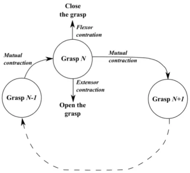
The first publication on myoelectric prosthesis dates 1948 and is made by physics student at Munich University, Reinhold Reiter [57]. This potentially revolutionary concept didn’t achieve clinical nor commercial use [58] until the year 1960 when Kobrinsky [59,60] has proposed a more practical design, which became possible due to advances in transistor technology. This device was capable of performing two functions: closing and opening the hand, both triggered by activity in one of two EMG electrodes placed above a pair of antagonist muscles groups of the forearm: flexors and extensors of the wrist and fingers. EMG was amplified, low-pass filtered and rectified to produce an estimate of the signal envelope, which was then compared with a certain pre-defined threshold in order to trigger either closure or opening of the gripper. The controller was also capable of detecting simultaneous contraction in both muscles and interpreting it as a command to stop the motion.
Even nowadays, many commercial prosthetic systems are still based on this strat-egy [61, 62]. Improvements mostly consider the mechanical part of the prosthesis: its weight, robustness and wider range of possible movements. The control strategy, on the other hand, undergone only a few modifications. Current strategies provide the user with a choice among several grasps and gestures, although the switching be-tween them is still triggered by the simultaneous contraction in both muscle groups. Having chosen a gesture, the user may adjust it by the contraction of one of the two muscle groups, e.g. flexion/extension for closing/opening the hand (see Figure 1.5). According to the classification provided above, this is a proportional control strategy with a more restricted sequential approach. Such a technique is robust and easy for the user to learn. However, it requires to remember which motor function is currently chosen, as well as the number of co-contractions needed to activate the next one.
Figure 1.5: Graph representing the classic dual-channel myoelectric control strategy. The user switches to the next motor function (grip) in the graph by performing a co-contraction of antagonistic muscle groups. While a function is chosen, the user can adjust it to the current task by contraction one of the muscle groups.
1.2.3
sEMG pattern recognition myoelectric control
strat-egy
The first attempts to avoid using the cyclic graph control were based on an increased number of sEMG channels in combination with machine learning techniques. The family of such strategies can be referred to as myoelectric pattern recognition. Then involve placement of several electrodes around the forearm (usually around 8) either symmetrically, or above specific muscles (targeting) [63], signal windowing, feature extraction, and subsequent supervised classification.
The variety of all possible hand movements is discretized to form an acceptable number of classes ([56,64]). These classes are designed to cover the most important motor functions and, at the same time, to be distinguishable by a classification algorithm. The most frequently used classes are cylindrical grasp, hand rotations, wrist flexions and extensions, radial and ulnar deviations ([65]).
In order to apply classification techniques to EMG, one should first reduce its dimensionality. This is usually achieved by means of feature extraction and feature selection. Features are scalars or vectors that represent temporal or spectral pa-rameters of the signal, taken in a short window (usually not longer than 250 ms). The number of feature types presented in the literature is large ([64]), and it is shown that some of them are more e↵ective than others when applied to myoelectric pattern recognition [66].
amplitude, spectral band, etc. It is hard and in most cases not possible to bond these parameters into one physiology-based model. Thus, multivariate statistical analysis and machine learning were considered to be suitable techniques for finding such models automatically. Numerous approaches were investigated during last two decades: Linear Discriminant Analysis [67, 68], Neural Networks [69] and Support Vector Machines [70].
This strategy is essentially a sequential on-o↵ control. Its advantage over the previous one is that the grip graph, compared to Figure 1.5 is fully connected. That is, each grip or gesture is accessible from any other grip. However, the main challenge for this approach is the fact that surface electrodes, that provide the input for pattern recognition, shift during daily activities, which causes a drop of the recognition accuracy since patterns are learned in the original position of the electrodes [71, 72].
It took almost three decades for this strategy to be recognized and implemented by companies producing myoelectric prostheses. At this moment, it is being imple-mented in commercial prostheses [73], [74]. Rate of acceptance of such devices by the amputees should be analyzed in the future. This is important since even the classic two-electrode strategy sometimes fails to satisfy the user when exposed to such factors as variable posture, sweat, and fatigue [75].
1.2.4
Other sEMG-based myoelectric control strategies
A strategy for simultaneous and proportional control of at least two motor functions was presented in [76]. This approach supposes that each motor function is regulated by a single excitatory input to the synergistic muscles that actuate it. Then, the authors propose a model that links these inputs to the sEMG signal in electrodes placed in the vicinity of the muscles. The model parameters are estimated from training data using non-negative matrix factorization. Training data consists of the motor function values and simultaneously acquired sEMG.
This method, since it is based on sEMG, may su↵er from the electrode shifts and fatigue (as for the variation of contraction force, we note that it is included in the model). However, authors have provided a separate study [77] on that cause and, using high-density electrode matrices, have shown that both longitudinal and transversal shifts do not significantly a↵ect the accuracy of this control strategy. This method was tested on a commercial prosthesis and have provided considerable acceleration on classic motor tests1 .
Another variant of pattern recognition and non-negative matrix factorization arises from the use of high-density electrode matrices. As an example, for pattern recognition, such features as spatial correlation, specific to these kinds of recordings, are extracted and classified [78]. Model for non-negative matrix factorization can be adapted to high-density electrodes [77].
Further improvements of existing strategies can be achieved using physiology-based modelling of how the user’s intent transforms into sEMG in each of the
able channels. In [79], a neuromusculoskeletal model defined to, first, transform sEMG features to muscle contraction forces and, second, transform muscle contrac-tion forces to a set of feasible prosthesis movements. In order to collect calibracontrac-tion data for the model, the user is asked to mimic prosthesis movements with his or her phantom limb. As authors suggest, neuromusculoskeletal modelling permits to con-straint the derived intent to a set of feasible movements and provide more natural control to the user.
To conclude, at the moment sEMG stays the only choice for interfacing the user’s motor system in commercial prostheses. Electrode shifts, being one of the most challenging issues, can in some cases be countered by choosing specific electrodes and control strategy [77]. The lack of spatial selectivity in sEMG makes it impossible to record solely from target muscles (so-called cross-talk ), which complicates the building of the control model. Incorporation of the cross-talk into the model, as in non-negative matrix factorization approaches [76], can slacken this problem. Thus, the full potential of sEMG-based strategies presented in the literature is yet to be realized in market prostheses.
However, cross-talk and electrode shift problems can also be alleviated by using a di↵erent acquisition approach, which is, for example, intramuscular EMG. In the following section, let us consider existing applications of iEMG in prosthetic control.
1.2.5
iEMG-based myoelectric control strategies
Intramuscular EMG (iEMG) is an invasive method that involves the placement of the electrodes either subcutaneously or in the interior of the muscle tissue. In the prosthetic control perspective, iEMG has a number of advantages compared to much more conventional sEMG, such as high spatial selectivity, access to deeper muscles and lesser exposure to ambient noise and interference.
High spatial selectivity permits to record from a target muscle, avoiding interfer-ence from the surrounding ones. As we have mentioned earlier, the last is a common problem of sEMG-based control strategies. Moreover, intramuscular electrodes per-mit to acquire iEMG from deeper muscles, such as pronator or supinator of the hand.
Despite the aforementioned advantages, iEMG is considered impractical for pros-thetic control due to its invasive nature that brings risks of infection, discomfort, and pain to the user. However, there exists a number of studies [18, 19, 20, 21, 22] that propose to surgically implant wireless acquisition system into the muscle. These implantable myoelectric sensors (IMES) usually have cylindrical form, with a length of 10 mm and diameter of 3 mm [18]. They consist of a single-channel analog in-strumentation amplifier, pass-band filter, analog-digital converted, and a magnetic coil for the wireless battery recharging and iEMG data transmission.
In such systems, as it is shown in [21], the total sampling frequency shared by all channels may reach 14 kHz. A high sampling frequency is of interest when complex iEMG processing approaches are used, such as decomposition. Alternatively, as in a more recent study [22], the signal can be rectified and low-pass filtered on the IMES and then transmitted with a much lower sampling frequency (e.g., 67 Hz in
[22]). This is an optimal solution for strategies where rectified sEMG is used as an estimate of muscle contraction force.
Rectified and low-pass filtered EMG was already used as an estimate of the proportional intent in studies on iEMG applied to prosthetic control. For example, [80] associate each motor function with one single iEMG channel and calculates the proportional input for the prosthesis as a low-pass filtered rectified signal. In [81], a slightly di↵erent approach is proposed, in which the proportional input is estimated using the di↵erence between the rectified signals from two antagonistic muscles.
However, the small acquisition area of intramuscular electrodes can also be a disadvantage in this case. This is due to the fact that observed muscle fibers may not represent the muscle as a whole [82]. The same authors [83] propose to use an artificial neural network to establish a non-linear regression model that relates iEMG features to the proportional input.
To conclude, in prosthetic control, such advantages of iEMG as spatial selectivity and access to deep muscles, come together with the need for complex technologi-cal solutions, such as chronic implants with wireless data transmission. However, the last ones are becoming more and more available since the technology is being explored and improved by several independent research and engineer groups.
1.2.6
Existing decomposition-based control strategies
A real-time decomposition of hdEMG and subsequent gesture classification is pre-sented in [84]. The decomposition is achieved using an iterative version of blind source separation technique [85], reaching the decomposition of approximately up to 15 MUs. Resulting individual firing rates are then low-pass filtered and passed as an input to a support vector machine classifier, recognizing four di↵erent gestures.
The approach proposed in [86] decomposes a high-density EMG (hdEMG) of a TMR1 subject using the convolution kernel compensation algorithm [50]. It groups
decomposed MUs into four categories depending on their approximate location in the observable part of the muscle, corresponding to four quadrants of the HD elec-trode. Their firing frequencies, along with their categories are then used as features for a support vector machine classifier. Alternatively, in this study they use a neu-romusculoskeletal model, similarly to the one we have mentioned in Section 1.2.4, that is able to transform decomposed spike trains into joint torques of a calibrated musculoskeletal model, that then are projected to plausible movements of the pros-thesis.
In [87], it is proposed to estimate the common input to a motor neuron pool by combining its individual estimates obtained from the decomposed spike train of each 1Targeted Muscle Reinnervation (TMR) [12], a surgical technique consisting of, during trans-humeral (shoulder) amputation, recovering the nerves of the amputated limb and placing them in an intact muscle, such as pectoralis major (chest). During the first months after the operation, the relocated nerve innervates the host muscle. Intended movements of the phantom limb, in that case, result in the contraction of host muscle with coincident EMG generation, which can be captured and analyzed for prosthetic control. TMR is an example of the more general concept that perceives the muscle as a biological amplifier of the spinal command.
single motor neuron. The authors use the common assumption that motor neurons of one pool receive a common input that regulates their joint production of muscle contraction force and, therefore, the mechanical parameter of the movement (in their case - the angle of wrist rotation). They directly replace the common input by angle measurements and model the angle-spike relationship as linear. Since angle estimates obtained from di↵erent motor neurons will not be identical due to the presence of individual inputs to the MNs, and decomposition errors, the authors use median value across the pool as a global estimate of the angle.
Despite not being directly related to prosthetic control, [88] studies the rela-tionship between smoothed composite spike train (CST1), as a measure of neural
drive to the muscle, and contraction force of a muscle. They suggest that CST is better correlated with the force, especially at higher frequencies, than convenient the integrated sEMG.
While previously mentioned approaches work with hdEMG decomposition pro-viding spike trains of 10 up to 20 MNs, [89] aims to infer the contraction force from a smaller number of MNs (1 to 10). Such numbers of decomposed trains are usu-ally observed in the decomposition of fine-wire or needle iEMG at low contraction forces. The authors propose to, first, estimate force-rate relationships for all decom-posed MNs, then to use the inverse of these relationships to estimate force from each currently observed inter-spike interval.
This approach can be applied either to the estimation of a single muscle con-traction level or to the proportional control of an entire motor function. Former will be of use in such control strategies as [79], where the contraction level of each muscle feeds a musculoskeletal model that then produces motor commands for the prosthesis. The latter application will be of use when EMG of only one muscle of a synergistic muscle group is being observed, so that the neural drive to the muscle group is estimated from this single muscle.
1.3
Problematic of decomposition-based
myoelec-tric control
Studies of decomposition-based control strategies appear much rarer compared to those on pattern recognition or other sEMG-based approaches. This is due to the high complexity of the decomposition task itself, both in terms of implementation difficulty and of computation power that this procedure demands.
Prosthetic applications require that all the processing applied to the signal should be performed online with a maximum delay of about 250 ms [90]. Up to this mo-ment, there’s only three existing real-time full decomposition approaches [91,84,92] that can provide decomposition with such a delay. The first is a recursive version of the convolution kernel compensation algorithm [50]. It is able to decompose an experimental signal acquired by a 6⇥ 5 matrix of sEMG electrodes in real-time. 1CST (cumulative or composite spike train) is the spike trains of all decomposed MNs summed up into one train as if it was produced by one single neuron. It usually serves as an estimate of neural drive or excitation applied to the motor neuron pool.
More specifically, their signal was generated during a 10% MVC isometric contrac-tion of the tibialis anterior muscle. The algorithm was capable of decomposing 4±2 MUs, correctly identifying at least 90% of their discharges. The second is a recursive version of blind source separation method [85], achieving decomposition of approx-imately up to 14 MUs at contraction levels of up to 30%MVC on a standard PC, using a matrix of 64 electrodes. Experiments were conducted on tibialis anterior and flexor digitorum superficialis muscles.
Approach presented in [93] is based on [94] and is being constantly developed by our team. It is based on state-space modelling of the MNs spiking activity, sampling of possible spiking scenarios and Bayesian filtering of the state vector. The advantages of this approach compared to the two previous ones are the use of a single-channel signal, the capability of tracking the slowly-varying changes of MUAPs’ shapes and adaptation to the additive noise level. Also, single channel intramuscular EMG is potentially better adapted to the variety of muscle geometries, while approaches based on hdEMG tend to perform worse on muscles whose fibers are not parallel to each other and to the skin.
Originally not adapted to changes in the number of active MUs, our approach was recently modified to decompose iEMG of dynamic contractions [93]. Next step of its development [92] is a GPU1-based implementation permitting to perform
real-time decomposition of up to 10 concurrently active MUs during dynamic contraction with global performance scores reaching 95%.
All of the presented methods are limited to the decomposition of a relatively small number of MUs (up to 6 in [91], 10 in [92] and 14 in [84]), which questions their applicability in prosthetic control. That is, these numbers are small compared to the total numbers of MUs even in small muscles, such as first dorsal interosseous (120 MUs [95]). Therefore, it is a question, whether such small number of MUs can be a representative subset of the entire MU pool, compared to, e.g. the rectified sEMG that integrates the activity of tens and hundreds of MUs.
In the case of [92], the number of decomposed MU is limited both by the avail-able computational power and by the acquisition volume of the intramuscular EMG electrode. An increase in the number of decomposed MUs by running several inde-pendent instances of the algorithm on several spaced out electrodes does not seem viable, at least for prosthetic control applications. This is due to the fact that prosthesis-embedded GPU-based decomposition2 of one single channel is a
challeng-ing task by itself and is not yet achieved. This leads us to the problematic of this research, which is: how to estimate the level of muscle contraction or of the neural drive to the muscle, using a very limited number of decomposed MU ? More detailed reasoning about this problem will be given in the following section.
1GPU - Graphics processing unit, or graphic card. Originally destined to PC screen image rendering, they are now widely used for parallel computations.
2 Possible approaches to this task may involve recently developedNvidia Jetson Nano, a de-veloper kit for embeddable GPU applications.