This thesis is wholeheartedly dedicated to the many people who made it possible.
To my Mother
Khadija Ennaji
A gentle and strong soul who taught me to believe in hard work, trust in Allah,
look at the bright side and enjoy little things in life.
To my Father
Ahmed Drissi
For being my first teacher and for encouraging and supporting me to believe in
myself.
To my Brother
Rachid Drissi
We do not choose our family, but I could not have dreamed of a better way to be
fulfilled.
To Dr. Sara El Guessabi
I would love to thank you for your time and for teaching me a lot about cell
culture.
To My friends from faculty of Medicine and Pharmacy of Rabat
I can not find the right and sincere words to express my affection to you. You
have always set the example of caring, faithful, and helpful friends.
To all my friends
You who have accepted to board the train of my life to share my sorrows and my
joys, the adventure is still going on.
To our Professor and President of the thesis jury
Professor Madam Naima ALAMI OUHABI
Professor of molecular biology and biochemistry
It is a great honor for us to have you as the chair of our thesis jury. In spite of your many solicitations, you have kindly agreed to take part.Would you please accept, dear Professor, the expression of our deep gratitude, our great respect and our deep appreciation.
To our Professor and Thesis Rapporteur
Professor Mister Azeddine IBRAHIMI
Head of Biotechnology Lab and Professor of Medical Biotechnology
The trust you have placed in me by assigning me this work, affects me in a special way.I would like to express my special appreciation and thanks to you Mr. IBRAHIMI Azeddine for allowing me to realize this thesis.
Your advice and your kindness were considerably precious to me. You have always welcomed me in spite of your professional obligations. I am very pleased to be able to express my deep gratitude for all the efforts you have made in order to ensure that this
work can succeed.
Would you please accept my infinite gratitude for having given me the chance to benefit from your scientific experience in this field.
To our professor and judge
Professor Madam Naima EL HAFIDI
Professor of Pediatrics
Professor, it is a great honor for us to have you as a member of our thesis jury Would you please accept, dear Professor, the expression of our deep gratitude, our great
To our Professor and judge
Professor Mister Jaouad HARTI
Professor of Therapeutic Chemistry
You have kindly accepted to judge our thesis, despite your multiple occupations. We thank you for your attention to this work.
Would you please accept, dear Professor, the expression of our deep gratitude, our great respect and our deep appreciation.
CAR : Chimeric Antigen Receptor
COBRA-FISH
: Combined Binary Ratio labelling - Fluorescence in situ hybridization
CPDs : cumulative population doublings
DMSO : Dimethyl Sulfoxide
DNA : Deoxyribonucleic Acid
ESCs : Embryonic Stem Cells
FBS : Fetal Bovine Serum
FDG : Fluorodeoxyglucose
HLA : Human Leukocyte Antigen
HSCs : Hematopoietic Stem cells
iPSCs : Induced Pluripotent Stem Cells
IVF : In Vitro Fertilization
L-DOPA : L-3, 4- dihydroxyphenylalanine
LIF : Leukemia Inhibitory Factor
MAPCs : Multipotent Adult Progenitor Cells
MIAMI : Marrow Isolated Adult Multilineage Inducible Cells
MSCs : Mesenchymal Stem Cells
NIH : National Institute of Health
NK : Natural killer
NOD : Non Obese Diabetic
PBMCs : Peripheral Blood Mononuclear Cells
PBS : Phosphate Buffered Saline
RNA : Ribonucleic Acid
List of figures
Figure 1:Isolating mononuclear cells from peripheral blood by density gradient
centrifugation using ficoll . ... 12
Figure 2: The pellet obtained after double centrifugation with PBS to wash off
any remaining platelets. ... 12
Figure 3:Malassez counting chamber to count the cells. ... 13 Figure 4:cultivation in a flask of 25 cm². ... 13 Figure 5: Microscopic observation... 14 Figure 6:The process of fertilization ... 19 Figure 7: Pre-embryonic cleavages ... 21 Figure 8: Germ layers fates in embryo ... 22 Figure 9: Different stages of stem cells differentiation ... 25 Figure 10: Adult stem cells plasticity ... 32 Figure 11: Hematopoietic stem cells differentiation ... 34 Figure 12: Example of manipulations in a closed system ... 44 Figure 13: Therapeutic cloning . ... 46 Figure 14: Hayflick limit. ... 50 Figure 15: Autologous stem cell-based therapy on burn injuries ... 60 Figure 16:Sources for cardiac stem cells . ... 67
Figure 17:Production of insulin-secreting pancreatic-like cellsfrom mouse
embryonic stem cells ... 75
List of Charts
Introduction ... 1 First part : cell culture ... 4
I. The history of cell culture ... 5 II. Cell Culture ... 5 1. Definition, Concepts, and Characteristics of Cell Culture ... 5 2. Primary culture and establishing a cell line ... 7 3. Applications of Cell Culture ... 9 4. Cell Culture experiment ... 10
Second Part : Stem Cells ... 16
I. History of Stem Cells ... 17 II. Human biological development ... 18 1. Fertilization ... 19 2. Segmentation ... 19 3. Gastrulation ... 21 4. Organogenesis ... 22 III. Stem Cells ... 23 1. Definition of Cell ... 23 2. Stem Cells ... 23 3. Different types of human stem cells ... 25 3.1. Embryonic stem cells ... 25 3.2. Induced pluripotent stem cells ... 27 3.3. Fetal stem cells ... 30 3.4. Adult stem cells ... 31 4. Timeline of stem cell research ... 37
I. Definition and perspectives ... 41 II. Stem cells in cell therapy ... 42 1. Principle and process of cell therapy ... 42 2. Cell therapy laboratory ... 43 3. Sources of stem cells samples ... 44 3.1. Sources of embryonic stem cells ... 44 3.2. Sources of fetal stem cells ... 45 3.3. Sources of adult stem cells ... 45 3.4. Therapeutic cloning ... 45 4. Culture and amplification of stem cells ... 47 4.1. Optimal conditions for culture ... 47 4.2. Stem cells’ culture limits ... 48 4.3. Culturing adult stem cells ... 52 4.4. Culturing embryonic stem cells ... 52 5. Stem cells banking ... 53 5.1. Bank of embryonic stem cells ... 53 5.2. Bank of fetal stem cells ... 54 5.3. Bank of adult stem cells ... 55 III. Applications of cell therapy ... 56 1. Cell therapy on the skin ... 57 2. Cell therapy and Parkinson’s disease ... 61 3. Cell therapy for heart failure ... 64 4. Cell therapy for type I diabetes ... 72 5. Cell therapy and cancer ... 77
Fourth Part: The Ethical, Legal, and Religious Aspects of Stem Cell
I. The Ethical Context ... 84 1. For the embryonic stage ... 85 2. Fetal stem cells samples ... 86 3. Post-natal stage ... 86 4. Therapeutic cloning ... 86 II. The Legal Context ... 87 1. Legal situation in Morocco ... 87 2. Legal situation in European countries ... 88 3. Legal situation in United States ... 89 III. The Religious Context... 91 1. Islam ... 91 2. Judaism ... 92 3. Christianity ... 92
Conclusion ... 94 References ... 100
1
2
Cell culture is a very useful and versatile technique in the study of basic scientific research issues. Its origins date back to the early 20th century, when it has been introduced to study the growth and maturation of tissues, the role of genes in health and disease, virus biology and vaccine production, and the use of large scale hybrids of cell lines to develop biopharmaceuticals. The experimental uses of cultured cells are as various as the types of cells that may be grown in vitro. There is a rising interest in optimizing stem cells culture, not only because cell culture has become widely used in fundamental research to study stem cells biology, but also because of the potential therapeutic applications of cultivated stem cells. Stem cell culture parameters are crucial and require continual refinement as our understanding of stem cell biology advances and the latest technological developments.
Stem cells have a unique potential to self-renew and to differentiate into different specialized cell types in the organism. Recent advances in stem cells biology offered new hopes in treatment of disorders and diseases that can not be treated yet. Therefore, in recent years, stem cells therapy is becoming a very promising and advanced topic of scientific research.
There are various interesting types of stem cells with their own advantages and disadvantages. Embryonic stem cells have a great proliferation and differentiation potential. Despite their potential contribution to science, the use of embryonic stem cells in research raised sharp ethical, legal and religious concerns.
3
The use of stem cells in therapeutic applications requires defining optimal conditions for these cells amplification in vitro and in vivo, in order to ensure the absence of tumorgenic potential as well as their functional integration within the damaged tissue.
The objective of this work is to focus on the discovery of various stem cells and the potential therapies that are based on these cells as well as the perspectives, issues and limits of stem cells biology.
The first part of this work is an explicit presentation of cell culture. The second part is a study of different stem cells.
The third part is an explanation of cell therapy and its therapeutic applications. In particular, the application of this therapy in dermatology, Parkinson’s disease, type I diabetes, heart failure and cancer.
The fourth and last part discusses the national and international legislation, ethical and religious issues concerning the use of stem cells.
4
5
I. The history of cell culture
Cell culture development has passed through three important periods.
In the early 1900s, the professor Ross Harisson developed the first method for culturing tissues outside of the body. A biologist, Harisson was able to grow frog neuroblasts on a culture dish and thus opened the door for current stem cell research.
Beginning in 1902, the biologist and surgeon Alexis Carrel attempted to develop the state of animal tissue cultures with experiments on a chick embryo tissue culture. He improved tissue harvesting techniques, developed aseptic principles, and studied nutritional requirements [1, 2].
In 1952, the biologist Aron Mosconaintroduced trypsinization of tissues for cell culture. Moscona developed a method for digesting a chick embryo tissue using trypsin in order to obtain dissociated cells that are capable of dividing in vitro [3].
II. Cell Culture
1. Definition, Concepts, and Characteristics of Cell Culture
Cell culture is the process through which cells are raised under particular conditions outside their natural environment. The term « in vitro » is used for tissue and cell cultures under artificial physiological conditions.
Cell culture provides a sufficient cell number to serve as an experimental material. The main challenge of cell culture is that it must be realized under sterile conditions because it is contamination-sensitive.
6
an appropriate medium that supplies the essential nutrients and growth factors under controlled conditions (temperature, pressure, pH, humidity, etc). Cell isolation varies depending on cell type. That is to say, cells purified from bloodare isolated by ficoll density gradient centrifugation whereas cells obtained from solid tissues are isolated by enzymatic digestion using trypsin [4].
Cells can be grown either free floating in suspension or on a surface adherent or monolayer cultures.
Cellular proliferation happens in four phases in the cell culture. Firstly, the lag phase, in which no cell division is observed because cells adapt to the culture conditions. In the subsequent phase, the cells actively divide. At this phase, cell population is considered to be the most viable. Then comes the plateau or stationary phase in which the cell death rate is balanced with the cell division rate due to the cell population becoming confluent and nutrients depletion. Lastly, there is the decline phase in which the number of viable cells decrease and cell death increases due not only to nutrient reduction but also to the natural evolution of the cellular cycle [5].
In-vitro cells are characterized by two fundamental properties, their proliferative capacity and their differentiation function. These properties tend to vary regardless of the culture method used.
Usually, cells maintain their division potential which can be stimulated at the beginning of the culture by growth factors whereas cells in the culture often change or lose their differentiation function. This de-differentiation phenomenon can be morphologically visible. However, some cell types reveal their specific function only when grown slowly by gradually depleting nutrients in the culture medium or by using a cell division inhibiting substance [6].
7
Most isolated primary cells go through the process of senescence and can no longer divide. The life span of most cells is limited, but some cells have been transformed into immortal cells and acquired the ability to reproduce indefinitely [7].
2. Primary culture and establishing a cell line
There are different types of culture which can be divided into primary culture and established cell lines culture.
Primary culture is a cell culture method that is established when culture cells are directly derived from their tissue of origin. Primary culture starts with the removal of cells from pieces of tissue and then depriving cells via mechanic or enzymatic digestion techniques from this biopsy. Despite the fact that primary cells provide a heterogeneous population, have a low proliferation rate, and the difficulty of the techniques used, cells in primary cultures are significantly closest in form to cells present in normal tissue. Cells that are reproduced with primary culture have a limited life span and their replication stops when one nutrient of the culture medium is depleted. In this situation, cells of primary culture are transferred to a new culture medium in appropriate conditions and continue to reproduce. This cell culture is referred to as secondary culture. At a certain time, secondary culture cells lose the capacity to proliferate and survive due to the changes that happen physiologically and pathologically. This process is similar to cellular senescence that occurs in the organism.
The drawbacks of primary culture are that when cells are removed from the in vitro environment and transferred into the culture medium, they could lose their structural and functional properties. At this point, cells that have a different morphology at the tissue level can present similar morphologies in the culture
8
medium. Another important problem is the cells’ limited lifespan and consequently, these cells must be replicated with serial passages and saved. Nonetheless, in some cases the number of cells of interest might be less than other cells and for this reason their reproduction could be pressed. In this situation, providing special mediums and growth factors might be required [5].
The second type of cell culture is the established or immortal cell lines culture. Most of these cells are obtained from tumors or transformed cells in vitro. Cell lines have the potential to divide indefinitely and to be grown for an extended period of time, retaining certain phenotypes and functions.
These cell lines have assumed an important role in studying the control of cell cycle and vaccine production. Cell lines have a number of advantages that make them useful in vitro. The obtained cells display a homogenous population, are easy to grow, and can be subcultured continually via an acceptable number of passes to produce a large cell number in a short time. This has made cell lines an attractive model for high-throughput drug screening. Nevertheless, the main drawback is that these cell lines are transformed which can alter their functional levels compared to those found in primary cells and some important functions are even missed entirely. Furthermore, cell lines cannot be used to evaluate genotypic and phenotypic differences in response to drugs in humans because they originate from a single ancestor cell [8].
Recently, cell lines have been evolved with the aim of retaining a normal phenotype combined with the capacity to grow the cell indefinitely in culture. This can be achieved by establishing the cell line from stem cells that can be induced to proliferate into a terminally differentiated cell type in culture. These lines are more difficult to handle in vitro [9].
9
For both scientific and economic reasons, cells might be needed again in separate studies, which reveal the need for freezing and storing cells (cryopreservation). Cells that provide a sufficient growth cycle can be frozen. Through this process cells are frozen in liquid nitrogen in the presence of cryoprotectant agents such as DMSO (dimethyl sulfoxide) or Glycerol. Thawing frozen cells is fast at 37 °C [5].
3. Applications of Cell Culture
Cell culture is the key technique for cellular and molecular biology. Cell culture is used as a model system to study cell functions and their normal physiology and biochemistry as well as the effects of drugs, cosmetics, chemicals, and toxic compounds on the cells and mutagenesis and carcinogenesis mechanisms.
Animal cell cultures are fundamental to the production of viral vaccines and important genetically engineered proteins (insulin, hormones…). These cell cultures are also used to insert new genetic material (DNA or RNA) into the cell, which can be used to study its effects on the cell. Furthermore, cell culture is utilized in drug screenings and development to study the cytotoxicity of new drugs.
Human stem cell cultures are used to increase the cell number and differentiate the cells into different types of somatic cells for transplantation which can be used to treat a variety of medical conditions [10, 11].
10
4. Cell Culture experiment
Experiment Design:
Cell culture samples can be obtained from different sources such as peripheral blood, adipose tissue, or the umbilical cord blood.
This experiment is about isolating cells and conducting a culture of human peripheral blood mononuclear cells (PBMCs) by density gradient centrifugation using ficollhistopaque.
Materials and Reagents:
All materials and reagents that come into contact with cells must be sterile. - Freshlycollectedheparinizedblood.
- Phosphate Buffered Saline (PBS). - Auto pipettes.
- Cell medium: 10% Fetal Bovine Serum (FBS), 1% L-Glutamine and 1% Antibiotic.
- Ficoll Histopaque. - Trypanblue.
- Malassez counting chamber or automated cell counter. - Culture flasks.
Equipment :
- Laminar air flow cabin. - CO₂ incubator.
11
- Air conditioner, the lab temperature should be at 17 °C. - Centrifuge machine with swing-out bucket rotors. - Inverted microscope connected to a computer. - Sterile centrifuge tube
Methodology:
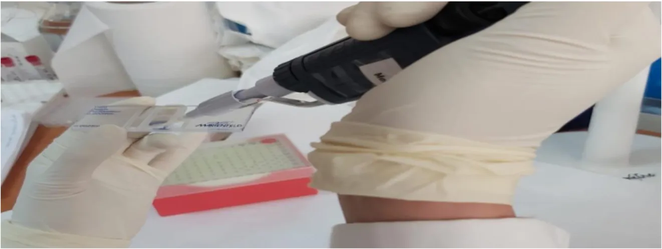
The method for PBMC’s isolation is based on density differences between PBMCs and different components of the blood. The most commonly used density gradient medium is Ficoll. This medium is less dense than granulocytes and erythrocytes, but denser than lymphocytes, monocytes, and platelets.
After receiving the heparinized blood sample, dilute the blood with the same volume of PBS. Prepare a sterile tube filled with an equal blood volume of ficoll, then hold this tube at a tilted angle and place the tip of the pipette against the tube wall and carefully layer the diluted blood over ficoll without intermingling the two phases. Centrifuge for 20 min at 400g without a brake. During this time, four layers are formed with each one containing different blood cell types. From bottom to top you find erythrocytes and granulocytes, followed by Ficoll, the layer containing PBMCs which appears white and cloudy, and the uppermost layer which is plasma. Collect the cloudy-looking material by pipetting and transfer PBMCs into a new tube and add PBS. Centrifuge for 10 min at 300g two times to wash off any remaining platelets. Remove the supernatant using the auto pipette and resuspend the pellet in the cell medium that was prepared beforehand. Take a drop of cell suspension using a micropipette and transfer it to paraffin paper, and then mix it with the trypan blue solution which isolates viable cells from dead cells. Take a drop from the
12
mix and put it in the malassez counting chamber to count the cells using the inverted microscope that is connected to a computer to facilitate cell counting. Finally, mark on the flask the date and number of cells, then culture the PBMCs in a flask of 25 cm ² and incubate the cells at 37 °C and 5% CO₂.
Figure 1: Isolating mononuclear cells from peripheral blood by density gradient centrifugation using ficoll [12].
Figure 2: The pellet obtained after double centrifugation with PBS to wash off any remaining platelets.
13
Figure 3: Malassez counting chamber to count the cells.
Figure 4: cultivation in a flask of 25 cm².
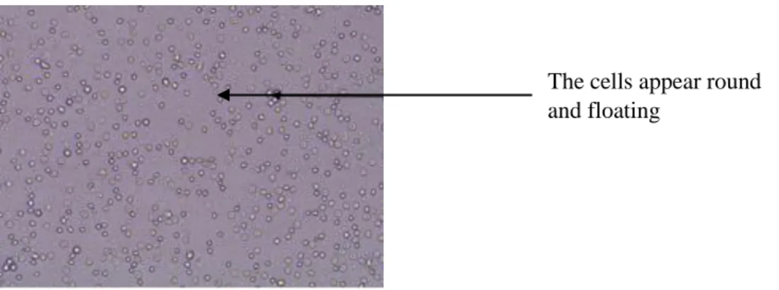
Results:
In the early stages of the culture, the mononuclear cells appear round and floating. After incubating the cells for 3 days, the culture medium color turns to purple which means that the culture medium is depleted. The viable cells appear adherent on a surface.
14
At this stage, the viable cells can be transferred into a new culture medium through a trypsinization process and pass from primary culture to secondary culture.
Figure5: Microscopic image of the early stages of the culture.
Figure 5: Microscopic observation
The system of cell culture is an important field of biotechnology. Currently, all kinds of cells can be isolated, reproduced, and manipulated in culture conditions. Furthermore, these cultured cells can be transplanted to treat some diseases. However, aseptic techniques are still key for cell culture, a simple contamination can easily ruin the study. Cell culture offers great potential for
The cells appear round and floating
15
innovative applications in cell therapy and regenerative medicine and for expanding our knowledge of human biology [5].
16
17
I. History of Stem Cells
Stem cells were first discovered as a consequence of the atomic bombs in World War II.
The atomic bombs dropped on Hiroshima and Nagasaki during the Second World War resulted in unusual destruction and loss of lives. The days to weeks after the bombing, victims began to die, not from burns or injuries, but from mysterious symptoms such as low white blood cell counts, hair loss, Anemia, in some cases, even destruction of the stomach and intestinal lining. This has caused scientists to look into the cause of death of these people, and they found out radiation to be the perpetrator [13].
This sinister report and the biological comparison of normal individuals with those affected by radiation has been able to further scientific work and lead to The concept of « Stem » cells, which are capable to divide when the rate of circulating cells decreases and gives daughter cell lines. By an intercellular message, each stem cell division helps to maintain the global capital of cells and stem cells, The concept of cell regeneration capacity of the skin and the liver in particular, and The concept of transferring bone marrow stem cells by transfusion [14].
Radiation damages cells, which causes cell death and interferes with the cell’s ability to divide to give rise to new cells. As a result, body parts like blood and hair cannot be regenerated. Therefore, early radiation experiments lead to the discovery of stem cells, in particular hematopoietic stem cells [15].
18
The discovery of stem cells is attributed to Drs. Ernest McCulloch and James Till, who have published a number of publications describing the regenerative and multipotent characteristics of hematopoietic stem cells. They were able to demonstrate that a sole stem cell generates all three types of mature blood cells: white blood cells, red cells and platelets [16].
Despite their potential contribution to science, the use of embryonic stem cells in research is a debated topic. Some scientists declare the benefits of pluripotent cells for research. Embryonic stem cells have the potential to treat genetic disorders, neurodegenerative diseases, injuries, and even generate new organs for those requiring a transplant. However, the ethical issues of deriving embryonic stem cells from human embryos have hampered progress [16].
II. Human biological development
Embryology is the science that studies embryonic development from an egg cell to an adult human being.
The time required for the complete development of a fetus in the womb is called gestation. It can be divided into different periods of gestation. The first two weeks of pre-natal development are referred to as the preembryonic stage. A growing human being is referred to as an embryo during weeks 3–8 and a fetus from the ninth week of pregnancy until birth. The preembryonic and embryonic periods of development are both characterized by cell division, migration, and differentiation. At the end of the embryonic stage, all organ systems are structured in a rudimentary form, despite the fact that the organs themselves are either non-functional or only semi-functional [17].
19
1. Fertilization
Fertilization is a process in which the male-haploid gamete (sperm) and the female-haploid gamete (oocyte or egg), fuse together to generate a diploid zygote. It usually happens in the uterine tube. The function of Fertilization is to enables genes to be transferred from parents to offspring and the process of development to initiate [18].
Figure 6: The process of fertilization [17]. 2. Segmentation
After fertilization, the zygote continues to be propelled towards the uterus. As it travels to the uterus, the zygote passes through five or six mitotic cell divisions. Despite the fact that each cleavage generates additional cells, the overall size of the zygote stays the same. Each daughter cell generated is called a blastomer.
20
Mitotic divisions continue for about 3 days, a 16-cell conceptus reached the uterus. The cells that were loosely grouped together are now compacted and look more like a solid mass. This structure is called morula.
Once in the uterus, the morula continues to divide for four to five days, forming a ball of about 100 cells. The ball of now tightly bound cells begins to secrete fluid and organize themselves around a fluid filled cavity, called blastocoel. At this stage of development, the conceptus is referred to as a blastocyst. Within this structure, a group of cells is formed into an inner cell mass, which is destined to become the embryo. The cells that constitute the outer shell are called trophoblasts. These cells will develop into the chorionic sac and the fetal part of the placenta.
The inner mass of embryonic cells is totipotent in this stage, which means that each cell has the capacity to differentiate into any cell type in the organism. Totipotency lasts only a few days before the cells’ fates are fixed as being the precursors to specific cell lineages [17].
21
Figure 7: Pre-embryonic cleavages [17]. 3. Gastrulation
The inner cell mass in the third week, the human embryo consists of two layers, epiblast and hypoblast. Gastrulation turns these two layers into three ones. An upper layer called ectoderm, a middle one called mesoderm, and a lower one referred to as endoderm [19].
Through the process of gastrulation, the cells go from totipotency to multipotency.
Each of these germ layers will differentiate into multiple human tissues in the embryo. The ectoderm develops into nervous system and brain, epidermis, sensory organs, hair, and nails. The mesoderm cells will become the muscles, bones, cardiovascular system, blood vessels, dermis, gonads, and the excretory systems. The endoderm proceeds to give rise to the inner lining of digestive tract, lining of lungs, and glands [17, 19].
22
Figure 8: Germ layers fates in embryo [17].
4. Organogenesis
Organogenesis is the process of formation of the embryo’s organs and tissues from the ectoderm, mesoderm, and endoderm. This occurs between the fourth and eighth week of gestation.
Through the process of neurulation, the neural tube is formed, which gives rise to the future brain and most of the length of the spinal cord. The ectoderm thickens which leads to the development of the neural plate- the precursor of the neural tube.
At this stage, most cells lose their ability to be pluripotent, and become unipotent. This means that the cells differentiate into a single cell type.
In this stage, the embryonic annexes are formed, which maintain the embryonic development and make the future placenta and the umbilical cord. These structures contain multipotent stem cells.
23
Within the eighth week, the organogenesis is mostly complete but the organs still need to develop to become functional. At this stage, the embryo is referred to as the fetus [17, 18].
III. Stem Cells
1. Definition of Cell
The cell is the fundamental structure of all living thing. It is the smallest element of life.
The cell is composed of cytoplasm, which is a jelly-like fluid consists of organelles such as mitochondria, ribosome…. That surrounds the nucleus. The nucleus contains the genetic material DNA and it is surrounded by a membrane named the nuclear envelope. The cytoplasm and the nucleus are enclosed in a membrane called the plasma membrane. It separates the cell from its environment and allows materials to enter and leave the cell.
The human body is composed of trillions of cells. The cells that divide to maintain the total number of cells and replace the ones who have died, are referred to as stem cells [20].
2. Stem Cells
Stem cells are defined by the ability to divide indefinitely in culture and the capacity to generate mature specialized cell types. Stem cells are characterized by two different properties, the potential to self-renew and to differentiate into a variety of specialized cell lineages. When stem cells divide, the daughter cells can either generate a differentiated specialized cell or self-renew to continue as an undifferentiated cell, a stem cell. This mode of cell division characteristic of stem cells is asymmetric and is a necessary physiological mechanism to maintain tissues and organs cells number in the body [21, 22].
24
Stem cells are classified as totipotent, pluripotent, multipotent, and unipotent:
Totipotent stem cells have the ability to produce all cell types of the conceptus, including the fetus and placenta. These cells are able to form the whole organism. Totipotent cells exist in the organism during the first four days after fertilization [21, 23].
Pluripotent stem cells have the capacity to form different cell types of all three germ layers (ectoderm, mesoderm, and endoderm) but not the entire organism. There are four categories of pluripotent stem cells which are embryonic stem cells, embryonic germ cells, embryonal carcinoma cells, and the multipotent adult progenitor cell [16, 21, 23].
Multipotent stem cells have the potential to give rise to limited range of cells and tissues appropriate to their emplacement. For example, blood stem cells give rise to red blood cells, white blood cells and platelets, whereas skin stem cells produce several types of skin cells [21].
Unipotent stem cells have the ability to renew itself but they can give rise to a single cell type [23].
25
Figure 9: Different stages of stem cells differentiation [24]. 3. Different types of human stem cells
3.1. Embryonic stem cells
Stevens is known as the innovator of embryonic stem cells research. Stem cell lines were formed from the teratocarcinomas called embryonic carcinoma cell lines and studied for their pluripotent characteristics [16].
Embryonic stem cells are pluripotent and are arised from the pre-implantation blastocyst that is composed of an outer layer of trophoblast cells and an inner cell mass. Embryonic stem cells are obtained from these cells at the fifth day of embryonic development. These cells can produce cells representative of all three embryonic germ layers (endoderm, mesoderm, and ectoderm) [25, 26].
26
In 1998 Dr. James Thomson harvested embryonic stem cells and tested the accumulated knowledge of cell culture conditions for stem cells on human fertilized eggs. The eggs were donated by couples who were undergoing treatment for infertility [16, 27].
Embryonic stem cells can be obtained from spare embryos that have been fertilized in vitro and not held for a parental project, or by a technique called therapeutic cloning, through this process a cell from the patient is fused with an enucleated donor oocyte applying the nuclear transfer technique. These cells are isolated from the resulting clone, and then differentiated in vitro into different type of cells and tissues for transplantation to help patients with disorders such as Parkinson’s disease, diabetes and cancer [23, 28].
Embryonic stem cells are defined by the possession of two key properties: embryonic stem cells can be grown in vitro and expanded in number indefinitely in the primitive undifferentiated state characteristic of the embryonic cells from which these cells are obtained, and throughout long periods of cultivation in vitro these cells retain a key property of those cells – pluripotency [27].
Furthermore, embryonic stem cells have the ability to self-renew and the capacity to preserve and expand the cells in culture for an extended time while maintaining their normal karyotype [29].
One major problem concerning the use of embryonic stem cells as a model system is their genetic and karyotypic integrity during long term culture and differentiation. Cytogenetic analysis of different human embryonic stem cell lines cultivated in different laboratories has shown that the euploid karyotype remains substantially stable, although recurrent gains of chromosome arms 12p and 17q or trisomy of chromosomes 12 and 17 have been described in some
27
subclones. Chromosomal abnormalities happen very frequently during embryonic stem cells derivation and culture [26].
The use of embryonic stem cells has major disadvantages. First, the use of human embryonic stem cells requires lifelong use of immunosuppressant drugs to prevent tissues rejection. Second, using embryonic stem cells can generate tumors from rapid growth when injected into adult patients. Finally, scientific progress is restricted by ethical issues about deriving human ES cells from aborted fetuses or from human embryos [30, 31].
An important stem cell discovery that may alter the ethical debate is induced pluripotent stem cells (iPSCs). This recent discovery has opened up new avenues of research, such as the induction of different somatic cell types into stem cells, comparisons of iPSCs to ESCs, and the potential of iPSCs for treating diseases [16].
3.2. Induced pluripotent stem cells
iPSCs are somatic cells that were reprogrammed to express specific genes, thereby causing them to transform from a differentiated state to a pluripotent state. The discovery was the result of many research groups working to determine what gave cell “stemness.” Shinya Yamanaka found out four genes that, when expressed together, could turn fibroblasts into iPSCs [16].
In 2006 Takahashi and Yamanaka launched the concept of induced pluripotent stem cells by producing stem cells that had having properties related to embryonic stem cells. iPSCs were generated by using a combination of four reprogramming factors, including Oct4 (Octamer binding transcription factor-4), Sox2 (Sex determining region Y) box 2, Klf4 (Kruppel Like Factor-4), and
c-28
Myc. These cells were demonstrated that they have the ability to self-renew and to differentiate like embryonic stem cells, and thus, could be used as an alternative for human embryonic stem cells in various research and clinics [32].
The production of iPSCs from somatic cells requires the incorporation of reprogramming factors into somatic cells. This can be realized by two types of methods— Integrative Viral Vector Systems and Non-Integrative Systems. Integrating systems are those in which the viral vector gets integrated into the host cell genome. The use of lentivirus and retrovirus comes under this category. Non-Integrating systems are those in which no integration in host cell genome happens. Various approaches such as viral vectors (Sendai virus and Adeno virus), Plasmid DNA, and the use of RNA and proteins come under this category.
iPSCs have been used since their discovery in many research and clinical studies including disease modeling, regenerative medicine, and drug discovery and or drug cytotoxicity studies. For disease modeling, somatic cells possessing the characteristics of patient’s diseased cells are used for the production of iPSCs and are further used for studying the diseases. Similarly, iPSCs are widely used for the regeneration of tissue-specific cells for the transplantation to patients suffering from different injuries or degenerative diseases. Studies such as drug discovery which implies small molecules screening, toxicity testing for assessment of safety, have successfully utilizediPSCs based technique [32].
29
Induced pluripotent stem cells afford various advantages:
The ethical concerns are eliminated through the use of iPSCs; these cells can be obtained from an adult patient instead of the destruction of the embryos resulting from the use of embryonic stem cells in research.
Reduced chances of immunorejection; iPSCs are produced from the somatic cells of patient’s own body and therefore the risk of immunorejection of these autologous cells is eliminated.
Throughput screening to predict toxicity and therapeutic responses of
new drugs
Reduce the global cost and risk of clinical trials; The cost of clinical trials could be lowered by using iPSCs to provide details of the drug’stoxicity through different cytotoxicity assays, and could reduce the cost related to providing animal models which will ultimately lower the total costs of the clinical trials.
Gene targeting and correction technologies;somatic cells reprogramming from any genetic background to iPSCs has enabled the generation of cell lines that possess diseasecausing mutations. The ability of modifying the specific sites in a genome to alter the genes of interest becomes very important here.
There are some drawbacks associated with iPSCs as well, the generation of iPSCs uses retroviral or lentiviral systems, so the concern is if the viral systems get incorporated with the host genome. The genetic material inserted by retroviral vectors could randomly integrate into the genome of the host which can lead to genetic aberration and teratoma formation. It has also been reported
30
that the altered expressions of the basal reprogramming factors can cause diseases. The overexpression of Oct4 can cause epithelial cell dysplasia. Furthermore, it has been reported that the aberrant expression of Sox2 cause mucinous colon carcinoma. Klf4 has a role in the development of breast tumors. C-Myc plays an important role in the formation of approximately 70% of human cancers. For this reason, retroviral insertion of c-Myc is also advised to be avoided for clinical applications [32].
3.3. Fetal stem cells
In 1998, John Gearhart and his team were the first who have been able to isolate and culture fetal stem cells which are known as primordial germ cells. These stem cells are the precursors of gametes, like eggs and sperms, and were isolated from fetal tissues such as the placenta, amniotic fluid, and umbilical cord blood, of 5-9 week old fetuses obtained by therapeutic abortion. The isolation of embryonic germ cells from fetal stem cells has two major problems. First, these cells are derived only from 8-9 week fetuses. Second, embryonic germ cells have limited proliferation ability [33, 34].
The concept of fetal stem cells is not new. Fetal stem cells have been used in clinics over the past years. Fetal neural tissue has been widely utilized therapeutically in Parkinson’s disease. Fetal stem cells have many characteristics which make them better than adult cells for use in regenerative medicine applications, such as greater plasticity in differentiation capacity, rapid growth in culture, and increased survival at low oxygen tension. These cells have also the ability to make high levels of angiogenic and trophic factors, which enhance growth in vivo and facilitating the surrounding host tissues regeneration. Also the use of stem cells derived from umbilical cord doesn’t make any ethical problems [34, 35].
31
The only problem associated with these stem cells is that the umbilical cord blood contains very few stem cells. The reason it is important to study these cells and their needs in order to improve the yield and the supply of those cells for therapeutic purposes [36].
3.4. Adult stem cells
Adult stem cells also called somatic stem cells are undifferentiated cells that take place in a differentiated tissue or organ. Adult stem cells are able to renew itself and can differentiate to yield all the specialized cell types of the tissue in which they reside. These stem cells exist in different tissues and organs such as the brain, liver, bone marrow, spinal cord, skin, epithelial lining of the digestive tract, skeletal muscle and heart. These cells are gathered in a microenvironment called niche. Somatic stem cells are multipotent or unipotent but they could exceed the limits and give rise to cell types of a different tissue, this phenomenon is referred to as plasticity [21, 33, 37].
Adult stem cells play an important role by replenishing dead or damaged cells to maintain the tissue or the organ. Some experiments in mice have shown that neural stem cells could differentiate into heart, blood, skeletal muscle, lung, and skin after transplantation. This phenomenon is called cell plasticity [33].
32
Figure 10: Adult stem cells plasticity [38].
There are three plasticity mechanisms of adult stem cells: transdifferentiation, dedifferentiation, and cell fusion.
The transdifferentiation phenomenon is defined by the conversion of a differentiated cell type into another cell type [39].
Through the process of dedifferentiation, a differentiated cell transforms into a differentiated cell of another cell type in condition that it dedifferentiate into progenitor [40].
The cell fusion mechanism is often observed in the organism: for example, myoblasts fuse to form multinucleated myofibers. When a differentiated cell fuse with an undifferentiated cell, this last one is as if reprogrammed by the differentiated cell.
33
Therefore, there could be confusion between the suspected plasticity phenomenon and the observed cell fusion phenomenon. In fact, transplanted stem cells showed a high fusion frequency with parenchyma cells acquiring the phenotype of these cells, and giving the illusion of plasticity or a transdifferentiation. As an example, replenishing a sick mouse’s liver by hematopoietic stem cells obtained from a normal mouse. Nevertheless, researchers estimate that the frequency of cell fusion phenomena is very low, at least in normal tissues and does not seem able to explain all published results [41].
Adult stem cells reside in a cell microenvironment referred to as niche which controls stem cell self-renewal and differentiation. These niches are consisted not only of stem cells but also of other adjacent differentiated cell types which generate a rich environment of extracellular matrix and other factors that allow maintaining the undifferentiated state of stem cells.
There is considerable change in niche design, in the world of stem cell niches. Most of stem cells reside within complex niches, but some stem cells are not surrounded by this cell environment. Stem cells of adult muscle called satellite cells are quiescent and attached to the basal lamina that enclose each muscle fiber bundle, and only reactivate to proliferate and fuse into differentiated myotubes when damaged fibers need repairing.
The niche has an anti-tumor role, by controlling stem cells self-renewal and division, in normal conditions. However, mutations in stem cells which make these cells insensitive to the niche control or a change in the signals emitted by the niche could contribute to a cancer development [42].
34
Scientific researchers are making significant efforts to explore the niche, which offers an opportunity to affect regenerative medicine and anticancer treatments.
Nowadays, it appears that most if not all adult organs contain stem cells. The bone marrow incorporates different populations of adult stem cells. Hematopoietic stem cells (HSCs)
HSCs were the first stem cells to be characterized, isolated and used clinically. These stem cells are capable to make all the types of blood cells, and are isolated from the bone marrow and umbilical cord blood. HSCs and many of the cytokines and growth factors are widely used clinically. For example, G-CSF helps mobilize HSCs from the bone marrow into the blood where they can be collected and used for the treatment of cancer patients [43].
35
Mesenchymal stem cells (MSCs)
MSCs are of embryonic origin and exactly from a structure of the embryo called mesenchyme. MSCs are distributed in the connective tissues of the body and have been derived from bone marrow, adipose tissue, placenta, lung, blood, and the umbilical cord. These stem cells have the capacity to stimulate blood vessels establishment, produce cytokines and growth factors, restrict T-lymphocytes activation and modulate innate immune response, and differentiate into different cell types such as chondroblasts, osteoblasts, neuroectodermal cells, adipocytes, and hepatocytes [44].
Endothelial progenitor cells
Endothelial progenitor cells have been found in bone marrow and blood. They have the ability to differentiate into blood vessels. Endothelial progenitor cells are useful due to their properties of neovascularization and restoring blood flow, in brain, retinal and heart damage [45].
Some reports claimed that there are rare cells within mesenchymal stem cells cultures which are pluripotent and can produce not only mesodermal but endodermal tissues. These cells are referred to as multipotent adult progenitor cells.
Multipotent adult progenitor cells (MAPCs) exists not only in bone marrow but in all organs. MAPCs are nonhematopoietic stem cells and have similar properties to those of embryonic stem cells. However, these cells seem not to give rise to tumor when injected into immunodeficient adult mice. MAPCs have the ability to grow extensively and to produce cells with phenotypic characteristics which distinguishes MAPC from mesenchymal stem cells, and
36
therefore suggests that MAPCs might be an ideal cell for in vivo therapies for damaged tissues and organs [46].
Recently another pluripotent cells population has been identified, from adult bone marrow referred to as marrow-isolated adult multilineage inducible cells (MIAMIs). MIAMI cells express the transcription factors oct-4 and rex-1 and telomerase. Also, they have the ability to differentiate into mesodermal, neural and pancreatic cells [47].
Adult stem cells offer different advantages:
Adult stem cells are not rejected by the immune system as a consequence of performing an autologous transplant.
Adult stem cells are chromosomally stable which does not cause a problem for therapeutic efficiency.
Harvesting techniques and where most of adult stem cells are obtained have little effect on the individual, which causes less ethical problems.
These stem cells are able to be reprogrammed and transformed into pluripotent stem cells.
Adult stem cells seem the best choice for cell therapy. However, the use of these stem cells is challenging, it is hard to isolate these cells because their numbers are small, their locations are hard to predict, they are difficult to culture in vitro and hard to maintain, and there is a low risk of cancer development after autologous transplant of stem cells [48].
37
4. Timeline of stem cell research
These are some important dates which mark advancements in human stem cell research:
1860 – 1920: The existence of stem cells is discovered from the analysis of embryo development and bone marrow microscopy [49].
1948 – 1958: Greater understanding is reached about stem cell mechanisms from sperm development and intestinal epithelium replacement [50].
1961: The first blood stem cells are discovered [51].
1964: Stem cells are detected in the embryonal carcinoma [52].
1968: The first bone marrow transplant is conducted [53].
1981: scientists begin obtaining and culturing mouse embryonic stem cells [54].
1992: The presence of nerve stem cells are recognized in the adult human brain [55].
1994: Scientists distinguish stem cells from the majority of other cancer cells. Doctors begin using corneal stem cells to treat patients with damaged cornea [56].
1994: Scientists begin the isolation and culture of inner cell mass cells from human blastocysts [57].
1995: Scientists begin the isolation of a primate embryonic stem cell’s line. These embryonic stem cells are diploid, pluripotent and have a normal karyotype. These cells have the potential to develop into derivatives of all three embryonic germ layers. Primate embryonic stem cells resemble
38
human embryonal carcinoma cells which have led to the possibility to produce and maintain human embryonic stem cells in vitro [58].
1996: The beginning of mammalian cloning and the birth of the cloned sheep dolly [59].
1998 – 2000: The beginning of the production of human embryonic stem cells from the inner cell mass of a human blastocyst donated by a couple who had undergone IVF treatment. Embryonic stem cells proliferate in vitro over extended periods while maintaining a normal karyotype. These cells have the ability to developinto somatic cell lines derived from the three embryonic germ layers and form teratomas when injected into immunodeficient mouses [60].
2000: The first production of human embryonic stem cells and their differentiation into neurons [60].
2003: The first production of human embryonic stem cells from human exfoliated deciduous teeth [61].
2004: Human embryonic stem cells were obtained by transferring a woman’s somatic cell nucleus into her enucleated ovum [62].
2004: The first successful bone marrow transplant was carried out at CHU Ibn Rochd of Casablanca [63].
2006: Human embryonic stem cells are derived from human embryos considered as naturally dead [64].
2008: The first production of stem cell lines from nuclear transferred and parthenogenetically activated mouse oocytes for therapeutic cloning [65].
39
2010: Scientists reprogram adult cells directly into neurons, heart muscle cells, and blood cells [66].
2010: Scientists successfully isolate human pluripotent blood stem cells that have the ability to form all types of blood cells [54].
Currently, stem cell research focuses on the mechanisms of intercellular communication, cell differentiation, and specialization. Scientists are studying the conditions under which these stem cells are harvested, cultured, proliferated, and administered locally or systemically. This effort seeks to understand the regenerative potential of these cells which allow many therapeutic applications like tissues and organs regeneration.
40
41
I. Definition and perspectives
Cell therapy can be defined as an approach which uses a biological product that has a therapeutic effect derived from human or viable animal cells. Cell therapy is a therapy in which viable human cells are injected in order to prevent and treat a disease. Cellular therapy is based on regenerating damaged tissues with new functioning cells [67].
Regenerative medicine is a field of medicine which deals with the process of replacing and engineering damaged tissues with cells or tissues obtained from stem cells or modified biological materials [68].
The cell types used in cell therapy are differentiated or mature cells obtained from an organ and precursor cells derived from stem cells. These cells have the best differentiation and proliferation capabilities, stem cells are the center of research and development of new treatments in cell therapy [67].
Many diseases cause cell destruction, and the only solution would be organs and tissues transplantation. This solution is challenging because of compatibility problems between donors and patients and because patient demand for donors outpaces availability. Cell therapy is an alternative to organs and tissues transplantation, which can not treat all pathologies relative to cells degeneration or cell destruction. Through this therapy, tissues could be produced from adult stem cells obtained from the patient (autologous cells), and thus the donors problem is solved and the risk of rejection is avoided.
42
The main issues encountered in cell therapy are to control the culture and to use adult stem cells, to induce their proper transformation; in terms of quality and quantity. Also, there is a need to develop an injection technique that will ensure the cells survival once implanted.
Cell therapy has already achieved many advances, and this therapy still offers therapeutic possibilities for many severe pathologies [69].
II. Stem cells in cell therapy
1. Principle and process of cell therapy
Cell therapy is based on cell’s transplantation. The principle of this therapy involves collecting cells which are either directly reinjected into the patient or in most cases undergo some manipulations in the laboratory (sorting, amplification, selection or depletion of certain cell populations). Some cells transplants can be stored at an extremely low temperature in stem cells banks, then thawed when needed for therapies.
Cell therapy can be done in five steps.
Cell therapy starts by harvesting the cells. The cells can be taken from the same patient (autograft).
If needed, a purification step of the collected cells is realized in order to keep only one cell type. Then these cells are modified or treated according to the therapeutic objective aimed.
If the collected cells are from a patient whose disease is genetic. In this situation, the collected cells should be corrected by gene transfer.
43
The cultured cells must be amplified in order to proliferate and then reimplant them in the patient at the damaged tissue or cells.
The last step is the administration of the biological product into the patient and its follow-up [70].
2. Cell therapy laboratory
Cell therapy laboratory should include a production laboratory, a research and development laboratory, a cold room, a freezing room, and the bank where cells are stored in liquid nitrogen. Monitoring of environmental conditions (oxygen concentration, storage spaces temperature, levels of liquid nitrogen) is necessary to ensure that cell therapy processes run smoothly and safely.
Manipulation of cell for clinical use requires respect of good manufacturing practices to guarantee the safety of cell transplants, individuals and the environment, which implies the use of closed systems. The closed system is a process where the biological material is not exposed to the open environment, in order to reduce the risk of contamination.
Closed culture system is a system that employs:
Bags for samples and for manipulation of different products.
Culture bags made of a special plastic allowing gas exchange between cells and the incubator atmosphere.
Sterile connection system, which connect the different bags by tubes that can be sterilely cut and reattached by welding at 320 °C.
44
The use of a closed culture system for production of therapeutic cell products batches has proved its efficiency concerning the risk of contamination [71].
Figure 12: Example of manipulations in a closed system [71]. 3. Sources of stem cells samples
3.1. Sources of embryonic stem cells
Embryonic stem cells are important for cell therapy as an unlimited cells source. Embryonic stem cells can be isolated by three different methods. These cells are isolated from aborted embryos or those lost in miscarriages, or by therapeutic cloning, or in vitro fertilization (IVF).
45
IVF, used for research purposes, is ethically inacceptable because this method ends up establishing embryos industrialization. However, the law of bioethics allows the use of supernumerary embryos systematically produced during IVF or abortion, and that are no longer the subject of a parental project and with the agreement of the parents. The point is not to create embryos but to use the cells of these spare embryos [72].
3.2. Sources of fetal stem cells
Fetal tissue contains a significant number of stem cells and progenitor cells which makes it useful for certain treatments.
Fetal stem cells are harvested after voluntary termination of pregnancy or after abortion at a later stage (5-9 weeks) than the blastocyst stage. The three most accurate sources of fetal stem cells are amniotic fluid, the placenta and umbilical cord blood. These sources are interesting in that their stem cells are collected in a less invasive way from the fetus [73].
3.3. Sources of adult stem cells
Adult organism always holds stem cells as mentioned above. These cells obtained from adult tissues do not generate any controversy from ethics committees. Research on other tissues that contains stem cells is still in progress [74].
3.4. Therapeutic cloning
Therapeutic cloning is the transfer of nucleus obtained from the patient somatic cell into an enucleated egg cell. Six days after in vitro nuclear transplantation the blastocyst is formed. At this stage, embryonic stem cells can be harvested and cultured.
46
Somatic cell nuclear transfer products are genetically and immunologically compatible with the nuclear donor which circumvents the problem of rejection. The harvest of stem cells from transnuclear unfertilized oocyte seems to generate fewer ethical problems than harvest of embryonic stem cellsfrom fertilized eggs derived from fertility treatment.
Therapeutic cloning, via the production ofan histologically compatible stem cells, offers considerable therapeutic potential and great promises for regenerative medicine [75].
47
4. Culture and amplification of stem cells
The establishment of stem cells population follows the rules of animal cell culture. Primary cultures are obtained either by cells migration from explant tissue or by tissue dissociation with proteolytic enzymes followed by culturing the cell suspension obtained. Some cells of the suspension adhere to the plastic of the culture bags, then proliferate under the effect of serum growth factors.
The resulting cell population will proliferate exponentially performing a limited number of cumulative population doublings (CPDs) of primary cells until reaching the crisis phase. This phase is characterized by a slow down (presenescence) and then a cessation of proliferation (senescence) of cell population. The maximum number of CPDs, or hayflick’s limit, attained by cells in culture before reaching the crisis phase depends on the species, the tissue/organ and especially on the age of the donor.
In fact, the younger the donor, the more CPDs number of a certain cell type is high. For stem cells cultures, if the culture conditions required to maintain the stem cells population are met, then a high CPDs number could be recorded without major signs of senescence. If, on the other hand, the culture conditions are not optimal, passage after passage, the number of stem cells within the population decreases [76, 77].
4.1. Optimal conditions for culture
Optimal culture conditions have to be defined and standardized in order to maintain reproducible cultures in various laboratories. Furthermore, the cells have to maintain a stable karyotype and phenotype under these conditions, as well as the ability to differentiate.
48
Plating density
Plating density is a critical parameter to ensure an optimal cell expansion as well as preserving the cells potential to differentiate. Prockop’s team has shown that most stem cells develop at low seeding densities. Increasing the seeding density per unit area can decrease the cells expansion rate [78].
Passage numbers
The passage number is a simple manner to reduce the samples volume collected from the donor. Each passage increase the culture surface area, the cell passage number is a technique based on cells dissociation by the action of a proteolytic enzyme (trypsin), then replating these cells on a new culture dish at lower density, the successive passages are enough to ensure stem cells purification because of the elimination of other initial adherent cells (macrophages, endothelial cells). This technique has its limits, the passages numbers affect the cells differentiation potential and can lead to cell senescence [79].
Culture medium and growth factors
Stem cells end use is important to define the appropriate medium for the culture and growth factors for the desired application [78].
4.2. Stem cells’ culture limits
Cell senescence
Senescence is the sum up of the cell mistakes in biosynthesis mechanisms. Senescence is an irreversible phenomenon. The senescent cells phenotype was described as the nuclear DNA replication machinery that is unable to complete the neostrands synthesis at telomeric ends of chromosomes, cycle after cycle,
![Figure 1: Isolating mononuclear cells from peripheral blood by density gradient centrifugation using ficoll [12]](https://thumb-eu.123doks.com/thumbv2/123doknet/15040370.691490/46.892.100.769.273.533/figure-isolating-mononuclear-peripheral-density-gradient-centrifugation-ficoll.webp)
![Figure 6: The process of fertilization [17].](https://thumb-eu.123doks.com/thumbv2/123doknet/15040370.691490/53.892.177.725.382.770/figure-the-process-of-fertilization.webp)
![Figure 7: Pre-embryonic cleavages [17].](https://thumb-eu.123doks.com/thumbv2/123doknet/15040370.691490/55.892.211.638.112.485/figure-pre-embryonic-cleavages.webp)
![Figure 8: Germ layers fates in embryo [17].](https://thumb-eu.123doks.com/thumbv2/123doknet/15040370.691490/56.892.184.697.121.449/figure-germ-layers-fates-embryo.webp)
![Figure 9: Different stages of stem cells differentiation [24].](https://thumb-eu.123doks.com/thumbv2/123doknet/15040370.691490/59.892.142.763.107.417/figure-different-stages-stem-cells-differentiation.webp)
![Figure 10: Adult stem cells plasticity [38].](https://thumb-eu.123doks.com/thumbv2/123doknet/15040370.691490/66.892.126.766.105.445/figure-adult-stem-cells-plasticity.webp)
![Figure 11: Hematopoietic stem cells differentiation [38].](https://thumb-eu.123doks.com/thumbv2/123doknet/15040370.691490/68.892.170.747.617.963/figure-hematopoietic-stem-cells-differentiation.webp)
![Figure 12: Example of manipulations in a closed system [71].](https://thumb-eu.123doks.com/thumbv2/123doknet/15040370.691490/78.892.146.774.221.668/figure-example-manipulations-closed.webp)
