Biochem. J. (1989) 262,457-462 (Printedin Great Britain)
Active-site and membrane
topology of
the
DD-peptidase/penicillin-binding
protein
no.
6
of
Enterococcus
hirae
(Streptococcus
faecium) A.T.C.C.
9790
Aboubaker EL KHARROUBI,* Graziella PIRAS,* Philippe JACQUES,* Istvan SZABO,*t
JozefVAN BEEUMEN,t Jacques COYETTE* andJean-Marie GHUYSEN*§
*Service deMicrobiologie, Universite de Liege,Institut de Chimie, B6, B-4000 Sart Tilman (Liege 1), Belgium,
and t Laboratoriumvoor Microbiologie, Rijksuniversiteit-Gent, K. L. Ledeganckstraat 35, B-9000 Gent, Belgium
The membrane-bound 43000-Mr penicillin-binding protein no. 6 (PBP6) ofEnterococcus hiraeconsists of
a 30000-MrDD-peptidase/penicillin-binding domain and a - 130-residue C-terminal appendage. Removal
of thisappendage bytrypsin proteolysishasnomarkedeffectonthecatalytic activity and penicillin-binding
capacity of the PBP. Anchorage ofthe PBP in the membrane appears to be mediated by a short
15-20-residue stretch at the C-terminal endof the appendage. The sequence of the 50-residue N-terminal region
of thePBP shows high degree of homology with thesequences of the corresponding regions of the PBPs5
ofEscherichia coli and Bacillus subtilis. On this basis the active-site serine residueoccurs at position 35 in
the enterococcal PBP.
INTRODUCTION
All the bacteria possess, bound to the plasma
mem-brane, proteins that upon reaction with ,J-lactam
antibiotics form stable serine ester-linked acyl-enzymes
and,for thisreason,arecalledpenicillin-binding proteins
orPBPs [1]. On the basis ofvariouscriteria, Mr,amino
acid sequence, nature of the catalysed reaction,
mem-brane topology and physiological function, several
groups of PBPs are distinguished [1]. The low-Mr
(25000-49000) PBPs/DD-peptidases (i) catalyse
acyl-transfer tractions onwell-definedsimple peptidessuchas
Ac2-L-Lys-D-Ala-D-Ala [2], (ii)have theactive-siteserine
residue located close to the N-terminal of the protein, usually in the position region 40-70 [3], and (iii) are
inserted into the plasma membrane by a short
non-cleaved C-terminal signal-like peptide while the bulk of
the polypeptide chain is on the periplasmic side of the
membrane. This type of membrane topology has been
established for the PBPs5 of Escherichia coli[4,5],Bacillus
subtilis and Bacillus stearothermophilus [6]. It probably
appliestoPBP2b and PBP3 ofStreptococcuspneumoniae
[7]. Mention should also be made ofStreptomyces R61,
which excretes a low-Mr PBP-DD-peptidase during
growth. Excretion involves cleavage ofa 26-residue
C-terminal stretch from theprotein precursor [8].
Enterococcus hirae (formerly Streptococcus faecium)
A.T.C.C. 9790 possesses seven PBPs, the Mr values of
whichrangefrom 119000to43000[9,10].The43000-Mr
PBP6 performs DD-peptidase activity on
Ac2-L-Lys-D-Ala-D-Ala[11].As describedbelow,it also fulfils thetwo
other criteriathat define thelow-MrPBPs/DD-peptidases:
it is anchored in the membrane by a small C-terminal
peptidesegmentand its active-siteserineresidue is located
closeto the N-terminal end of the protein.
MATERIALS AND METHODS
Growth conditions andmembranes
Enterococcus hiraewas grownat37°C inunshaken
1-litre flasks containing 500ml of SB medium [12]. Cells
werecollected atthe lateexponential phase (A550 =6.0)
andthe membraneswereprepared essentiallyasdescribed
previously [13]. Cells from 500 ml cultures were
sus-pended in 20 ml of 5mM-sodium phosphate buffer,
pH 7, containing 1 mM-MgCl2 and lysed witha mixture
of 2mgoflysozyme, 40#sgofDNAase, 20,tgof RNAase
and 0.2mg ofmuramidase I (10,tg/ml). Muramidase I
from Streptomyces globisporus 1828 [14]was agiftfrom
Dr.K. Yokogawa,DainipponPharmaceuticalCo., Osaka, Japan. It was further purified by f.p.l.c. (P. Jacques, unpublished work). The membranes were suspended
in 40 mM-sodium phosphate buffer, pH 7, containing
1 mM-MgCl2 and 5% (v/v) glycerol. DD-Carboxypeptidase activity
Thecatalysed reaction was:
Ac2-L-Lys-D-Ala-D-Ala+H2O-+
D-Ala+Ac2-L-Lys-D-Ala
The released D-alanine was measured as described
pre-viously [11].
Labelling with radioactive benzylpenicillin
All the PBPs, except the highly resistant PBP5, were
quantitatively labelled by reaction with 50
luM-benzyl['4C]penicillin (54 Ci/mol; The Radiochemical
Centre, Amersham, Bucks., U.K.) for 30min at 37'C.
The second-order rate constant of PBP acylation was
derived from the benzyl['4C]penicillin concentration
required to cause half-saturation [15,16].
Abbreviations used:PBP(s), penicillin-binding protein(s);tPBP(s), tryptic penicillin-binding protein derivative(s).
t Presentaddress: Biological Institute,Medical University,H-4012Debrecen, Hungary.
§ To whomcorrespondenceshouldbeaddressed.
A.El Kharroubi and others
SDS/polyacrylamide-gel electrophoresis and
fluorography
Discontinuous gel electrophoresis [17]was carried out
at constant voltage, in an LKB 2001 apparatus (Pharmacia, Uppsala, Sweden). The acrylamide/ bisacrylamide ratio was 37.5:1 for the stacking gel and
60:1 for the separating gel. Dependingonthe Mrofthe
proteins, 7.2%,
100%
and 12% acrylamide separatinggels (16cm x18 cm or 32 cm x 18cm) were used. The
Coomassie Blue-stained gels were submitted to
fluorography [9] and exposed for 2-7 days at -70'C. Hydrophobicity
Partitioning between the upper aqueousphase and the lower detergent phase in the presence of Triton X-114
[18] was used as an index of the hydrophilic versus
hydrophobic character of the PBPs.
Amino acidsequencing
Automated sequence analysis was carried out on a
477 A pulsed liquid Sequenator with on-line analysis of the amino acid phenylthiohydantoin derivatives with a
120 A analyser (Applied Biosystems, Foster City, CA,
U.S.A.). Two samples of solublepurifiedtPBP6* (3and 4.5nmol respectively) were applied to glass-fibre filters
carrying3 mgoftwice-precycled Polybrene. Asampleof
the native PBP6 (600pmol) was sequenced after SDS/ 12%-polyacrylamide-gel electrophoresis and
electro-blotting on a Millipore Immobilon
[poly(vinylidene
di-fluoride)]membraneby usingaBio-Rad Mini Trans-Blot cell. The initial yields were about 30% for the two
tPBP6*runsand about 10% for theelectroblotted PBP6
run. The blotting experimentwascarried out according
to the procedure described in ref. [19].
Analytical polyacrylamide-gel isoelectric focusing Experiments were carried out on a 111 Mini IEF cell
following the manufacturer's (Bio-Rad Laboratories) instructions and with pH 2-pH 5.5 Pharmalyte (Pharmacia, Uppsala, Sweden).
RESULTS
Eachofthe seven PBPs in Enterococcus hirae A.T.C.C. 9790 represented about 0.2 % of the total membrane proteins. Aeration conditions during bacterial growth hadlittleeffectontheirabsolute and relative abundance. Cellsweregrownandcollected,and themembranes were
prepared as described above.
Proteolytic conversion of PBP6 into water-soluble tPBP6a and tPBP6*
Membranes, previously labelled with benzyl[1C]-penicillin, were extracted with Triton X-100, and the
extracts containing theradioactivelylabelled PBPs were
submittedtopartitioningwith thetwo-phasesystem with Triton X-114. SDS/polyacrylamide-gel electrophoresis and fluorography of samples of the lower detergent phase and the upper aqueous phase showed that the PBPs, including PBP6, occurred only in the detergent phase, i.e. were hydrophobic.
It wasknown from previousstudies [9,10] that (i) the
membrane-bound PBPs of Enterococcus hirae had
differing
susceptibilities to proteolysis by trypsin, PBP6beingthe mostresistant one, and
(ii)
theend product oftrypsin degradation of PBP6 was a water-soluble catalytically active30000-Mr tPBP6* derivative. In order
tocheck whether conversion of PBP6 into tPBP6*might involve the transitory formation of a water-soluble intermediate, benzyl["4C]penicillin-labelled membranes (total proteins 3.6 mg) were incubated for 30 min at
37°C in 300 ,u of 10mM-Tris/HCl buffer, pH 7.8, containing 150 mM-NaCl and 0.750
(v/v)
Triton X-100 and then centrifuged at40000g for 1 h. Portions ofthesupernatant fraction (50 ,ul; total proteins 480 ,Clg) were
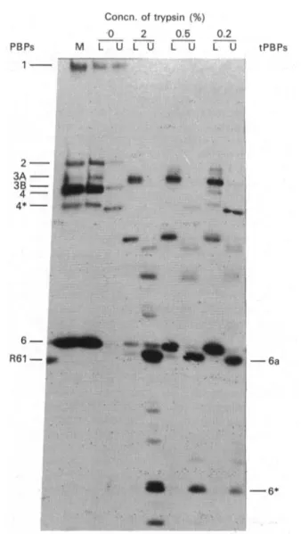
incubated assuch andwith 0.2 00, 0.50 and 20 (w/w) trypsin for 10 minat37°C and then extractedtwicewith Triton X- 114. Analysis of the aqueous and detergent phases by SDS/polyacrylamide-gel electrophoresis and
fluorography (Fig. 1) revealed the transitory occurrence of a radioactive 41000-Mr hydrophilic tPBP6a. On the
basis of radioactivity measurements, the sum of the
amounts of the 43 000-Mrhydrophobic PBP6, 41000-Mr
Concn. of trypsin (%) 0 2 0.5 PBPs M L U L U L U 1 *~~~~~~~~R :? S'.. 0.2 L U tPBPs 23A -3B - 4-4*-
q,-
~~~~~~~~~~~~~~~~~~~~~~~~~~~~~~~~~~~~~~~~~~...
...
... 0 . . . . .. ..:~~~~~~~~~~~.
II .. ;:...6* 'Fig. 1.Tryptic conversion ofthemembrane-bound
benzyll14CI-penicilloyl-PBP6 ofEnterococcushirae into radioactive
water-soluble tPBP6a andtPBP6*
All samples, except sample M (for membrane control),
weresubmittedtopartitioningwiththe Triton X-114
two-phasesystem. Key: L, lower, lipophilic, phase; U,upper,
hydrophilic, phase. Forconditionssee thetext.
Enterococcus hirae penicillin-binding protein no. 6
hydrophilic tPBP6a and 30000-Mr hydrophilic tPBP6* remained constant irrespectiveof the condition oftrypsin treatment. Hence loss of a - 20-residue peptide stretch wassufficient to release PBP6 from the membranes in the form of tPBP6a.
Quantitative conversion ofPBP6 into tPBP6* and
purification of tPBP6*
On the basisof preliminary studies of the effects of pH, buffer nature, trypsin concentration and incubation time, the following optimal conditions were used [10]. Each step of the procedure described below was monitored by estimating the amounts of tPBP6* on the basis of its benzyl["4C]penicillin-binding capacity and DD-carboxypeptidase activity.
A membrane suspension (total proteins 1.35 g; 62.1 nmol of PBP6) made in 90 ml of 40 mM-sodium phosphate buffer, pH 7.0, containing 1 mM-MgCl2 and
5 0 (v/v)glycerol was incubated with 13.5 mg of trypsin (type XI; Sigma Chemical Co.) for 10min at 37°C and thencentrifuged at 40 000 g for 30 min. This pretreatment had virtually no effect on the membrane-bound PBP6 but caused substantial degradation of the other PBPs into water-soluble fragments, and thus yielded a PBP6-enriched membrane pellet. This pellet was resuspended in 40 mlof 100 mM-ammonium bicarbonate buffer, pH 7.8, containing 0.1 mM-CaCl2 and incubated with 130 mg of
trypsinfor 2 hat 37 'C. Centrifugation yielded a
super-natant SI fraction that contained 5900 of the original
DD-carboxypeptidase activity. The pellet thus obtained
was re-incubated for 2h at 37 'C in the same
trypsin-1.00 0.75 1- r-5 0.r-50 z 0.25 0 20 40 60 Fraction no.
containing buffer as above. Centrifugation yielded a supernatant S2 fraction that contained 26
%
of theDD-carboxypeptidase activity.
The combined SI and S2 fractions (80 ml; total proteins 1.2 g) weresupplemented with an equal volume of 50mM-Tris/HCl/40mM-sodium phosphate buffer, pH 8.0.The solution was filtered through a Pharmacia
Q-SepharoseFastFlowcolumn (2.6 cmx40cm;gel volume 58ml) and the enzyme was eluted (flow rate 5 ml/min; fraction volume 10 ml) with the help of an NaCl gradient made in 25mM-Tris/HCl/ 20mM-sodium phosphate buffer, pH 8.0 (Fig. 2). Fractions 78-88 (I10 ml) con-tained 670 of the DD-carboxypeptidase activity. They were combined and concentrated to 10 ml by ultra-filtration on an Amicon YM membrane. The resulting preparation was supplemented with 10 ml of 50
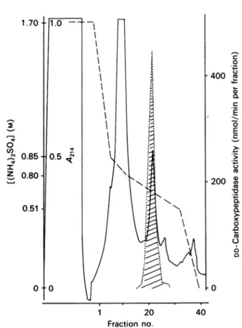
mM-sodium phosphate buffer, pH 7.0, containing 1.7 M-(NH4)2SO4,and 6 mlportionsof the solutionwerefiltered separately through a Pharmacia 1 ml phenyl-Superose HR5/5 column. The enzyme was eluted (flow rate
0.5ml/min; fraction volume 0.5ml) with the help ofa
decreasing (NH4)2SO4 gradientmade in thesame buffer.
Fractions 20-25 contained thepurified tPBP6* (Fig. 3). The overall yield of the operation in terms of
DD-carboxypeptidase activity was
42.50%
with a 490-fold enrichment. Fig. 4 illustrates the efficacy of thepurification procedure in terms of
benzyl[14C]penicillin-2000 C .) c; 1 600 01 0 1200 E° -.E 800 Xu 0 Cu 10 0.. 0) 400 xs 0 0 Cu U 0 .0 1-len I z L--80 100 c 0 0 400 co 0. C 0 E C.) c; 200 O ._ Q 0.a x 0 1.0 Cu I0
Fig. 2. Purification ofthe Enterococcus hirae tPBP6*by
anion-exchangechromatography onaPharmacia Q-Sepharose Fast Flowcolumn
Samples of each fraction were incubated with 4.5
mm-Ac2-L-Lys-D-Ala-D-Alainafinalvolumeof 20
,ul
for 20min at 37 'C. Results are expressed in nmol of D-alaninereleased/minper total fraction. ,
A280'
---, enzymeactivity. Thegradient (---) wasmadeby mixingbuffer
A (25mM-Tris/HCI/20mM-sodium
phosphate
buffer,pH 8.0) with bufferB(buffer Acontaining 1
t-NaCl).
1 20 40
Fraction no.
Fig. 3. Purification of the EnterococcushiraetPBP6*by
hydro-phobic chromatographyonaPharmaciaphenyl-Superose
HR5/5column
DD-Carboxypeptidase activityisexpressed asindicated in Fig.
2 legend. , A214; ---, enzyme activity. The gradient
(----)wasmadebymixingbuffer C[50mM-sodiumphosphate
buffer, pH 7.0, containing 1.7
M-(NH4)2S041]
with buffer D[buffer C without(NH4)2SO4].
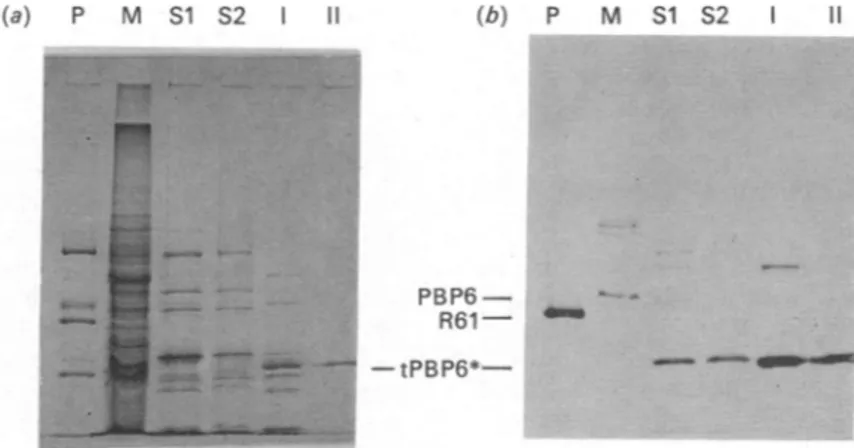
(a) P M Si S2 II (b) P M S1 S2
PBP6-R61 ..
-tPBP6*-Fig. 4.SDS/polyacrylamide-gel electrophoresisillustratingthepurificationsteps of the Enterococcus hirae tPBP6*:(a)Coomassie Blue staining; (b) fluorography
Key:M, original membranes(120jtgofprotein); SI (48jugofprotein)andS2(34ugofprotein),supernatantfractions obtained
after two successive trypsin treatments of the membranes; I (14.7,ug ofprotein) and 11(0.5 ,ug ofprotein), activefractions
obtained afterQ-Sepharose and phenyl-Superose chromatography respectively; P, standard proteins(bovine serum albumin,
ovalbumin, benzyl['4C]penicilloyl-DD-peptidase/PBP ofStreptomyces R61 and carbonic anhydrase).
Esch. coli PBP5 Ec. hirae PBP6* B.subtilis PBP5 P O 10 P 0 ii : IS } S F CK v 1 2
Esch. co/i PBP5 i P A S
Ig
4[] [_IiJ
S m IV C G Q[ r K40 50
Ec. hirae PBP6* 3} MP (D)T| A SI(T)K|IHGI H v L e I'. T)QS (v)
30 40
B. subtilis PBP5
PRL
1
I
I
AL SFM
T
3L1
T0
30-
40 50Fig. 5. Amino acid sequences of the N-terminal regions of theEnterococcus hirae PBP6 and the Escherichia coli 1191 and Bacillus subtilis
1201 PBPs5
Thereare 11 identitiesbetweenthepairEnterococcus hirae PBP6-Escherichia coli PBP5, 18 between the pair Enterococcushirae
PBP6-Bacillussubtilis PBP5and21 between thepairEscherichia coliPBP5-BacillussubtilisPBP5. The Enterococcus hirae
N-terminal sequenceshownin theFigureisthat of the water-soluble derivative tPBP6*. The20-residue N-terminalregion of the
purifiedmembrane-bound PBP6 hasexactlythe same sequence.Amino acidsinparentheseswerenotdeterminedwith certainty.
bindingactivity. The absoluteamountofpurifiedtPBP6*
was 0.8 mg with a 98% estimated degree of
purity.
tPBP6* migrated as a single compound onSDS/polyacrylamide-gel electrophoresis and exhibited
an apparent 30000 Mr. When
compared
withPBP6,
tPBP6* was slightly less sensitive to inactivationby
benzylpenicillin (second-order rate constant of enzyme
acylation 445M-1*s-1
compared
with 155M-'s-1)
andslightly more sensitive to
p-chloromercuribenzoate,
a 5.5/LM
instead of 30,M concentrationbeing
sufficientto inhibit the enzyme
activity by
50%.Preparation of
benzyll'4Clpenicilloyl-PBP6
for aminoacid sequencing
PBP6-enriched membranes
(total
proteins
1.92g)
obtained by thetrypsin
pretreatment described above were extracted with 60 ml of 25mM-Bistris/HCl
buffer,
pH 6.3, containing 1% (v/v) TritonX-100.Allthe steps describedbelow,except step4,werecarriedoutinbuffers
containing 0.05 % (v/v) Triton X-100. The extracted PBP6wasisolated from the supernatant fraction by the
following procedure: (1) filtration on a Pharmacia Q-Sepharose Fast Flow column, elution with an NaCl concentration gradient in 25mM-Bistris/HCl buffer,
pH 6.3, and concentration by ultrafiltration on an
Amicon YM membrane; (2) filtration on an ampicillin-Sepharose column [11], elution with a 0.5M-Tris/HCl buffer, pH8.0, containing 1 M-hydroxylamine and
dialysisagainst 5mM-sodium
phosphate
buffer,
pH 7.0;(3) reaction of the PBPs with radioactive penicillin,
filtration on a Mono Q HR5/5column, elution withan
NaCl concentration gradient in 25mM-Bistris/HCl buffer, pH6.3, and concentration by filtration on a
Centricon 10 filter; (4) separation of the radioactive
-
_-EHQ i
D Q D
Enterococcus hirae penicillin-binding protein no. 6
PBPs by SDS/polyacrylamide-gel electrophoresis and electroblotting of the gel on a Millipore Immobilon transfermembrane as described above.The
benzyl["4C]-penicilloyl-PBP6 band, lightly stained with Coomassie Blue, was cut and used for amino acid sequencing. Sequence of the N-terminal region of PBP6 and tPBP6*The amino acid sequence wasdetermined up to residue
50 for thepurified tPBP6* (two runs) and up to residue
20 for the purified electroblotted PBP6. Both PBPs had exactly the same 20-residue N-terminal region (Fig. 5). Electrofocusing of tPBP6* (pH gradient from 2 to 5.5) yielded two polypeptides with isoelectric points of 4.08 and 4.15 occurringin amolar ratioof1: 2.SincetPBP6*
had one single N-terminal amino acid, itwasconcluded that these two polypeptides had slightly different C-termini.
DISCUSSION
Conversion of the membrane-bound 43000-Mr PBP6
of Enterococcus hirae into the water-soluble 30000-Mr
tPBP6* by trypsinaction is made byloss of a 13000-Mr
polypeptide segment without alteration of the
N-terminal region of theprotein but withgeneration of
a ragged C-terminal, asevidenced by the occurrence of two tPBP6* species with slightly different isoelectric
points. Since tPBP6* performs DD-carboxypeptidase
ac-tivity and bindspenicillin,itis concluded thatthe -
130-residue segment that is eliminated from the C-terminal region of the protein exertslittleinfluence, ifany,onthe
polypeptide scaffolding of the protein and the
con-formation of the active site. The role of this long
C-terminal extension is unknown exceptthat the last
15-20-residue stretch anchors the protein into the plasma membrane. Loss of this latter small segment appears to
be sufficient to convert PBP6 into a 41000-Mr
water-soluble derivative tPBP6a.
It has been proposed [3] that the penicillin-binding
domain of the active-site-serine penicillin-recognizing
enzymes
(,f-lactamases
and DD-peptidases/PBPs) startsapprox. 60 residues upstream of the conserved tetrad
Ser-Xaa-Xaa-Lys, where Ser is the active-site serine
residue, andterminates approx. 60 residuesdownstream
of the conservedtriadHis-Thr-Gly, Lys-Thr-Glyor
Lys-Ser-Gly. On the basis of this definition, the Escherichia
coliand Bacillus subtilis DD-peptidases/PBPs5 also
pos-sess downstream of their
penicillin-binding
domain a long C-terminal stretch of about 105 and 125 residues respectively, the last 15-20 residues of which serve asmembrane-anchoring device [4-6].
The active site of theEnterococcushirae PBP6 hasnot
been identified chemically. But on the basis of the high degree of homology that exists between the amino
terminalregion ofPBP6and thecorresponding regionof
both Escherichiacoli[20]and Bacillussubtilis
[21]
PBPs5,itcanbesafelyconcluded that Ser-35 of the tetrad
Ser35-Ile-(Thr)-Lys38 is the active-site serine residue of the
enterococcal PBP. The 50-residue N-terminal region of
theEnterococcushirae PBP6 possesses five
lysine residues,
atpositions 5,9, 20,29and 38. Thefact that theadjacent peptidebonds escape
trypsin cleavage
suggests that theseresiduesbelong to
highly organized
structured elements.ThecorrespondingN-terminal
region
of the,-lactamases
of class A consists of one a-helix followed by two ,-strands and then
by
a second ac-helix. The active-siteserine residue is located at the N-terminal end of this
lattera-helix [22,23]. Thesame disposition of secondary
structures is thought to occur in the low-Mr
DD-peptidases/PBPs [3,24].
Both the Enterococcus hirae PBP6 [9] and the
Escherichia coli PBP5 [25]are susceptible to inactivation
bythiol-blockingreagents.Inactivation of the Escherichia
coli PBP5 is by blocking of the thiol group of Cys- 1 15,
which is located 70 residues downstream of the active-siteserine residue.The position of the susceptible cysteine residuein the enterococcal PBP6 remains unknown, but
it is located downstream of the 50-residue N-terminal
region.
The work inLiegewassupported in part by the Fonds de la
Recherche Scientifique Medicale (Contract no. 3.4507.83), an
Action Concert6e with the Belgian Government (Convention
86/91-90), a convention with the Region Wallonne
(C2/C16/Conv. 246/20428), the Fonds de Recherche de la
Faculte de Medecine ULg and a contract with the E.E.C. (BAP-0197-B). G. P. and P. J. were recipients of a Fellowship from the Institut pour l'Encouragement de la Recherche
Scientifique dans l'Industrie et l'Agriculture, Brussels. The
work in Gent was supported by the Belgian National Incentive Program of Fundamental Research in LifeSciences, initiated
by the Belgian State-Prime Minister's Office, Science Policy
Programming Department, and by the Fund for Joint Basic Research (Contact no. 2.0042.85). Some of the work described inthis paper forms part of a dissertation by A.E.K. presented as partial fulfilment for a Ph.D. degree at the University of
Li.ege.
REFERENCES
1. Ghuysen, J. M. (1988) Rev. Infect. Dis. 10, 726-732 2. Nguyen-Disteche, M., Leyh-Bouille, M., Pirlot, S., Frere,
J. M. & Ghuysen, J. M. (1986) Biochem. J. 235, 167-176
3. Joris, B.,Ghuysen, J. M., Dive, G., Renard, A., Dideberg,
O., Charlier, P., Frere, J. M., Kelly,. J. A., Boyington,
J. C.,Moews, P. C. & Knox, J. R. (1988) Biochem. J. 250, 313-324
4. Ferreira, L. C. S., Schwarz, U., Keck, W., Charlier, P., Dideberg, 0. & Ghuysen, J. M. (1988) Eur. J. Biochem. 171, 11-16
5. Jackson, M. E. & Pratt, J. M. (1988) Mol. Microbiol. 2,
563-568
6. Waxman, D.J. &Strominger, J.L. (1981) J. Biol. Chem.
256, 2067-2077
7. Ellerbrok, H. & Hakenbeck, R. (1984) Eur. J. Biochem.
144, 637-641
8. Duez, C.,Piron-Fraipont,C., Joris, B., Dusart, J., Urdea,
M.S.,Martial,J.A., Frere, J.M.&Ghuysen, J. M. (1987)
Eur. J. Biochem. 162, 509-518
9. Coyette, J., Ghuysen, J. M. &Fontana, R. (1980) Eur. J.
Biochem. 110, 445-456
10. El Kharroubi, A., Jacques, P., Piras, G., Coyette, J. &
Ghuysen,J. M.(1988)in AntibioticInhibitionofBacterial
Cell Surface Assembly and Function (Actor, P., Daneo-Moore, L.,Higgins, M.L.,Salton, M. R. J.&Shockman,
G. D., eds.), pp. 367-376, American Society for
Micro-biology, Washington
11. Coyette, J., Ghuysen, J.M. &Fontana, R. (1978) Eur. J.
Biochem. 88, 297-305
12. Coyette, J., Perkins, H.R., Polacheck, I., Shockman,
G.D. &Ghuysen,J. M. (1974)Eur. J. Biochem. 44,
459-468
13. Coyette, J., Ghuysen,J. M. & Fontana, R. (1977) Eur.J.
Biochem. 75, 225-229
14. Yokogawa, K., Kawata, S., Takemura, T. &Yoshimura,
Y. (1975) Agric. Biol. Chem. 39, 1533-1543
15. Ghuysen, J. M., Frere, J. M., Leyh-Bouille, M., Nguyen-Disteche, M. &Coyette,J.(1986)Biochem.J.235, 159-165
16. Leyh-Bouille, M.,Nguyen-Disteche,M.,Pirlot, S., Veithen, A., Bourguignon, C. & Ghuysen, J. M. (1986)Biochem. J. 235, 177-182
17. Laemmli, U. K. & Favre, M. (1973) J. Mol. Biol. 80, 575-599
18. Bordier, C. (1981) J. Biol. Chem. 256, 1604-1607 19. Matsudaira, P. (1987) J. Biol. Chem. 262, 10035-10038
20. Broome-Smith, J.K., Edelman, A. &Spratt, B.G. (1983)
in The Targetof Penicillin(Hakenbeck,R.,H6ltje,J. V. &
Labischinski, H., eds.), pp. 403-408, Walter de Gruyter,
Berlin
21. Todd, J. A., Roberts, A.N., Johnston, K., Piggot, P.L.,
Winter, D. & Ellar, D. (1986) J. Bacteriol. 167,
257-264
22. Herzberg, 0. & Moult, J. (1987) Science 236,
694-701
23. Dideberg, O., Charlier, P., Wery, J. P., Dehottay, P.,
Dusart, J., Erpicum, T., Frere, J. M. & Ghuysen, J. M.
(1987) Biochem. J. 245, 911-913
24. Kelly,J.A., Dideberg,O., Charlier, P., Wery,J.P., Libert,
M., Moews, P. C., Knox, J. R., Duez, C., Fraipont, C.,
Joris, B., Dusart, J., Frere,J. M. &Ghuysen, J. M. (1986)
Science 231, 1429-1437
25. Broome-Smith, J. & Spratt, B. G. (1984) FEBS Lett. 165,
185-189