The Raie of the Dileucine Motif in Helix VIII of the BLTl Receptor and RhoA in Neutrophil Degranulation.
By
Waqar Yunus Haider
Immunology Division, Department of Pediatrics Faculty of Medicine and Health Sciences
Mémoire présenté à la Faculté de médecine et des sciences de la santé en vue de l'obtention du grade de
maître ès sciences (M.Sc.) en Immunologie
NOTICE:
Branch
395 Wellington Street Ottawa ON K1A ON4 Canada
The author has granted a
non-exclusive license allowing Library and Archives Canada to reproduce, publish, archive, preserve, conserve, communicate to the public by
telecommunication or on the Internet, loan, distribute and sell theses worldwide, for commercial or non-commercial purposes, in microform, paper, electronic and/or any other formats.
The author retains copyright ownership and moral rights in this thesis. Neither the thesis nor substantial extracts from it may be printed or otherwise reproduced without the author's permission.
ln compliance with the Canadian Privacy Act some supporting forms may have been removed from this thesis.
While these forms may be included in the document page count, their removal does not represent any loss of content from the thesis.
•
••
Canada
Patrimoine de l'édition 395, rue Wellington Ottawa ON K1A ON4 Canada
AVIS:
Your file Votre référence ISBN: 978-0-494-65628-0 Our file Notre référence ISBN: 978-0-494-65628-0
L'auteur a accordé une licence non exclusive permettant
à
la Bibliothèque et Archives Canada de reproduire, publier, archiver, sauvegarder, conserver, transmettre au public par télécommunication ou par l'Internet, prêter, distribuer et vendre des thèses partout dans le monde, à des fins commerciales ou autres, sur support microforme, papier, électronique et/ou autres formats.L'auteur conserve la propriété du droit d'auteur et des droits moraux qui protège cette thèse. Ni la thèse ni des extraits substantiels de celle-ci ne doivent être imprimés ou autrement
reproduits sans son autorisation.
Conformément
à
la loi canadienne sur la protection de la vie privée, quelquesformulaires secondaires ont été enlevés de cette thèse.
Bien que ces formulaires aient inclus dans la pagination, il n'y aura aucun contenu manquant.
Table of contents ---
IDiagrams and
Tables---~---
VIIFïgu.res ---
VIIIAbb . .
rev1at1ons ---
.
IXSu.mmary --- x
In trod uctio n ---
11.0 Human Neutrophils ---
I }:>Description ---
1}:>
Physiological and pathological role
in
the immune system---
22.0 1'eu:k.otrienes ---
32.1
Arachidonic acid ---
3}:>
Metabolism ---
32.2 Leukotriene B
4(L TB
4) ---J 4);>-
Discovery ---
4);>-
Functions of L TB
4 --- 43.0 Leukotriene B
4receptors BL Tl and BL T2 ---
63.1 BLTl receptor ---
6);>-
Discovery, structure and cloning ---
6);>-
Expression of BLTI ---
83.2
BLT2 receptor ---
10);>-
Discovery and cloning ---:---
10);>-
Gene location ---
113.3
BLTl and BLT2 signalling ---
12);>-
Antagonists of BLTI and BLT2---
12);>-
Binding and signal transduction ---
133.4 Helix VIII of BL Tl receptor ---
15);>-
Structure ---
15);>-
Mutations and truncation ---
16);>-
Role in intracellular signalling ---
174.0 RhoGTPases ---
18);>-
Definition and structure ---
18);>-
Most characterized RhoGTPases ---
18~
Role of RhoGTPases in downstream signalling ---
195.0 Neutrophils Granules and Degranulation ---
215.1 Granule Population
in
Neutrophils ---
21~
Types of granules ---
il
~Granules Content ---
215.2 Degranulation ---
22~
Exocytosis ---
225.3 Degranulation mechanisms in. Neutrophils ---
23~
Stages of the de granulation process ---:---
235.4 Actin cytoskeletal dynamics in exocytosis. ---
25~
Role of actin rearrangement in de granulation ---
255 5 C
. a signa ng in exocy os1s ---.:---
2+ •li .
t .
27 ~Hierarchy of granule release in response to Ca
2+. --- 27~
Ca
2+as a crucial second messenger ---
275.6 Phospholipid signalling in Degranulation •••••••••••••••••••••••••••••••••
28~
Role of PIP2 and Pl3K in exocytosis ···-···-···
285.7 Role for src family kinases in Neutrophil Degranulation. ••••••••••••
30)- Definition ···-···-···
30 ~Expression and function of src family kinases in Neutrophils ••••••••••••••••••••••
315.8 Arrestin 2 and 3 functions in regulating exocytosis •.•....•...•
33~
Requirement of Arrestins for granule release •••••••••••••••••••••••••••••••••••••••••
33~
Dual action of Arrestins ···-···
33•
Hypothesis and objectives ••••••••••••••••••••••••••••••••••••••••••••••••••
34•
Materials and Methods ··••···•···•·•••···•·•·•·•··•··••·•···
36•
RE!slllts ... .
41•
D.
ISt:llSSIC>ll ••••••••••••••••••••••••••••••••••••••••••••••••••••••••••••••••••••••••
.
65• Conclusions
75'Talll~
1···
9'Ta.l:>le
~
•···••••••••·•·•••••••··•••••••••·•····•••••••••••••••••••••••••••••••••••···•··••·
13List of Figures
Figure 1 ···•··•··•···•···•···•···
43Figure 2 •···•···•···•·•··•···••••···•···•··
44Figure 3 •···•···•···••···•···•••••·•·••·
48Figure 4 •••••••••••··•·•••••••••••••••••••••••·••••·•··•••••••••••••·•··•••·••••••·••••·•••••
49Figure 5 ···•···••···•···•····•···•··••·•···••
50Figure 6 •···••··•·•···•····••···•···•·•·••··•••····•···••···•···••···•····•····
53Figure 7 ···•··•···•···•··••·•••···•····•·•··•····
54Figure 8 ··•···•·•···•·••···••·•·•·•·••••···••··•···•·•···•·••
55Figure 9 ··•···••···•···•··•••••••····•···•···••···•···
56F'igurE! 10 •·•••••••••••••••••••••••••••••••••••••••••••••••••••••••••••••••••••••••••••••••••
60Figure 11 •••••····•••••••••·•·•·••••·••·•·••··•••••••••••••••••••••••••••·••·••··•••••••••••
61F'igure 12 •••••••••·•••••••••••••··••••••••••••••••••••••••••••••••••••••••••••••·•·•••••••••
63F'igurE!13 ••••••••••·••·••••••••••••••••••••••••••••••••••••••••••••••·•••••••••••••••••••••••
64Abbreviations:
PLB-BLTI (wt): PLB-985 cells stably transfected with BLTl cDNA.
PLB-BLTI (2LA): PLB-985 cells stably transfected with mutant 2LA(304-305) cDNA.
GPCR: G protein-coupled receptor.
P AFR: platelet-activating factor receptor.
G418: geneticin.
EtOH: ethanol.
PTX: Bordetella pertussis toxin.
PP 1: 4-amino-5-( 4methylphenyl)-7-(tert-butyl)pyrazolo[3,4-d]pyrimidine.
AG490: N-benzyl-_3,4-dihydroxylbenzylidenecyanoacetamide.
SB203 5 80: 4-( 4-fluorophenyl)-2-( 4-methylsulfinylphenyl)-5-( 4-pyridyl) IH-imidazo le.
PD98059: 2'-amino-3'-methoxyflavone.
AG 14 78: 4-(3-chloroanilino )-6, 7-dimethoxyquinazolin.
SUMMARY
Neutrophil degranulation involves a number of well-orchestrated structural and
biochemical events. We have investigated the mechanism of intracellular
signalling · involved in neutrophil de granulation that was mediated by the high
affinity leukotriene (LT)B
4receptor, BLTI. The model systems used were
consisted of Peripheral blood neutrophils as well as promyeloid PLB-985 cells,
stably transfected with human BLTl cDNA (PLB-BL T) or a substitution mutant
(2L(304-305)/A) of the distal dileucine motif in helix VIII of BLTl, and
differentiated into a neutrophil-like phenotype. The degranulation of these cells
was measured in the presence and absence of factors that would affect the
signaling pathway. The results show that Degranulation responses to LTB
4were
similar for differentiated PLB-BLTl and neutrophils. However, the degranulation
response of cells bearing the dileucine mutation in helix VIII of BLTl was
significantly reduced in response to L TB
4.Pretreatment of differentiated
PLB-BL Tl cells and neutrophils with Y-27632, a pharmacological inhibitor of p
160-ROCK, the down-stream effector of the small GTPase RhoA, abrogated their
degranulation in response to L TB
4.The degranulation defect observed with the
mutation with a constitutively active form of RhoA. Taken together, our results
sug~est
an essential role for the distal dileucine motif in helix VIII of BL Tl
involving RhoA which allows normal neutrophil degranulation in response to
Key·words: Neutrophil, BLT1, helix VIII, distal dileucine motif, RhoA signaling,
Introduction
1.0 Human Neutrophils
Neutrophils are key cellular elements of the innate immune system, which provide protection from invading bacteria. When normal regulatory mechanisms fail, the neutrophil is also responsible for immunologically induced tissue injury (Faist and Kim
1998). Following activation by bacterial by-products or other immune stimuli such as lipopolysaccharides, glycolipids, and methylated DNA, neutrophils execute several specialized functions that include chemotaxis, phagocytosis and the generation ofreactive oxygen metabolites. All of these processes are required for the elimination of invading micro-organisms or cellular debris.
Neutrophils are highly mobile, short-lived white blood cells that are densely packed with secretory granules. They emigrate from the bone marrow into the blood and infiltrate tissues in response to injury or infection. In healthy individuals, peripheral blood neutrophils make up the majority of white blood cells (40-80%).
The lungs have the largest marginated pool of neutrophils in the body; they fulfill an important role in maintaining alveolar sterility. As a major effector cell in innate immunity neutrophils act as a "double-edged sword". If neutrophils are absent, for example in congenital neutropenia, or the more common "cyclic neutropenia" opportunistic infections result from overgrowth ofnormally resident skin and gut bacteria
and fungi at sites of injury, or exposed mucosal tissues. At the other extreme, accumulation and over activation of neutrophils can be fatal in disorders such as in septic shock or acute respiratory distress. The tissue-damaging effects of neutrophils are completely dependent on their degranulation.
2.0 Leukotriene
B4
(L
TB4)
2.1 Arachidonic acid
Arachidonic acid plays a central role in a biological control system where such oxygenated derivatives as prostaglandins, thromboxanes, and leukotrienes are mediators. The leukotrienes are formed by transformation of arachidonic acid into an unstable epoxide intermediate, leukotriene Ai, which can be converted enzymatically by hydration to leukotriene B4, and by addition of glutathione to leukotriene C4. Leukotriene C4 is metabolized to leukotrienes D4 and E4 by successive elimination of a gamma-glutamyl
residue and glycine. The Slow-reacting substance found during anaphylaxis consists of leukotrienes C4, D4, and E4. The cysteinyl-containing leukotrienes are potent bronchoconstrictors which increase vascular permeability in postcapillary venules, and stimulate mucus secretion. Leukotriene B4 causes adhesion and chemotactic movement of leukocytes and stimulates aggregation, enzyme release, and generation of superoxide in neutrophils. Leukotrienes C4, D4, and E4, which are released from the Jung tissue of
asthmatic subjects exposed to specific allergens, seem to play a pathophysiological role in immediate hypersensitivity reactions. These leukotrienes, as well as leukotriene .B4, have pro-inflammatory effects.
2.2 Leukotriene B4 (L TB4)
LTB4 was initially discovered by Borgeat and Samuelsson,(Borgeat and Samuelsson 1979) and found to be a potent neutrophil chemoattractant by Ford-Hutchinson et al. (Ford-Hutchinson, Bray et al. 1980). A high-affinity binding site for LTB4. on human neutrophils was initially detected by Goldman and Goetzl in 1982 with a
K-0.
of 0.39nM (Goldman and Goetzl 1984),others detected this binding site withKil
of 0.46nM (Lin, Ruppel et al. 1984), and, 1.5nM (Bomalaski and Mong 1987). Leukotriene B4 (LTB4) is an extren;iely potent lipid inflammatory mediator derived from membrane phospholipids by the sequential actions of cytosolic phospholipase A2, 5-lipoxygenase (5-LO) and LT ~hydrolase.
The major activities of LTB4 include the recruitment and activation of leukocytes, suggesting that it has considerable functional overlap with the chemokine family of chemoattractant peptides, which also direct the recruitment of leukocytes. Though structurally completely different, the lipid LTB4 and the peptide chemokines mediate their function through the same class of receptors, the G protein-coupled seven transmembrane domain receptor (GPCR) superfamily.
Two GPCRs for LTB4 have been identified, BLTl and BLT2. By mediating the activities of LTB4, these receptors participate bath in the recruitment and activation of leukocytes as part ofhost immune responses to invading pathogens, as well as in the pathogenesis of inflammatory diseases in which LTB4 has been implicated.
3.0 Leukotriene B
4receptors BL Tl and BL T2
3.1 BLTl
Molecular identification of a receptor for LTB4 eluded investigators for many years,
however, human high-affinity LTB4 receptor was finally cloned by Y okomizo et al. in
1997 from retinoic acid-differentiated HL-60 cells using a subtraction strategy (Yokomizo, Izumi et al. 1997), and the mouse ortholog by Huang et al. (Huang, Garcia-Zepeda et al. 1998). This receptor was initially named BLTR, and subsequently renamed BLTI when a second LTB4 receptor was identified. Membrane fractions ofCOS-7 cells
transfected with the BLTl cDNA demonstrated LTB4 binding with a Kd comparable to
that observed in retinoic acid-differentiated HL-60 cells, and CHO cells stably transfected with the BLTl cDNA demonstrated LTB4-induced increases in intracellular calcium and
chemotactic responses, indicating that this cDNA encoded the high-affinity LTB4
receptor (Yokomizo, Izumi et al. 1997).
Interestingly, this sequence had been previously cloned using degenerate PCR strategies by two independent groups as an orphan receptor gene that appeared to encode a member of the G protein-coupled seven transmembrane domain receptor (GPCR) superfamily. At. that time, the receptor was called R2 by Raport et al. (Raport, Schweickart et al. 1996) and chemoattractant receptor-like 1 (CMKRLI) by Owman et al. (Owman, Nilsson et al. 1996).
The mouse ortholog of BLTI was independently cloned by Huang et al. In 1998, by performing degenerate PCR with primers directed to well-conserved transmembrane domains of chemoattractant GPCRs, using cDNA isolated from murine eosinophils (Huang, Garcia-Zepeda et al. 1998).
Martin et al. identified the identical murine sequence by screening a mouse genomic library with a fragment of the human cDNA identified previously as encoding the P2Y1 receptor (Martin, Ronde et al. 1999).
Human BLTl gene was localized to chromosome 14 (Raport, Schweickart et al. 1996; Owman, Nilsson et al. 1996; Akbar, Dasari et al. 1996). Kyte-Doolittle hydrophobicity analysis of the amino acid sequences of human (Owman, Nilsson et al. 1996; Raport, Schweickart et al. 1996) and mouse (Huang, Garcia-Zepeda et al. 1998) BLTl showed the presence of seven hydrophobie transmembrane demains common to GPCRs. The amino acid sequences also contain other motifs characteristic of this family of receptors (Huang, Garcia-Zepeda et al. 1998; Owman, Nilssonet al. 1996), including (a) conserved praline residues in several of the transmembrane domains, which are thought to induce flexibility in the he1ix formations; (b) conserved cysteine residues in two of the extracellular loops for intra-molecular chain disulfide bonding; (c) a serine and thr~onine
rich C-terminal intra-cytoplasrnic segment, which in other GPCRs are sites of phosphorylation involved with receptor desensitization and intemalization; and ( d) consensus sequences for
N-linked glycosylation near the N-terminus and in one of the extracellular loops (Murphy 1994).
Mause BLTl has been demonstrated to be N-linked glycosylated, as in-vitro translation of the cDNA in the presence of dog pancreatic microsomes revealed an upward shift in mobility of the protein product on SDS-PAGE of ,..._4kD compared with the protein product translated in the absence of microsomes (Huang, Garcia-Zepeda et al. 1998). The highly conserved DRYLAIV motif at the end of the third transmembrane region of the GPCR family of receptors is present as DRSLA V in bath human and mouse BL T 1 (Yokomizo, Izumi et al. 1997; Huang, Garcia-Zepeda et al. 1998).
Human BLTI is 352 amino acid residues in length, and mouse BLTl is 351 amino acids, similar to the lengths of other chemoattractant receptors (Murphy 1994). The primary structures of human and mouse BLTl are 78% identical at the amino acid level. Interestingly, the three intra-cytoplasmic loops are identical across these species. In contrast; these sequences are not conserved across the subfamily of chemoattractant receptors, suggesting that human and mouse BLTl may be coupled to a unique, well-conserved, signaling pathway among the chemoattractant receptors (Huang, Garcia-Zepeda et al. 1998).
Leukocyte BLTl expression is up-regulated in inflammation. Mause BLTI transcription, which is not detectable in resting peritoneal cells, is dramatically increased in bath activated macrophages and neutrophils elicited into the peritoneum of mice by sodium
casein injection (Huang, Garcia-Zepeda et al. 1998). IFNy and glucocorticoids have also been shown to induce ?Lîl expression. Mouse BLTl transcription is induced in the RA W 264.7 macrophage cell line by IFNy (Huang, Garcia-Zepeda et al. 1998), and human BLTl transcription is induced in peripheral blood neutrophils by dexamethasone (Stankova, Turcotte et al. 2002).
Table 1: Regulators ofBLTl expression
Regulators of BL Tl expression
• Increased by LTB4 IL-10, Dexamethasone IL-2 IL-15Petterson A., et al, 2005, Stankova J. et al, 2002
• Decreased by
IFN-y, · LPS, TNFa
3.2BLT2
Binding studies of L TB4 suggested that in addition to the presence of a high-affmity receptor, a second, low-affinity LTB4 receptor was also present on human neutrophils (Goldman and Goetzl 1984; Lin, Ruppel et al. 1984) with a Ka of 61nM (Goldman and Goetzl 1984). BLT2, a second seven transmembrane spanning G protein-coupled LTB4 receptor, was identified by Yokomizo et al. (Yokomizo, Kata et al. 2000).
Membrane fractions of HEK 293 cells transfected with the cloned human BLT2 demonstrated specific and saturable LTB4 binding with a Ka of 22. 7nM, approximately 20-fold higher than human BLTl transfectants. CHO cells stably transfected with human BLT2 also demonstrated L TB4-induced increases in intracellular calcium and chemotactic responses, indicating that this sequence encoded an L TB4 receptor. This was confirmed when human BLT2 was independently cloned to be an L TB4 receptor by three additional groups (Tryselius, Nilsson et al. 2000; Kamohara, Takasaki et al. 2000; Wang, Gustafson et al. 2000).
The two LTB4 receptor genes forma cluster in bath the human (Nilsson, Tryselius et al. 2000; Y okomizo, Kata et al. 2000) and mouse (Y okomizo, Kata et al. 2000) genomes. The human BLT2 gene being located approximately 3 kb 5' of the human BLTl gene, and overlaps the 5' untranslated region of one of the 4' identified splice variants of BLTl (Nilsson, Tryselius et al 2000). Bath genes are transcribed in the same direction. The mouse BLT2 gene similarly is located approximately 4 kb 5' of the BLTl coding
sequence (Yokomizo, Kata et al. 2000). The identity between human BLTl and BLT 2 at the amino acid level, of 45.2%, is lower than that between CXCRl and CXCR2 (77%) or between FPRl and FPRLl (69%) or FPRL2 (58%), suggesting that the duplication event that generated the two L TB4 receptors may have occurred earlier than the events that
3.3
BLTl and BLT2 signaling
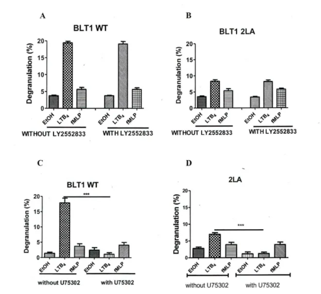
BLTl and BLT2 can be distinguished phannacologically. Multiple LTB4 receptor antagonists have been developed, including CP-105,696 (Showell, Pettipher et al. 1995), CP-195,543 (Showell, Conklyn et al. 1998), U-75302 (Lawson, Wishka et al. 1989), L Y255283 (Herron, Goodson et al. 1992), ZK 158252 and ON0-4057 (Kishikawa, Tateishi et al. 1992; Yokomizo, Kato et al. 2000). Sorne of these agents selectively antagonize BLTI or BLT2, whereas others antagonize both receptors. CP-105,696 and U-75302 compete with LTB4 binding in a dose-dependent manner to membrane fractions of CHO cells expressing human BLTl but not human BLT2, LY 255283 competes with LTB4 binding to human BLT2 but not human BLTI, and ZK 158252, CP-195,543, and ONO 4057 compete with LTB4 binding to both receptors (Yokomizo, Kato et al. 2000; Yokomizo, Kato et al. 2001).
There are two principal signal transduction pathways involving the G-protein coupled receptors: the cAMP signal 'pathway and the Phosphatidylinositol signal pathway. When a ligand binds to the GPCR it causes a conformational change in the GPCR which allows it to actas a guanine nucleotide exchange factor (GEF). The GPCR can then activate an associated G-protein by exchanging its bound GDP for a GTP. The G-protein's a subunit, together with the bound GTP, can then dissociate from the
p
andy
subunits to furtheraffect intracellular signaling proteins or target functional proteins directly depending oii the a subunit type (Gas ,Gai, Gaq111 , Ga12/13).
Table2: Characteristics ofBLTl and BLT2.
BLTl BLT2
Gene location chromosome! 4q
11.2 ~q 12
Amino acids
352 amino acids 358 amino acids Identity between humanand mouse 78.6% 92.7% Agonists LTB4 LTB4 12 epi-LTB4 12 epi-LTB4 12(s)-HTETE 12(s)HPETE 12(R)HTETE 20-hydroxy L TB4 Pharmaco logic CP105696 CP195543 inhibitors CP195543 ZK158252 ZK158252 ON04057 U75302 LY2442843 ON04057
Sorne differences in G protein-coupling have been observed between the two receptors. BLTI and BLT2 receptors appear to use different G proteins in mediating different cellular events, and receptor G protein coupling may differ in different cell types. LTB4-induced chemotaxis of CHO cells stably transfected with either human BLTI or human BLT2 is completely abolished by Bordetella pertussis toxin (PTX), suggesting that chemotatic responses directed by both receptors involve Gairo subunit signaling. In contrast, increases in intracellular calcium induced by LTB4 in CHO-human BLTI and CHO-human BLT2 transfectants are only partially blocked by PTX, indicating that calcium responses directed by the two receptors are mediated by both PTX-sensitive and PTX-insensitive G proteins in CHO cells (Yokomizo, Izumi et al. 1997; Yokomizo, Kato et al. 2000).
Experiments co-transfecting BLTI with different Ga subunits into COS-7 cells indicate that BLTI can couple to Ga16, a PTX-insensitive a subunit of the Gq class of G proteins
(Gaudreau, Le Gouill et al. 1998). As opposed to the results with CHO transfectants, L TB4-induced increase in intracellular calcium in human neutrophils was largely PTX-sensitive (Powell, MacLeod et al. 1996), suggesting that the L TB4 receptors couple to different types of G proteins in different cell types. Both LTB4 receptors also mediate inhibition of forskolin-induced adenylyl cyclase activity, but do so using different G proteins. Whereas inhibition of forskolin-induced adenylyl cyclase activity by LTB4 was reduced approximately 80% by PTX in CHO-human BLTl transfectants, PTX had a
negligible effect on LTB4-induced inhibition of forskolin-induced adenylyl cyclase activity in CHO-human BLT2 transfectants (Yokomizo, Kata et al. 2000).
3.4 Helix VIlI of BLTl receptor
BLTl belongs to the rhodopsin subfamily of GPCRs analysis of the crystal structure of rhodopsin con:finned the presence .of 7 transmembrane (TM) helices and revealed the existence of an Sth helix (helix VIII) that projects at a right angle from the C terminus of TM7 (Palczewski, Kumasaka et al. 2000) . This short helix of rhodopsin is anchored by a palmitoyl group to the cytoplasmic leaflet of the cell membrane, and a hypothetical model has predicted that helix VIII interacts with the N-terminal helix of the Ga and Gy subunits (Lu, Saldanha et al. 2002). BLTI was demonstrated to contain the helix VIII in its intracellular C terminus (Okuno, Aga et al. 2003; Gaudreau, Beaulieu et al. 2004).
BL T 1 mutants with a truncated or substituted helix VIII showed much higher L TB4 binding than wild-type (Wf) receptor in HEK293 and CHO cells, albeit with comparable expression on the cell surface (Okuno, Aga et al. 2003). Similar to the Wf receptor, in mutant receptors LTB4 pr01~oted binding of GTPyS. However unlike Wf-BLTl, the
The mutant receptors maintained a high affinity for LTB4, even in the presence of an
excess amount of GTPyS. They also showed more prolonged intracellular signaling (e.g. calcium mobilization and metabolic activation) after LTB4 treatment. The BLTI model
predicts a helix VIII extending from TM7, similar to the one observed in rhodopsin. The BLTI model suggests that a pair of aromatic residues (Tyr-285 and Phe-300), which are positioned similarly to the conserved Tyr-306 ànd Phe-313 pair in rhodopsin, may stabilize the inactive form of the receptor by holding TM7 and helix VIII at almost a right angle to each other.
Gaudreau et al. (Gaudreau, Le Gouill et al. 2002) proposed that Thr-308 is involved in GRK.6-mediated desensitization of BLTl signaling. This suggested that Phosphorylation at that site may also play arole in inactivation ofBLTl.
In addition Gaudreau et al. (Gaudreau, Beaulieu et al. 2004) also proposed that helix VIII is involved in a hydrophobie core containing other hydrophobie residues in helix 1 and . disruption of this hydrophobie core may facilitate the irreversible activation of BLTl. Therefore Cell surface expression levels of helix VIII mutants of BLT2 are considerably reduced. The helix VIII of BLT2 might also be important for receptor sorting, Severa! studies suggest that helix VIII of BLTI may play an important role in the inactivation of BLTI after G-protein activation, possibly by sensing Ger subunits as being GTP-bound.
Itoh et al. 2003) showed that trimer-ic G-proteins were required for maintenance of the high affinity state of BL Tl. The binding affmity of GPCR is thus critically dependent on the status of G-proteins, which suggest that further studies will reveal an important role of helix VIII in downstream signaling.
4.0 Rho GTPases
Rho GTPases are members of the Ras superfamily of monomeric 20-30 kDa GTP-binding proteins. Ten different mammalian Rho GTPases, some with multiple isoforms, have been identified to date (Jones, Ridley et al. 2000). The most extensively characterized members are RhoA, Racl and 2 and Cdc42. Each ofthese GTPases actas a molecular switch, cycling between an active GTP-bound, and an inactive GDP-bound, state. In the GTP-bound form they are able to internet with effector or target molecules to initiate a downstream response, while an intrinsic GTPase activity returns the proteins to the GDP-bound state, to complete the cycle and terminate signal transduction.
The major function of Rho GTPases is to regulate the assembly and organization of the actin cytoskeleton (Hall 1998). The effects of 3 members of the Rho family RhoA, Rac and Cdc42 were initially described using quiescent Swiss3T3 fibroblasts, a cell line in which serum starvation creates a very low background of organized F-actin structures. Addition of lysophosphatidic acid induce the formation of contractile actin-myosin stress fibres and associated focal adhesions, and which was blocked by C3 transferase (Ridley and Hall 1992).
The dominant-negative Rac specifically inhibits the response (Ridley, Paterson et al. 1992).to Growth factors, such as platelet-derived growth factor, insulin or epidermal growth factor (EGF) which induce the formation of actin-rich lamellipodia and membrane ruffles associated with focal contacts.
Bradykinin a nine amino acid peptide induces the formation of peripheral microspikes or filopodia, which are also associated with focal contacts, and this can be inhibited by expression of dominant-negative Cdc42 (Kozma, Ahmed et al. 1995).
Rho GTPases have also been found to play a role in a variety of cellular processes that are dependent on the actin cytoskeleton, such as cytokinesis (Mabuchi, Hamaguchi et al. 1993; Prokopenko, Saint et al. 2000) phagocytosis (Cox, Chang et al. 1997; Caron and Hall 1998), pinocytosis (Ridley, Paterson et al. 1992), cell migration (Nobes and Hall 1999; Allen, Zicha et al. 1998), morphogenesis (Settleman 1999) and axon guidance (Luo, Jan et al. 1997).
In addition to their effect on the actin cytoskeleton, they also regulate a variety of other biochemical pathways, including serum response factor (SRF) and nuclear factor kB (NF-kB) transcription factors (Hill, Wynne et al. 1995; Perona, Montaner et al. 1997), the
c-jun N-terminal kinase (JNK) and p38 mitogen-activated protein kinase pathways (Coso, Chiariello et al. 1995; Mir\den, Lin et al. 1995), the phagocytic NADPH oxidase complex (Abo, Pick et. al. 1991), G1 cell-cycle progression (Oison, Ashworth et al. 1995), the
assembly of cadherin-containing cell-cell contacts (Braga 1999), (Kaibuchi, Kuroda et al. 1999), secretion in mast cells (Norman, Price et al. 1996), cell polarity (Johnson 1999) and cell transformation (Van Aelst and D'Souza-Schorey 1997).
Therefore, although Rho GTPases are best characterized for their effects on the actin cytoskeleton, there is now much interest in their ability to affect cell proliferation and
'
gene transcription and degranulation of neutrophils. Thus studies using inhibition always show some effect and are hard to analyse
5.0 Neutrophils Granules and Degranulation
5.1 Granule Population in Neutrophils.
Neutrophils contain at least four different types of granules identified by subcellular fractionation and transmission electron microscopy. These are: primary granules, also known as azurophilic granules, secondary granules, also known as specific granules, tertiary granules, and secretory vesicles. The primary granules are the main storage site of the most toxic neutrophil-derived mediators, including elastase, myeloperoxidase, cathepsins, and defensins. The secondary and tertiary granules contain lactoferrin and matrix metalloprot~ase-9 (l\.fMP-9, also known as gelatinase B), respectively (Borregaard
and Cowland 1997).
The secretory ves!cles in human neutrophils contain human serum album.in, suggesting that they derive from endocytosis. The secondary and tertiary granules have similar contents but can be discriminated by their different intrinSic buoyant densities when centrifuged on gradient media (Kjeldsen, Sengelov et al. 1994). Ali ofthese granule types are retained in the cell cytoplasm and are not released until receptors in the plasma membrane or phagosomal membrane signal to the cytoplasm to activate their movement for secretion oftheir contents by exocytosis. Thus, the receptor-linked secretory pathway is an important control mechanism, as the neutrophil is highly enriched in tissue-destructive proteases and other enzymes.
5.2 Degranulation
Degranulation is defined as the secretion by receptor-mediated exocytosis of granule-derived substances. Upon receptor-mediated stimulation of neutrophils, granules are mobilized triggering their docking and content secretion which can occur intracellularly with microbe-laden phagosomes, or extracellularly at the plasma membrane. Upon exocytosis of granules, neutrophils release a diverse array of antimicrobial proteins and enzymes, rnany of which also possess tissue-damaging properties. At the same time, neutrophils release reactive oxygen species by respiratory burst, to aid in pathogen clearance, in addition to cytokines, which recruit additional leukocytes to the region of infection or inflammation.
A recent study by Brinkmann et al (Brinkmann, Reichard et al. 2004) described a novel mechanism causing elimination of microbial pathogens by neutrophils. Upon activation by ligands such as interleukin-8 (IL-8), lipopolysaccharide, and interferon-y with complement 5a (Martinelli, Urosevic et al. 2004), neutrophils generated a web of extracellular fibres known as "neutrophil extracellular traps" (NETs), composed of DNA, histones, and antimicrobial granule proteins, which were highly effective at trapping and killing invasive microbes. The authors proposed that NETs amplified the effectiveness of antimicrobial components by concentrating them in a fibrous, net-like structure and reducing their exposure to host tissues.
Even though this report did not suggest the molecular mechanisms responsible for NET formation and its association with granular protein, it opened a new horizon in the field of neutrophil biology in relation to mediator release and bactericidal activity. Moreover, it has been recently suggested that NET formation may be an important mechanism for control of sepsis (Clark, Ma et al. 2007).
In many in:flammatory disorders, such as acute lung injury, ischemia, reperfusion injury, severe asphyxie asthma, rheumatoid arthritis, and septic shock, excessive neutrophil degranulàtion is a common feature. Recent findings have identified a number of important signaling pathways in neutrophils that may be essential for neutrophil exocytosis. .
5.3 Degranulation mechanism in neutrophils
Receptor stimulation of neutrophils by a secretagogue triggers granule translocation to the phagosomal or plasma membrane, where they dock and fuse, releasing their contents. The release of granule-derived mediators from neutrophils occurs by a tightly controlled receptor-coupled mechanism termed "regulated exocytosis". Exocytosis is postulated to take place in four discrete steps (Toonen and Verhage 2003).
The first step of exocytosis is granule recruitment from the cytoplasm and translocation to the target membrane, which is dependent on actin cytoskeleton remodeling and microtubule assembly (Burgoyne and Morgan 2003). This is followed by the second step of granule tethering and docking, leading to contact of the outer surface of the granule lipid bilayer membrane with the inner surface of the target membrane. Granule priming then follows as the third step to make granules fusion-competent and ensure that they fuse rapidly, to form a reversible pore structure· between the granule and the target membrane.
The fourth and final step, granule fusion, occurs by rapid expansion of the fusion pore, leading to complete fusion of the granule membrane with the target membrane and expulsion of the granular contents to the outside of the cell.
This increases the total surface area of the cell, and exposes the interior surface of the granule membrane to the exterior. Tethering and docking steps require the sequential action of Rab and SN ARE proteins (Stow, Manderson et al. 2006); whether these factors are also needed for subsequent steps of fusion remains to be determined.
Translocation and exocytosis of granules from neutrophils requrre, as a llllll1Il1um, increases in intracellular Ca2+ and guanosine triphosphate (GTP) as well as hydrolysis of
Not surprisingly, there are numerous target molecules for these effectors, including Ca2+
-binding proteins such as annexins and calmodulin, and GTP--binding proteins such as heterotrimeric and small monomeric G proteins. The role of ATP in exocytosis as well as kinases, is to act as an energy source for SNARE complex reorganization and as a phosphate donor for phosphorylation of downstream effector molecules.
5.4 Actin cytoskeletal dynamics in exocytosis
Actin remodeling is clearly a prime downst1;eam target of activated effector molecules during receptor-mediated exocytosis. The actin cytoskeleton forms a mesh around the periphery in many different kinds of secretory cells (i.e., neutrophils, mast cells, neurons and endocrine cells). This may actas a shield against aberrant granule docking and fusion at the plasma membrane. So in this case the actin cytoskeletal mesh would have to be disassembled during exocytosis (Norman, Price et al. 1994; Muallem, Kwiatkowska et al. 1995; Takao-Rikitsu, Mochida et al. 2004).
However, some studies suggest that F-actin has normally a very diffuse distribution in
resting neutrophils, and only assembles into a cortical ring upon stimulation by f-Met-Leu-Phe (Downey, Elson et al 1991; Filippi, Harris et al. 2004). In the latter studies, live
neutrophils were first adhered to poly-L-lysine-coated glass slides (which itself leads to activation), before stimulation with f-Met-Leu-Phe for a period of time prior to fixation. Therefore, this procedure may lead to F-actin remodeling prior to stimulation.
Several other studies report that actin remodelling is needed to direct neutrophil migratory responses (Affolter and Weijer 2005). Directed neutrophil movements, or chemotaxis towards sites of infection, are driven by polarized F-actin formation, with the leading edge of cells showing enhanced levels of actin polymerization. Interestingly, neutrophils produce a polarized response even when subjected to uniform concentrations of chemoattractants, and maintain this polarity via continued production of activated effector molecules such as 3-phosphoinositol lipids in the cell membrane (Zigmond, Levitsky et al. 1981; Xu, Wang et al. 2003). A recent comprehensive study of neutrophil granule proteins revealed that actin associates with ail granule subsets, which suggests that actin may play an active role in regulating neutrophil exocytosis (Jog, Rane et al. 2007).
5.5 Ca2+ signaling in exocytosis
Increases in intracellular Ca2+ alone are sufficient to induce the release of granules from neutrophils, particularly if the concentration of Ca2+ is elevated to sufficiently high levels
by the use of Ca2+ ionophores such as A23187 or ionomycin. A hierarchy of granule release exists in response to elevating concentrations of Ca2+ (Pinxteren, O'Sullivan et al.
2000; Sengelov, Kjeldsen et al. 1993). The order of release is secretory vesicles > tertiary granules > secondary granules > primary granules (Sengelov, Kjeldsen et al. 1993; Bentwood and Henson 1980).
The release of each type of granule appears to be differentially regulated by unique intracellular signaling pathways. Activation of many neutrophil receptors causes elevated Ca2
+ levels, including the seven transmembrane-spanning G protein-coupled receptors
such as the fqrmyl peptide receptor (that binds the bacterial tripeptide, f-Met-Leu-Phe) and chemokine receptors (such as the interleukin-8 receptor, CXCRl) (Andersson, Dahlgren et al. 1986; Itagaki, Kannan et al. 2002). Although Ca2+ is a crucial second
messenger in the activation of exocytosis, the specific target molecules for Ca2+ in
neutrophil degranulation have not yet been identified. Several candidates have been suggested, notably annexins, protein kinase C, and calmodulin, all ofwhich bind Ca2+ to
modulate their activities (Brown, Reed et al. 1991; Sjolin, Stendahl et al. 1994; Steadman, Petersen et al. 1996; Donnelly and Moss 1997; Haribabu, Richardson et al. 2000). Neutrophils have been shown to require these Ca2+-binding proteins for
5.6 Phospholipid signaling in degranulation
Numerous studies have indicated a role for phospholipids, particularly polyphosphoinositides, in regulation of neutrophil degranulation. Polyphosphoinositide production, such as phosphatidylinositol 4,5-bisphosphate (PIP2), induced by the activation of the hemat0poietic cell-specific isoform phosphatidylinositol 3-kinase-g (P13Kg ) has been shown to be required for granule exocytosis in permeabilized neutrophilic-like cells, HL-60 cells (Fensome, Cunningham et al. 1996).
Intracellular sites of PIP2 formation in neutrophils are not known, but are likely to occur both at the plasma and granule membranes. Regions of PIP2 enrichment in the membrane form essential binding sites for many intracellular signaling molecules, particularly those that contain pleckstrin homology domains, such as Rho guanine exchange factors including Vav, which signal downstream to Rho guanosine triphosphatases (GTPases) (Kim, Marchal et al. 2003; Gakidis, Cullere et al. 2004; Fumagalli, Zhang et al. 2007) .
Phospliatidylinositol transfer protein (PI-TP) has been shown to be essential for the transport of phosphatidylinositol to cellular membranes as a substrate for PBK activity to generate PIP2, and is also capable of restoring exocytotic responses in HL-60 cells
In addition, arole for phospholipase D has been indicated in neutrophil degranulat~on,
particularly for primary and secondary granule rèlease, as its product, phosphatidic acid, induces the release of these granules (Kaldi, Szeberenyi et al. 2002). Phosphatidic acid and its lipid derivative, lysophosphatidic acid, may act as second messengers for downstream reactions, but they clearly aid fusion through the generation of membrane curvature (Kooijman, Chupin et al. 2003). Thus, membrane lipid remodelling is also an essential component of degranulation in neutrophils.
5.7 Role for src family kinases in n~utrophil degranulation
Protein phosphorylatiop is a critical event in neutrophil activation leading from receptor stimulation to exocytosis. Phosphorylation is carried out by kinases which are themselves frequently activated by phosphorylation of molecules situated upstream. This specifically involves the attachment of a phosphate molecule, donated by intracellular ATP, to a key site in the effector molecule, leading to conformational changes that cause activation. Receptor stimulation through the formyl peptide receptor by f-Met-Leu-Phe leads to phosphorylation of a wide range of kinases which then activate their respective effector pathways.
Protein kinases can be discriminated based on their affinity for different amino acid residues in effecfor molecules. Thus, serine/threonine kinases and tyrosine kinases have been characterized as two distinct types of kinases involved in receptor signaling. Tyrosine kinases are further differentiated for their intrinsic association with the intracellular domain of receptors (receptor tyrosine kinases) or as cytosolic enzymes (nonreceptor tyrosine kinases). The src family of nonreceptor tyrosine kinases have been implicated in the control of exocytosis of granule products from neutrophils.
Three src family members, Hck, Fgr, and Lyn, are expressed in neutrophils and activated by f-Met-Leu-Phe receptor stimulation. Hck translocates to the myeloperoxidase-positive primary granule population following cell activation (Mohn, Le Cabec et al. 1995;
Kooijman, Chupin et al. 2003; Gutkind and Rabbins 1989) , while Fgr becomes associated with the lactoferrin-containing secondary granules during exocytosis (Mocsai, Jakus et al. 2000). The selective recruitment of src kinases indicates that different signaling pathways exist in neutrophils to induce the release of each granule population. Treatment of human neutrophils with the src farnily inhibitor PPI has led to inhibition of the release of primary granules, secondary granules, and secretory vesicles in response to f-Met-Leu-Phe (Barlic, Andrews et al. 2000). Neutrophils isolated from hcëfgr-1-zyn-1
-triple knockout rnice also showed a deficiency in secondary granule release of lactoferrin, although it was not possible to determine primary granule release of glucuronidase from murine cells(Barlic, Andrews et al. 2000) .
Hck and Fgr were recently shown to regulate the activation of the Rho GEF and Vavl that signais through the Rho GTPase and Rac inducing actin polymerization and superoxide release (Fu~agalli, Zhang et al. 2007). The deficiency in secondary granule
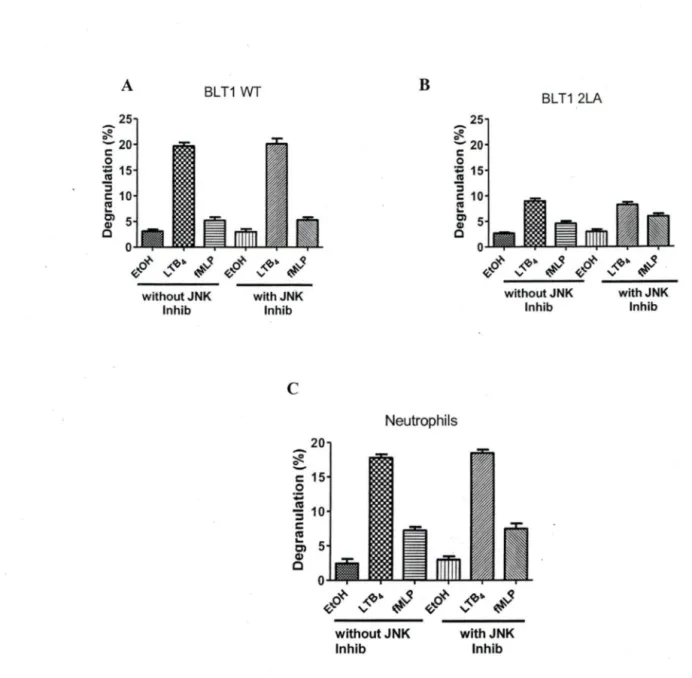
release in triple knockout neutrophils correlated with reduced p38 rnitogen-activated protein (MAP) kinase activity, suggesting that s_rc kinases act upstream of p38 MAP kinase. Indeed, treatment of human neutrophils with the p38 MAP kinase inhibitor, SB203580, led to reduced primary and secondary granule exocytosis in response to fMLP (Barlic, Andrews et al. 2000).
However, another MAP kinase inhibitor, PD98059, which blacks ERK.112 activity, did not affect release of primary and secondary granules or secretory vesicles (Barlic, Andrews et al. 2000). These findings indicate that src kinases, Hck, Fgr, and Lyn, along with p38 MAP kinase, but not ERK.112, play arole in regulating the release of granules.
Gaudreault et al. (Gaudreault, Thompson et al. 2005) demonstrated that there is partial involvement of src family kinase potentially Yes kinase in LTB4 mediated neutrophil
5.8 Arrestin fonction in regulating exocytosis
A group of scaffolding proteins known as arrestins 2 and 3 have also been shown to be required for activating signaling pathways leading to exocytosis of primary and secondary granules in neutrophils (Ferguson, Downey et al. 1996; Barlic, Andrews et al. 2000). Arrestins 2 and 3 are cytosolic phosphoproteins that were previously characterized for their role in endocytosis of ligand-bound chemokine receptors, particularly CXCRl, which is the high affinity receptor for the neutrophil chemotactic factor, IL-8. Arrestins 2 and 3 act by uncoupling activated G protein-coupled receptors from their associated heterotrimeric G proteins, they bind directly to the cytoplasmic tail of the CXCRl receptor (Barlic, Andrews et al. 2000; Ferguson, Downey et al. 1996).
Dominant negative mutants of arrestin 2 and 3 were shown to inhibit the release of granules when transfected into rat basophilie leukemia (RBL) cells, a cell line resembling mast cells and basophils (Barlic, Andrews et al. 2000). Interestingly, arrestins 2 and 3 also associate with the primary and secondary granules in IL-8-activated neutrophils, and they do so by binding to Hck and Fgr, respectively (Barlic, Andrews et al. 2000). Thus, arrestins 2 and 3 act at two sites in the cell during chemokine activation; one at the receptor in the plasma membrane and a second on granule membranes.
Hypothesis
The Role of helix VIII of BLTI in neutrophil responses to LTB4 is well documented (Shimizu, Yokomizo et al. 2000; Okuno, Ago et al. 2003; Gaudreau, Beaulieu et al. 2004). Our lab and others have shown that truncation or mutation in BLTI can lead to abnormal signaling in neutrophils (Baneres, Martin et al. 2003; Gaudreau, Beaulieu et al. 2004; Okuno, Yokomizo et al. 2005). Gaudreau et al. (Gaudreau, Beaulieu et al. 2004) have demonstrated that a mutation in helix VIII leads to significant reduction in BLTI internalization. Gaudreault, et al. have shown that receptor endocytosis is crucial for LTB4-mediated neutrophil degranulation, whereas Lambert et al. demonstrated that a defect in helix VIII of BLTI leads to multiple pseudopod formation and defective chemotaxis. They also proposed that a member of the Rho family of GTPases, RhoA, is important for LTB4-mediated chemotaxis. ·
Since the intracellular granule movement and granule exocytosis in neutrophils require cytoskeleton rearrangement in a similar fashion as in chemotaxis which has been shown to be RhoA-dependent and receptor endocytosis is crucial for LTB4-mediated degranulation, we hypothesised that the dileucine motif in helix VIII of BLTI receptor plays a role in RhoA-dependent neutrophil degranulation.
Objectives
To study the role of the dileucine motif and Rho A in neutrophil degranulation.
•:• To test the PLB985 cell line stably transfected with BLTI (wt) or BLTI (mutant) as a mode! for L TB4 -induced neutrophil degranulation.
•:• To determine whether the 2LA mutation in BLTI effects LTB4-mediated degranulation.
•:• To determine the signaling pathways involved in wt and mutant BLTI receptor-mediated degranulation using pharmacological inhibitors.
•:• To d.etermine whether the defective signaling in mutant BLTl is reversible by co-expression of constitutively active RhoA.
Materials and Methods Reagents
RPMI 1640 and geneticin (G418) were from Invitrogen Canada. FBS, cytochalasin B, Y-27632 and Triton were from Sigma-Aldrich Canada. DMSO was fromFischer Scientific.
Dextran and Ficoll-Paque PLUS, were from Amersham Biosciences. U75302,4-amino-5-( 4methylphenyl)-7-(tert-butyl)pyrazolo[3,4-d] pyrimidine (PP 1 ), N-benzyl-3,4-dihydroxylbenzylidenecyanoacetamide (AG490), ( fluorophenyl)-2-( 4-methylsulfinylphenyl)-5-( 4-pyridyl) IH-imidazole (SB203580), 2'-amino-3'-methoxyflavone (PD98059), L Y294002, and p-nitrophenyl N-acetyl-P-glucosaminide, Wortmannin and Pertussis toxin were from BIOMOL. LTB4 was from
Isolation of human neutrophils
Neutrophils were obtained from peripheral blood of healthy medication-free volunteers after informed consent in accordance with an Internai Review Board-approved protocoL as described previously (Stankova, Turcotte et al. 2002). Briefly, peripheral blood leukocytes were enriched by dextran sedimentation, layered over a Ficoll-Hypaque cushion, and centrifuged at 4000 x g for 20 min. mononuclear leukocytes were collected at the interface, whereas neutrophils were obtained from the pellet. The neutrophils preparation was depleted of erythrocytes by osmotic shock, then washed and resuspended in PBS until used.
Jl-Hexosaminidase release assays
Degranulation was determined by measunng the release of a granule marker,
13-hexosaminidase, as described previously by Ali et al. (Al~ Richardson et al. 1993), withsome modifications. For PLB-BLT cells, after 3 days of differentiation, 2.5 x 10⁵ cells were washed once with 1 ml of PBS. For human neutrophils, 4 x 10⁶ cells were used.
When necessary, cells were incubated with inhibitors for indicated times at 3 7°C. Cell pellets were resuspended in 250 µI of PBS containing cytochalasin B (4.8 µg/ml) and incubated for 5 min. Cytochalasin B was used to facilitate degranulation without priming
cells with other reagents that might interfere directly in intracellular signaling pathways. Cells were then stimulated with L TB4 ( 1 OO nM) or other stimuli for 10 min.
After stimulation, 1>-hexosaminidase activity was measured in 50 µI of cell-free supematant by spectrophotornetric analysis using 50µ1 of 2 rnM p-nitrophenyl-N-acetyl-P-glucosaminide as chromogenic substrate. Cell supematant and substrate were incubated for 1hat37°C. The reaction was stopped by adding 150 µI ofa O.lMNa2C03-NaHC03
buffer at pH 9.5. OD was then read at 405 nm using a spectrophotometer (BioRad). Values were expressed as percentages of total 1>-hexosaminidase, which was determined in cells lysed with 0.1 % Triton X-100. Ail percentages were corrected by subtracting spontaneous ll-hexosaminidase release in cell supematants. Ali assays were performed in triplicate, and OD was read three times.
Construction of Myc-tagged wild type and mutant receptors.
The cloning of WT BLTl-cDNA and generation of the mutant 2L (304-305)A in which leucines were substituted with alanines was previously described (Gaudreau, Le Gouill et al. 1998; Gaudreau, Beaulieu et al. 2004). Ali constructions were subcloned into pcDNA3
Cell culture and transfection
PLB-985 cells were grown in RPMI 1640 supplemented with 5% FBS and gentamicin sulfate (40 µg/ml). All cells werecultured at 37°C in a humidified 5% C02 incubator.
PLB-985 cells were stably transfected with a pcDNA3 vector containing a construct encoding for a tagged BLTl sequence (Gaudreau, Le Gouill et al. 2002) or myc-tagged BLTl mutant sequence (Gaudreau, Beaulieu et al. 2004) . In short, 30 x 106 cells were electroporated at 320 V using 30 µg of pcDNA3-BLT. Cells were then cultured for 3 wk in medium containing G418 at a concentration of 800 µg/ml. After 2 wk of G418 selection, cells were sorted twice using a FACSVantage cell sorter (BD Biosciences). These cells arereferred to as PLB-BLTl (WT) and PLB- BLTl (2LA).
Differentiated PLB-BLTl (wt) cells or PLB-BLTl (2LA) (10 x 106 cells), loaded in
nucleofection buffer, were nucleofected with 10 µg of plasmid containing constitutively active RhoA or an empty vector pcDNA3 using nucleofector program U02 (Amaxa Biosystems). For degranulation experiments, cells were used 8 h post transfection PLB-BLTl (WT) and PLB- PLB-BLTl (2LA) cells were cultured in medium supplemented with 1.25% DMSO for 3 days before each experiment to induce cell differentiation into a neutrophil-like phenotype, unless mentioned otherwise.
Statistical analyses
Data were analyzed by using PRISMS software. Paired Student's t test was performed
Results
The Dileucine motif in helixVIII of the BL Tl receptor plays an important role in LTB4 mediated degranulation in neutrophils.
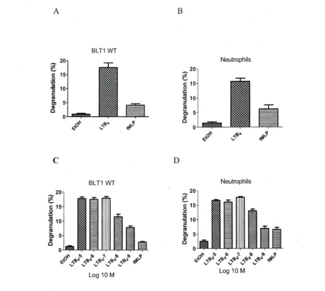
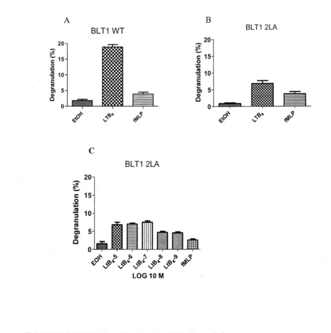
We and others (Yokomizo, Masuda et al. 2000; Okuno, Ago et al. 2003; Gaudreau, Beaulieu et al. 2004; Okuno, Yokomizo et al. 2005) have shown that the distal 2LA (304-305) motïf: which is in helix VIII of BL Tl receptor plays an important role in phospholipase C activation, receptor desensitization, internalization and chemotaxis. To investigate whether mutation of dileucine motifs of BLTI affects degranulation, we used PLB-985 cells stably transfected with BLTI-wt and the mutant 2LA (304-305). Those cells were cultured in medium supplemented with DMSO for 3 days to induce cell differentiation into a neutrophil-like phenotype. Differentiated PLB-BLTl (wt) cells and freshly acquired human neutrophils were stimulated with L TB4, its respective vehicle ethanol and fMLP as a control. LTB4 mediated degranulation levels in PLB-BLTl (wt) (figlA) and freshly acquired human neutrophils (figlB) were comparable. The degranulation levels in PLB-BLTI (wt) (figlC) and human neutrophils (figlD) were concentration dependent. Degranulation levels in PLB-BLTI (2LA) mutant cells were 60±5% less (fig2B) compared to PLB-BLTl (wt) cells (fig2A) and human neutrophils (fig lB). We also observed that PLB-BLTI (2LA) did not degranulate in a concentration
concentration of LTB4 failed to bring about any change in de granulation levels of PLB-BLTl (2LA) cells (fig 2C). This strongly suggests that the distal dileucine motif in helix VIII plays a crucial part in LTB4 mediated downwards signaling in neutrophil
degranulation. It was interesting to see that there remained a residual LTB4 dependent
20 ~ c: 15 .!2
..
.!!l 10 ::::i c: ~ en Cii cc
~20c
c: 15 0 :; 10 ::J ' c: ni c, 5 Cii c 5 0 ~ ~o BLT1 WT .... ~·"
~s BLT1 WT Log 10 M D ~ e.... c: 20 15 .5! ~ ::J 10 c: ni c, 5 Cii c 20 c: 15 0 :;::;-3
10 c: ni c, 5 ~ Neutrophils Neutrophils 0 .J..mllllL....ll'l"'-"'i"'J..U,1.U...., o~~j>~'J>'i'J:,$ tÇ .... ~· .... ~· .... ~· ,,,,<Q• ,,,,<Q• ~" "
Log 10 M" "
"
Fig 1: PLBB-BLTl (Wf) and Neutrophil degranulation.
A) PLB-P85 cells stably transfected with BLT 1 (Wf) differentiated for 0-3 days using DMSO. (2.5 X 10⁵) and, B) freshly isolated human neutrophils (3 X 10⁻⁶) were stimulated with 1
o-
7M of LTB4 or its appropriate vehicle (EtOH) or 1o-
7M fMLP (positive control) for 10 min. After stimulation, P-hexosaminidase release was assessed. C) PLB-P85 cells stably transfected with BLTl (Wf) and D) Freshly isolated human neutrophils (3 X10⁶) were stimulated with of LTB4 (10-5- 10-9M) orits appropriate vehicle ( ethanol EtOH) or 1
o-
7M fMLP (positive control) ·for 10 min. After stimulation, P-hexosaminidase release was assessedA 20 ~ 0 -; 15 0 ;
-5
10 c: Cii...
CJ) 5 ~ c 0 BLT1 WT ~ /ÇO ~ ~c
20 c 15 .2 -.!!! ::::1 10 c !? Cl 5 Q) c ,,_IQ~"
~.s BLT1 2LA 0 -'--1""--DqX'-"=;=l-u..y.Ll.-o~ ~ ~ ~ !b ~ ~ ~-!'"'
v-<Q"'-v-<Q"'
-!'"' -!'"'
~ LOG 10 M B 20 ~ ~ c: 15 0..
.!!! 10 ::::1 c: ra...
5 Cl Q) c 0 ~ <ÇoFIG 2: PLB BLTl (2LA) and PLB.BLTl (WT) degranulation
BLT1 2LA
.... ~~
"
~s
A) PLB-P85 cells stably transfected with BLTl (WT) and, B) PLB-985 cells stably transfected with BLT 1 (2LA) (2.5 X 10⁵) were differentiated for 0-3 days using DMSO and were stimulated with 10-7M of LTB4 or it appropriate vehicle (EtOH) or
1
o-
7M fMLP (positive control) for 10 min. A:fter stimulation, P-hexosaminidaserelease was assessed. C) PLB-P85 cells stably transfected with BLTl (2LA) were differentiated for 0-3 days using DMSO (2.5 X 10⁵) were stimulated with (10-5-
10-9M) of LTB4 or its appropriate vehicle (EtOH) or 10-7M fMLP (positive control) for
10 min. After stimulation, P-hexosaminidase release was assessed. n= 3
A Pertussis -sensitive Gai protein subunit, PI3k and Src family kinases are involved in L TB4 mediated neutrophil degranulation.
Signaling of BLTI through Gai protein subunit and its involvernent in neutrophils degranulation has been shown previously (Masuda, Itoh et al. 2003; Gaudreault, Thompson et al. 2005). In order to assess whether the residual degranulation in PLB-BLTI (2LA) cells was also dependent on Gai protein subunit signaling, we used pertussis toxin, an inhibitor of Gai and GaO signaling.
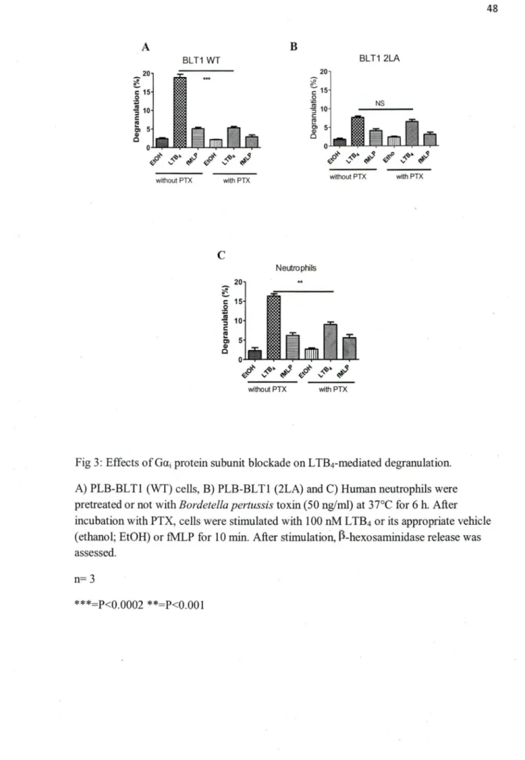
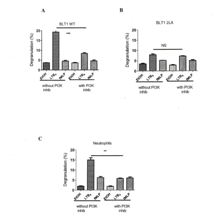
PLB-BLTI (wt), PLB-BLTI (2LA) and human neutrophils cell were pretreated with PTX (50nM) for 6 h, followed by 10 min stimulation with LTB4 (IOOnM). Pretreatment with PTX resulted in 80 % reduction in degranulation levels of PLB-BLTI (wt) (fig3A) and 40% reduction in degranulation levels of neutrophils (fig3C), while PTX pretreatment did not have any significant effect on PLB-BLTI (2LA) residual degranulation (fig 3B). Previously it has been shown that BLT-1 mediated degranulation in RBL-2H3 cells (lto, Yokomizo et al. 2002) and PLB985 cells (Gaudreault, Thompson et al. 2005) stably transfected with BLTI cDNA was partially dependent on PI3k To see if the residual degranulation in PLB-BLTl (2LA) cells was dependent on PI3K activation, we used the specific PI3k inhibitor wortmanin (lµM).
Pre-incubation for lh with wortmanin followed by stimulation with LTB4 led to 60% inhibition of degranulation of PLB-BLTl (wt) (fig4A) and 60% inhibition of human neutrophils (fig4C) degranulation. Incubation with wortmanin did not produce any reduction in degranulation levels in PLB-BLTl (2LA) cells (fig4B).
Gai protein subunit activation is known to play an important role in activation of several kinases including src kinase by different GPCR. Src family kinases are also known to be involved in certain neutrophil functions including granule secretion (Gaudreault, Thompson et al. 2005). To assess whether the residual degranulation in PLB-BLTl (2LA) is dependent on scr kinase activation, PLB-BLTlwt and PLB-BLT12LA cells were pre-incubated or not with src kinase inhibitor PPl (lOµM) for lh and then stimulated with LTB4. PLB-BLTl (wt) (fig5A) cells showed 40% reduction of degranulation levels compared to non pre-incubated cells, whereas PLB-BLTl (2LA) (fig5B) cells did not show a significant reduction in degranulation levels. Human neutrophils pre-incubated with the inhibitor ppl (30 µM) for lh showed 45% reduction of degranulation levels after stimulation with LTB4 (fig5C). Moreover PPI inhibition of PLB-BLTl (wt) was concentration dependent, reaching 65% at the inhibitor concentration of 50µM (fig5D). There was no effect of increased concentration on PLB-BLTl (2LA) mutant cells. The degranulation rate remained at similar level from 0-50µM (fig5E).
These results indicate that the residual degranulation by 2LA mutant BLTl is not the result of downstream signaling of Ga~
0
, PBK, or src kinase activation.20 ~ c: 15 .S! ~ :::i 10 c:
..
c, 5 GI 0 without PTX with PTX ~ ~ 15 0 ~ :l c: 10 ~ g> 5 Cl NS 0 ..._ ... "l""--'=i="--W-iui--~o~ ~~· ~S ~~0 ~~· ~S without PTX with PTXc
20 c: 15 .2 ] 10 :::i c:..
c, 5 Cl c Neutrophils 0 ..._ ... """"" __ ... ol"L-<v~~ ~· ~s ~o~ ~~· ~s without PTX with PTXFig 3: Effects of Gai protein subunit blockade on LTB4-mediated degranulation.
A) PLB-BLTl (WT) cells, B) PLB-BLTl (2LA) and C) Human neutrophils were pretreated or not with Bordetella pertussis toxin (50 ng/ml) at 37°C for 6 h. After incubation with PTX, cells were stimulated with 1 OO nM L TB4 or its appropriate vehicle
(ethanol; EtOH) or fMLP for 10 min. After stimulatioll, rl-hexosaminidase release was assessed.
n= 3
BLT1 WT 20 ~ ~ c 15 .Q ]1 :::J 10 c ro ~ 5 0
c
without Pl3K inhib *** 20 ~ ~ 15 c 0 ~ :::J 10 c ~ Cl 5 Q) 0 0 with Pl3K inhib Neutrophils ** ~ ~o 'V ,,_'lJ~ ~ ~ <f2 .p~ without Pl3K inhib 20 .-.. ~ ~ c 15 .QN
:::J 10 c Cil Ci 5 Q) 0 ,,_'lJ~ ~ 'V ~ with Pl3K inhib BLT1 2LA without P13K inhib NSFig 4. Effects of PBK inhibition on LTB4-mediated degranulation.
with Pl3K inhib
A) PLB-BLTl (WT) cells, B) PLB-BLTl (2LA) and C) human neutrophils were pretreated or not with wortmannin (1 µM) at 37°C for 1 h. After incubation with the inhibitor, cells were stimulated with 1 OO nM of LTB4, its appropriate vehicle
( ethanol) or fMLP for 10 min. After stimulation, r>-hexosaminidase release was assessed.
n= 3
~ ~ c 0