Université de Montréal
The effect of lesion size on cortical reorganization
in the ipsi and contralesional hemispheres
Boris Touvykine
Département de Physiologie
Faculté de Médecine
Mémoire présentée à la Faculté de Médecine en vue de l’obtention du grade de maîtrise (M.Sc.)
en Sciences Neurologiques 2-530-1-0
Décembre 2013 © Boris Touvykine, 2013
i
RÉSUMÉ
Bien que la plasticité ipsilesionnelle suite à un accident vasculo-cérébral (AVC) soit bien établie, la réorganisation du cortex contralésionnel et son effet sur la récupération fonctionnelle restent
toujours non élucidés. Les études publiées présentent des points de vue contradictoires sur le rôle du cortex contralésionnel dans la récupération fonctionnelle. La taille de lésion pourrait être le facteur déterminant la réorganisation de ce dernier. Le but principal de cette étude fut donc d’évaluer l’effet des AVC de tailles différentes dans la région caudal forelimb area (CFA) du rat sur la réorganisation physiologique et la récupération comportementale de la main. Suite à une période de récupération spontanée pendant laquelle la performance motrice des deux membres antérieurs fut observée, les cartes motrices bilatérales du CFA et du rostral forelimb area (RFA) furent obtenues. Nous avons trouvé que le volume de lésion était en corrélation avec le niveau de récupération comportementale et l’étendue de la réorganisation des RFA bilatéraux. Aussi, les rats ayant de grandes lésions avaient des plus grandes représentations de la main dans le RFA de l’hémisphère ipsilésionnel et un déficit de fonctionnement plus persistant de la main parétique. Dans l’hémisphère contralésionnel nous avons trouvé que les rats avec des plus grandes représentations de la main dans le RFA avaient des lésions plus grandes et une récupération incomplète de la main parétique. Nos résultats confirment l’effet du volume de lésion sur la réorganisation du cortex contralésionnel et soulignent que le RFA est l’aire motrice la plus influencée dans le cortex contralésionnel.
Mots-clés : accident vasculo-cérébral, réorganisation contralésionnelle, microstimulation intracorticale, récupération fonctionnelle, taille de lésion.
ii
ABSTRACT
While our understanding of ipsilesional plasticity and its role in recovery of hand function following ischemic stroke has increased dramatically, the reorganization of the contralesional motor cortex and its effect on recovery remain unclear. Currently published studies offer contradictory views on the role of contralesional motor cortex in recovery. Lesion extent has been suggested as the factor determining the type of reorganization of the contralesional motor cortex. The primary goal of this study was thus to evaluate the effect of unilateral strokes of different sizes in caudal forelimb area (CFA) of the rat on both physiological reorganization and behavioral recovery. At the end of a period of spontaneous recovery during which we monitored motor performance of both limbs, we obtained bilateral maps of the CFA and the putative premotor area of the rat – rostral forelimb area (RFA). We found that lesion volume in the CFA correlates with both the extent of behavioral recovery of the paretic hand and the extent of both ipsi and contralesional cortical reorganization. We found that rats with bigger lesions had larger hand representations in the ipsilesional hemisphere and more
persistent deficits of the paretic hand. In the contralesional hemisphere we found that rats with larger hand representation in the RFA had bigger lesions and incomplete recovery of the paretic hand. Our results confirm the effect of lesion volume on the reorganization of the contralesional motor cortex and highlight contralesional RFA as the motor cortical area most influenced by lesion volume for future investigations.
Key words: cortical stroke, contralesional reorganization, intracortical microstimulation, functional recovery, lesion size.
iii
TABLE OF CONTENTS
RÉSUMÉ ..………..i
ABSTRACT……….ii
TABLE OF CONTENTS ………..iii
LIST OF FIGURES ….……….………v
LIST OF ABBREVIATIONS….……….……….…vi
ACKNOWLEDGEMENTS ….……….……….………vii
CONTRIBUTION OF AUTHORS ……….………..viii
CHAPTER 1: GENERAL INTRODUCTION AND LITERATURE REVIEW….……….………1
1.1 General Introduction….……….……….1
1.1.1 Motor areas of the frontal cortex….……….3
1.1.2 Organization of primary motor cortex….………6
1.2 Plasticity in the ipsilesional hemisphere….………8
1.2.1 Release of local inhibition can support rapid changes of motor outputs in M1….………….8
1.2.2 Primary motor cortex plasticity and motor learning….……….9
1.2.3 Cortical reorganization after stroke in M1….………10
1.2.4 Early changes in the ipsilesional hemisphere after stroke….……….11
1.2.5 Late changes in the ipsilesional hemisphere after stroke….………..12
1.3 Plasticity in the contralesional hemisphere….……….14
1.3.1 Interhemispheric interactions in healthy adults….……….……….14
1.3.2 Early changes of interhemispheric interaction after stroke….……….……….16
1.3.3 Late changes of interhemispheric interaction after stroke….……….………..17
1.4 Effect of lesion size on contralesional reorganization….……….……….…….18
1.4.1 Effect of lesion size on physiological, anatomical and functional reorganization in the contralesional hemisphere….……….………..……….…………18
iv
1.4.2 Rational for the set of experiments conducted in the present study….…..……….…….20
CHAPTER 2: THE EFFECT OF LESION SIZE ON CORTICAL REORGANIZATION IN THE IPSI AND CONTRALESIONAL HEMISPHERES….……….……….………….…..….22
CHAPTER 3: GENERAL SUMMARY AND DISCUSSION………….………….….………….………….…..…..………….60
3.1 General summary………….………….….………….……….………….….……….……….…..…..………….60
3.2 Relation between the reorganization of the contralesional RFA and behavioral recovery.… 62 3.2.1 Detrimental plasticity………….………….….………….………….…..…..……….………….….……...62
A) Detrimental effect of contralesional RFA on behavioral recovery…….….………..…...62
B) Expansion of RFA due to learned non-use…….….……...…….….…….…….….………..65
3.2.2 Compensatory plasticity…….….……...…….….……...…….….……...…….….……...…….….…..…...67
C) Increased importance of contra and ipsilateral corticospinal projections from contralesional RFA…….….……...…….….……...…….….……...…….….……...…….….…..…...67
D) Contralesional RFA contributing to the function of the ipsilesional RFA………...72
3.3 General conclusion…….….…..………...…….….…..………...…….….…..………...…….….…..…….74
v
LIST OF FIGURES
CHAPTER 2: THE EFFECT OF LESION SIZE ON CORTICAL REORGANIZATION IN THE IPSI AND CONTRALESIONAL HEMISPHERES
1. Experimental timeline..………...…….….…..…….….…..………..…...…….….…….47
2. Experimental design………...…….….…..…….….…..………..…...…….….…..…….48
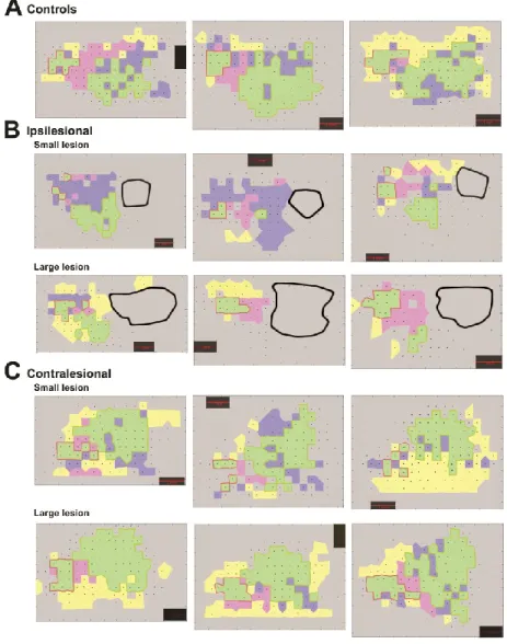
3. Histological reconstruction of lesions.…..…….….…..………..…...….….…..………49
4. Effect of lesion size on the final recovery of the paretic hand….…..………….…..………..….…..……..50
5. Examples of motor maps….…..………51
6. Motor representations in the ipsilesional CFA………..……50
7. Examples of motor maps of lesioned animals………..……52
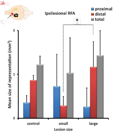
8. Motor representations in the ipsilesional RFA………..53
9. Motor representations in the contralesional CFA………54
10. Motor representations in the contralesional RFA………55
11. The effect of lesion size on motor representations……….56
12. The relation between motor representations and final recovery………..………57
13. Schematic summary of results………..………..………59
CHAPTER 3: GENERAL SUMMARY AND DISCUSSION 14. Stereotaxic coordinates for RFA lesion………...…….….…..…….….…..…………...…….….…..…….65
vi
LIST OF ABBREVIATIONS
1a afferent fibers - Primary afferent fiber
AP - Anterior-posterior
CFA - Caudal forelimb area
EMG - Electromyographic
ET-1 - Endothelin-1
GABA - Gamma-Aminobutyric acid
H-reflex - Hoffmann's reflex
Hz - Hertz
ICMS - Intracortical microstimulation
M1 - Primary motor cortex
MAP2 - Microtubule-associated protein 2
MCA - Middle cerebral artery
MCAo - Middle cerebral artery occlusion
ML - Medial-lateral
NMDAR1 - N-methyl-D-aspartate subunit 1
PMd - Dorsal premotor cortex
PMv - Ventral premotor cortex
RFA - Rostral forelimb area
rTMS - Repetitive transcranial magnetic stimulation
S1 - Primary somatosensory cortex
SMA - Supplementary motor area
vii
ACKNOWLEDGEMENTS
I would like to first of all thank my supervisor Dr. Numa Dancause. His guidance, mentoring, knowledge and passion for science not only made me better as a scientist, but also inspired me to want to continue in research. Most of all I would like to thank him for giving me a second chance. I would also like to thank Dr. Stephan Quessy for the motivation he provided to work harder and try to better oneself. As a fellow student with a lot more research experience Dr. Babak Mansoori helped make sense of things I could not with both scientific and personal advice for which I am grateful.
Janine El Helou, thank you for not stopping to believe in me. When I am under you pull me out and help me get back on track. Thank you for continuing to share this pursuit of knowledge with me. To my sisters: Vera and Mila, to my father and mother I could never thank you enough for everything. I hope I will continue to make you proud.
viii
CONTRIBUTION OF AUTHORS
Chapter 2 contains a manuscript ready for submission to Neurorehabilitation and Neural Repair: Touvykine B., Mansoori B. K., Jean-Charles L., Deffeyes J., Quessy S., Dancause N. The effect of lesion size on cortical reorganization in the ipsi and contralesional hemispheres. For this article, the
nature of my contribution is as follows: task familiarization of animals, lesion inductions, histological analysis, behavioral testing and preliminary data analysis. In addition I participated in all the terminal ICMS experiments, and was the lead surgeon for all of control group and half of experimental
animals. I also wrote the first draft of the Methods and Results sections. Dr. Babak Khoshkrood Mansoori participated in most terminal experiments and was the lead surgeon in half of experimental animals. Loyda Jean-Charles assisted in all the control ICMS experiments as well as most terminal ICMS experiments. Dr. Joan Deffeyes created the software in MATLAB, which I used to analyze experimental data. Dr. Stephan Quessy provided guidance and oversight with the behavioral component of the experiment, as well as running additional statistical analysis on the data. My supervisor Dr. Numa Dancause provided guidance and instruction throughout the study, including data collection, data analysis and interpretation, and the preparation of the final manuscript.
1
Chapter 1
General introduction and literature review
1.1 General introduction
Stroke is a cardiovascular disease, which damages a part of the brain due to a disruption of normal functioning of the cardiovascular system. It is the leading cause of disability worldwide. In Canada alone each year there are approximately 50000 strokes (PHAC 2011). While many people survive stroke, they are left with multiple behavioral and cognitive deficits. Currently there are approximately 315,000 Canadians dealing with post-stroke complications (Hakim, Silver, and Hodgson 1998). To contribute to the design of more successful treatments for individuals with stroke-induced deficits, it is important that we gain a better understanding of the basic mechanisms of cortical reorganization that occur after stroke.
There are two types of strokes, hemorrhagic and ischemic. Hemorrhagic stroke is neuronal death due to a rupture of a blood vessel. This type of stroke accounts for approximately 13% of all stroke cases. The second type of stroke is ischemic, also known as cerebral infarction. Ischemic stroke is neuronal death due to a blockage of a blood vessel, most often by a blood clot. This either
significantly slows down the blood flow or stops it completely, interrupting vital oxygen and nutrients supply to the brain. This type of stroke is much more common and accounts for approximately 87% of all stroke cases. There is also a phenomenon that has been identified as mini-strokes which are often asymptomatic. They are due to a very transient blockage of a minor blood vessel that does not last long enough to lead to significant neuronal damage. The major difference between mini-stroke (also
2
known as Transient Ischemic Attack) and ischemic stroke is the amount of damage done to the brain. Our study investigated cortical reorganization following the most prevalent type - ischemic stroke.
As much as 80% of ischemic stroke cases are due to blockage of the middle cerebral artery (MCA), which is the largest artery in the brain or one of its branches (Harrison 1994). MCA supplies multiple cortical (frontal, parietal and temporal lobes) and subcortical (basal ganglia and the internal capsule) regions of the brain. The extent of initial ischemic damage depends on whether the whole of the MCA or one of its multiple branches will be blocked. This leads to variability of lesion size and location, creating differences from patient to patient and complicating prognosis.
The overwhelming majority of strokes are unilateral and therefore result in a lesion in one hemisphere. Many stroke survivors are left with persistent deficits in motor control, from such extreme cases as hemiparalysis to milder cases such as difficulties in fine motor control. In a classic study in 1951 Twitchell observed that unilateral stroke affects the upper limb more than the lower limb, and recovery of the upper limb is worse.
While research into stroke recovery and rehabilitation has made great progress in the past decade, numerous stroke survivors with motor deficits of the upper limb are left with significantly lower quality of life and a large strain on the health care system. In particular, motor deficits of the hand following stroke are some of the most resilient motor impairments after stroke, meaning such survivors are unable to do even simple manipulations. As a consequence, better knowledge of how reorganization following stroke permits the recovery of hand is needed. To help us better understand the recovery process this study was designed to investigate motor recovery of the hand in the rat. Rats are able to grasp and manipulate small objects with their forelimbs. Vasoconstrictor endothelin-1 (ET-endothelin-1) was used for lesion induction protocol. It is an endogenous molecule, which binds to
3
Vasoconstriction results in hypoxia, which in turn induces cortical lesions replicating the mechanism of ischemic stroke. One advantage of using a rat model is that there is incredibly high variability of lesion size and location in human patients, whereas inducing stroke in the rat circumvents this problem. The size of focal lesions we induce in the motor cortex of the rat can be controlled by injecting small amounts of ET-1 to limit its spread. This allows for examination of reorganization and recovery induced by a cortical lesion in the motor cortex.
1.1.1 Motor areas of the frontal cortex
In humans motor cortex is responsible for the planning and execution of voluntary
movements. It is the region in the caudal part of the frontal lobe of the cerebral cortex. Currently the motor cortex is separated into a primary motor cortex (M1) and a variety of non-primary motor cortical regions (Fulton 1935; Penfield and Welch 1951). The execution of voluntary movements is through the corticospinal tract, the vast majority of which originates in M1 (Dum and Strick 1991). Most of the corticospinal tract consists of fibers originating from the large pyramidal neurons in Layer V of the motor cortex. The axons of these neurons form pyramids in the brainstem, and then most of those axons cross over to the side contralateral to their hemisphere of origin (approximately 80% of pyramidal fibers) (Nathan and Smith 1973). In the spinal cord these axons form synapses with excitatory and inhibitory interneurons, which in turn synapse on motoneurons enervating the muscles. Humans, great apes, and some higher order non-human primates (e.g. Macaca) have corticomotoneuronal connections. In these cases, there is only one synapse between a cortical neuron and a motoneuron. This feature is limited to the hand and finger muscles of the forelimb and may support high manual dexterity of these species (Porter 1985).
In many primates, a series of non-primary motor areas are found rostral to M1. To date, at least six premotor areas have been described, which include the premotor ventral (PMv), premotor
4
dorsal (PMd), supplementary motor area (SMA) and three cingulate motor areas. Ablation studies in primate SMA have resulted in significant impairment of performance of bimanual tasks, suggesting its involvement in preparation and coordination of sophisticated bimanual movements (Brinkman 1984). PMv has been shown to be involved in the processing and transformation of visual information into internal set of coordinates which are consequently passed on to M1, which executes the motor command (Rizzolatti, Fogassi, and Gallese 2002; Davare et al. 2009). PMd is currently thought to process temporal demands of a task and prepare the necessary sequence for muscle activation (Davare et al. 2006). Cingulate motor areas have not been studied as well as other non-primary motor areas. Rostral cingulate motor area has been implicated in evaluating the reward benefit of the available motor repertoire and subsequent selection of the most rewarding movement (Shima and Tanji 1998). The authors were not able to distinguish between dorsal and ventral cingulate motor areas and grouped them into caudal cingulate motor area. The authors propose that it is involved in movement initiation and motor preparation. In summary planning and preparations of movement are understood to be performed by the higher order (non-primary) motor areas.
By comparison, rodents have a much simpler motor cortex. Currently, only two forelimb cortical regions have been identified. There is a larger caudal forelimb area (CFA), and a smaller rostral forelimb area (RFA). The connection patterns of CFA and RFA are different and suggest that these areas play different roles in the control of the forelimb. The first exhaustive examination of these two areas in the rat came from a study by Rouiller and colleagues (1993). This study examined and compared the pattern of connections to and from RFA and CFA. They found a significant
difference in the pattern of incoming and outgoing connections between the two motor cortical areas. Among those was a segregation of both corticocortical and thalamocortical projections. RFA was interconnected with the insular cortex while the CFA was not, a pattern also seen for SMA and
5
the premotor cortex in primates (Matelli et al. 1986). In addition RFA and CFA were interconnected with different nuclei in the thalamus similar to segregation of thalamic input to the cortex between M1 and non-primary motor areas (SMA, premotor cortex) (Schell and Strick 1984). CFA is also the area from which the majority of the corticospinal neurons projecting to the cervical segment of the spinal cord originate (Starkey et al. 2012). The proportion of corticospinal projections from RFA is much smaller. This mirrors what has been found in primates, in which M1 is the area from which the most corticospinal neurons originate. The projections to the cervical enlargement from any single non primary motor area are significantly smaller (Dum and Strick 1991). These anatomical findings further support the proposed role of RFA as a non-primary motor area acting as either premotor cortex or SMA, with CFA acting as M1. Thus, based on these anatomical data, the RFA is likely to be homologue of a premotor motor area, while the CFA is likely to be a homologue of M1 (Rouiller 1993). However, to date the functional role of RFA is still is not clear, but lately with the advent of optogenetics different researchers have started to explore the functional significance of these anatomical differences in the pattern of connections. There is an increasing body of evidence that RFA acts as a higher-order motor cortical area comparable to non-primary motor areas in primates (Smith et al. 2010; Hira et al. 2013). Smith and colleagues (2010) found that inactivation of RFA leads to increased response time, but does not increase premature responding. Inactivation of the medial prefrontal cortex (mPFC) produced the opposite results. The response time did not change, but premature responding increased. Evaluating these results together with anatomical studies previously done on the interconnectivity of RFA, the authors propose that RFA acts as a premotor cortex and competes with mPFC for action selection. Hira and colleagues (2013) found that RFA and CFA have an
asymmetrical pattern of reciprocal connections where the majority of corticocortical connections originating in layer 5b of RFA project towards Layer 5b of CFA. However the majority of corticocortical connections from CFA to RFA originate in layer 2/3 and projection towards layer 5b of RFA. Arguing
6
that there is laminar hierarchy in the motor cortex with neurons in layer 5b being the final outputs of corticospinal networks, the authors propose that the asymmetrical reciprocity of corticocortical connections between RFA and CFA suggests that RFA is a higher order motor area.
As of yet it is still unclear if RFA functions as a specific non-primary motor area or a fusion of two or more of them. Nonetheless the proposed hierarchical organization of the rat motor cortex makes the organization of the rat motor cortex significantly more relevant to primates than
previously thought (Rouiller, Moret, and Liang 1993). All of these factors make the findings on cortical reorganization in the rat more clinically relevant.
1.1.2 Organization of primary motor cortex
Primary motor cortex is organized somatotopically for large regions of the body. The cortical area responsible for evoking movements for different segments of the body, such as upper limb, trunk, face and leg are always oriented the same way relative to one another. For example, the face representation is always found lateral to the forelimb representation. This type of organization was discovered by Penfield and Boldrey (1937) in the somatosensory and motor cortex. In 1957
Mountcastle described the organization of the somatosensory cortex by proposing the concept of the cortical column. According to this hypothesis, a cortical column is the basic processing unit of the somatosensory cortex. In a column, all the neurons have the same receptive fields and there is no overlap of receptive fields between cortical columns. In 1975, based on his previous work using intracortical microstimulation (ICMS) Asanuma proposed the cortical column as the basic functional unit in the motor cortex as well. In his view each cortical column in the primary motor cortex would project to a single muscle. This interpretation was based on his work with ICMS. This technique uses an insulated stimulation electrode to penetrate the cortex and to pass a train of pulses to evoke muscle contractions. By doing so, the volume of stimulated cortex is very small, potentially limited to
7
a single column. In his experiments using ICMS in primates Asanuma and Rosén (1972) observed that stimulation at threshold current typically induced contractions to a single muscle.
However a number of studies have cast doubts over the columnar organization of M1 corticospinal outputs. In 1980 Fetz and Cheney performed a study where the muscle activity of monkeys doing a simple manual task was correlated to single-neuron activity in M1. After averaging the EMG activity that followed the firing of cortical neurons, they found that several muscles can show facilitation after firing of a single neuron. They proposed that this effect is due the divergent connectivity of cortical tract neurons, which would synapse on different motoneuron pools,
innervating different muscles. An anatomical study by Shinoda and colleagues (1981) supported this view by demonstrating that a single large pyramidal neuron originating in the motor cortex has collaterals at several levels of the spinal cord suggesting connections with multiple motoneurons.
The question remained as to how M1 manages to elicit specific muscle contractions that produce movements, considering that its projections are so divergent. The answer was provided by Schieber and Hibbard in 1993, when they recorded isolated neurons as the monkey moved its individual fingers. They found that neurons with activity related to the movements of the different fingers were intermingled and that there was no clear localization of neurons involved in the control of movements of one finger in relation to the others. Their conclusion was that the control of the digits is widely distributed through the hand area of M1, with no apparent clusters dedicated to single muscles. This divergent distribution of the origin of corticospinal projections in M1 and their
destination in the spinal cord suggests that for a muscle contraction to take place there should be a temporal convergence of inputs onto appropriate motoneurons. This highly redundant organization of the corticospinal projections is thought to underlie the plasticity and rapid reorganization in the motor cortex, and is considered to be one of the underlying substrates that allow stroke recovery.
8
1.2 Plasticity in the ipsilesional hemisphere
1.2.1 Release of local inhibition can support rapid changes of motor outputs in M1
Fast acquisition of new motor skills is a huge evolutionary advantage. Motor cortex plasticity is thought to underlie mammalian capacity to quickly acquire new motor behavior. What permits this ability for rapid motor cortical plasticity? Reversal of cortical inhibition has been shown to play a very important role in the reorganization of the motor cortex. In a culmination of a series of experiments Jacobs and Donoghue (1991) assessed reorganization of motor cortex due to release of local
GABAergic inhibition. In this study using ICMS the authors identified stimulation sites that evoked either only vibrissae or forelimb movements in the rat. They then applied a GABA antagonist (bicuculine) in the forelimb region to remove the effect of local inhibition on the motor outputs of that region. After the injection of the GABA antagonist, they stimulated sites from which vibrissae movements were evoked again. Along the border of the two representations, as early as 15 minutes after local application of GABA antagonist the stimulation of a vibrissae site started to also evoke forelimb movements. This time window is too short for synaptogenesis or any other anatomical changes to occur. Their results thus strongly suggest that there were already present, functional (but silenced) corticocortical connections between the vibrissae and the forelimb regions, which were supressed by tonic GABAergic inhibition. By removing the tonic inhibition, the previously silenced synapses become responsive to stimulation. This suggests that there is a significant amount of redundancy in the pattern of connections in the motor cortex. This mechanism is faster than establishing new synapses. By taking advantage of the high redundancy of both the descending projections from M1 and the local corticocortical connections within M1, the modulation of local inhibition would allow for fast cortical reorganization. The inherent plasticity of M1 is likely an
important factor in the reorganization of the motor cortex after stroke that allows functional recovery of many patients.
9 1.2.2 Primary motor cortex plasticity and motor learning
Before looking at stroke-induced plasticity it is important to examine plasticity intrinsic to healthy individuals. Plasticity in the motor cortex is believed to support motor learning in adults. Indeed, several experiments have shown that motor learning is associated with cortical
reorganization. In a study in squirrel monkeys, animals had to develop a new motor skill to perform precision pinch with an index and thumb to grasp food pellets in a small well (R. J. Nudo et al. 1996a). Following motor learning and an increase in performance, the digit representation in M1 of these animals expanded. Subsequently, the same animals were trained at a task that required the animals to engage in the skilled use of the forearm and not the digits. Cortical motor maps obtained after the training at the second task showed a decrease and return to baseline of the size of the digit
representation in M1. Even though monkeys still had to use their fingers to perform the second task, the animals were performing an already acquired behavior and thus it did not require an increase in the size of the digit representation in M1. Thus cortical reorganization seems to be very dynamic and dependent on active learning of a new motor skill.
It has been previously demonstrated that there is an increase in excitability of the motor cortex at the initiation of motor skill learning (Rioult-Pedotti et al., 1998). This is further supported by an experiment in which hyperexcitation of M1 was achieved through application of high frequency repetitive transcortical magnetic stimulation (rTMS), and resulted in the improvement of sequential learning (Kim et al. 2003). What is the functional significance of this increased excitability of the motor cortex? As was previously discussed, there are plenty of potentially functional synapses in M1, which are suppressed by the inhibitory interneurons. The increased excitability of M1 could reflect that a certain number of previously “masked” synapses become functional. During the initial stage of motor skill learning there is an increase in muscle contraction (Osu et al. 2002). This increased
co-10
contraction is thought to increase task accuracy as it offers tighter control over limb dynamics and its placement in space and likely warrants larger corticospinal output.
Hikosaka and collaborators (2002) proposed that after initial learning, basal ganglion and cerebellum would come into play and mediate consolidation. These structures would reinforce the synapses in M1 that caused muscle contraction resulting in accurate performance of the task in a process not unlike “tuning”. As learning of the motor task proceeds, co-activation decreases without loss in accuracy, because limb dynamics have been optimized to the task. Eventually this process would result in a new set of functional synapses that are activated for the execution of this task. This can be seen as a consolidation, when synapses involved in the activation pattern necessary to produce muscle contractions to the right degree and at the right time, have been selectively reinforced.
Therefore, during motor learning, existing but silenced connections are activated. Those that best contribute to the new skill performance are selectively reinforced to be engaged in the particular motor skill. After the completion of motor learning, tonic inhibition in the motor cortex returns to normal. It is important to note that motor learning may not require axonal sprouting. It can simply take advantage of the redundant anatomic infrastructure already present and selectively reinforcing parts of it, while inhibiting other parts. This aforementioned redundant anatomical organization of M1 is thought to fast allow acquisition of new motor skills, and it is thought that it can also be used to support motor recovery after stroke.
1.2.3 Cortical reorganization after stroke in M1
Following injury, stroke patients recover at different speeds. After examining 46 stroke patients Fuji and Nakada (2003) separated the patients into three distinct groups. The first group demonstrated almost complete recovery a month after stroke, and was deemed the “fast” recovery
11
group. The rest of the patients demonstrated “slow” recovery. By three months post stroke some of these patients recovered to a level approaching that of the “fast” recovery group. They were thus classified as the “slow and good” recovery group. The remainder of patients did not recover much, even by the end of the three months period and were reclassified into “slow and bad” recovery group. The authors suggest that independent of the extent of recovery, the patients who recover slower do so through a different pattern of reorganization. Whereas the patients who recover quickly undergo one type of reorganization, the patients in both “slow” groups undergo a different type of reorganization that may or may not lead to good recovery of hand function.
1.2.4 Early changes in the ipsilesional hemisphere after stroke
We know that as early as one day after stroke there is widespread cortical disinhibition (Schiene et al. 1996). However the disinhibition appears to last longer than one day. Indeed, one week after injury, global down-regulation of GABA binding was reported (Qü et al. 1998). As discussed previously there are plenty of synapses in the cortex that are functional, but supressed by the tonic GABA inhibition (Jacobs and Donoghue 1991). Global disinhibition after stroke could allow for re-tuning of existing, but previously non-functional connections and selectively strengthen those which would result in return of function. This process can result in recovery if enough of M1 was spared by the lesion. In this case, at least part of the behavioural recovery would be sustained by physiological reorganization of the surviving M1 and would not require significant anatomical reorganization. This process would likely take advantage of the endogenous anatomical organization, and utilise the innate plasticity of the mammalian motor cortex which has evolved for fast acquisition of new motor skills. This could be the major route of reorganization of the “fast” recovery group described by Fujii and Nakada (2003).
12
1.2.5 Late changes in the ipsilesional hemisphere after stroke
However as Fujii and Nakada (2003) have demonstrated the majority of patients do not recover within a month. So what sort of processes might be involved in “slow, but good” recovery? Lashley (1938) proposed that it is the extent of the damage to the cortex that would drive subsequent reorganization. Thus if the damage to M1 is too extensive, where not enough of M1 remains, this would trigger significant reorganization of distant cortical areas. In particular non-primary cortical motor areas are the best candidates for where this reorganization takes place, as they are already heavily interconnected with M1 and form part of the corticospinal tract. This functional
reorganization of distal areas was demonstrated by transiently inhibiting the premotor cortex in monkeys that recovered after stroke (Liu and Rouiller 1999). Following recovery from lesions in the sensorimotor cortex of macaque monkeys, inhibition of the premotor cortex in the ipsilesional side with muscimol, a GABA agonist, can re-instate behavioral deficits in the paretic hand. When the inhibition was done in the contralesional premotor cortex, there was no decrease in the task
performance for the paretic hand. These results support the idea that during post stroke recovery the ipsilesional premotor cortex has taken on some of the function of M1.
Frost and colleagues (2003) looked at physiological reorganization of PMv following lesions in M1. They found that after large ischemic lesions in the hand area of M1, the hand area of PMv underwent expansion, presumably as part of compensatory functional reorganization. Building up on these results Dancause and colleagues (2005) conducted a study which looked into anatomical changes associated with stroke recovery and with the physiological reorganization of PMv. Following recovery, they injected the neuroanatomical tracer into PMv and compared the pattern of
connections to the one found in intact animals. Injections of neuroanatomical tracer in PMv of control animals did not result in any significant labelling of either neuronal cell bodies or axonal terminals in
13
primary somatosensory cortex (S1). This indicates a lack of direct projections between S1 and PMv. Tracer injections in PMv of animals that recovered from the ischemic lesions resulted in a larger number of labelled axonal terminals and cell bodies in S1. Furthermore the orientation of labelled axons originating in PMv was towards S1 in experimental animals, but not in controls. As M1 is reciprocally connected to both PMv and S1, but PMv does not project directly to S1, the authors proposed that as part of compensatory reorganization, PMv needs to re-establish these connections with S1 to take on some of the function of M1. The expansion of the hand representation of PMv along with the long distance anatomical rewiring (which appear to try to reproduce the connectivity pattern of M1) strongly support that PMv is undergoing compensatory reorganization. This type of reorganization could explain the novel role of the premotor cortex following recovery from stroke and the return of deficits in the paretic hand following inactivation of ipsilesional PMv in recovered animals (Liu and Rouiller 1999). Furthermore, such mechanisms could be the major route of recovery of the “slow, but good” group of Fujii and Nakada (2003).
In summary, there are multiple processes taking place in the ipsilesional hemisphere
following a lesion in M1 (R. Nudo 2006). Depending on the extent of damage, the motor cortex might reorganize relatively quickly, taking advantage of redundancy particular to the motor cortex. This would result in relatively fast recovery. However if the damage to M1 is too extensive, significant anatomical reorganization is required to achieve an adequate degree of functional recovery. The need to generate new axons and guide them to the right targets is significantly more demanding and takes longer. Therefore while recovery after relatively extensive damage to the motor cortex is possible, it takes significantly longer.
14
1.3 Plasticity in the contralesional hemisphere
1.3.1 Interhemispheric interactions in healthy adultsThe majority of projections composing the corticospinal tract originate from neurons within the motor cortex to the forelimb. However the ipsilateral motor cortex could also participate in the control of the forelimb by sending signals through corpus callosum, the largest bundle of nerve fibers in the mammalian brain, which connects the two hemispheres. One hypothesis is that the motor cortex of one hemisphere exerts inhibitory influence over its homologue in the other hemisphere to allow unimanual movements (Beaulé, Tremblay, and Théoret 2012). Supporting this hypothesis are studies demonstrating that stimulation of the motor cortex of one hemisphere with TMS produces suppression of EMG activity in the hand ipsilateral to the stimulation (Ferbert et al. 1992; Harris-Love et al. 2007). In these experiments they examined the effect of a subthreshold conditioning pulse in M1 of one hemisphere on the electromyographic (EMG) output of a suprathreshold pulse in M1 of the other hemisphere. In both studies the authors observed that the conditioning stimulus resulted in a consistent suppression of muscles in the arm contralateral to M1 stimulated with a suprathreshold pulse. To determine if the interhemispheric inhibition takes place at the spinal cord, the effect of the conditioning stimulus on the Hoffmann's reflex (H-reflex) was established. The H-reflex is EMG activity due to an electrical stimulus administered to 1a afferent fibers which are known to have a
monosynaptic connection with alpha-motoneurons (Palmieri, Ingersoll, and Hoffman 2004). In other words, the H-reflex is analogous to an electrically evoked stretch reflex. In these two studies (Ferbert et al. 1992; Harris-Love et al. 2007), they used the H-reflex to examine changes in spinal cord
motoneuron excitability. They found that conditioning stimulus to the ipsilateral M1 did not modulate the H-reflex response, suggesting that interhemispheric inhibition takes place in the supraspinal structures.
15
In a 2009 study, Kobayashi and collaborators looked at the effect of low frequency
subthreshold rTMS on motor learning. Subjects in all groups had to learn a unimanual sequential task after receiving the rTMS treatment. The first group received low frequency rTMS in M1 contralateral to the hand performing the task, the second group in M1 ipsilateral to the hand performing the task; the control group received rTMS treatment to the control scalp position (Cz). The subjects who received the rTMS treatment to the contralateral M1 did not learn the task as effectively as the control subjects. This was expected as low frequency rTMS is thought to be inhibitory. However the subjects who received rTMS to the ipsilateral M1 showed slight but significant improvements in motor skill learning compared to controls. Another study achieved similar results by exciting the contralateral motor cortex (Kim et al. 2003). In this study high frequency rTMS, thought to cause cortical hyperexcitability, was applied to the M1 contralateral to the hand performing the task and resulted in improvement of motor learning. It thus appears that either decreasing the activity of the M1 ipsilateral to the hand involved in skilled motor learning or increasing the activity of the M1 contralateral to the hand used improves motor skill learning. These studies further support the functional importance of interhemispheric inhibition for motor control.
There is a convergence of opinions that are singling out the corpus callosum as the important actor through which interhemispheric inhibition takes place (Ferbert et al. 1992; Harris-Love et al. 2007). Mayer and colleagues (1995) compared interhemispheric interactions of healthy subjects to patients with a complete or partial damage of corpus callosum. In both groups they found
suppression in tonic muscle activity after ipsilateral stimulation of M1. However in patients with callosal damage such as partial agenesis and hypoplasia, this suppression appeared later and was weaker than in healthy subjects. These findings are further corroborated by results from a study in cats in which Asanuma and Okamoto (1959) observed that in most recorded large pyramidal neurons
16
the stimulation of corpus callosum resulted in suppression. While these findings do not isolate the corpus callosum as the sole structure through which interhemispheric inhibition takes place, they do point to it as the major mediator.
The current assumption as to the role of the inhibitory interhemispheric activity is thought to be the lateralization of movement (Grefkes et al. 2008). This inhibitory network would allow us to perform unimanual tasks without simultaneous movements of the other arm. Whereas healthy adult humans can easily perform such unilateral movements, children up to the age of ten often show engagement of the other forelimb during performance of a unilateral task (Mayston, Harrison, and Stephens 1999). It is suggested that the difficulty encountered by children might come from an immature interhemispheric network. These unintentional and unwanted movements of the opposite hand during a tentative unimanual task are called mirror movements. As the child’s brain matures they tend to disappear. Mirror movements are also observed in some stroke patients and something that has been proposed to be due to the disruption of the normal functioning of interhemispheric inhibition (Kim et al. 2003).
1.3.2 Early changes of interhemispheric interaction after stroke
When a region of the sensorimotor cortex is destroyed or silenced the input from that particular region to the contralesional hemisphere is lost. Even if it is a temporary lesion caused by transient inactivation there is a release of inhibition in the contralesional hemisphere. In monkeys, inactivating part of the motor cortex has resulted in expansion of receptor fields in the contralateral somatosensory cortex immediately after inactivation (Clarey, Tweedale, and Calford 1996). In the rat, Maggiolini and colleagues (2008) documented acute changes in the contralateral motor cortex. Immediately after lidocaine inactivation of the motor cortex in one hemisphere, they obtained an ICMS map of contralesional motor cortex. These motor maps were bigger than in sham animals that
17
did not receive cortical lidocaine injection. Thus, due to loss of input from the inhibited motor cortex, there is an expansion of motor representation in the opposite hemisphere. The short interval
between the inactivation and the effect seen in the contralesional hemisphere suggests an unmasking of “dormant” connections. This acute disinhibition is most likely due to the loss of interhemispheric input that has been shown to be mostly inhibitory in healthy subjects. As part of the same study, Maggiolini and colleagues (2008) mapped the contralesional forelimb sensorimotor cortex 3 and 14 days after a chemical lesion in the forelimb motor cortex and found no difference from controls. These results suggest that the expansion of the motor map happens rapidly after the lesion and is transitory.
1.3.3 Late changes of interhemispheric interaction after stroke
As was discussed previously the ipsilesional motor cortex undergoes reorganization to recover functionality of the paretic limb. The contralesional motor cortex also undergoes reorganization to re-establish the interhemispheric balance disrupted by stroke (van Meer et al. 2012). While it might appear that disrupting the cortical reorganization might be detrimental to recovery, studies show that inhibiting the contralesional motor cortex with low frequency rTMS improves the recovery of the paretic hand (Takeuchi et al. 2005; Mansur et al. 2005). It is thought that the mechanism employed is through further disinhibition of the ipsilesional motor cortex which might act to speed up the reestablishment of a new interhemispheric balance. As discussed
previously stroke recovery has been compared to learning a new motor skill by a healthy person. Just as motor skill acquisition improves after inhibition of the motor cortex ipsilateral to the task in a healthy person, suggesting hyper-excitation of the contralateral motor cortex, a similar mechanism is thought to be responsible for the beneficiary effect of contralesional inhibition in stroke patients. In fact hyper-exciting the ipsilesional cortex with 5 Hz rTMS resulted in improvement of functional
18
recovery of the paretic hand, similar to supressing the contralesional motor cortex with 1 Hz rTMS (Emara et al. 2010). All of these studies offer support for the detrimental effect of interhemispheric inhibition exerted by the contralesional motor cortex.
Nonetheless there is also some data that contradicts these conclusions on the adverse role of the contralesional hemisphere in recovery of the paretic limb. A patient who successfully recovered from a unilateral stroke and then suffered another one in the previously intact hemisphere had the functional deficits of the initial paretic hand reinstated (Song Y 2005). This suggests that the contralesional hemisphere can indeed contribute to control of the paretic hand. In fact there are studies showing that after stroke recovery, the contralesional hemisphere of patients exert a more facilitatory effect on the ipsilesional motor cortex, in contrast to healthy subjects (Bütefisch et al. 2003). It appears that with time after stroke, the contralesional motor cortex can assume a positive or a negative role in the recovery of the paretic hand. In the face of these contradictory results coming from multiple studies it becomes clear that we are most likely missing a key factor which would influence the kind of role the contralesional hemisphere would play in stroke recovery.
1.4 Effect of lesion size on contralesional reorganization
1.4.1 Effect of lesion size on physiological, anatomical and functional reorganization in the CL hemisphere
Why is there such conflicting data about the role of the contralesional motor cortex in stroke recovery? A potential explanation could be that the contralesional cortex participates in stroke recovery differently depending on the how much of the ipsilesional motor cortex remains intact following stroke. There is a body of evidence indicating that lesion size influences reorganization in the contralesional motor cortex. In a functional magnetic resonance imaging (fMRI) study in rats
19
Dijkhuizen and colleagues (2003) demonstrated that the extent of contralesional activity correlates positively with the lesion size. In this experiment after inducing a middle cerebral artery occlusion (MCAo) in rats, hemodynamic activity in both hemispheres in response to paw stimulation was evaluated with fMRI. The results show a strong correlation between the hemodynamic activity in the contralesional hemisphere and the lesion size. On the anatomical level we know that certain proteins, such as MAP2 and NMDAR1 are associated with cortical plasticity (Derksen et al. 2007; Carroll and Zukin 2002). These proteins were found to be expressed at different levels in the contralesional hemisphere after lesions of different size (Hsu and Jones 2006). MAP2 and NMDAR1 were expressed at higher levels in rats with larger lesions. This suggests that larger lesions in the ipsilesional cortex induce more extensive reorganization in the contralesional cortex.
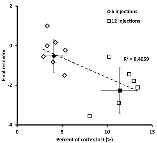
As lesion size has already been shown to influence both physiological and neuroanatomical activity in the contralesional motor cortex, Biernaskie and colleagues (2005) looked into the interaction of these factors with behavior. After inducing stroke in the rat and letting the animals recover, the contralesional motor cortex was inhibited by lidocaine right before the test of the performance of the paretic hand. They found that the inhibition of the contralesional motor cortex in the rats with larger lesions resulted in significantly greater deficits of the paretic hand than in the rats with smaller lesions. These results suggest that the contralesional motor cortex contributes more to the functional recovery of the paretic limb after a large lesion, than after a small lesion.
In the ipsilesional hemisphere, the physiological reorganization of areas distant from the lesion has been found to be affected by the size of lesion. Using motor maps obtained with ICMS the authors found that lesions that destroyed less than 30% of the hand representation of M1 caused a contraction in the hand area of PMv. In contrast lesions almost completely destroying the hand area of M1 caused a 50% expansion of the hand area of PMv (Frost et al. 2003; Dancause et al. 2006;
20
Dancause et al. 2005) . While the monkeys with small lesions recovered within three weeks, the monkeys with large lesions still had mild behavioral deficits 5 months after stroke induction. The authors proposed that the capability of the hand area of M1 to reorganize was exhausted by large lesions. In these cases, PMv, a premotor area heavily interconnected with M1 and with its own corticospinal projections, underwent expansion of its hand area to support recovery.
Similarly if a lesion is large enough to eliminate the capacity of the ipsilesional cortex to reorganize, the contralesional motor cortex would then undergo adaptive reorganization to contribute to the recovery of the paretic hand. Summarising all the evidence presented above I propose that a lesion in the motor cortex will trigger a reorganization in the contralesional motor cortex. However the functional outcome of this reorganization and the observable physiological and anatomical changes will be influenced by the volume of the lesion.
1.4.2 Rationale for the set of experiments conducted in the present study
We wanted to investigate how lesions of different sizes in the motor cortex influence cortical reorganization in the contralesional motor cortex. Currently there are only two studies which
examined the effect of lesion size on physiological reorganization in the contralesional hemisphere. The first one by Dijkhuizen and colleagues (2003) was discussed previously. Unfortunately the resolution of fMRI in rodents does not allow the separation of the rat motor cortex into rostral (RFA) and caudal forelimb regions (CFA), which are suspected to play different roles in motor control (Rouiller, Moret, and Liang 1993) and thus might play different roles in stroke recovery. Additionally an increase in hemodynamic activity does not actually reveal what sort of reorganization is taking place.
The only other study which looked at the effect of lesion size on contralesional reorganization was done by Gonzalez and colleagues (2004) using ICMS, a well-established technique that allows us
21
to examine cortical organization within each motor cortical region at high resolution. In this study unilateral stroke was induced in the sensorimotor cortex in rats with one of two methods:
devascularisation of surface vessels or electrocoagulation of the middle cerebral artery (MCA). The strokes caused by MCAo were larger and more lateral when compared to strokes resulting from the devascularisation of the surface vessels. The authors did not find any effect of lesion size on the contralesional motor cortex. However, in this study not only size, but also lesion location varied between the two groups. Indeed, due to difference in rodent vascular anatomy, MCAo routinely leaves the motor cortex intact (Gharbawie et al. 2005). Thus, it is yet not clear what would be the effect of lesion of different sizes in M1 on the reorganization of the contralesional motor areas.
Our objective was to evaluate the effect of lesion size in the CFA of rats on cortical reorganization of both hemispheres and behavioral recovery of the paretic hand. We predict that lesions of different sizes should result in different reorganization patterns in the contralesional motor cortex. Our results will further the understanding of the physiological reorganization following ischemic stroke. In particular, as discussed in the sections above while the role of the ipsilesional motor cortex in functional recovery has been an area of active research, there is a current gap in understanding how the contralesional motor cortex contributes to recovery. Furthermore, there are clinical interventions that are currently being designed that rely on untested assumptions of how the ipsi and contralesional motor cortices interact. As a consequence, this study seeks to contribute to closing this gap and provides a better understanding of the processes that take place in the
contralesional hemisphere following stroke and their relation to the functional recovery of the paretic limb.
22
Chapter 2
The effect of lesion size on cortical reorganization in the ipsi and
contralesional hemispheres
Manuscript prepared for submission to Neurorehabilitation and Neural Repair
Authors: Touvykine B, Mansoori BK, Jean-Charles L, Deffeyes J, Quessy S, Dancause N
Introduction
Cortical lesions, such as may occur following stroke, trigger plasticity in diverse, distant
regions of the brain that are spared from the injury. In humans, corticospinal tract disruption
is a good predictor of motor impairments (Schaechter, et al., 2009, Stinear, et al., 2007,
Ward, et al., 2006). In addition, patients with greater deficits show more activation in diverse
areas of the ipsi and contralesional cortex during movement of the paretic limb (Cramer, et
al., 1997, Ward, et al., 2007, Ward, et al., 2006).
In animal studies, comparable effects of lesion size have been reported. Following
middle cerebral artery occlusions (MCAo) in rats, the reorganization of the pattern of
hemodynamic activity (Dijkhuizen, et al., 2003) and of the functional and structural
connectivity of the contralesional hemisphere (van Meer, et al., 2012) are more pronounced
in animals with larger lesions. Many neuroanatomical changes are also known to occur in the
contralesional hemisphere (Adkins, et al., 2004, Biernaskie and Corbett, 2001, Jones and
23
Schallert, 1992, Stroemer, et al., 1995) and are affected by the extent of injury (Hsu and
Jones, 2006, Kim and Jones, 2010). Reorganization of cortical motor representations, or
motor maps, in the ipsilesional hemisphere is also affected by the size of injury (Dancause, et
al., 2006, Frost, et al., 2003). Altogether, these data support that the size of lesion has
substantial effects on postlesion plasticity and recovery.
To date, the effect of lesion size on the reorganization of motor representations in the
contralesional cortex have not been studied. Moreover, there has been no complete
documentation of how the volume of the lesion affects the organization of cortical motor
maps in the two hemispheres. In the present study, our objective was to evaluate the effect
of cortical lesion size on the organization of motor areas of the ipsi and contralesional
hemispheres. In a rat model, we induced cortical lesions of different size in the caudal
forelimb area (CFA), the rodent equivalent of the primate primary motor cortex (M1) and the
main source of corticospinal neurons in adult rats (Brosamle and Schwab, 1997, Miller, 1987).
Following recovery, we used intracortical microstimulation techniques (ICMS) to study the
organization of motor representations in both hemispheres.
24
Methods
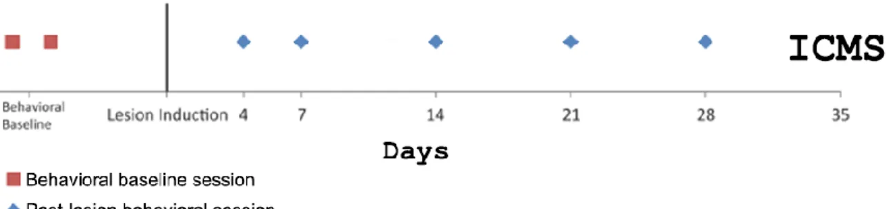
18 Sprague-Dawley rats of approximately 3 months of age weighing from 250g to 300g were
used for the study (Charles River Laboratories, Montreal, Québec, Canada). All animals were
housed separately in a reversed day-night light cycle and were only handled in the dark,
under red light. Animals were randomly assigned to one of three groups, controls (n= 5), a
‘small’ (Group
small; n=7) or a ‘large’ (Group
large; n=6) cortical lesion group. Animals in the
Group
smalland Group
largewere familiarized with banana flavored food pellets in the Montoya
Staircase task (Biernaskie and Corbett, 2001, Montoya, et al., 1991) for 10 work-days. Testing
chamber was made out of Plexiglas (6-cm wide, 12-cm high and 30cm long) with a central
platform (2.3-cm wide, 6-cm high and 19-cm long) which separates right and left forelimbs
(Biernaskie and Corbett 2001; Montoya et al. 1991). Prior to lesion induction animals were
familiarized with the task. Familiarization consisted of two sessions of Montoya staircase,
one in the morning and one in the afternoon. In a session a rat had 4 three-minute trials with
each hand (8 trials per day in totals). Number of pellets eaten per trial was established at the
end of three minutes, and all 7 wells refiled for the next trial (one pellet per well). On the last
two days of the familiarization period, the performance in terms of the number of eaten
pellets was recorded and used to establish if the animal reached our inclusion criteria. To be
included in the study, rats needed to eat 4 out of 7 pellets in 3 of the 4 trials on both days
with both forepaws. Each forepaw was testing separately (i.e. 4 three-minute sessions with
the right hand, then 4 three-minute sessions with the left hand and vice-versa). Prior to the
lesion, grasping performance of both forelimbs in the Montoya Staircase task was collected
25
on the 9
thand 10
thdays and averaged to establish a baseline performance. Following the
lesion, behavior was reevaluated twice in the first week and then once per week for the
three following weeks. At the end of this recovery period, motor mapping was conducted
(Figure 1). In control animals, the mapping procedures were done after 5 weeks of being
single housed in our facility. Controls did not undergo the familiarization period, as this was
showed to have no effect on motor maps (Barbay, et al., 2013). The familiarization and
behavioral data collection procedures have been described in detail previously (Mansoori, et
al., in revision).
Behavioral recovery was calculated using the following formula:
Our experimental protocol followed the guidelines of the Canadian Council on Animal Care
and was approved by the Comité de Déontologie de l'Expérimentation sur les Animaux of the
Université de Montréal.
Lesion induction surgery
Lesion surgeries were done aseptically. Animals were fixed in a stereotaxic frame in a prone
position. Anesthesia was induced with ketamine hydrochloride (80mg/kg; ip) and sustained
with ~2% isoflorane and 100% oxygen. The temperature was monitored and maintained
between 35.5°C and 36.0°C by a self-regulating heating mat (Harvard Apparatus, Holliston,
26
MA). The oxygen saturation was also monitored throughout the procedures (
Nellcor Puritan
Bennett, Model NPB-190, Mansfield, MA
). In both Group
smalland Group
large, lesions targeted
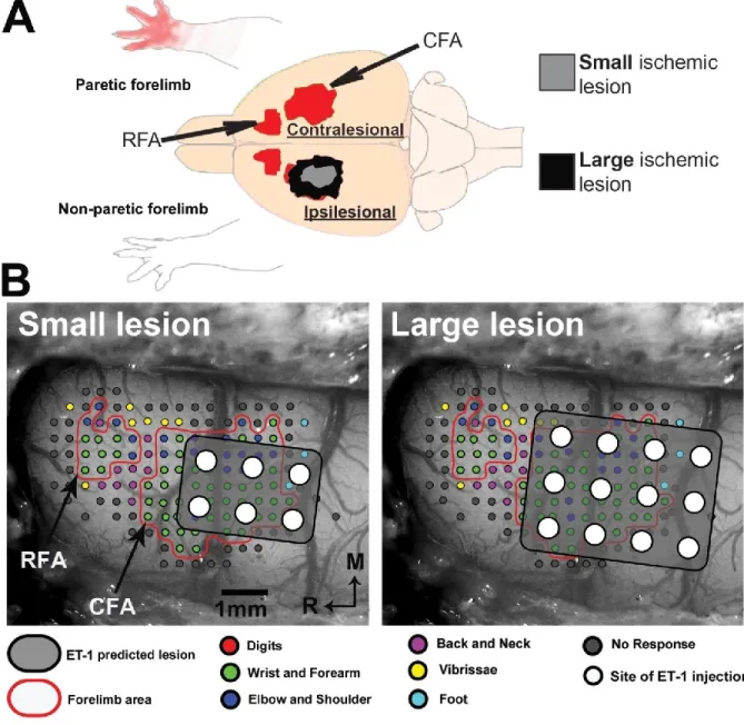
the CFA based on stereotaxic coordinates (Fang, et al., 2010, Mansoori, et al., in revision)
(Figure 2). For Group
small, six 0.7mm diameter holes were drilled through the skull (+1.5, +0.5,
-0.5mm anteroposterior, +2.5, +3.5mm mediolateral to bregma). In each hole, a Hamilton
syringe (Hamilton Company, Reno, Nevada, United States) was lowered at a depth of -1.5mm
in the cortex to inject 330nL of endothelin-1 (ET-1) (EMD chemicals, San Diego, CA, USA;
0.3µg/µL in saline) at a rate of 3nL/s with a microinjector (Harvard apparatus, Holliston, MA).
For Group
large, ET-1 was injected in a similar manner in twelve holes (+2.0, +1.0, 0.0, -1.0mm
anteroposterior, +2.0, +3.0, +4.0 mediolateral to bregma), doubling the area of targeted
cortex in the CFA. Our lesion protocol was specifically designed to increase the area of the
cortical gray matter damaged in Group
large, without damaging subcortical structures, which
occurs following ET-1 injections of bigger volumes (Biernaskie, et al., 2005, Hsu and Jones,
2006, Kim and Jones, 2010). Upon completion of injections, the holes in the skull were sealed
with bone wax and the skin sutured. After the surgery, animals received a regimen of pain,
anti-inflammatory and antibiotics medication and their recovery was closely followed for 48
hours.
Electrophysiological mapping surgery
Five weeks after the lesion, in a terminal acute experiment, ICMS techniques were used to
obtain cortical motor maps of forelimb movements in both hemispheres. A first craniotomy
27
and durectomy exposed the brain of the contralesional (CL) hemisphere under isoflurane
anesthesia. Mineral oil was applied over the opening to protect the cortex. A digital
photograph of the exposed brain was exported to Canvas 11 software (Seattle, Washington,
USA). A grid with a resolution of 0.333mm was overlaid onto the photograph and was used to
guide the electrode penetrations to generate the motor map (333µm interpenetration
distance). As it is impossible to evoke any motor response with cortical stimulation under
isoflurane, anesthesia was switched to ketamine hydrochloride (~10mg/kg/10 minutes;
intraperitoneal) for the collection of electrophysiological data. A glass insulated tungsten
microelectrode (~1.0 MΩ; FHC Bowdoin, ME USA) was lowered into the cortex to a depth of
1600 μm targeting cortical layer 5 using a microdrive (David Kopf Instruments
Model 2662,
Tujunga, CA
). Each stimulation train consisted of 13 monophasic square pulses (0.2ms
duration and 3.3ms interpulse interval) generated by a Master-8 stimulus generator (A.M.P.I.
Jerusalem, Israel). ICMS trains were delivered at 1Hz with a constant current stimulus isolator
(Bak Electronics, Model BSI-2, Sanford, FL, USA). At each stimulation site, the movement
evoked at threshold current intensity, defined as the current at which movements were
evoked by 50% of the stimulation trains, was used for subsequent analyses. If no movement
was evoked at a maximum current intensity of 100 μA, the site was qualified as
unresponsive.
Evoked movements were divided in three categories: distal forelimb, proximal
forelimb or other.
Movements of digits, wrist and forearm were included in the
distal
forelimb
and movements of the elbow and shoulder were included in
the proximal forelimb
representation (Dancause, et al., 2006, Kleim, et al., 1998, Nudo, et al., 1992)
. Movements of
the neck, back, vibrissae, hindlimb or non-responsive sites defined the borders of the CFA
28
and rostral forelimb area (RFA; rodents putative equivalent of a primate premotor area
(Rouiller, et al., 1993)). Following completion of the contralesional motor maps, the animal
was put back on isoflorane anesthesia and a second craniotomy exposed the ipsilesional
cortex. Similar ICMS mapping techniques were used to define motor areas in this
hemisphere. In some cases, due to complications during the experiment, the motor mapping
was limited to the contralesional hemisphere and was immediately followed by perfusion
(see results).
During mapping procedures, a small circle with a color specific to the movement
category was overlaid onto the image of the cortex in Canvas at each penetration site. At the
end of data collection, the digital image with color circles was used for analysis of the surface
area of each movement category. This analysis was performed with a custom-made program
in Matlab (MathWorks, MA, USA). The algorithm used nearest neighbor interpolation
between penetration points to assign each pixel to a movement category. Dimensions of
pixels were scaled according to a ruler placed on the brain in the digital picture of the cortex.
The total number of pixels with the same movement color was multiplied by the scaling
factor to obtain the cortical surface area of distal and proximal forelimb representations. The
distinction between pixels in the in the CFA and RFA was made using a k-means cluster
analysis of the distal forelimb representations. Surface areas for distal and proximal forelimb
representations in rats that recovered from small and large lesions were compared to each
other and to control, naïve rats.
29
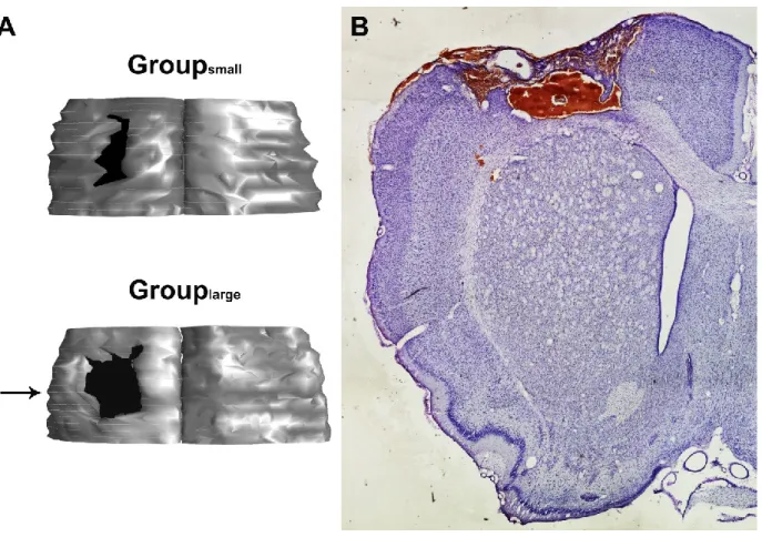
Histology
Upon completion of the electrophysiological data collection the animal was given a lethal
dose of sodium pentobarbital. It was transcardially perfused with heparinized saline solution
(1% NaCl in H
20; 0.2% heparine; total volume = 500ml), followed with a 4%
paraformaldehyde in 0.1M phosphate buffer saline (PBS) (total volume = 500ml). The brain
was extracted and cryoprotected with a 20% sucrose, 4% paraformaldehyde 0.1M PBS
solution overnight. It was then transferred to 20% sucrose, 2% dimethyl sulfoxide 0.1M PBS
for 2 hours and then in 20% sucrose 0.1M PBS for 48 hours. The brains were frozen and cut
coronally with a cryostat (40um thickness). One out of six sections were Nissl stained and
reconstructed using Neurolucida (MicroBrightField, Colchester, VT, USA). Reconstructed
sections were used to calculate the lesion extent with Neuroexplorer (MicroBrightField,
Colchester, VT, USA). Lesion volume was obtained by subtracting the volume of the
ipsilesional cortex to the volume of the contralesional cortex. The volume was then
transformed to percentage using the contralesional hemisphere according to the following
formula (Mansoori et al 2013):
Statistical Analysis
Statistical analyses of behavioral data were carried out with SigmaPlot Version 11 (Systat
Software, San Jose, CA). Repeated measure ANOVA was conducted using lesion size group,
30