Exp Brain Res (2006) 173: 318–321 DOI 10.1007/s00221-006-0500-0
R E S E A R C H A R T I C L E
Saïd Boujraf · N. Benajiba · F. Belahsen · S. Tizniti L. J. Garey
The impact of restricted diet on brain function using BOLD-fMRI
Received: 10 February 2006 / Accepted: 12 April 2006 / Published online: 19 May 2006 © Springer-Verlag 2006
Abstract We investigated the eVect of a restricted diet model on activity in the human motor cortex using func-tional magnetic resonance imaging (fMRI). Two series of blood oxygenation level-dependent (BOLD)-fMRI mea-surements were made in healthy subjects performing simple motor tasks using their right hands. The Wrst series was done 5–10 days prior to the restricted diet schedule (controls), and the second series was performed after 25–28 days of restricted diet, in the form of a reli-gious fast (Ramadan). The size and intensity of the acti-vated area in the motor cortex increased during the time of restricted diet versus the controls. We conclude that restricted diet has a signiWcant eVect on cerebral activity, as shown by BOLD-fMRI, although the exact relation-ship between the images and neuronal activity due to the restricted diet is still to be determined.
Keywords Restricted diet · Fasting · BOLD · fMRI · Motor cortex · Human
Introduction
Functional magnetic resonance imaging (fMRI) based on blood oxygenation level-dependent (BOLD) contrast mechanisms has become a powerful tool to investigate the functional organisation of human and animal brains and has been used to show functionally activated brain regions as a result of visual stimulation, as well as other paradigms (Ogawa et al.1990, 1992; Belliveau et al.
1991; Blamire et al. 1992; Kwong et al. 1992; Turner et al. 1993; Britsch et al. 1996; Kollias et al. 1996; Wild-gruber et al. 1996, 1997; Disbrow et al. 1998; Frahm et al. 2004).
Changes in the oxygenation of blood correlate with changes in neuronal activity (Raichle et al. 1994) and lead to MRI signal changes caused by alterations in the physical properties of the blood. While completely oxy-genated haemoglobin is diamagnetic, deoxyoxy-genated hae-moglobin is paramagnetic (Ogawa et al. 1990; Turner et al. 1991; Buxton et al. 1998). As the relevant properties of brain tissue are similar to those of blood, the magnetic Xux density is disrupted in the vicinity of vessels contain-ing deoxygenated blood. Haacke et al. (1997) demon-strated that as a result of diVerences associated with an increase in oxygenation state it is possible to show signal increases in brain tissue using T2*-weighted sequences. This is known as the BOLD eVect.
Diet is of major importance for general health, with particular impact on blood pressure, Xow and oxygena-tion, and tissue perfusion (Kim et al. 1993; Gati et al.
1997; Mattson et al. 2002, 2003; McGrath-Hanna et al.
2003). These parameters, which may therefore be in Xu-enced by a restricted diet, should aVect the BOLD signal, which reXects functional activity of the brain (Ogawa et al. 1990, 1992; Turner et al. 1991; Haacke et al. 1997). Nevertheless, to our knowledge no BOLD-fMRI study has been performed on the impact of nutritional behav-iour on human brain function in general and oxygena-tion of the cortex in particular. In this study, we looked for changes in the BOLD-fMRI signal representing
S. Boujraf
Biophysics and MRI Methods Department, Faculty of Medicine and Pharmacy, Fez, Morocco N. Benajiba
Nutrition and Food Department, University of Cordoba, Cordoba, Spain
F. Belahsen
Neurology Department, University Hospital of Fez, Fez, Morocco
S. Tizniti
Radiology Department, University Hospital of Fez, Fez, Morocco
L. J. Garey
Centre for Psychiatric Neuroscience, University of Lausanne, Lausanne, Switzerland
L. J. Garey (&)
Route Suisse 43, 1166 Perroy, Switzerland E-mail: [email protected]
Tel.: +41-21-8253115 Fax: +41-21-8253115
319
neural activity during a period of restricted diet (reli-gious fasting) using a simple motor paradigm.
Methods
Subjects
Fourteen healthy male right handed volunteers were recruited for this study, but only six were retained for the Wnal analysis, as several conditions had to be taken into account. To be admitted, volunteers had to have no sig-niWcant history of neurological pathology or trauma, have a consistent, equilibrated and healthy daily nutri-tional habit, as well as a generally healthy lifestyle, in terms of regular sleep and rest during the whole period of the study and had to be non-smokers. All participants were briefed on MRI scanner safety and gave consent before taking part.
Diet model
The restricted diet model used in this study was based on fasting during daytime for one month during the Muslim month of Ramadan. This involved voluntary avoidance of food and drink from sunrise to sunset, meaning a daily fast of some 14 h.
BOLD-fMRI paradigm
The paradigm consisted of a motor task involving open-ing and closopen-ing the dominant (right) hand at a frequency of 1.5 Hz, according to the scheme shown in Fig.1. The total time of each scan session was 3 min in which 3 cycles were acquired each consisting of 30 s “on” alter-nating with 30 s “oV”. The volunteers were trained out-side the scanner to keep exercising the same power level during the course of the motor task. They also rehearsed their task in the scanner before each session. Two BOLD-fMRI scan sessions were performed, the Wrst between the 5th and 10th days preceding the start of the fast and the second between days 25 and 28 of the fasting month. In each session, the same BOLD-fMRI measure-ment was done twice to control the reproducibility of the required motor task. All sessions were performed between 1530 and 1730 hours. BOLD-sensitive fMRI measurements were carried out on a 1.5 Tesla magnet (Signa, General Electric) using a single shot Gradient
Echo, Echo-Planar Imaging (EPI) sequence. The echo time was 50 ms, the repetition time was 3,750 ms and the Xip angle was 90°. Twenty axial slices covering the whole motor cortex were acquired at a slice thickness of 4 mm and a planar resolution of 2 £ 2 mm2. The activated area in each case was mainly situated in the dorsal part of the pre-central gyrus, but extending slightly to the post-cen-tral area. We refer to the activated area as simply “motor area”. Individual diVerences were compensated by the averaging process for residual signal diVerences, explained below.
Data post-processing
All data underwent identical post-processing using Sta-tistical Parametric Mapping (SPM 99; Wellcome Department of Cognitive Neurology, London, UK;
http://www.Wl.ion.ucl.ac.uk/spm; Friston et al. 1995; Matlab version 6.1, Mathworks Inc., USA). The func-tional images from each subject were analysed individu-ally. The echo-planar images were realigned using a rigid body transformation to the Wrst volume. A spatial nor-malisation routine was performed. A box-car convolved with a hemodynamic response function was used. T-sta-tistics were calculated for each voxel. Raw data were Wltered with a Gaussian Wlter of 5 mm FWHM (full width at half maximum) in all directions. SigniWcantly activated areas were shown at a threshold of P < 0.01.
Individual maximal BOLD signal changes in the motor area were calculated for each subject for each BOLD-fMRI session. Then the diVerences in maximal BOLD signal in the same area between the pre-fasting and fasting sessions were calculated for each subject. Finally the size of the activated motor area was calcu-lated for both pre-fasting and fasting sessions. Activa-tion maps were calculated and overlaid on anatomical images. A group analysis of the data was performed, and subtraction maps of the averages of both sessions were calculated.
Results
All individual results showed consistent and signiWcant increase of activity in the motor cortex during fasting. Figure2 shows the size of the activated motor area expressed in pixels (1 pixel = 2 £ 2 mm2) during pre-fasting and pre-fasting. The size of the activated area varied
Fig. 1 Motor paradigm used in
both restricted diet (fasting) and normal diet (non-fasting)
320
from 49 § 25 to 108 § 25 pixels across the six subjects prior to fasting, while during fasting it was from 114 § 16 to 157 § 16 pixels. The average increase in size was 53 pixels (P = 0.0012).
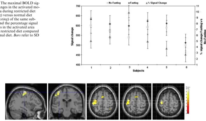
Figure3 shows the maximal BOLD signal changes in the activated motor area during fasting versus normal diet of the same subjects. All subjects showed an increase of the maximal BOLD signal during the restricted diet protocol, reXecting an increase of activation. The signal in the non-fasting sessions varied from 499 § 39 to 601 § 39 (arbitrary units) over the subjects, while during fasting it was from 513 § 43 to 633 § 43. The average
increase was 33 § 15. Although non-signiWcant, this sig-nal increase in the activated brain during the restricted diet of from 2.7 to 9.9% was consistent across subjects. Figure4 shows the residual BOLD signal changes in the activated motor area. The activation represents the diVerence between the average maps of all subjects with restricted compared to normal diet. These residual maps reXect the strength of the average activation resulting from the restricted diet versus that from the normal diet protocol.
Discussion
The size of the activated area in the motor cortex was signiWcantly increased near the end of the fasting month versus a few days prior to the start of the fast (P < 0.01). Our results also show a tendency to an increase of the intensity of the maximal BOLD signal reXecting an increase in the oxygenation level of the motor cortex.
Our study suggests that a restricted diet, in this case fasting, can have a marked eVect on brain activity. Matt-son et al. (2002, 2003) suggested that dietary restriction could improve functional deWcits in the brain and reduce the risk in major neurodegenerative disorders such as Alzheimer and Parkinson disease. Kennedy and Zochodne (2005) showed that pathological sensory and motor function may recover following spontaneous remission from diabetes with near-euglycaemia. Laijiani et al. (2003) reported a signiWcant decrease in glycaemia
Fig. 2 The size of the activated motor area (in pixels) achieved
dur-ing restricted diet (fastdur-ing) versus normal diet (non-fastdur-ing) of each subject. Bars refer to SD
Fig. 3 The maximal BOLD
sig-nal changes in the activated mo-tor area during restricted diet (fasting) versus normal diet (non-fasting) of the same sub-jects, and the percentage signal changes in the activated area during restricted diet compared to normal diet. Bars refer to SD
Fig. 4 The residual BOLD signal changes in the activated motor area. The activation shown represents the diVerence between the average
321
by the end of one month of fasting. However, increased oxygenation and activation of neurons in the motor cor-tex as recorded by BOLD require energy. In other words, if restricted diet decreases blood glucose concentration, to maintain suYcient oxygenation and energy supply to the brain cerebral blood Xow and perfusion would need to be enhanced. Thus restricted diet can have a signi W-cant eVect on cerebral activity as shown by BOLD-fMRI. However, the exact relationship between restricted diet, increased neuronal activity and possible physiological beneWt remains to be determined.
References
Belliveau JW, Kennedy DN, McKinstry RC, Buchbinder BR, WeisskoV RM, Cohen MS, Vevea JM, Brady TJ, Rosen BR (1991) Functional mapping of the human visual cortex by mag-netic resonance imaging. Science 254:716–719
Blamire AM, Ogawa S, Ugurbil K, Rothman D, McCarthy G, Eller-mann JM, Hyder F, Rattner Z, Shulman RG (1992) Dynamic mapping of the human visual cortex by high-speed magnetic res-onance imaging. Proc Natl Acad Sci USA 89:11069–11073 Britsch PM, Grodd W, Klose U, Ackermann H (1996) Functional
mapping of the motor system during voluntary movements with whole brain multislice EPI. Proc ISMRM 4:1865
Buxton RB, Wong EC, Frank LR (1998) Dynamics of blood Xow and oxygenation changes during brain activation: the balloon model. Magn Reson Med 39:855–864
Disbrow E, Buonocore M, Antognini J, Carstens E, Rowley H (1998) Somatosensory cortex: a comparison of the response to noxious thermal, mechanical, and electrical stimuli using func-tional magnetic resonance imaging. Hum Brain Map 6:150–159 Frahm J, Dechent P, Baudewig J, Merboldt KD (2004) Advances in
functional MRI of the human brain. Prog Nucl Mag Reson Spectro 44:1–32
Friston KJ, Holmes AP, Worsley KJ, Poline JP, Frith CD, Frac-kowiak RSJ (1995) Statistical parametric maps in functional imaging: a general linear approach. Hum Brain Map 2:189–210 Gati JS, Menon RS, Ugurbil K, Rutt BK (1997) Experimental
deter-mination of the BOLD Weld strength dependence in vessels and tissue. Magn Reson Med 38:296–302
Haacke EM, Lai S, Reichenbach JR, Kuppusamy K, Hoogenraad FGC, Takeichi H, Lin WL (1997) In vivo measurement of blood oxygen saturation using magnetic resonance imaging: a direct validation of the blood oxygen level-dependent concept in func-tional brain imaging. Hum Brain Map 5:341–346
Kennedy JM, Zochodne DW (2005) Experimental diabetic neurop-athy with spontaneous recovery. Is there irreparable damage? Diabetes 54:830–837
Kim SG, Ashe J, Georgopoulos AP, Merkle H, Ellermann JM, Me-non RS, Ogawa S, Ugurbil K (1993) Functional imaging of hu-man motor cortex at high magnetic Weld. J Neurophysiol 69:297– 302
Kollias SS, Valavanis A, Golay X, Boesiger P, McKinnon GC (1996) Functional magnetic resonance imaging of cortical activation. Int J Neuroradiol 2:450–472
Kwong KK, Belliveau JW, Chesler DA, Goldberg IE, WeisskoV RM, Poncelet BP, Kennedy DN, Hoppel BE, Cohen MS, Turner R et al (1992) Dynamic magnetic resonance imaging of human brain activity during primary sensory stimulation. Proc Natl Acad Sci USA 89:5675–5679
Larijani B, Zahedi F, Sanjari M, Amini MR, Jalili RB, Adibi H, Vas-sigh AR (2003) The eVect of Ramadan fasting on fasting serum glucose in healthy adults. Med J Malaysia 58:678
Mattson MP, Chan SL, Duan W (2002) ModiWcation of brain aging and neurodegenerative disorders by genes, diet, and behavior. Physiol Rev 82:637–672
Mattson MP, Duan W, Guo Z (2003) Meal size and frequency aVect neuronal plasticity and vulnerability to disease: cellular and molecular mechanisms. J Neurochem 84:417–431
McGrath-Hanna NK, Greene DM, Tavernier RJ, Bult-Ito A (2003) Diet and mental health in the arctic: is diet an important risk fac-tor for mental health in circumpolar people? Int J Circumpolar Health 62:228–241
Ogawa S, Lee T, Nayak AS, Glynn P (1990) Oxygenation sensitive contrast in magnetic resonance image of rodent brain at high magnetic Welds. Magn Reson Med 14:68–78
Ogawa S, Tank DW, Menon R, Ellermann JM, Kim SG, Merkle H, Ugurbil K (1992) Intrinsic signal changes accompanying sensory stimulation: functional brain mapping with magnetic resonance imaging. Proc Natl Acad Sci USA 89:5951–5955
Raichle ME (1994) Images of the mind: studies with modern imag-ing techniques. Ann Rev Psychol 45:333–356
Turner R, Le Bihan D, Moonen CT, Despres D, Frank J (1991) Echo-planar time course MRI of cat brain oxygenation changes. Magn Reson Med 22:159–166
Turner R, Jezzard P, Wen H, Kwong KK, Le Bihan D, ZeViro T, Balaban RS (1993) Functional mapping of the human visual cor-tex at 4 and 1.5 Tesla using deoxygenation contrast EPI. Magn Reson Med 29:277–279
Wildgruber D, Erb M, Klose U, Grodd W (1997) Sequential activa-tion of supplementary motor area and primary motor cortex during self-paced Wnger movement in human evaluated by func-tional MRI. Neurosci Lett 227:161–164
Wildgruber D, Klose U, Grodd W, Erb M, Ackermann H (1996) Dorsolateral prefrontal activation during reversal of automated word orders. Proc ISMRM 4:1866