Author contributions: V.N.: Conception and design, collection and/or assembly of data, data analysis and interpretation, manuscript writing, final approval of manuscript.; D.C.: Data analysis and interpretation, final approval of manuscript.; C.C.: Data analysis and interpretation, final approval of manuscript.; B.R.: Conception and design, final approval of manuscript, financial support, administrative support.; R.F.: Data analysis and interpretation, final approval of manuscript.; S.W.-G.: Conception and design, manuscript writing, final approval of manuscript, financial support, administrative support.
Correspondance informations: Sabine Wislet-Gendebien, PhD, GIGA – Neuroscience, University of Liège, Tour de Pathologie 2, Avenue de l'Hôpital, 1, 4000 Liège, Tel. 32 4 366 39 88, Fax 32 4 366 23 14, s.wislet@ulg.ac.be; Received July 08, 2013; accepted for publication September 27, 2013; 1066-5099/2013/$30.00/0 doi: 10.1002/stem.1579
This article has been accepted for publication and undergone full peer review but has not been through the copyediting, typesetting, pagination and proofreading process which may lead to differences between this version and the Version of Record. Please cite this article as doi:
10.1002/stem.1579
S
TEM
C
ELLS
R
EGENERATIVE
M
EDICINE
Spinal Cord Injuries – How Could Adult Mesenchymal and Neural
Crest Stem Cells Take Up the Challenge?
Virginie Neirinckx1, Dorothée Cantinieaux1, Cécile Coste1, Bernard Rogister1,2,3, Rachelle Franzen1, and Sabine Wislet-Gendebien1.
(1) Groupe Interdisciplinaire de Génoprotéomique appliquée (GIGA), Neurosciences Unit, University of Liège, Liège, Belgium.; (2) GIGA, Development, Stem Cells and Regenerative Medicine Unit, University of Liège, Liège, Belgium.; (3) Neurology Department, Centre Hospitalier Universitaire de Liège, Liège, Belgium.
Key Words. Adult mesenchymal and neural crest stem cells • cell therapy • spinal cord injury.
ABSTRACT
Since several years, adult/perinatal mesenchymal and neural crest stem cells have been widely used to help experimental animal to recover from spinal cord injury. More interestingly, recent clinical trials confirmed the beneficial effect of those stem cells, which improve functional score of patients suffering from such lesions. However, a complete understanding of the mechanisms of stem cell-induced recovery is seriously lacking. Indeed, spinal cord injuries gathered a wide range of biochemical and physiopathological events (such as inflammation, oxidative stress, axonal damage, demyelination, etc)
and the genuine healing process after cell transplantation is not sufficiently defined. This review aims to sum up recent data about cell therapy in spinal cord lesions using mesenchymal or recently identified neural crest stem cells, by describing precisely which physiopathological parameter is affected and the exact processes underlying the observed changes. Overall, although significant advances are acknowledged, it seems that further deep mechanistic investigation is needed for the development of optimized and efficient cell-based therapy protocols.
INTRODUCTION
1. Mesenchymal and neural crest stem cells - Definition and rationale for SCI treatment
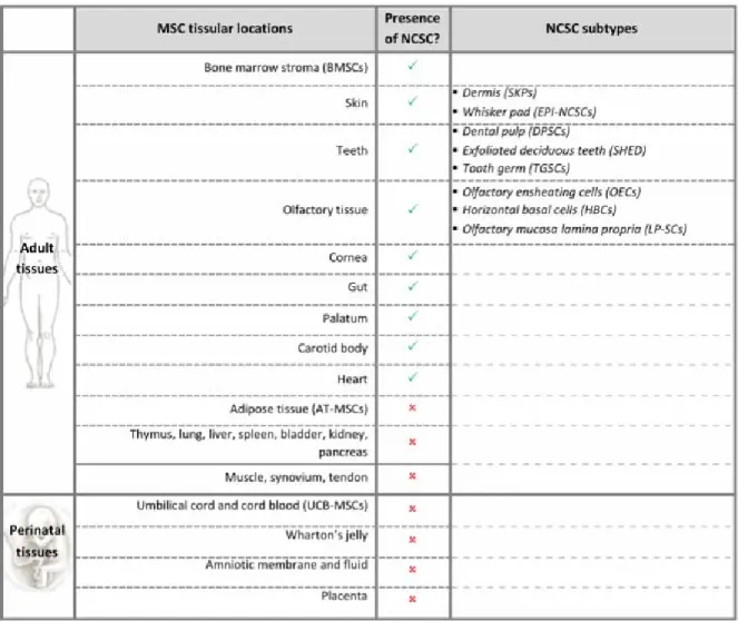
Mesenchymal stem cells (MSCs) in the adult bone marrow stroma were first identified in the last 70’s by Friedenstein et al. [1, 2] as colony-forming unit fibroblast-like cells. Those mesoderm-derived cells were subsequently described to be self-renewable and highly multipotent, giving rise to different cells of mesodermal origin such as adipocytes, chondrocytes, and osteocytes[3, 4]. In the following years, MSC were isolated from other adult tissues (ex: fat tissue [5], muscle [6],
synovium [7], peripheral blood and circulatory system [8],…), where they contribute and regulate organ physiology and homeostasis. In addition, it was shown that MSCs were also present in perinatal tissues like umbilical cord blood [9] or Wharton’s jelly [10], amniotic fluid [11] and placenta [12] (Table 1).
Bone marrow stromal cells (BMSCs) were especially considered for cell therapy in neurological lesions regarding their capacity to give rise to neural-like cells [13-15]. However,
in vivo neural differentiation is currently matter
of debate and it seems that adult BMSCs would rather help lesion recovery through many other
2 mechanisms than in-host differentiation [16, 17]. Indeed, beside the immunomodulatory effects and the secretion of several neurotrophic factors, those cells have an anti-inflammatory effect, making them attractive candidates as the most efficient treatment for SCI, so far, is a high dose of methylprednisolone (anti-inflammatory drug) [18]. Moreover, a recent clinical trial showed that BMSCs transplantation inside the cerebrospinal fluid of spinal cord-injured patients modestly enhanced motor and sensitive functions. As this work provided first clues about BMSCs relevancy for SCI treatment, the underlying ways of recovery observed in these cases were not explained [19].
More recently, neural crest stem cells (NCSCs) were identified inside the adult bone marrow [20, 21]. Those cells initially derive from neural crest, which arises at the borders of the neural tube during embryonic development of the nervous system. NCSCs then migrate towards different organs, where they differentiate to give rise to peripheral neurons and glia, melanocytes, chondrocytes, smooth muscle cells, etc. Adult NCSCs were identified in several post-natal organs [22], intermingled with MSCs inside the bone marrow [15, 20, 23, 24], skin [25-27], gut [28], teeth [29, 30], heart [31], palatum [32], cornea [33], and olfactory tissue [34-36] (Table 1).
Inside the bone marrow stroma, both MSCs and NCSCs accumulate at the bone epiphysis, and they are consequently often studied together and referred as bone marrow stromal cells or BMSCs. This fact makes BMSCs even more attractive for SCI treatment as beside the inflammatory modulation effects provided by MSCs, NCSCs may be the perfect candidate for cell replacement therapy. Indeed, due to their neural origin, neural crest cells are closely related to neural tube stem cells, which makes them in close ontological relationship with the spinal cord [37]. Furthermore, NCSC express the neural crest stem cell molecular signature genes that were initially used to create induced pluripotent stem cells (iPS) [38]. This last fact makes adult NCSCs more attractive than iPS or ES cells according to the guidelines for the
clinical translation of stem cells, published by the International Society For Stem Cell Research - ISSCR : « …maximum effort should be made to minimize the risks for all possible adverse events associated with stem cell based therapy. » Therefore, adult bone marrow NCSCs are more suitable as they are not genetically modified like iPS and are less immunogenic (similarly to BMSCs) than ES cells.
According to the last update reported by Lee et
al. [39], the global incidence of traumatic
spinal cord injuries (SCI) was estimated in 2007 at 23 cases per million worldwide. Reported SCI cases mainly concern young adults, among them 80% of men, who are for the most part victim from motor vehicle accidents and falls [40]. The cervical spine and lumbar spine are the most commonly affected regions, and regarding the level of injury, those patients could suffer from para- or tetraplegia. Besides locomotor impairments, neuropathic pain, reflexive and sensitive troubles also occur, accompanied by highly disabling social and financial issues. In the last decade, although numerous reports have shown significant improvements in medical management and clinical recuperation after SCI, there is still no effective treatment that completely allows functional recovery.
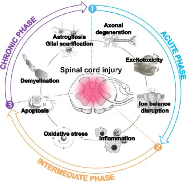
The development of such effective treatments should first be based on the full understanding of SCI physiopathological events, which are gathered in three major phases. Briefly, the first events after traumatic SCI and spinal shock, named the acute phase, encompass neuronal necrosis, and axonal disruption, blood supply default, edema and accumulation of electrolytes, calcium but also potassium and excitotoxic neurotransmitters. The intermediate phase starts a few minutes after the lesion, and lasts for several weeks. It is characterized by further ischemia, oxidative stress taking place by free-radical production and lipid peroxidation, as well as by the recruitment of neutrophils and lymphocytes which secrete cytokines and promote the development of an inflammatory environment. The chronic phase arises after few months, covering continuous alteration of ionic balance, apoptosis of
3 oligodendrocytes and demyelination, formation of cavities, and astroglial scar formation (reviewed by Oyinbo et al., in 2011 [41] and by Ronaghi et al., in 2010 [42]) (Figure 1). Altogether, those unfavorable events hamper axonal regrowth and functional recovery. Therefore, in this review, we will detail updated evidences (coming from either in vitro or in vivo pre-clinical experiments performed in the seven last years) about the different abilities of adult MSCs and NCSCs to manage neural tissue-associated oxidative stress, inflammation or apoptosis, neurodegeneration and demyelination, which could be highly relevant in spinal cord injury cases and provide new clues about the precise mechanisms that could trigger recovery. All the data gathered in this review are classified in an order which follows the temporal progression of spinal cord injuries as suggested by the most relevant studies in that field. Still we keep in mind that transplanted MSCs/NCSCs can act on different events concomitantly, soon after the injury or later.
2. Various properties of mesenchymal and neural crest stem cells and spinal cord injury treatment
2.1. Acute phase events: Axonal
degeneration, ion balance disruption and excitotoxicity.
2.1.1. Neurotrophic support by MSCs/NCSCs and protection against glutamate toxicity.
Traumatic damages to the spinal cord induce axonal interruptions that are more or less complete according to the degree of injury. Regrowth of disrupted nervous fibers are highly hampered first by the hostile environment and later on, by the astroglial scar, which significantly inhibits nerve regeneration and prevents axons to go through. Synergistic strategies could be considered in order to enhance axonal regrowth, combining neurotrophic factor supply to degenerated axons; and afterwards, degradation of extracellular matrix/glial scar and reconstitution of their myelin sheaths (see below).
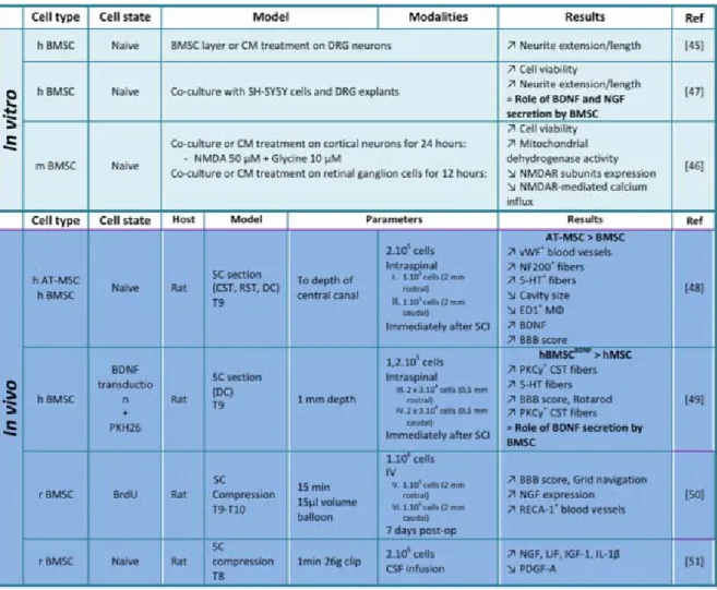
Different papers described that MSCs/NCSCs could serve as trophic mediators in diverse situations of tissue lesions [43, 44], which make their use relevant in the context of spinal cord damages. Indeed, several studies reported that MSCs/NCSCs were able to secrete neurotrophic factors that exhibit substantial effects on neuron survival and neurite outgrowth, in both in vitro and in vivo paradigms (Table 2). For instance, BMSCs were able to support and direct axonal growth of adult DRG neurons in vitro, by secreting extracellular matrix that orientate neurite extension, but especially by secreting molecules (as BMSCs-conditioned medium (BMSCs-CM) treatment generated the longest neurites) [45]. Besides, it was shown that BMSCs and BMSCs-CM (even when microvesicle-depleted) were able to protect cortical neurons against N-methyl-D-asparte (NMDA) excitotoxicity [46]. Unfortunately, no details about the precise molecules present inside the conditioned medium were provided. Crigler et al. identified those factors and showed that BMSCs secreted significant levels of brain-derived neurotrophic factor (BDNF) and nerve growth factor (NGF), thereby partially favoring neurite extension when co-cultured with neuroblastoma cells or DRG neurons [47]. The involvement of BDNF and NGF were then confirmed in animal models of SCI. Indeed, grafts of adipose tissue-derived MSCs (AT-MSCs) into the injured spinal cord of mice improved functional recovery, in correlation with increased BDNF expression and enhanced regrowth of serotonergic fibers. They also noticed that improvements were greater compared to BMSCs transplantation conditions [48]. Neuronal rescue coupled with pathological and behavioral progresses were also observed after graft of BDNF-hypersecreting-BMSCs in spinal cord-injured rats [49], suggesting a trophic role for grafted cells. NGF also seemed to be involved in SCI motor recovery and tissue sparing [50]. Finally, Hawryluk et al. showed that the expression levels of NGF, leukemia inhibitory factor (LIF), insulin-like growth factor (IGF)-1 and transforming growth factor (TGF)-β1 were significantly reduced in the rat spinal cord, three weeks after being compressed, and that
4 intrathecal transplantation of BMSCs induced an up-regulation of those trophic factors [51].
2.2. Intermediate phase events:
Inflammation, oxidative stress and apoptotic death of host cells
2.2.1. Immunomodulating properties of MSCs/NCSCs and regulation of
inflammation
The immunomodulatory properties of MSCs/NCSCs have been associated with both molecule secretions and cell-cell contact. First of all, it was shown that MSCs/NCSCs were able to suppress T-cell proliferation [52] and monocyte maturation into dendritic cells [53]. Moreover, MSCs/NCSCs impaired the functionality of dendritic cells, their antigen-presenting properties and cytokine secretion [54], and also hampered the proper function of natural killer cells and their interleukin (IL)-2 secretion [55]. Many other studies also reported immunomodulating functions of MSCs/NCSCs which were of significant importance in graft-versus-host disease cases [56], and reviewed in details by Uccelli et al. [57], Prockop et al. [58] and Singer and Caplan [59].
In the past few years, several papers also described more precisely the immunomodulative properties of MSCs/NCSCs in the particular case of SCI (Table 3). Globally, all those experimental studies reported a potent anti-inflammatory action of MSCs/NCSCs when transplanted in spinal cord injured animals.
Indeed, Seo et al. demonstrated that rats with contused spinal cord directly followed by umbilical cord blood MSCs (UCB-MSCs) intra-venous (IV) injection, presented significant improvements in motor/sensory score. These improvements were correlated with a reduction of inflammatory events, as both a decrease in IL-1β and IL-6 expression and an increase in IL-10 expression were observed. Moreover, the number of activated macrophages was reduced in those conditions [60]. Those results were consistent with previous observations from Abrams et al., showing that BMSCs transplantation decreased astrocytic reactivity and microglial activation
inside the lesioned spinal cord, associated with a reduced injury-induced response to mechanical stimuli [61].
Still, those experiments did not provide any clue on the real interaction of MSCs/NCSCs with host cells underlying these anti-inflammatory effects. Nakajima et al. showed that MSC/NCSCs transplantation into the epicenter of a spinal cord contusion was associated with a switch in host macrophage phenotype, from classically-activated (M1) to alternatively-activated (M2) [62] (which are known to have anti-inflammatory properties), and even promoted regrowth of sensory axons in a SCI model. This was also associated with an elevation of IL-4 and IL-13 levels of expression and reduction in IL-6 and tumor necrosis factor (TNF)-α levels, at the lesion site. All those events were correlated with a significant recovery of locomotor function in contused rats, as assessed by the Basso Beattie Bresnahan (BBB) scoring which appraises motor function, especially based on walk evaluation [63]. Busch et al. previously reported this M1 to M2 switch, while studying the beneficial effects of multipotent adult progenitor cells or MAPCs (a very immature BMSCs subtype) [64] on dorsal roots ganglia (DRG) dystrophic neurons, in vitro. Besides, this study also described a valuable effect of MAPCs transplantation on macrophage activation after dorsal column crush lesion, associated with enhanced axonal growth [65]. At the opposite, Hawryluk et al. demonstrated that intrathecal injection of BMSCs induced an increase in IL-1β in the epicenter of a spinal cord compression [51] which rather reveals an induction of inflammation.
2.2.2.Anti-oxidative actions of MSCs/NCSCs.
Free radical formation, lipid peroxidation and further oxidative damage events are generated during acute SCI and are tightly linked with mitochondrial dysfunctions, disruption in ion balance, glutamate-release and associated excitotoxicity. Different therapeutical strategies based on antioxidant compounds are therefore currently tested in experimental animal models and in clinical trial procedures. Unfortunately those drugs often lack in
5 selectivity and trigger side effects (reviewed by Hall et al. [66]). Very few detailed data about anti-oxidant potency of stem cells are recorded in SCI preclinical models. Still, several in vitro studies provided clues on the mechanisms involved in the protection against oxidative damage and reactive oxygen species (ROS) (Table 3).
First of all, BMSCs were reported to be able to efficiently manage in vitro-induced oxidative stress. Indeed, it was shown that cell viability was not altered by ROS [67], and that resistance to ROS was linked to glutathione availability. Therefore it would be conceivable to use BMSCs to reduce oxidative stress-induced damages, in vivo. However, other studies about UCB-MSCs showed that these cells suffered from senescence and genomic alterations once undergoing oxidative stress [68].
In vitro tests showed that stem cells could
protect different types of cells against oxidative stress. For instance, tooth-germ stem cells (TGSCs) are able to protect SH-SY5Y cells (human neuroblastoma-derived neuronal cell line) from amyloid-beta (Aβ) peptide or 6-hydroxydopamine (6-OHDA)-induced cell death in culture, by increasing the activity of antioxidant proteins such as catalase, glutathione-peroxidase (GP) or superoxide dismutase (SOD) [69]. Similarly, AT-MSCs-CM helped dermal fibroblasts to resist to free radicals in culture by inducing an enhancement of their SOD and GP activity with a decrease of apoptotic cells [70]. In the same line of evidence, Oh et al. demonstrated that the co-culture of neural stem cells (NSCs) with AT-MSCs protected NSCs against hydrogen peroxide and serum deprivation-insult, in both normoxia and hypoxia conditions (as showed by increased survival and decreased apoptosis). Likewise, they showed that the co-transplantation of NSCs and AT-MSCs enhanced the survival of grafted NSCs inside the compressed spinal cord of rats [71]. Finally, Lin et al.’s study showed that neuroglobin-expressing BMSCs grafting improved functionality after SCI in rabbits, and this was associated with a decrease in lipid peroxidation (as assessed by reduced
malondialdehyde levels) and decreased apoptotic index [72].
2.2.3.Anti-apoptotic properties of MSCs/NCSCs.
Another aspect that has been quite well-described in injured spinal cord concerns apoptotic events. Indeed, after SCI, post-traumatic tissue necrosis occurs but is followed by apoptotic events from myelinating oligodendrocytes, microglial cells and neurons [73]. Apoptosis pathways subsequent to SCI imply either Fas-dependent and/or TNF-α signalization, followed by activation of caspase cascade or JNK pathway [74, 75].
As described above, different studies showed that MSCs/NCSCs treatment are able to lessen cell apoptosis, either in a SCI model or under unfavorable culture conditions. Dasari et al. characterized more accurately the diverse anti-apoptotic actions of MSCs/NCSCs in the context of SCI (Table 3). They demonstrated the decrease of caspase-3 expression by oligodendrocytes and neurons, lowering the number of TUNEL-positive cells (TUNEL - Terminal deoxunucleotidyl dUTP Nick End Labeling – is a method to detect cells undergoing apoptosis) when an impacted spinal cord was transplanted with BMSCs. The expression of Fas was down-regulated in neural/glial cells inside the lesion site, triggering the decrease of 8, caspase-10 and caspase-3 activation [76]. Besides, BMSCs treatment up-regulated the expression of FLIP and XIAP (two inhibitors of apoptosis) and nuclear cleavage of poly [ADP-ribose] polymerase 1 (PARP-1) was also reduced. They further completed those data by evidencing the activity of UCB-MSCs on TNF-α and NF-κB-mediated apoptosis pathway [77] and the implication of the upregulation of the phosphoinositide-3-kinase (PI3K)/Akt pathway in anti-apoptotic activity of UCB-MSCs once grafted in SCI rats [78]. PI3K/Akt pathway was also activated in embryonic neurons by MSCs in vitro and was associated with a neuroprotective effect [79].
6
2.3. Chronic phase events – Demyelination of axons and glial scar formation.
2.3.1.Pro-myelinating abilities of MSCs/NCSCs.
Different in vitro evidences established that MSCs are able to promote maturation of glial precursors into myelinating cells (Table 4). Indeed, BMSCs-CM primes oligodendroglial cell fate decision of proliferating neural precursors cells in vitro (assessed by A2B5, NG2, O4, galactocerebroside [GalC] and
2',3'-Cyclic-nucleotide 3'-phosphodiesterase [CNPase] staining) by modulating Id2/Olig2
expression [80]. This was previously observed in co-cultures of BMSCs with NSCs [81] and in co-transplantation experiments with NSCs on hippocampal slices [82]. Interestingly, the same group compared BMSCs-CM to ciliary neurotrophic factor (CNTF) treatment, and showed that if CNTF only influences differentiation/maturation of neural precursor cells into oligodendrocytes, BMSCs-CM also instructs cell fate decisions and commitment [83].
Lamina propria stem cells (LP-SCs) were identified to present typical BMSCs features, but LP-SCs or LP-SCs-CM are more efficient (compared to BMSCs of BMSCs-CM) to promote in vitro myelination of dissociated spinal cord neurons [84]. Additionally, LP-SCs and LP-SCs-CM enhanced proliferation and process extension of oligodendrocyte progenitor cells (OPCs) and olfactory ensheathing cells (OECs).
Remyelination abilities of MSCs were further described in in vivo experimental models (Table 4). Neurotrophin-3 (NT3)-overexpressing BMSCs were injected in a rat model of ethidium bromide-induced spinal cord demyelination [85]. The cell implantation was coupled to strong myelin-basic protein (MBP) expression in or around the demyelinated area. NT3-BMSCs seemed to participate to remyelination, but more specifically promoted the enrollment of endogenous myelinating oligodendrocytes, hence improving significantly locomotor function. Park et al. also described enhanced oligodendrogliogenesis inside the contused
spinal cord of rats, once they were treated with UCB-MSCs [86].
Another strategy consists into differentiating MSCs/NCSCs into myelinating cells just before transplanting them inside the lesioned spinal cord. For instance, neuroprotection and motor improvements were much more drastic using skin-derived precursor (SKPs) or BMSCs previously differentiated into Schwann cells than non-predifferentiated cells [87, 88]. Indeed, results revealed that both cell types reduced the size of the contusion cavity, myelinated host axons and recruited endogenous Schwann cells. SKPs-derived Schwann cells also provided a bridge across the lesion site, increased the extent of spared tissue, promoted myelination of spared axons, reduced gliosis, and provided an environment that highly favors the axonal growth. Finally, SKPs/BMSCs-derived Schwann cells provided enhanced locomotor recovery relative to native cells. In the same way, co-cultivating BMSCs with Schwann cells improves their therapeutic effects when grafted in spinal cord-injured mice [89].
2.3.2. Degradation of extracellular matrix by MSCs/NCSCs.
Matrix metalloproteinases (MMPs) are endopeptidases that degrade extracellular matrix proteins, proteinases and membrane receptors, and play a dual role in cases of SCI. After SCI, MMPs contribute to secondary pathogenesis by promoting inflammation, inducing blood-brain-barrier disruption, degrading myelin, etc. On the other hand, it has been shown that they are able to regulate axonal guidance and regrowth by remodeling the extracellular matrix [90, 91].
UCB-MSCs upregulate the expression of MMP-2 inside a lesion, three weeks following rat spinal cord contusion [92] (Table 4). The activity of MMP2 was increased in the treated spinal cords and this was coupled to a reduction in the glial scarring, confirming the already-reported beneficial effect of MMP-2 on astrocyte reactivity, and chondroitin sulfate proteoglycans accumulation after SCI [93].
7
2.4. Pro-angiogenic abilities of MSCs/NCSCs.
Managing blood supply and revascularization is mandatory for the repair of hypoxic and inflamed environment of spinal cord lesioned tissue. The secretion of pro-angiogenic factors such as vascular endothelial growth factor (VEGF) by MSCs/NCSCs could help in this vascular remodeling (Table 5). New blood vessels and enhanced blood supply could provide valuable help in lesion recovery whatever the acute, intermediate or chronic timing,
The study of Quertainmont et al. revealed a higher number of blood vessels stained for rat endothelial cell antigen 1 (RECA-1) in the epicenter of spinal cord lesions after BMSCs graft, even if VEGF was not overexpressed in MSC-treated spinal cord extracts [50]. The same observation was reported after AT-MSCs transplantation inside acutely injured spinal cord, as assessed by the higher number of von Willebrand Factor (vWF)-positive blood vessels (which was superior compared to BMSC transplantation conditions) [48].
Oh et al. showed that AT-MSCs enhanced vasculogenesis after being transplanted in the lesion-epicenter of spinal cord injured rats [94]. Moreover, the vWF, CD31 or smooth muscle actin (SMA)-positive areas were especially higher when the AT-MSC were transplanted as a 3D-cell mass (inserted in maltose-binding protein-fibroblast-growth factor 2 surface).
The same concept of 3D-cluster was used by Zeng et al., who removed a segment of the spinal cord of rats and replaced it by a gelatin sponge scaffold containing BMSC. They observed that regenerated blood vessels crossed the junction between host spinal cord tissue and scaffold. vWF-positive area was also higher in those conditions compared with a scaffold without cells. They also showed that MSC-surrounding blood vessels expressed HIF-1α and VEGF [95].
3CONCLUSIONS
Traumatic spinal cord injuries represent a critical issue in clinical situation nowadays, and injured patients undergo severe physiological, psychological and social failures. Even if slight recovery can be achieved with intensive training programs and treatments, this recuperation is slow and limited, and depends on the degree of injury. Cell-based therapies could consequently provide additional expectations in the case of spinal cord lesions. Recent remarkable papers reviewed the interest of stem cell therapy in spinal cord injuries and other neurological diseases, from experimental models to clinical application (iPS cells and ES cells [42, 96, 97]; MSCs [98-100]; comparison of diverse types of stem cells [101-103]).
However, after considering all those papers that were somehow informative about several aspects of stem cell therapies, we wanted to dig deeper about the precise mechanisms of stem cell-associated positive effects, and more particularly stromal stem cells. Indeed, as initially mentioned, we are convinced that MSCs and NCSCs, both isolated from adult bone marrow could constitute a great tool for stem cell therapy and we will discuss their advantages thereafter.
In the past few years, several prospective clinical trials confirmed that AT-MSC or BMSC transplantation in spinal cord injured-patients did not induce tumors or any other side effects, but promoted preliminary motor and somatosensory improvements, which still have to be confirmed with further investigations in randomized trials, at a larger scale [19, 104, 105]. Nonetheless, it seems that those clinical analyses were somewhat prematurely executed. Indeed, the exact(s) way(s) by which those stem cells exert their beneficial effects are not fully understood and still need to be characterized.
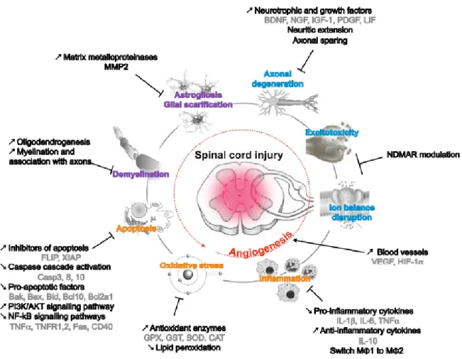
All experimental data described in this review summarized the diverse properties of adult mesenchymal or neural crest stem cells that
8 could be relevant in spinal cord injury context Still, is has to be highlighted that (Figure 2): (1) BMSCs could modulate immune response and inflammatory reaction after SCI by adjusting cytokine secretion and macrophage activation, resulting in a potent anti-inflammatory effect for the most part. This inflammatory modulation has been observed also for AT-MSCs and UCB-MSCs suggesting that these effects should be attributed to the presence of MSCs, rather than NCSCs, as no NCSC has ever been identified in adipose tissue or umbilical cord blood so far.
(2) BMSCs also protect cells against oxidative damage by increasing the expression of antioxidant proteins such as glutathione-associated enzymes, catalase and superoxide dismutase. All those neuroprotective activities are obviously associated with a decrease in apoptotic events, which appear to be mediated through different pathways (Caspases, PI3K, Akt…), and well regulated by stem cell graft. (3) Besides inducing this newly favorable lesion environment, MSCs are also able to promote axonal sparing and sprouting of new neurites, by means of neurotrophic support and efficient remyelination of fibers. The formation of new myelin sheaths can be both due to recruited endogenous glial cells and to transplanted BMSCq themselves. Interestingly, this last characteristic has only been demonstrated for BMSC or SKPs. As both populations contain NCSCs, it is tempting to suggest that this last property could be attributed to those cells.
To the light of those observations, it appears that MSCs and NCSCs could be used concomitantly: 1) MSCs could be injected in acute phasis to modulate inflammation, whereas NCSCs could then be injected in chronic phasis to improve neuronal recovery and axonal sprouting. Adult MSCs/NCSCs present enormous interest regarding SCI therapy and could efficiently regulate a multitude of phenomena occurring at the time of a lesion. Moreover, it is interesting to highlight that both cell types could be isolated
from the same patient, limiting the risk of immune reaction in case of heterologous graft. Nonetheless, we think that this wide-ranging beneficial effect of MSCs/NCSCs lacks in systematic and mechanistic explanations. Although the different molecules that are secreted by MSCs were largely studied and identified in vitro (for example in conditioned medium analyses) [106], details are missing about the genuine mechanisms by which the physiopathological and clinical recovery of experimental SCI animals are achieved.
A common criticism concerning BMSCs is the lack of reproducibility. Therefore, several points have to be taken into consideration: 1) Donor age: It appears that for human above 30 years old, MSCs have a drastically decreased secreting factors and stemness characteristics [107, 108]. It is therefore suggested to use MSCs from young donors, excluding autologous graft for oldest patients. 2) MSC and NCSC isolation protocols: since NCSCs are systematically mistaken for MSCs, new specific panels of MSC or NCSC markers should be defined. Indeed, all CD markers classically defined by the International Society for Cellular Therapy to characterize clinical grade MSCs [109] are not specific enough to discriminate NCSCs. Similarly, technical questions remain regarding the design of MSCs/NCSCs transplantation and its procedural parameters. 3) Technical questions remain regarding the design of MSCs/NCSCs transplantation and its procedural parameters. (a) The finest way to implant cells still needs to be defined. It was recently published that intracisternal transplantation of BMSCs led to the best motor recovery in rats, followed by intralesional and intravenous administration [110], while another study described the same functional improvement after intralesional or intravenous cell injection [111]. (b) The impact of immunosuppressive treatment has to be evaluated, in order to allow or prevent allografts. (c) The right timing of injection should be identified, as well as the importance of SCI kinetics on the transplantation time. As suggested above, injection of MSCs in acute phasis, then NCSCs in chronic phasis might be the right protocol. (d) Finally, the maturation
9 state of transplanted stem cells should be characterized. As several studies use stem cells under their initial state; other papers have used pre-differentiated cells in SCI models. Both neuron-differentiated and glia-differentiated cells have shown valuable benefits in experimental animals. Questions also remain concerning cell culture conditions before grafting: confluence, number of passages, etc. All those technical issues should be deeply studied in order to answer these questions and develop better strategies for cell therapy protocols.
In conclusion, as recently suggested by Vawda and Fehlings [112], the explosion in the number of studies examining the secretomes of stromal cells as well as the number of submitted patent reflect a recognition by the scientific communities of the vastly untapped potential of MSCs/NCSCs, however, evidence
on mechanistic organization and technical procedures should be provided in order to ensure efficient and accurate cell-therapy protocols in SCI patients.
ACKNOWLEDGMENTS
This work was supported by a grant from the Fonds National de la Recherche Scientifique (FNRS) of Belgium, by the Belgian League against Multiple Sclerosis associated with the Leon Frédericq Foundation and by the Fonds Spéciaux à la Recherche of the University of Liège.
4. Disclosure of potential conflicts of interest
The authors indicate no potential conflict of interest.
REFERENCES
1. Friedenstein AJ. Stromal mechanisms of bone marrow: cloning in vitro and retransplantation in vivo. Haematol Blood Transfus. 1980;25:19-29. 2. Friedenstein AJ, Deriglasova UF, Kulagina NN, et al.
Precursors for fibroblasts in different populations of hematopoietic cells as detected by the in vitro colony assay method. Exp Hematol. 1974;2:83-92.
3. Bianco P, Riminucci M, Gronthos S, et al. Bone marrow stromal stem cells: nature, biology, and potential applications. Stem Cells. 2001;19:180-192. 4. Pittenger MF, Mackay AM, Beck SC, et al.
Multilineage potential of adult human mesenchymal stem cells. Science. 1999;284:143-147.
5. Zuk PA, Zhu M, Ashjian P, et al. Human adipose tissue is a source of multipotent stem cells. Mol Biol Cell. 2002;13:4279-4295.
6. Dellavalle A, Sampaolesi M, Tonlorenzi R, et al. Pericytes of human skeletal muscle are myogenic precursors distinct from satellite cells. Nat Cell Biol. 2007;9:255-267.
7. Fan J, Varshney RR, Ren L, et al. Synovium-derived mesenchymal stem cells: a new cell source for musculoskeletal regeneration. Tissue Eng Part B Rev. 2009;15:75-86.
8. Caplan AI, Correa D. The MSC: an injury drugstore. Cell Stem Cell. 2011;9:11-15.
9. Erices A, Conget P, Minguell JJ. Mesenchymal progenitor cells in human umbilical cord blood. Br J Haematol. 2000;109:235-242.
10. Wang HS, Hung SC, Peng ST, et al. Mesenchymal stem cells in the Wharton's jelly of the human umbilical cord. Stem Cells. 2004;22:1330-1337.
11. Sessarego N, Parodi A, Podesta M, et al. Multipotent mesenchymal stromal cells from amniotic fluid: solid perspectives for clinical application. Haematologica. 2008;93:339-346.
12. Yen BL, Huang HI, Chien CC, et al. Isolation of multipotent cells from human term placenta. Stem Cells. 2005;23:3-9.
13. Sanchez-Ramos J, Song S, Cardozo-Pelaez F, et al. Adult bone marrow stromal cells differentiate into neural cells in vitro. Exp Neurol. 2000;164:247-256. 14. Wislet-Gendebien S, Hans G, Leprince P, et al.
Plasticity of cultured mesenchymal stem cells: switch from nestin-positive to excitable neuron-like phenotype. Stem Cells. 2005;23:392-402.
15. Morikawa S, Mabuchi Y, Niibe K, et al. Development of mesenchymal stem cells partially originate from the neural crest. Biochem Biophys Res Commun. 2009;379:1114-1119.
16. Prockop DJ, Oh JY. Medical therapies with adult stem/progenitor cells (MSCs): a backward journey from dramatic results in vivo to the cellular and molecular explanations. J Cell Biochem. 2012;113:1460-1469.
17. Neirinckx V, Coste C, Rogister B, et al. Concise review: adult mesenchymal stem cells, adult neural crest stem cells, and therapy of neurological pathologies: a state of play. Stem Cells Transl Med. 2013;2:284-296.
18. Bracken MB. Methylprednisolone in the management of acute spinal cord injuries. Med J Aust. 1990;153:368.
19. Karamouzian S, Nematollahi-Mahani SN, Nakhaee N, et al. Clinical safety and primary efficacy of bone marrow mesenchymal cell transplantation in
10 subacute spinal cord injured patients. Clin Neurol Neurosurg. 2012;114:935-939.
20. Wislet-Gendebien S, Laudet E, Neirinckx V, et al. Mesenchymal stem cells and neural crest stem cells from adult bone marrow: characterization of their surprising similarities and differences. Cell Mol Life Sci. 2012;69:2593-2608.
21. Neirinckx V, Marquet A, Coste C, et al. Adult Bone Marrow Neural Crest Stem Cells and Mesenchymal Stem Cells Are Not Able to Replace Lost Neurons in Acute MPTP-Lesioned Mice. PLoS One. 2013;8:e64723.
22. Achilleos A, Trainor PA. Neural crest stem cells: discovery, properties and potential for therapy. Cell Res. 2012;22:288-304.
23. Nagoshi N, Shibata S, Kubota Y, et al. Ontogeny and multipotency of neural crest-derived stem cells in mouse bone marrow, dorsal root ganglia, and whisker pad. Cell Stem Cell. 2008;2:392-403.
24. Glejzer A, Laudet E, Leprince P, et al. Wnt1 and BMP2: two factors recruiting multipotent neural crest progenitors isolated from adult bone marrow. Cell Mol Life Sci. 2011;68:2101-2114.
25. Fernandes KJ, McKenzie IA, Mill P, et al. A dermal niche for multipotent adult skin-derived precursor cells. Nat Cell Biol. 2004;6:1082-1093.
26. Toma JG, McKenzie IA, Bagli D, et al. Isolation and characterization of multipotent skin-derived precursors from human skin. Stem Cells. 2005;23:727-737.
27. Hu YF, Zhang ZJ, Sieber-Blum M. An epidermal neural crest stem cell (EPI-NCSC) molecular signature. Stem Cells. 2006;24:2692-2702.
28. Kruger GM, Mosher JT, Bixby S, et al. Neural crest stem cells persist in the adult gut but undergo changes in self-renewal, neuronal subtype potential, and factor responsiveness. Neuron. 2002;35:657-669. 29. Gronthos S, Mankani M, Brahim J, et al. Postnatal
human dental pulp stem cells (DPSCs) in vitro and in vivo. Proc Natl Acad Sci U S A. 2000;97:13625-13630.
30. Miura M, Gronthos S, Zhao M, et al. SHED: stem cells from human exfoliated deciduous teeth. Proc Natl Acad Sci U S A. 2003;100:5807-5812.
31. Tomita Y, Matsumura K, Wakamatsu Y, et al. Cardiac neural crest cells contribute to the dormant multipotent stem cell in the mammalian heart. J Cell Biol. 2005;170:1135-1146.
32. Widera D, Zander C, Heidbreder M, et al. Adult palatum as a novel source of neural crest-related stem cells. Stem Cells. 2009;27:1899-1910.
33. Yoshida S, Shimmura S, Nagoshi N, et al. Isolation of multipotent neural crest-derived stem cells from the adult mouse cornea. Stem Cells. 2006;24:2714-2722.
34. Barraud P, Seferiadis AA, Tyson LD, et al. Neural crest origin of olfactory ensheathing glia. Proc Natl Acad Sci U S A. 2010;107:21040-21045.
35. Suzuki J, Yoshizaki K, Kobayashi T, et al. Neural crest-derived horizontal basal cells as tissue stem cells in the adult olfactory epithelium. Neurosci Res. 2013;75:112-120.
36. Tome M, Lindsay SL, Riddell JS, et al. Identification of nonepithelial multipotent cells in the embryonic olfactory mucosa. Stem Cells. 2009;27:2196-2208. 37. Mujtaba T, Mayer-Proschel M, Rao MS. A common
neural progenitor for the CNS and PNS. Dev Biol. 1998;200:1-15.
38. Sieber-Blum M, Hu Y. Epidermal neural crest stem cells (EPI-NCSC) and pluripotency. Stem Cell Rev. 2008;4:256-260.
39. Lee BB, Cripps RA, Fitzharris M, et al. The global map for traumatic spinal cord injury epidemiology: update 2011, global incidence rate. Spinal Cord. 2013.
40. Spinal cord injury facts and figures at a glance. The journal of spinal cord medicine. 2012;35:480-481. 41. Oyinbo CA. Secondary injury mechanisms in
traumatic spinal cord injury: a nugget of this multiply cascade. Acta Neurobiol Exp (Wars). 2011;71:281-299.
42. Ronaghi M, Erceg S, Moreno-Manzano V, et al. Challenges of stem cell therapy for spinal cord injury: human embryonic stem cells, endogenous neural stem cells, or induced pluripotent stem cells? Stem Cells. 2010;28:93-99.
43. Caplan AI, Dennis JE. Mesenchymal stem cells as trophic mediators. J Cell Biochem. 2006;98:1076-1084.
44. Phinney DG, Hill K, Michelson C, et al. Biological activities encoded by the murine mesenchymal stem cell transcriptome provide a basis for their developmental potential and broad therapeutic efficacy. Stem Cells. 2006;24:186-198.
45. Fuhrmann T, Montzka K, Hillen LM, et al. Axon growth-promoting properties of human bone marrow mesenchymal stromal cells. Neurosci Lett. 2010;474:37-41.
46. Voulgari-Kokota A, Fairless R, Karamita M, et al. Mesenchymal stem cells protect CNS neurons against glutamate excitotoxicity by inhibiting glutamate receptor expression and function. Exp Neurol. 2012;236:161-170.
47. Crigler L, Robey RC, Asawachaicharn A, et al. Human mesenchymal stem cell subpopulations express a variety of neuro-regulatory molecules and promote neuronal cell survival and neuritogenesis. Exp Neurol. 2006;198:54-64.
48. Zhou Z, Chen Y, Zhang H, et al. Comparison of mesenchymal stromal cells from human bone marrow and adipose tissue for the treatment of spinal cord injury. Cytotherapy. 2013;15:434-448.
49. Sasaki M, Radtke C, Tan AM, et al. BDNF-hypersecreting human mesenchymal stem cells promote functional recovery, axonal sprouting, and protection of corticospinal neurons after spinal cord injury. J Neurosci. 2009;29:14932-14941.
50. Quertainmont R, Cantinieaux D, Botman O, et al. Mesenchymal Stem Cell Graft Improves Recovery after Spinal Cord Injury in Adult Rats through Neurotrophic and Pro-Angiogenic Actions. PLoS One. 2012;7:e39500.
51. Hawryluk GW, Mothe A, Wang J, et al. An in vivo characterization of trophic factor production
11 following neural precursor cell or bone marrow stromal cell transplantation for spinal cord injury. Stem Cells Dev. 2012;21:2222-2238.
52. Bartholomew A, Sturgeon C, Siatskas M, et al. Mesenchymal stem cells suppress lymphocyte proliferation in vitro and prolong skin graft survival in vivo. Exp Hematol. 2002;30:42-48.
53. Jiang XX, Zhang Y, Liu B, et al. Human mesenchymal stem cells inhibit differentiation and function of monocyte-derived dendritic cells. Blood. 2005;105:4120-4126.
54. Aggarwal S, Pittenger MF. Human mesenchymal stem cells modulate allogeneic immune cell responses. Blood. 2005;105:1815-1822.
55. Spaggiari GM, Capobianco A, Becchetti S, et al. Mesenchymal stem cell-natural killer cell interactions: evidence that activated NK cells are capable of killing MSCs, whereas MSCs can inhibit IL-2-induced NK-cell proliferation. Blood. 2006;107:1484-1490.
56. Le Blanc K, Rasmusson I, Sundberg B, et al. Treatment of severe acute graft-versus-host disease with third party haploidentical mesenchymal stem cells. Lancet. 2004;363:1439-1441.
57. Uccelli A, Moretta L, Pistoia V. Mesenchymal stem cells in health and disease. Nat Rev Immunol. 2008;8:726-736.
58. Prockop DJ, Oh JY. Mesenchymal stem/stromal cells (MSCs): role as guardians of inflammation. Mol Ther. 2012;20:14-20.
59. Singer NG, Caplan AI. Mesenchymal stem cells: mechanisms of inflammation. Annu Rev Pathol. 2011;6:457-478.
60. Seo JH, Jang IK, Kim H, et al. Early Immunomodulation by Intravenously Transplanted Mesenchymal Stem Cells Promotes Functional Recovery in Spinal Cord Injured Rats. Cell Medicine. 2011;2:55-67.
61. Abrams MB, Dominguez C, Pernold K, et al. Multipotent mesenchymal stromal cells attenuate chronic inflammation and injury-induced sensitivity to mechanical stimuli in experimental spinal cord injury. Restor Neurol Neurosci. 2009;27:307-321. 62. Kigerl KA, Gensel JC, Ankeny DP, et al.
Identification of two distinct macrophage subsets with divergent effects causing either neurotoxicity or regeneration in the injured mouse spinal cord. J Neurosci. 2009;29:13435-13444.
63. Nakajima H, Uchida K, Guerrero AR, et al. Transplantation of mesenchymal stem cells promotes an alternative pathway of macrophage activation and functional recovery after spinal cord injury. J Neurotrauma. 2012;29:1614-1625.
64. Jiang Y, Jahagirdar BN, Reinhardt RL, et al. Pluripotency of mesenchymal stem cells derived from adult marrow. Nature. 2002;418:41-49.
65. Busch SA, Hamilton JA, Horn KP, et al. Multipotent adult progenitor cells prevent macrophage-mediated axonal dieback and promote regrowth after spinal cord injury. J Neurosci. 2011;31:944-953.
66. Hall ED. Antioxidant therapies for acute spinal cord injury. Neurotherapeutics. 2011;8:152-167.
67. Valle-Prieto A, Conget PA. Human mesenchymal stem cells efficiently manage oxidative stress. Stem Cells Dev. 2010;19:1885-1893.
68. Ko E, Lee KY, Hwang DS. Human umbilical cord blood-derived mesenchymal stem cells undergo cellular senescence in response to oxidative stress. Stem Cells Dev. 2012;21:1877-1886.
69. Yalvac ME, Yarat A, Mercan D, et al. Characterization of the secretome of human tooth germ stem cells (hTGSCs) reveals neuro-protection by fine-tuning micro-environment. Brain Behav Immun. 2013.
70. Kim WS, Park BS, Kim HK, et al. Evidence supporting antioxidant action of adipose-derived stem cells: protection of human dermal fibroblasts from oxidative stress. J Dermatol Sci. 2008;49:133-142.
71. Oh JS, Kim KN, An SS, et al. Cotransplantation of mouse neural stem cells (mNSCs) with adipose tissue-derived mesenchymal stem cells improves mNSC survival in a rat spinal cord injury model. Cell Transplant. 2011;20:837-849.
72. Lin WP, Chen XW, Zhang LQ, et al. Effect of neuroglobin genetically modified bone marrow mesenchymal stem cells transplantation on spinal cord injury in rabbits. PLoS One. 2013;8:e63444. 73. Crowe MJ, Bresnahan JC, Shuman SL, et al.
Apoptosis and delayed degeneration after spinal cord injury in rats and monkeys. Nat Med. 1997;3:73-76. 74. Yu WR, Liu T, Fehlings TK, et al. Involvement of
mitochondrial signaling pathways in the mechanism of Fas-mediated apoptosis after spinal cord injury. Eur J Neurosci. 2009;29:114-131.
75. Nakahara S, Yone K, Sakou T, et al. Induction of apoptosis signal regulating kinase 1 (ASK1) after spinal cord injury in rats: possible involvement of ASK1-JNK and -p38 pathways in neuronal apoptosis. J Neuropathol Exp Neurol. 1999;58:442-450.
76. Dasari VR, Spomar DG, Cady C, et al. Mesenchymal stem cells from rat bone marrow downregulate caspase-3-mediated apoptotic pathway after spinal cord injury in rats. Neurochem Res. 2007;32:2080-2093.
77. Dasari VR, Veeravalli KK, Tsung AJ, et al. Neuronal apoptosis is inhibited by cord blood stem cells after spinal cord injury. J Neurotrauma. 2009;26:2057-2069.
78. Dasari VR, Spomar DG, Li L, et al. Umbilical cord blood stem cell mediated downregulation of fas improves functional recovery of rats after spinal cord injury. Neurochem Res. 2008;33:134-149.
79. Isele NB, Lee HS, Landshamer S, et al. Bone marrow stromal cells mediate protection through stimulation of PI3-K/Akt and MAPK signaling in neurons. Neurochem Int. 2007;50:243-250.
80. Steffenhagen C, Dechant FX, Oberbauer E, et al. Mesenchymal stem cells prime proliferating adult neural progenitors toward an oligodendrocyte fate. Stem Cells Dev. 2012;21:1838-1851.
81. Rivera FJ, Couillard-Despres S, Pedre X, et al. Mesenchymal stem cells instruct oligodendrogenic
12 fate decision on adult neural stem cells. Stem Cells. 2006;24:2209-2219.
82. Rivera FJ, Siebzehnrubl FA, Kandasamy M, et al. Mesenchymal stem cells promote oligodendroglial differentiation in hippocampal slice cultures. Cell Physiol Biochem. 2009;24:317-324.
83. Rivera FJ, Kandasamy M, Couillard-Despres S, et al. Oligodendrogenesis of adult neural progenitors: differential effects of ciliary neurotrophic factor and mesenchymal stem cell derived factors. J Neurochem. 2008;107:832-843.
84. Lindsay SL, Johnstone SA, Mountford JC, et al. Human mesenchymal stem cells isolated from olfactory biopsies but not bone enhance CNS myelination in vitro. Glia. 2013;61:368-382.
85. Zhang YJ, Zhang W, Lin CG, et al. Neurotrophin-3 gene modified mesenchymal stem cells promote remyelination and functional recovery in the demyelinated spinal cord of rats. J Neurol Sci. 2012;313:64-74.
86. Park SI, Lim JY, Jeong CH, et al. Human umbilical cord blood-derived mesenchymal stem cell therapy promotes functional recovery of contused rat spinal cord through enhancement of endogenous cell proliferation and oligogenesis. J Biomed Biotechnol. 2012;2012:362473.
87. Biernaskie J, Sparling JS, Liu J, et al. Skin-derived precursors generate myelinating Schwann cells that promote remyelination and functional recovery after contusion spinal cord injury. J Neurosci. 2007;27:9545-9559.
88. Kamada T, Koda M, Dezawa M, et al. Transplantation of human bone marrow stromal cell-derived Schwann cells reduces cystic cavity and promotes functional recovery after contusion injury of adult rat spinal cord. Neuropathology. 2011;31:48-58.
89. Xu X, Geremia N, Bao F, et al. Schwann cell coculture improves the therapeutic effect of bone marrow stromal cells on recovery in spinal cord-injured mice. Cell Transplant. 2011;20:1065-1086. 90. Zhang H, Chang M, Hansen CN, et al. Role of
matrix metalloproteinases and therapeutic benefits of their inhibition in spinal cord injury. Neurotherapeutics. 2011;8:206-220.
91. Duchossoy Y, Horvat JC, Stettler O. MMP-related gelatinase activity is strongly induced in scar tissue of injured adult spinal cord and forms pathways for ingrowing neurites. Mol Cell Neurosci. 2001;17:945-956.
92. Veeravalli KK, Dasari VR, Tsung AJ, et al. Human umbilical cord blood stem cells upregulate matrix metalloproteinase-2 in rats after spinal cord injury. Neurobiol Dis. 2009;36:200-212.
93. Hsu JY, McKeon R, Goussev S, et al. Matrix metalloproteinase-2 facilitates wound healing events that promote functional recovery after spinal cord injury. J Neurosci. 2006;26:9841-9850.
94. Oh JS, Park IS, Kim KN, et al. Transplantation of an adipose stem cell cluster in a spinal cord injury. Neuroreport. 2012;23:277-282.
95. Zeng X, Zeng YS, Ma YH, et al. Bone marrow mesenchymal stem cells in a three-dimensional gelatin sponge scaffold attenuate inflammation, promote angiogenesis, and reduce cavity formation in experimental spinal cord injury. Cell Transplant. 2011;20:1881-1899.
96. Lukovic D, Moreno Manzano V, Stojkovic M, et al. Concise review: human pluripotent stem cells in the treatment of spinal cord injury. Stem Cells. 2012;30:1787-1792.
97. Nakamura M, Okano H. Cell transplantation therapies for spinal cord injury focusing on induced pluripotent stem cells. Cell Res. 2013;23:70-80. 98. Joyce N, Annett G, Wirthlin L, et al. Mesenchymal
stem cells for the treatment of neurodegenerative disease. Regen Med. 2010;5:933-946.
99. Uccelli A, Laroni A, Freedman MS. Mesenchymal stem cells for the treatment of multiple sclerosis and other neurological diseases. Lancet Neurol. 2011;10:649-656.
100. Wright KT, El Masri W, Osman A, et al.
Concise review: Bone marrow for the treatment of spinal cord injury: mechanisms and clinical applications. Stem Cells. 2011;29:169-178.
101. Mothe AJ, Tator CH. Advances in stem cell
therapy for spinal cord injury. J Clin Invest. 2012;122:3824-3834.
102. Gogel S, Gubernator M, Minger SL. Progress
and prospects: stem cells and neurological diseases. Gene Ther. 2011;18:1-6.
103. Tetzlaff W, Okon EB, Karimi-Abdolrezaee S,
et al. A systematic review of cellular transplantation therapies for spinal cord injury. J Neurotrauma. 2011;28:1611-1682.
104. Ra JC, Shin IS, Kim SH, et al. Safety of
intravenous infusion of human adipose tissue-derived mesenchymal stem cells in animals and humans. Stem Cells Dev. 2011;20:1297-1308.
105. Pal R, Venkataramana NK, Bansal A, et al. Ex
vivo-expanded autologous bone marrow-derived mesenchymal stromal cells in human spinal cord injury/paraplegia: a pilot clinical study. Cytotherapy. 2009;11:897-911.
106. Cantinieaux D, Quertainmont R, Blacher S, et
al. Conditioned medium from bone marrow-derived mesenchymal stem cells improves recovery after spinal cord injury in rats: an original strategy to avoid cell transplantation. PLoS One. 2013;8:e69515.
107. Wang J, Liao L, Wang S, et al. Cell therapy
with autologous mesenchymal stem cells-how the disease process impacts clinical considerations. Cytotherapy. 2013;15:893-904.
108. Brohlin M, Kingham PJ, Novikova LN, et al.
Aging effect on neurotrophic activity of human mesenchymal stem cells. PLoS One. 2012;7:e45052.
109. Dominici M, Le Blanc K, Mueller I, et al.
Minimal criteria for defining multipotent mesenchymal stromal cells. The International Society for Cellular Therapy position statement. Cytotherapy. 2006;8:315-317.
13
110. Shin DA, Kim JM, Kim HI, et al. Comparison
of functional and histological outcomes after intralesional, intracisternal, and intravenous transplantation of human bone marrow-derived mesenchymal stromal cells in a rat model of spinal cord injury. Acta Neurochir (Wien). 2013.
111. Kim JW, Ha KY, Molon JN, et al. Bone
marrow-derived mesenchymal stem cell
transplantation for chronic spinal cord injury in rats: comparative study between intralesional and intravenous transplantation. Spine (Phila Pa 1976). 2013;38:E1065-1074.
112. Vawda R, Fehlings MG. Mesenchymal cells in
the treatment of spinal cord injury: current & future perspectives. Curr Stem Cell Res Ther. 2013;8:25-38.
14
Figure 1. Physiopathological events occurring after spinal cord injury, contributing to the harmful
15
Figure 2. Adult MSCs/NCSCs properties and the different ways they can contribute to functional
16
Table 1: MSCs and NCSCs in adult and perinatal tissues: AT-MSCs: Adipose tissue
mesenchymal stem cells, BMSCs: bone marrow stromal cells, DPSCs: Dental pulp stem cells; EPI-NCSCs: Epidermal neural crest stem cells; HBCs: Horizontal basal cells; LP-NCSC: Lamina propria neural crest stem cells; OECs: Olfactory ensheating cells; SHED: Stem cells from human exfoliated deciduous teeth; SKPs: Skin-derived precursors; UCB-MSCs: Umbilical cord blood mesenchymal stem cells
17
Table 2: ACUTE PHASE EVENTS - Neurotrophic properties of MSCs/NCSCs, axonal regrowth and protection against excitotoxicity. 5-HT: 5-hydroxytryptamine (serotonin):
AT-MSC: Adipose tissue mesenchymal stem cells; BBB : Beattie Basso Bresnahan; BDNF : Brain-derived neurotrophic factor; BMSC : Bone marrow stromal cells; CM : Conditioned medium; CSF: Cerebrospinal fluid; CST: Corticospinal tract; DC: Dorsal column; DRG : Dorsal root ganglion; GFAP: Glial fibrillary acidic protein; h : Human; IGF: Insulin-like growth factor; IL : Interleukin; IV : intravenous; Leukemia inhibitory factor; MΦ : Macrophages; NGF: Nerve growth factor; NF : Neurofilament; NMDA: N-methyl-D-aspartate; NMDAR : NMDA receptor; PDGF: Platelet-derived growth factor; PKC: Protein kinase C; r : Rat; RECA-1: Rat endothelial cell antigen 1; RST: Rubrospinal tract; SC : Spinal cord; T: Thoracic; vWF: von Willebrand factor
18
Table 3: INTERMEDIATE PHASE EVENTS– Immunomodulation, regulation of inflammation, anti-oxidative and anti-apoptotic abilities of MSCs/NCSCs.
6-OHDA: 6-hydroxydopamine; Aβ: Amyloid peptide β; AT-MSC: Adipose tissue mesenchymal stem cells; Bad: Bcl- 2 associated death promoter; Bak: Bcl-2 homologous antagonist/killer; Bax: B-cell lymphoma 2-associated X protein; BBB : Beattie Basso Bresnahan; Bcl-…: B-cell lymphoma ... ; Bid;-: BH3 interacting-domain death agonist ; Bik: Bcl-2 interacting killer; BMSC : Bone marrow stromal cells; C: Cervical; Casp: Caspase; CAT: Catalase; CM: conditioned medium; CSF : Cerebrospinal fluid; DC: Dorsal column; DRG : Dorsal root ganglion; eGFP: enhanced green fluorescent protein; ERK: Extreacellular signal-regulated kinase; FLIP : FLICE-inhibitory protein ; g: Gerbil; GFAP: Glial fibrillary acidic protein; GPX: Gluthatione peroxidase; GST: Gluthathione-s-transferase; h : Human; Iba1: Ionized calcium-binding adapter molecule 1; IGF: Insulin-like growth factor; IL : Interleukin; iNOS: inducible nitric oxide synthase; IV : intravenous; ; LIF : Leukemia inhibitory factor; MΦ : Macrophages; MAPC : multipotent adult progenitor cells; MAPK: Mitogen-activated protein kinase; MDA : malondialdehyde ; NGF: Nerve growth factor; NSC: Neural stem cells; p53 : protein 53 ; PARP-1: Poly [ADP-ribose] polymerase 1 ; PDGF: Platelet-derived growth factor; r : Rat; SC : Spinal cord; SSEP: Somato-sensory evoked potentials; SOD: Superoxide dismutase; T: Thoracic; tbOOH: tert-butyl hydroperoxide; TGSC: Tooth germ stem cells; TNF: Tumor necrosis factor; TNFR: TNF receptor; TUNEL: Terminal deoxynucleotidyl transferase dUTP nick end labeling; UCB-MSC : Umbilical cord blood mesenchymal stem cells; WM: White matter ; XIAP: X-linked inhibitor of apoptosis protein
20
Table 4: CHRONIC PHASE EVENTS - Pro-myelinating properties and matrix-degrading actions of MSCs/NCSCs for promoting axonal regrowth.
5-HT: 5-hydroxytryptamine (serotonin); APC: Adenomatous popyposis coli; BBB : Beattie Basso Bresnahan; BMSC : Bone marrow stromal cells; CA: Cornu ammonis; Caspr: Contactin-associated protein; CM : Conditioned medium; CNPase: 2',3'-Cyclic-nucleotide 3'-phosphodiesterase; DC: Dorsal column; DG: Dentate gyrus; DRG : Dorsal root ganglion; EtBr: Ethidium bromide; GAP43: Growth-associated protein 43; GFAP: Glial fibrillary acidic protein; h : Human; Id2 : Inhibitor of DNA-binding protein 2 ; LP-MSCs: Lamina propria mesenchymal stem cells; MBP : Myelin basic protein ; MMP: matrix metalloproteinase; NF : Neurofilament; NPC: Neural precursor cells; NSC: Neural stem cells; NT-3: Neurotrophin 3; OEC: Olfactory ensheating cells; Olig2 : Oligodendrocyte transcription factor 2 ; OPC: Oligodendrocyte precursor cells; P0 : Protein 0; r : Rat; RIP : Receptor interaction protein; SC : Spinal cord; SCEP: Spinal cord evoked potentials; SKP: Skin-derived precursors; T: Thoracic; TH : Tyrosine hydroxylase; TIMP: tissue inhibitor of metalloproteinases; UCB-MSC : Umbilical cord blood mesenchymal stem cells; ≠ : predifferentiated.
22
Table 5: Pro-angiogenic abilities of MSCs/NCSCs.
AT-MSC: Adipose tissue mesenchymal stem cells; BMSC: Bone marrow stromal cells; FGF2: Fibroblast growth factor 2; h : Human; HIF: Hypoxia-inducible factor; IL : Interleukin; MΦ : Macrophages; MBP: Maltose binding protein; r: Rat, SC : Spinal cord; SMA: Smooth muscle actin; T : Thoracic; TNF: Tumor necrosis factors; VEGF: Vascular endothelial growth factor; vWF: von Willebrand factor.