FERESHTEH FANI
Genomic analysis of β-lactam resistance mechanisms in
Streptococcus pneumoniae
Thèse présentée
à la Faculté des études supérieures et postdoctorales de l‟Université Laval dans le cadre du programme de doctorat en microbiologie-immunologie
pour l‟obtention du grade de Philosophiae Doctor (Ph.D.)
FACULTÉ DE MÉDECINE UNIVERSITÉ LAVAL
QUÉBEC
2013 © Fereshteh Fani, 2013
Résumé
Streptococcus pneumoniae est le pathogène bactérien le plus important des voies
respiratoires (pneumonie, bronchite et otite moyenne) chez les adultes et les enfants. Cette bactérie est responsable d‟une morbidité et une mortalité importantes. Bien que la pénicilline présente une activité contre de nombreux isolats de S. pneumoniae, la résistance à cet antibiotique est aujourd'hui, fréquemment rencontrés à la fois à l'hôpital et dans la communauté. La résistance à la pénicilline chez Streptococcus
pneumoniae est causée par un bloc de gènes codant pour des versions altérées de
protéines liant la pénicilline (PLP). Néanmoins, S. pneumoniae a également développé des mécanismes de résistance à la pénicilline indépendants des PLPs altérées.
L'objectif principal de cette thèse était d‟utiliser des approches génomiques pour comprendre le génotype et le phénotype de résistance aux ß-lactamines chez S.
pneumoniae.
Le travail présenté dans cette thèse a indiqué que des mutations dans les PLPs ne sont pas suffisantes pour obtenir une résistance de haut niveau à la pénicilline et au céfotaxime. Cette étude a indiqué également que la sélection de la résistance à la pénicilline chez S. pneumoniae peut impliquer l‟acquisition de mutations conférant une tolérance à l‟accumulation d‟oxydants causée par les antibiotiques. Cette tolérance peut se traduire par une augmentation de la survie qui permet possiblement la sélection des déterminants majeurs de résistance tels que des mutations dans les PLPs. Dans le cas des souches cliniques résistantes à la pénicilline, nous présentons également un nouveau rôle pour une alpha-amylase cytoplasmique conférant une résistance modérée à la pénicilline en présence d'altération des PLPs. Par ailleurs, nos travaux sur la résistance au céfotaxime chez S. pneumoniae a permis la découverte de nouveaux gènes impliquées dans la résistance au céfotaxime, y compris les gènes spr1333, spr0981, spr1704 et spr1098 qui codent respectivement pour un peptidoglycan GlcNAc déacetylase, une glycosyltransférase, un transporteur ABC et une sortase. Nos travaux génomiques ont permis de découvrir de nouveaux gènes de résistance aux β-lactamines chez S. pneumoniae.
Abstract
Streptococcus pneumoniae is the most important bacterial pathogen of the
respiratory tract (pneumonitis, bronchitis and otitis media) in adults and children resulting in significant morbidity and mortality. Although penicillin shows activity against many isolates of S. pneumoniae, resistance to this antibiotic is now frequently encountered, both at the hospital and in the community. Penicillin resistance in
Streptococcus pneumoniae is mediated by a mosaic of genes encoding altered
penicillin-binding proteins (PBPs). Nonetheless, S. pneumoniae has also developed non-PBP mechanisms implicated in penicillin resistance.
The principal objective of this thesis was to use global sequencing approaches to understand ß-lactam resistance genotype and phenotype in S. pneumoniae.
The work presented in this thesis indicated that mutations in PBPs are not sufficient to achieve high level resistance to penicillin and cefotaxime. This study also indicates that the selection of resistance to penicillin in S. pneumoniae involves the acquisition of mutations conferring tolerance to the antibiotic-induced accumulation of oxidants. This tolerance can translate into an increased survival that putatively enables the selection of major resistance determinants such as mutations in PBPs. In the case of clinical isolates, we also report a new role for a cytoplasmic alpha amylase in conferring moderate resistance to penicillin in the presence of altered PBPs. Furthermore, our works on cefotaxime resistance has allowed the discovery of novel cefotaxime resistance genes in S. pneumoniae including spr1333, spr0981, spr1704 and spr1098 coding respectively for a peptidoglycan GlcNAc deacetylase, a glycosyltransferase, an ABC transporter, and a sortase were implicated in resistance to cefotaxime. Our genomic approaches were useful to discover novel β-lactam resistance genes in S.
Avant-Propos
Over the years of carrying out the project detailed in the present thesis, I had the privilege of knowing wonderful individuals in the “Centre de Recherche en Infectiologie” at CHUL, without whose invaluable help this work would have never been materialized. I would therefore like to express my deeply-felt thanks to every one of them.
First and foremost, I would like to thank my supervisor,Professor Marc Ouellette, for giving me the opportunity to initiate my PhD programme in his laboratory, and for the confidence and trust he showed me during my academic career. His academic support as well as his incredible patience had a prime role in the success of this research project. Equally valuable to my academic development were his scaffolding at the start of my study, the scientific discussions he held with me on a regular basis, and his constant availability.
I would also like to thank the external reviewers of my thesis, Dr. Jacque Corbeil and Dr. Sylvain Moineau from Université Laval, and Dr. Patrick Trieu-Cuot from “Institut Pasteur de Paris”, for taking the time and trouble to provide careful and critical examinations of my thesis. I am also grateful to the director of Microbiology-Immunology program, Dr. Sylvain Bourgoin, and to Chantal Joubert, the “Agente de gestion des etudes”, for their collaborations during the submission process.
My special thanks would go to all the members of the team working under the supervision of Professor Marc Ouellette, the so-called “Mou Team”, whom I met over the years. I deeply appreciate their regular constructive comments on my work and for their valuable esprit de corps. I would like to thank Suzanne Avoine, the “mother of lab” as she is often called by colleagues, for her friendly support in answering my technical as well as language-related questions, not to mention the pleasant atmosphere she constantly created in the lab. Many thanks to Andréanne Lupien, Hélène Gingras and Dr. Dewan Billal, the members of Streptococcus group, for their scientific comments and their pleasant and valuable friendship. Equally thankful I am to Marie-Christine Brotherton for her help with setting up and running the SILAC experiment and with analyzing the related data, to Isabelle Girard, who gave me training on how to
work with the streptococcus, and to Danielle Legaré for her academic guidance over the first months of my work in the lab. I would also like to express my special thanks to Dr. Philippe Leprohon for his critical comments on the three research reports presented in this thesis. Without a doubt, I am indebted to “Fondation de l‟Université Laval” for the financial support it provided me with over a good part of my PhD programme.
Last but definitely not the least, I would like to thank the members of my family. I appreciate all the support and encouragement I received from my mother and my sisters during my study. A very special thank would go to my dear husband, Faramarz, without whose love and sacrifices as well as his editing assistance I would not have brought this thesis to its end. Finally I would like to thank my dear and lovely son, Daniel, for his patience and constant smiles that warmed my heart on a daily basis.
Contributions
The present thesis is the result of the work carried outunder the direct supervision of professor Marc Ouellette. Structurally, it compiles, among other parts, three research articles to which the author has contributed. These articles, which make up three
different chapters in the thesis, are listed below.
1. “Whole genome sequencing of penicillin-resistant Streptococcus pneumoniae reveals mutations in penicillin-binding proteins and in a putative iron permease” was published in 2011 in Genome Biology. This article makes up chapter VII. My contribution included designing and performing the experiment as well as analyzing the data under the supervision of Dr. Marc Ouellette. The integrative plasmids used in this project were also generated by me. Dr. Philippe Leprohon and Dr. Danielle Légaré revised the manuscript and provided critical comments.
2. “Genome analysis and reconstruction of cefotaxime resistant in Streptococcus
pneumoniae” was published in 2013 in Jounnal of Antimicrobial Chemotherapy. The
article is presented in chapter VIII of the thesis. I was responsible for designing,
performing and analyzing all of the biological experiments as well as writing up of the manuscript. Professor Marc Ouellette supervised the project and Dr. Philippe Leprohon revised the manuscript and provided critical comments.
3. “Genomic analyses of DNA transformation and penicillin resistance in
Streptococcus pneumoniae clinical isolates”, presented in chapter IX of the thesis,was
submitted to journal of Antimicrobial Chemotherapy for publication. Here again, I designed, performed andanalyzed all the biological experiments and produced the manuscript. Dr. Marc Ouellette supervised the project and Dr. Philippe Leprohon provided critical comments on manuscript.
For my son, Daniel, and my husband, Faramarz, whose love, supports and encouragements provided the impetus for this work
Table des matières
Résumé ... ii
Abstract ... iii
Avant-Propos ... iv
Table des matières... viii
List of figures ... x
List of abbreviation ... xi
Chapter I. Streptococcus pneumoniae... 1
1.1 Bacteriology ... 1
1.2 Identification ... 3
1.3 Pneumococcal colonization and disease ... 3
1.4 Virulence factors ... 3 1.4.1 Surface proteins ... 5 1.4.2 Capsule ... 6 1.4.3 Pneumolysin ... 6 1.4.4 Cell wall ... 6 1.5 Treatment ... 7 1.6 Vaccination ... 7
1.6.1 Pneumococcal polysaccharide vaccine (PPV) ... 8
1.6.2 Pneumococcal conjugate vaccine (PCV) ... 8
Chapter II. Genetic diversity in Streptococcus pneumoniae ... 9
2.1 Mechanisms of DNA exchange ... 9
2.1.1 Transformation (competence) ... 10
2.1.2 Conjugation (mobile genetic elements) ... 12
2.1.3 Transduction (bacteriophage) ... 14
Chapter III. Mode of action of antibiotics and resistance mechanisms ... 15
3.1 Mechanisms of action of antimicrobial agents ... 15
3.1.1 Interference with cell-wall biosynthesis ... 15
3.1.2 Inhibition of protein synthesis ... 17
3.1.3 Interference with DNA replication and repair ... 17
3.1.4 Inhibition of a metabolic pathway ... 17
3.1.5 New paradigm for the mode of action of antibiotics ... 18
3.2 General mechanisms of antimicrobial drug resistance ... 20
3.2.1 Reduced uptake ... 21
3.2.2 Efflux of the antibiotic ... 22
3.2.3 Inactivation of the antibacterial agents ... 22
3.2.4 Modification of the target ... 23
3.2.5 Target amplification ... 23
3.2.6 Alternative pathway metabolism ... 24
3.3 Specific mechanisms of resistance in S. pneumoniae ... 24
3.3.1 Macrolides resistance ... 25
3.3.2 Fluoroquinolones (FQ) resistance ... 25
3.3.3 Linezolid resistance ... 26
3.3.5 Trimethoprim-sulfamethoxazole resistance ... 27
Chapter IV. Streptococcus pneumoniae and ß-lactam resistance ... 28
4.1 Bacterial peptidoglycan ... 28
4.1.1 Stage I of PG biosynthesis ... 30
4.1.2 Stage II of PG biosynthesis ... 31
4.1.3 Stage III of PG biosynthesis ... 31
4.2 Penicillin-binding proteins (PBPs) ... 31
4.3 ß- lactam mode of action ... 32
4.4 Resistance mechanisms ... 34
4.4.1 PBPs mechanisms ... 34
4.4.2 Non-PBPs resistance mechanisms ... 40
Chapter V. Studying antibiotic resistance by global genomic approaches... 45
5.1 Genome sequencing ... 45
5.1.1 Comparative genome sequencing (CGS) ... 46
5.1.2 Next-generation sequencing ... 48
5.2 Transcriptomic techniques ... 51
5.3 Proteomic techniques ... 53
5.3.1 Two-dimentional gel electrophoresis (2D) ... 53
5.3.2 Isobaric tagging for relative and absolute quantification (iTRAQ) ... 54
5.3.3 Stable isotope labeling by amino acids in cell culture (SILAC) ... 54
Chapter VI. Rationale, Hypothesis and Objectives ... 55
Rationale ... 55
Hypothesis ... 55
Objectives ... 56
Chapter VII. Whole genome sequencing of penicillin-resistant Streptococcus pneumoniae reveals mutations in penicillin-binding proteins and in a putative iron permease ... 58
7.1 Résumé ... 58
7.2 Article ... 60
Chapter VIII. Genome analysis and reconstruction of cefotaxime resistant in Streptococcus pneumoniae ... 97
8.1 Résumé ... 97
8.2 Article ... 98
Chapter IX. Genomic analyses of DNA transformation and penicillin resistance in Streptococcus pneumoniae clinical isolates. ... 133
9.1 Résumé ... 133
9.2 Article ... 134
Chapter X. General discussion ... 155
10.1 Strain selection ... 155
10.2 Genomic analyses of resistance ... 158
10.3 Penicillin-binding proteins (PBPs) and ß-lactam resistance ... 160
10.4 The putative iron permease and penicillin resistance ... 164
10.5 The role of non-PBPs in cefotaxime and penicillin resistance ... 166
10.6 DNA transformation at the genome scale level ... 169
Conclusion ... 171
List of figures
Figure 1.1 S. pneumoniae Gram-stain
Figure 1.2 Infections caused by S. pneumoniae
Figure 2.1 Schematic representation of competence regulation in S. pneumoniae Figure 2.2 A DNA uptake machine
Figure 2.3 The maps of pFF3 series plasmids Figure 3.1 Structure of antibacterial drugs
Figure 3.2 Model for the common mechanism of killing by bactericidal drugs Figure 3.3 Mechanisms of genetic resistance to antimicrobial agents
Figure 4.1 Architecture of bacterial peptidoglycan
Figure 4.2 Schematic representation of bacterial peptidoglycan biosynthesis pathway Figure 4.3 Schematic structure of ß-lactam drugs (penicillins, cephalosporins) Figure 4.4 PBPs profile of S. pneumoniae
Figure 4.5 Crystal structure of PBP2x of S. pneumoniae R6
Figure 4.6 Site of mutations in PBP 2x, 2b and 1a found in altered affinity to ß-lactam Figure 5.1 CGS protocol illustration
Figure 5.2 GS-FLX system
Figure 5.3 Illumina genome analyzer sequencing
Figure 5.4 Schematic presentation of microarray technology
Figure 10.1 Whole genome transformation and resistance reconstruction in S.
pneumoniae.
List of abbreviation
ABC ATP-binding cassette AMP Adenosine monophosphate CBP Choline-binding protein CDM Chemically defined medium
cDNA Complementary deoxyribonucleic acid CGS Comparative genome sequencing
CIP Ciprofloxacin
CSP Competence stimulating peptide
CTX Cefotaxime
DAP Diaminopimelic acid
DGH Distributed genome hypothesis DHFR Dihydrofolate reductase
DNA Deoxyribonucleic acid
dNTP Deoxyribonucleotide triphosphate dsDNA Double stranded deoxyribonucleic acid
EF Elongation factor
FQ Fluoroquinolone
GAPDH Glyceraldehyde-3- phosphate dehydrogenase GT Glycosyltransferase
HMM High molecular mass HSP Heat shock protein
IPD Invasive pneumococcal disease
iTRAC Isobaric tagging for relative and absolute quantification
LMM Low molecular mass
LZD Linezolid
MIC Minimum inhibitory concentration mRNA Messenger ribonucleic acid
MRSA Meticillin-resistant staph aureus
NCSP Non-classical surface protein PBP Penicillin-binding protein PCR Polymerase chain reaction PCV Pneumococcal conjugate vaccine
PG Penicillin G
PGN Peptidoglycan
PNSP Penicillin non-susceptible streptococcus pneumoniae PPV Pneumococcal polysaccharide vaccine
PRS Penicillin-resistant streptococci
QRDR Quinolone-resistance determining regions RNA Ribonucleic acid
ROS Reactive oxygen species RPR Ribosomal protection proteins rRNA Ribosomal ribonucleic acid
SILAC Stable isotope labeling of amino acids in cell culture SNP Single-nucleotide polymorphism
ssDNA single stranded deoxyribonucleic acid
TP Transpeptidase
VRE Vancomycin-resistant enterococci WGS Whole genome sequencing
WHO World health organization
Chapter I. Streptococcus pneumoniae
Streptococcus pneumoniae is a Gram-positive coccus of the order Lactobacillales,
family Streptococcaeceae and genus Streptococcus. Streptococcus pneumoniae (the pneumococcus) belongs to the species of streptococci which include other pathogenic bacteria such as Streptococcus pyogenes, Streptococcus agalactiae, Streptococcus suis,
Streptococcus uberis, Streptococcus bovis and Streptococcus mutats. The taxonomy of S. pneumoniae (the pneumococcus) has evolved since separate descriptions in the
saliva by Pasteur (Pasteur, 1881) and as Micrococcus lanceolatus by Sternberg (Sternberg, 1881). In 1903 Schottmuller described Streptococcus mucosus (Schottmuller, 1903) but within a couple of years it was reclassified as pneumococcus (a term used since 1886 (Fraenkel, 1886)) on the basis of its biochemistry and specific agglutination and agglutinin absorption tests despite its markedly different mucoid morphology on solid agar. From 1926 the genus Diplococcus was used until 1974 when it was changed to Streptococcus pneumoniae.
1.1 Bacteriology
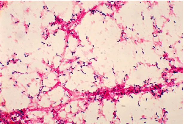
Streptococcus pneumoniae cells are Gram-positive; lancet shapes cocci (Figure
1.1). They are seen as a pair of cocci (diplococci), single cell or in short chains under microscopic examination. Streptococcus pneumoniae are α-hemolytic (partially hemolytic), so a green zone surrounds S. pneumoniae colonies on blood agar plates. Individual cells are between 0.5 and 1.25 micrometers in diameter. They do not form spore and they are not motile.
Streptococcus pneumoniae is a fastidious bacterium and it grows best in 5% CO2.
Nearly 20% of clinical isolates require fully anaerobic condition. In all cases, growth requires a source of catalase (e.g. blood) to neutralize the large amount of hydrogen peroxide produced by the bacteria. On blood agar, colonies characteristically produce a zone of alpha (green) hemolysis, which differentiates S. pneumoniae from group A
(beta haemolytic) streptococci but not from commensal alpha haemolytic (viridans) streptococci which are co-inhabitants of the upper respiratory tract.
Morphology of colonies varies and depends on the degree of encapsulation of
Streptococcus pneumoniae. The heavily capsulated strains create large colonies with
several millimeters in diameter, which look grey and very mucoid, while less capsulated strains usually form smaller colonies.
Figure 1.1. Streptococcus pneumoniae Gram-stain. Gram-stain of blood broth culture (Taken from Todar‟s online textbook, http://www.textbookofbacteriology.net)
1.2 Identification
Streptococcus pneumoniae can be identified and distinguished from other streptococci by its production of alpha hemolysis on blood agar, bile solubility, inhibition by optochin (ethyl hydrocupereine) and catalase negativity (MUSHER 2005). One or more of these tests may be inconclusive and further phenotypic tests such as agglutination with anti-pneumococcal polysaccharide capsule antibodies or genotypic tests can be required to distinguish the isolates from closely related oral streptococci (Whatmore et al. 2000; Hanage et al. 2005) .
1.3 Pneumococcal colonization and disease
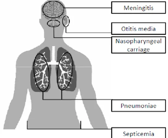
The human nasopharynx is a major ecological niche of S. pneumoniae (Figure 1.2). The nasopharyngeal carriage is higher among children mainly during the first years of life (Driver 2012b). However, S. pneumoniae remains as a leading cause of morbidity and mortality among the young children in developing country throughout the world. The World Health Organization (WHO) estimated that 10.6 million children less than 5 years of age present with pneumococcal disease every year and that 1.4 million children die due to pneumonia (WHO, 2011).
When S. pneumoniae spread from nasopharynx to lower respiratory tract or other sterile sites like blood, it may cause invasive pneumococcal diseases (IPD) such as bacteremic pneumoniae, meningitis, otitis media and bacteremia (Lynch and Zhanel 2010) (Figure 1.2).
1.4 Virulence factors
The pathogenicity of the pneumococcus has been linked mainly to its surface proteins. The capsule has a protective role and allows the pneumococcus to evade phagocytosis by immune cells. Other factors, including the cell wall polysaccharide
and pneumolysin are involved mainly in the inflammation caused by infection (AlonsoDeVelasco et al. 1995).
Figure 1.2. Infections caused by S. pneumoniae. All pneumococcal diseases are preceded by nasopharyngeal carriage, after which the bacterium can spread to the middle ear causing otitis media or disseminate to the lungs causing pneumonia. During infection of the lungs, bacteria may penetrate into the blood circulation, from which they can spread through the whole body. Otitis media might also cause infection of the meninges.
1.4.1 Surface proteins
There are four cluster of surface proteins in pneumococcus involved in virulence: proteins with an LPxTG motif (13 in R6 and 19 in TIGR4), the lipoproteins (42 in R6 and 47 in TIGR4), the choline-binding-protein (CBP) family (10 in R6 and 15 in TIGR4) and non-classical surface proteins that lack a classical leader peptide and membrane anchoring motifs have been identified on the pneumococcal surface (Bergmann and Hammerschmidt 2006; Perez-Dorado et al. 2012). A few examples of each category with major roles in colonization and virulence are discussed below: 1.4.1.1 LPxTG containing proteins
LPxTG proteins are covalently linked to the peptidoglycan after cleavage of their LPxTG motif by a sortase (Paterson and Mitchell 2006). The two genes, nanA and
nanB, that encode neuraminidase contain LPxTG anchoring motif. They cleave sialic
acid from glycoproteins, glycolipids and polysaccharides on host cell surfaces. Two zinc metalloproteases including IgA1-protease and Zmp-C that cleave mucosal IgA1 antibody and extracellular matrix respectively are two other examples of the surface proteins with an LPxTG motif (Paterson and Mitchell 2006).
1.4.1.2 Choline binding proteins (CBPs)
S. pneumoniae can produce 13 to 16 CBPs including three cell wall hydrolase that
are important for virulence including LytA, LytB and LytC. These autolysines hydrolyze murein of the cell wall (Paterson and Mitchell 2006). Interestingly autolysis induced by LytA is important for the release of major virulence factors, such as pneumolysin. CbpA, PspA and PcpA and is link to adhesion to host cells and colonization in mice.
1.4.1.3 Lipoproteins
Pneumococcal lipoproteins including PsaA, an ABC type manganese transport system, and Ami/AliAB, the oligopeptide permease have been shown to be essential for substrate transport and virulence (Kerr et al. 2004; Anderton et al. 2007).
1.4.1.4 Non classical surface proteins
Non classical surface proteins (NCSPs) are proteins lacking classical leader peptide and membrane- anchoring motifs. The enzymes glyceraldehyde-3-phosphate dehydrogenase (GAPDH) and α-enolase which are involved in carbohydrate metabolism, represent non-classical surface proteins (Hammerschmidt 2006). PavA protein is another example of NCSP which has been identified as a pneumococcal adhesion protein for fibronectin and as an important factor involved in adherence, invasion and modulation of meningeal inflammation (Holmes et al. 2001; Pracht et al. 2005).
1.4.2 Capsule
The most important virulence factor of S. pneumoniae is its capsule. A capsule composed of polysaccharide completely covers the pneumococcal cell, interfering with phagocytosis by preventing complement C3b opsonisation of the bacterial cells. During invasion the capsule is an essential determinant of virulence. Currently at least 90 different capsule types (serotypes) of pneumococci have been recognized and form the basis of antigenic serotyping of the organism (Driver 2012b).
1.4.3 Pneumolysin
Pneumolysin, which represents a cholesterol-dependent cytolysin, is another major virulence factor in the pneumococcus. Pneumolysin has multiple biological activities; it has pore-forming activity in host cell and it can trigger the immune system (i.e. complement activation and pro-inflammatory responses (Paterson and Mitchell 2006a).
1.4.4 Cell wall
The cell wall is a layer below the polysaccharide capsule and it consists of peptidoglycan, teichoic and lipoteichoic acid and phosphorylcholine. A variety of proteins with different function are anchored to this layer. Purified peptidoglycan is a
powerful stimulus for induction of inflammation similar to that seen after infection with whole pneumococci.
1.5 Treatment
Antibiotics are the mainstay for the treatment for Streptococcus pneumoniae infection. Until the 1970s, all S. pneumoniae isolates were sensitive to all commonly used antibiotics such as penicillin, macrolides, clindamycin, cephalosporin, vancomycin and trimethoprim-sulfamethoxazole. Beginning in the 1990s, many pneumococcal isolates showed decreased susceptibility to penicillin and other commonly used antibiotics. Drug resistant S. pneumoniae are now frequently encountered, both at the hospital and in the community and more than 20% of clinical isolates of S. pneumoniae in the USA are multidrug resistant (Doern et al. 2001). The three main classes of antibiotics used for the treatment of S. pneumoniae are ß-lactams, macrolides and fluoroquinolones (FQ).
1.6 Vaccination
In developed countries, serious disease occurs mainly in young children under two years of age and in the elderly. Growing resistance of S. pneumoniae to conventional antibiotics limits the treatment options, and emphasizes the urgent need for vaccines in order to prevent pneumococcal infections. Although there are more than 90 known serotypes of pneumococci, relatively few serotypes are responsible for serious diseases. Antibody to the capsular polysaccharide confers serotype specific protection against diseases. Vaccination is efficient in preventing invasive diseases (Whitney et al. 2003), but pneumococcal clones expressing capsular polysaccharides with serotypes not included in the current vaccines‟ formulations survive, and the incidence of invasive diseases induced by these clones is increasing (Hicks et al. 2007). There are two types of pneumococcal vaccines: polysaccharide and conjugated vaccines.
1.6.1 Pneumococcal polysaccharide vaccine (PPV)
The first licensed pneumococcal vaccine (pneumovax 23) in 1983 contained 23 pneumococcal serotypes associated with most infections. The main limitation of polysaccharide vaccines is that they are T-cell independent antigens so they do not stimulate a significant immune response in the infants under 2 years of age. In addition, PPV does not induce immune memory and does not have any effect on nasopharyngeal carriage (Driver 2012b).
1.6.2 Pneumococcal conjugate vaccine (PCV)
In 2000, a pneumococcal 7-valent PCV (PCV7, Prevnar) was licensed in the United States. This vaccine composed of seven different pneumococcal polysaccharides conjugated to a protein carrier that induces T-cell dependent immune response. Immunized infants would generate robust immune response and immune memory, with associated reduction in carriage (Driver 2012b). Since 2010, several expanded valency PCVs have been licensed such as 13-valent Prevnar or 10-valent with three different protein carriers (Moffitt and Malley 2011).
Chapter II. Genetic diversity in Streptococcus
pneumoniae
S. pneumoniae is an organism which exhibits a high degree of genomic diversity
and natural populations appear to preserve genetic exchange in order to adapt to their environment. Whole genome sequencing of two strains of S. pneumoniae R6 (Hoskins et al. 2001) and TIGR4 (Tettelin et al. 2001) in 2001 and comparing the genomes of these two strains was a milestone in realizing the potential extent of diversity of pneumococcal genomes. The two genomes differed in size (R6 being 2Mb and TIGR4 being 2.16Mb) and differed in 10% of their genes (Bruckner et al. 2004). The sequence comparison of 17 strains of S. pneumoniae (Hiller et al. 2007) revealed that only 46% genes were conserved among all 17 strains with each strain‟s genome containing between 21 and 32 % of noncore genes. The extensive genomic diversity data obtained from this study supports the distributed-genome hypothesis (DGH) which asserts that pathogenic bacteria possess a “supragenome” or gene pool that is much larger than the genome of any single isolate and that these pathogens generate genomic diversity by using genetic recombination and a large noncore set of genes as a resource of diversity generation (Hiller et al. 2007). Natural diversity in S. pneumoniae is generated by different mechanisms, and these will be discussed below.
2.1 Mechanisms of DNA exchange
Exchange of DNA among bacteria could occur by at least three different ways. In transformation, DNA released from one cell directly enters to another cell of the same species. In conjugation, the plasmids transfer DNA molecule from one cell to another, while in transduction, a bacterial virus (bacteriophage) picks up the DNA molecule from a cell it has infected and injected it to another cell.
2.1.1 Transformation (competence)
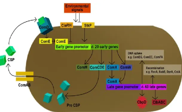
Bacterial transformation is widely distributed in nature and contributed to the adaptation and ecological diversification of several bacterial species (Johnsborg et al. 2007). Genetic transformation requires the development of competence, a genetic-programmed transient state in which the bacteria has the ability to transfer the DNA to the cytosol (Berge et al. 2002). Competence development in S. pneumoniae is carefully controlled by a comCDE operon which encodes a competence-stimulating peptide (CSP), a membrane bound histidine kinase, and a cognate response regulator (Havarstein et al. 1996; Pestova et al. 1996) (Figure 2.1). Pre-mature CSP (42-amino-acid prepeptide) encoded by comC, is processed in parallel with its export outside the cell by the comAB (ABC transporter) secretion apparatus (Hui, 1991, Havarstein, 1995). The extracellular concentration of CSP is monitored and is regulated by a two-component signal transduction system, ComDE (Stock et al. 2000) (Figure 2.1). When the concentration of CSP reaches a critical level, Com D is activated, phosphorylating its cognate regulator (ComE) leading to up-regulation of the early competence gene, including the gene encoding the alternative sigma factor ComX (Lee and Morrison 1999). ComX activates a second wave of gene expression (late competence genes), which are involved in DNA uptake and recombination process (Lee and Morrison 1999).
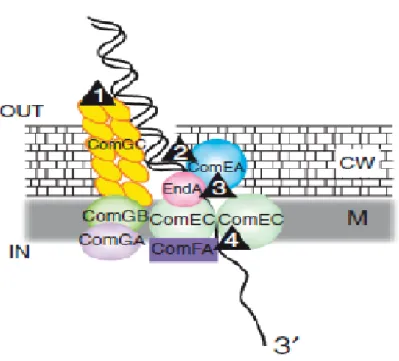
The DNA uptake machinery consists of a transformation pseudopilus, made of the major pilin like (ComGC) and minor pilin like (ComGD, ComGE, ComGG); dsDNA receptor protein (ComEA); an endonuclease EndA; a membrane channel protein for ssDNA, ComEC; and an ATP-binding protein, ComFA (Claverys et al. 2009) (Figure 2.2). All component of the DNA uptake except EndA are encoded by genes belonging to the competence regulon (Claverys et al. 2006). The initial binding of dsDNA to the cell surface of competent cells without any base sequence preference is the first step during transformation (Dubnau 1999). Then following retraction of the pseudopilus, the exogenous DNA is allowed to traverse peptidoglycan and to be in contact with ComE. Then this receptor delivers dsDNA to EndA, leading to degradation of one strand, thus allowing transport of other strand through a transmembrane channel,
ComEC. In the last step ComFA acts as an ATP-binding protein, mediating internalization of ssDNA (Claverys et al. 2009). So, uptake of exogenous dsDNA directly produces ssDNA in competent cells with an average length of 6kb in S.
pneumoniae (Claverys et al. 2009).
Internalized ssDNA can be integrated into the bacterial chromosome by RecA-dependent homologous recombination process. Two cytoplasmic products of late competence genes, an Ssb and RecA, have been involved in protecting ssDNA from nucleases hydrolysis and promoting its search for homologous partner (Berge et al. 2002).
Figure 2.1. Schematic representation of competence regulation in S. pneumoniae (Taken from (Johnsborg et al. 2007)). The CSP-precursor, which is encoded by comC gene, is processed during export by ComAB transporter, resulting in extracellular accumulation of mature CSP. Global regulators such as the serine/threonine protein kinase StkP and CiaHR two-component system regulate basal transcription of comCDE operon. CSP binds to ComD receptor, autophosphorylating ComD which subsequently transfers a phosphoryl group to the ComE response regulator. Then activated ComE, stimulates transcription of early competence genes. ComX which is part of these genes,
activates transcription of late competence genes encoding fratricide trigger factors CpbD and CibAB as well as the proteins involved in DNA
Figure 2.2. A DNA uptake machine. Uptake of exogenous dsDNA produces ssDNA in competent cells. Retracting of the pseudopilus, allowing dsDNA to traverse peptidoglycan (step1), thereby conveying dsDNA into contact with ComEA (step2), this latter delivers dsDNA to EndA (step3) to degrade one strand, then allowing transport of its complement through transmembrane channel, ComEC (step4).CW, cell wall; M, membrane.( Adapted from (Claverys et al. 2009))
2.1.2 Conjugation (mobile genetic elements)
The conjugative mechanism relies on exchange of mobile genetic elements (e.g. plasmids, conjugative transposons).
Conjugation-mediated chromosome transfer has been documented only in some limited species (e.g. Escherichia coli, Salmonella) (Claverys et al. 2009). The plasmids are not a common means by which diversity is introduced into the pneumococcal genome (Bruckner et al. 2004). The role of conjugation in pneumococcal diversity is also unclear (Bruckner et al. 2004). However, the acquisition and loss of plasmids by
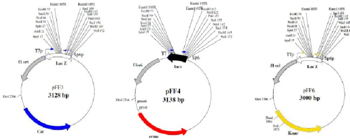
pneumococci has been demonstrated as comparison of the genome of R6 with its progenitor strain D39, shows loss of its pDP1 plasmid (Lanie et al. 2007). Nonetheless the plasmids are used as a tool for gene inactivation, over-expression of proteins in streptococci, allowing us to functionally validate the role of genes in resistance. One of the most frequently used non-replicative plasmid in S. pneumoniae is pEVP3 (Claverys et al. 1995) and it was used as a high throughput system to determine gene essentiality in S. pneumoniae (Thanassi et al. 2002). I have generated a series of plasmids, the pFF series (Figure 2.3) that can replicate in E. coli, but not in streptococci because of the absence of a streptococcal origin of replication. Once integrated, their resistance markers (chloramphenicol, Cm (pFF3); kanamycin, Km (pFF6); erythromycin, Em (pFF4) are expressed through the promoter of the S. pneumoniae amiC gene. The pFF plasmids have the advantages of being small, of high copy number (in E. coli) and in having an Eam1105I site allowing TA cloning. The low-copy replicative plasmid vector pDL289 (Buckley et al. 1995) is used to over-express proteins constitutively.
The conjugative transposable genetic elements (transposons) such as Tn916, Tn918, Tn5030 can promote, via their transposition functions encoded by excisase and integrase genes, their intracellular transposition or transfer as singlestranded DNA (ssDNA) to a recipient cell via a cell-to-cell contact (Rice 1998). As conjugative transposons frequently carry antibiotic resistance genes, e.g. tetQ on Tn5030 (Salyers et
al. 1995), tetM gene on Tn916 (Showsh and Andrews 1992) or ermB gene on Tn1545
and Tn917 (Okitsu et al. 2005), these are of great importance when considering human health threats. Moreover, Tn916 was shown to transfer even among phylogenetically distant (i.e. Gram-positive and Gram-negative) bacteria (Bertram 1991; Clewell 1995).
2.1.3 Transduction (bacteriophage)
Pneumococcal phages (pneumophages) were first isolated in 1975 from throat swabs of healthy children, by two independent groups (McDonnell et al. 1975; Tiraby et al. 1975). One year later the presence of bacteriophage has been also reported in half of pneumococci recovered from pediatric patients and one-third of isolates from adult patients (Bernheimer 1979). It has been proposed that the majority of clinical isolates of S. pneumoniae (around 75%) contain temperate bacteriophages (Ramirez et al. 1999; Romero et al. 2009) of which four have been sequenced (Lopez and Garcia 2004). While the complete genomic sequences of several pneumococcus prophages are available in GenBank, only two virulent phage have been sequenced, namely, phage Cp-1, a member of the Podoviridae family (Martin et al. 1996) and Dp-1 which belongs to the family Siphoviridae (Sabri et al. 2011). It was shown that the phages are important vehicles for the transmission of virulence genes within different species of bacterial populations (Wagner and Waldor 2002). Lytic phages appear to contribute to the natural transformation of the pneumococcus by expanding the reservoir of exogenous DNA available for incorporation into the pneumococcal genome (Ramirez et al. 1999; Lopez et al. 2000).
Chapter III. Mode of action of antibiotics and
resistance mechanisms
Each year infectious diseases kill 3.5 million people (WHO, 2012). Around the middle of the 20th century, the discovery of antimicrobial agents and other means of infection control such as improved hygiene and large scale vaccination approaches helped turn the tide and for a moment human seemed to have coped with their battle against infectious microorganisms. Regarding bacterial infection, the situation dramatically improved when penicillin became available for use in the 1940s. Almost as soon as antimicrobial drugs have been used, drug resistance appeared, however. The phenomenon of antimicrobial resistance has now reached epidemic proportion. Resistant infections are more often fatal and are responsible for prolonged illness, hence increasing the likelihood of transmission to others. It has been estimated that the annual cost of antibiotic resistance infections in the USA health care system is above $ 20 billion.
To understand why antimicrobial drugs stop to being effective, we need first to look at their mechanisms of action (Table 3.1).
3.1 Mechanisms of action of antimicrobial agents
Most antimicrobial agents (the structure of few of them is available in Figure 3.1) may be categorized according to their principal modes of action. As summarized in Table 3.1, there are 4 major mechanisms of action: (1) interference with cell-wall biosynthesis (2) inhibition of protein synthesis (3) interference with DNA replication and repair; and (4) inhibition of a metabolic pathway.
3.1.1 Interference with cell-wall biosynthesis
The peptidoglycan (PGN) is a meshwork of strands of glycan and peptide outside the plasma membrane of bacteria, forming the bacterial cell wall. This layer confers structural strength to the cell wall (Walsh 2000). Antibacterial drugs that act on cell-wall by inhibiting peptidoglycan synthesis include the ß-lactams, such as penicillins
and cephalosporins (McManus 1997). The ß-lactam agents inhibit the synthesis of bacterial cell wall by interfering with transpeptidase enzymes (also termed penicillin-binding proteins or PBPs) required for the synthesis of the peptidoglycan layer (Tenover 2006). In addition to ß-lactams, glycopeptides, including vancomycin and teicoplanin also target the peptidoglycan layer in the cell wall assembly. But rather than targeting PBPs, vancomycin binds to the D-alanyl-D-alanine peptide tail of the peptidoglycan precursor, preventing the incorporation of new PGN monomers into the cell wall and leads to cell death (Tenover 2006).
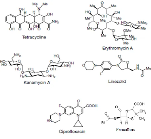
Figure 3.1. Structure of antibacterial drugs. a, Antibiotics include a set of natural products, penicillins, macrolides (represented by the erythromycin), tetracyclines, aminoglycosides (represented by kanamycin) and linezolid. These products inhibit protein synthesis. b, The fluoroquinolones (represented by ciprofloxacin) are synthetic
antibiotics, which kill bacteria by inhibiting DNA replication and repair (Taken from (Walsh 2000)).
3.1.2 Inhibition of protein synthesis
The RNA and protein machinery of bacterial ribosome is distinct from the analogous machinery in eukaryote cells, so there are many inhibitors of protein synthesis that selectively inhibit bacterial growth. Macrolides, aminoglycosides, tetracyclines, chloramphenicol inhibit protein synthesis by targeting different steps in ribosome action. Macrolides (erythromycin), aminoglycosides (streptomycin, kanamycin) and tetracyclines bind to the 30S ribosomal subunit, whereas chloramphenicol acts on the 50S subunit (Tenover 2006).
3.1.3 Interference with DNA replication and repair
The fluoroquinolones, such as ciprofloxacin are synthetic antibiotic structures that disrupt DNA synthesis and causes lethal double-strand DNA breaks during DNA replication; they do so by targeting the enzyme DNA gyrase (DNA topoisomerase type II) that is responsible for negative supercoiling of the double-stranded bacterial DNA. Quinolones antibiotics are inhibitors of DNA gyrase and act by forming a complex with the enzyme and the doubly cleaved DNA such that DNA gyrase in this complex cannot religate the cleaved DNA and lead to bacterial cell death (Walsh 2000).
3.1.4 Inhibition of a metabolic pathway
Antifolates including sulphonamides and trimethoprim block the pathway for folic acid synthesis. Trimethoprim is a folic acid analogue and prevents the formation of tetrahydrofolate by inhibiting the enzyme dihydrofolate reductase (DHFR), which catalyses the final stage in bacterial folate synthesis. Tetrahydrofolate is essential for purine and pyrimidine synthesis and its deficiency ultimately inhibits DNA synthesis (Sefton 2002; Tenover 2006).
Table 3.1. Targets and mode of action and mechanisms of resistance of the main classes of antibacterial drugs
Mode of action Antibiotic Potential resistance mechanisms cell wall synthesis
Transpeptidases/transglycosylases (PBPs) ß-lactams ß-lactamase, low affinity PBP D-Ala-D-Ala termini of peptidoglycan
precursor
vancomycin Reprogramming of D-Ala-D-Ala to D-Ala-D-Lac or D-Ala-D-Ser
protein synthesis
30S ribosomal subunit Macrolides rRNA methylation, drug efflux
30S ribosomal subunit Tetracycline Drug efflux
30S ribosomal subunit Aminoglycosides Enzymatic modification of drug, reduced uptake
50S ribosomal subunit Chloramphenicol Reduced uptake, drug inactivation, target modification
DNA replication/repair
DNA topoisomerase II (DNA gyrase) Ciprofloxacin Gyrase mutations, drug efflux
Folic acid synthesis
Dihydrofolate reductase (DHFR) Trimethoprim Mutations in DHFR
3.1.5 New paradigm for the mode of action of antibiotics
Classification of antibiotic mode-of-action is based upon drug-target interaction and upon its effect on cellular function. Antimicrobial drug targets fall into four main classes: cell-wall biosynthesis, protein synthesis, DNA replication and repair; and an essential metabolic pathway (Walsh 2000). Current antimicrobial therapies, which cover a broad range of targets (Walsh 2003), divide into two general categories: bactericidal drugs, which kill bacteria with an efficiency of more than 99.9%, and bacteriostatic drugs, which just inhibit growth (Pankey and Sabath 2004). The bactericidal antibiotic killing mechanisms have been attributed to class specific drug-target interactions, but understanding of the bacterial responses which occurs after this interaction is not complete (Tomasz 1979; Davis 1987; Lewis 2000). Recent work has revealed new mechanistic insights into how bacteria respond to bactericidal antibiotic
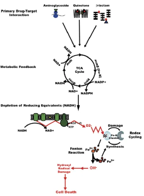
treatment (Dwyer et al. 2007; Kohanski et al. 2007; Kohanski et al. 2008; Davies et al. 2009; Dwyer et al. 2009; Kohanski et al. 2010). They reported that three classes of bactericidal drugs (quinolones, ß-lactams and aminoglycosides) induced a common mechanism of cellular death (Kohanski et al. 2007). This study has argued that bactericidal antibiotics, regardless of their primary targets, kill bacteria by inducing alterations in iron homeostasis, ultimately leading to the accumulation of hydroxyl radicals through the Fenton reaction (Kohanski et al. 2007) (Figure 3.2). They also showed that the bacteriostatic drugs, in contrast, do not produce hydroxyl radicals. This new paradigm indicate the importance of better understanding the mechanisms by which antibiotics kill bacteria in order to find alternative antibacterial therapies to combat bacterial infections
Figure 3.2. Proposed model for the common mechanism of killing by bactericidal drugs (Kohanski et al. 2007)
3.2 General mechanisms of antimicrobial drug resistance
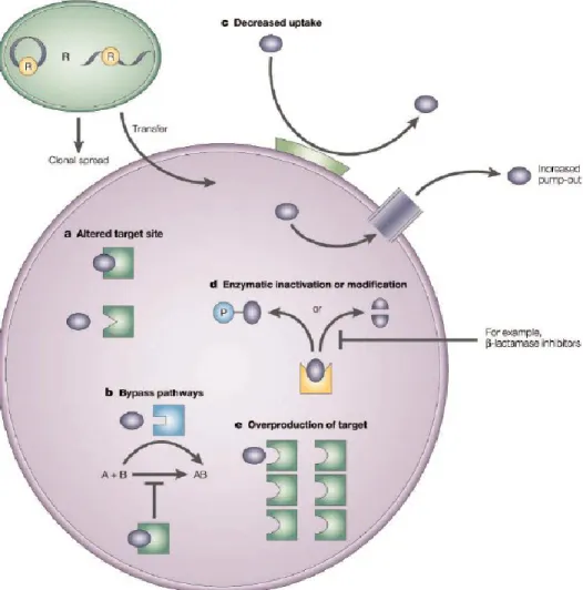
To survive, bacteria have developed a variety of antimicrobial resistance mechanisms. There are three types of antibiotic-resistance strategies, which deal with most of the main classes of antibacterial drugs listed in Table 1. These mechanisms of resistance are shown in Figure 3.3 and are discussed in detail in the rest of this chapter.Figure 3.3. Mechanisms of genetic resistance to antimicrobial agents adapted from. The five main mechanisms that bacteria use to resist antibacterial drugs are shown in the figure. a) The site of action (enzyme, ribosome or cell-wall precursor) can be altered. b) The inhibited steps can be by-passed. c) Bacteria can reduce the
intracellular concentration of the antimicrobial agent, either by reducing membrane permeability or by active efflux of the agent54. d) They can inactivate the drug. For example, some bacteria produce lactamase, which destroys the penicillin β-lactam ring. e) The target enzyme can be overproduced by the bacteria (Taken from (Coates et al. 2002)).
3.2.1 Reduced uptake
Antibiotic must reach their specific target and accumulate at high enough concentration to be effective, so one of the main mechanisms of resistance in bacteria is
a decreased uptake of the drug. Some strains of Pseudomonas aeruginosa and other non-fermenting gram-negative bacilli exhibit aminoglycoside resistance due to membrane impermeabilization. This mechanism is likely chromosomally mediated and results in cross-resistance to all aminoglycosides. The level of resistance that is seen is moderate (i.e. intermediate susceptibility) (Bryan et al. 1976; Bryan and Van den Elzen 1976; Stefani and Russo 1989; Mingeot-Leclercq et al. 1999).
3.2.2 Efflux of the antibiotic
Antibiotic must reach their specific target and accumulate at high enough concentration to be effective. Resistant cells take up the drug as rapidly as do sensitive ones but the drug is pumped out faster than it can diffuse in, whereby the intra-bacterial concentrations are kept low and ineffective. Efflux pumps are transport proteins involved in the extrusion of toxic substrates (including virtually all classes of clinically relevant antibiotics) from within cells into the external environment. These proteins are found in both Gram-positive and -negative bacteria as well as in eukaryotic organisms (Webber and Piddock 2003). The pumps that are not specific for one substrate, may transport a range of compounds including the antibiotics of multiple classes, so such pumps can be associated with multi- drug resistance (MDR).One of the mechanism of resistance to tetracycline in both Gram-positive and Gram-negative bacteria is over-production of some membrane proteins that act as efflux pump (Walsh 2000). In S.
pneumoniae, active efflux has been recognized as a mechanism of resistance to
macrolides (Klaassen and Mouton 2005; Wierzbowski et al. 2005), fluoroquinolones (Gill et al. 1999; Marrer et al. 2006). Efflux also implicated in the resistance of
Mycobacterium tuberculosis to ciprofloxacin and linezolid (Escribano et al. 2007).
3.2.3 Inactivation of the antibacterial agents
Another strategy of resistance is destruction of the chemical structure of the drugs. Some organisms may acquire genes encoding enzymes such as ß-lactamases, that hydrolyse the ß-lactam ring of penicillins and cephalosporins, whereby producing the
non-functional penicilloic acid product (Philippon et al. 1989), which is useless as an antibiotic. Other antibiotic classes, such as the aminoglycosides are neutralized by enzymes which modify the antibiotic, leading to chemically modified drugs which bind poorly to the ribosomes. These modifying enzymes are classified into three major classes according to the type of modification they introduced. It includes: (1) adenylyl transferases, which add AMP moieties, (2) phosphoryl transferases, which add phosphoryl group, or (3) acetyl transferases, which acetylate the amino groups of the antibiotic (Shaw et al. 1993).
3.2.4 Modification of the target
The third strategy of resistance focuses on modifying the target. Penicillin resistance can occur not only by β-lactamase expression, but also by mutation in penicillin-binding proteins, resulting in altered PBPs with lower affinity for the drug (Spratt 1994). Erythromycin resistance in resistant bacteria can emerge by a specific mutation in 23S rRNA component of the ribosome. This modification is performed by a methyl transferase enzyme Erm and leads to decrease in affinity of erythromycin for the RNA (Bussiere et al. 1998). An additional example of target modification is used by vancomycin-resistant enterococci (VRE) to escape from vancomycin effect. In VRE the vanHAX genes encode a new pathway of enzymes that adds alanine and D-lactate together to produce D-Lac as termini of peptidoglycan instead of D-Ala-D-Ala normal termini. This modification in termini of PGN lowers the binding affinity of vancomycin by 1000-fold (Bugg et al. 1991).
3.2.5 Target amplification
When a drug is administered, it is given at a prescribed dose so the active ingredient can bind and inhibit a particular enzyme, or target. If the target is amplified over and over, it reaches a point to where the drug is not binding to enough of the substrate to be effective. Essentially, gene amplification dilutes a normal dose and in order to combat it, higher dosages are needed. Target amplification has been detected in bacteria (Musher et al. 2002; Brochet et al. 2008; Sandegren and Andersson 2009) as well as in a number of eukaryotic microorganisms in response to drug exposure, reviewed in
(Guimond et al. 2003). The 13.5-kb tandem amplification carrying the five genes required for dihydrofolate biosynthesis has been detected in Streptococcus agalactiae which led to both trimethoprim and sulfonamide resistance. Resistance to sulfonamide probably resulted from the increased synthesis of dihydropteroate synthase, the target of this antibiotic, whereas the amplification of the whole pathway was responsible for trimethoprim resistance (Brochet et al. 2008).
3.2.6 Alternative pathway metabolism
The metabolic genotype of an organism can be changed through loss and
acquisition of enzyme-coding genes, allowing pathogens to evolve resistance to anti-metabolites (Barve et al.). Some sulfonamide-resistant bacteria do not require para-aminobenzoic acid (PABA), an important precursor for the synthesis of folic acid and nucleic acid in bacteria inhibited by sulfonamides, instead, like mammalian cells, they turn to using preformed folic acid (Skold 2000; Tenover 2006)..
3.3 Specific mechanisms of resistance in S. pneumoniae
Despite the availability of appropriate antimicrobial treatment, pneumococcal diseases kill 1.6 million people each year, of which one million are children under five years in developing countries (Levine, 2006). S. pneumoniae is the most common cause of community-acquired pneumonia, meningitis, and bacteremia in children and adults (Klugman 2001). The three main classes of antibiotic used for the treatment of S.
pneumoniae are ß-lactams, macrolides and fluoroquinolones (FQ). There are other
antibiotics such as ketolides, vancomycin, trimethoprim-sulfamethoxazole, tetracyclines and linezolid (LZD) that are used for treatment of pneumococcal disease. However, drug resistant S. pneumoniae are now frequently encountered both at the hospital and in the community and more than 20% of clinical isolate of S. pneumoniae in the USA are multi-resistant (Doern et al. 2001) and I will describe resistance mechanisms to both macrolides and fluoroquinolones in this chapter as well as resistance to secondary antibiotics and devote the next full chapter to β-lactam resistance.
3.3.1 Macrolides resistance
Macrolide resistance in S. pneumoniae is due to two main mechanisms, including modification of the target or efflux pumps, which are mediated through acquisition of the erm or mef genes respectively. Acquisition of the erm(B) gene encoding a 23S rRNA methylase is a major resistance determinant in pneumococci. Expression of this gene reduces the affinity of macrolides for 23S rRNA by the post transcriptional modification of 23S rRNA, resulting to high level macrolide resistance (> 64 μg/ml) (Cornick and Bentley 2012). The second major mechanism of macrolide resistance in pneumococci is the acquisition of mef genes, encoding an active efflux pump. There are at least three subclasses of mef gene, of which mef(A) and mef(E) are the most abundant (Daly et al. 2004). Pneumococci carrying both erm(B) and mef(A) (referred to dual phenotype) exhibit a high level of resistance to macrolides in addition to other antimicrobials, are increasingly being described worldwide and are becoming a serious potential public health problem (de la Pedrosa et al. 2008; Reinert et al. 2008) .
3.3.2 Fluoroquinolones (FQ) resistance
Fluoroquinolone introduced in the 1980s against Gram-negative pathogen, but increasing resistance to β-lactams and macrolides among pneumococci led to an increase usage of FQs against Gram-positive pathogens (Cornick and Bentley 2012). It has been shown that this increase in prescription led to a dramatic rise in ciprofloxacin-resistant S. pneumoniae in Canada (Adam et al. 2009). Resistance to fluoroquinolone in pneumococci is primarily due to accumulation of spontaneous mutations in regions known as quinolone-resistance determining regions (QRDR) of DNA gyrases (gyrA and gyrB) and DNA topoisomerases (parC and parE) (Adam et al. 2007). Most fluoroquinolone resistance > 16μg/ml originates from mutations in both parC and
gyrA, which confer resistance to newer fluoroquinolones (Brueggemann et al. 2002).In
addition to the accumulation of spontaneous mutations, efflux pumps may also contribute to resistance to ciprofloxacin (Jumbe et al. 2006). Indeed, over-expression of the major facilitator pmrA (Gill et al. 1999) and of the ABC transporter patA and patB (Marrer et al. 2006) was also shown to contribute to in vitro drug resistance and also in
clinical isolates (Piddock 2006; Garvey et al. 2010). Efflux pumps alone are not known to confer high fluoroquinolone resistance > 16μg/ml, but their presence increases the chance of acquiring additional mutations in parC/E and gyrA/B, which confer higher level of resistance to quinolones (Jumbe et al. 2006; Piddock 2006)
3.3.3 Linezolid resistance
Gram positive bacteria usually develop resistance to linezolid as a result of point mutations in the gene encoding 23S rRNA (Prystowsky et al. 2001). This is the most common mechanism of resistance in staphylococci. Although resistance in clinical strains of S. pneumoniae has not yet been reported, a deletion in ribosomal protein L4 has been shown to lead to non-susceptibility to linezolid (Wolter et al. 2005). Other mechanisms have been identified in S. pneumoniae in vitro resistant isolates. These include a mutation in a 23S rRNA methyltransferase and mutations causing over-expression of the ABC proteins patA and patB (Feng et al. 2009), along with mutations in the 23S rRNA.
3.3.4 Tetracycline resistance
Tetracyclines are antibiotics inhibiting the bacterial growth by stopping protein synthesis. Three major mechanisms of tetracycline resistance have been identified so far: ribosome protection, efflux pumps and enzymatic inactivation of tetracycline. Ribosome protection is the most important mechanisms in tetracycline resistance in both Gram-negative and Gram-positive bacteria. This mechanism of resistance is mediated by ribosomal protection proteins such as Tet(A) and Tet(O), which shares homology with two elongation factors EF-Tu and EF-G implicated in protein synthesis. These ribosomal protection proteins (RPRs) which are called tetracycline-resistance elongation factors can protect ribosome by dislodging tetracycline from ribosome (Connell et al. 2003). Tetracycline efflux is achieved by an export protein from the major facilitator superfamily (Chopra and Roberts 2001).
3.3.5 Trimethoprim-sulfamethoxazole resistance
Trimethoprim and sulfamethoxazole are extensively used in combination as co-trimoxazole and these two components work sequentially to inhibit enzyme systems involved in the bacterial synthesis of tetrahydrofolic acid (THF) (Adrian and Klugman 1997; Schmitz et al. 2001). Resistance to co-trimoxazole in clinical isolates of S.
pneumoniae has been reported due to mutations in dihydrofolate reductase which affect
Chapter IV. Streptococcus pneumoniae and ß-lactam
resistance
The discovery of penicillin by Alexander Fleming is unmatched with any other antibiotics, so ß-lactam antibiotics, which contain a ß-lactam nucleus in their molecular structure, are the most important drugs in the therapy of pneumococcal infections. Therefore, the emergence of the first penicillin-non-susceptible S. pneumonia (PNSP) in Australia in 1967 and rapid dissemination of penicillin resistant pneumococci is recognized as a serious public health problem (Izdebski et al. 2008).
ß-lactams share the same mode of action, inhibiting synthesis of the bacterial cell wall by binding to penicillin-binding proteins (PBPs), which are involved in extracellular assembly of bacterial peptidoglycan, the essential component of the bacterial cell wall. In this chapter, we will talk about the mode of action of ß-lactams by first focusing on peptidoglycan synthesis to better understand the function of penicillin-binding proteins and then we will describe PBPs and non-PBPs ß-lactam resistance mechanisms in S. pneumoniae.
4.1 Bacterial peptidoglycan
The cell wall is responsible for the bacterial shape, but also provides strength and rigidity to protect bacteria from lysis under internal osmotic pressure (Nanninga 1998). Peptidoglycan (PGN) is a main component of the bacterial cell wall, which serves as an attachment site for virulence and adhesion factors and helps bacteria in undergoing morphological transformation in response to different stresses, and its instability may lead to cell death (Holtje 1998; Nanninga 1998). Although the chemical composition of PGN is similar in both Gram-positive and Gram-negative bacteria, the PGN layer is thinner and less cross-linked in Gram-negative bacteria.
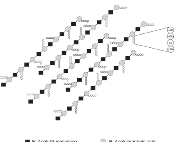
Peptidoglycan, also known as a murein layer, is a heteropolymer composing of ß-linked N-acetylglucosamine (GlcNAc) and N-acetylmuramic acid (MurNAc) polysaccharide chains (glycan), which cross-linked through short peptides chain (Figure 4.1) (Rogers HJ 1980; Barreteau et al. 2008). Attached to a MurNAc residue is
a peptide chain of 4 amino acids which has the consensus sequence (L-Ala, D-glutamic acid, meso-diaminopimelic acid (DAP) and D-Ala) in many bacteria (Figure 4.1) (Gautam et al. 2011). There is some variation in the number and nature of the amino acids in the cross-linkage in different bacteria but all bacteria contain both D and L amino acids and the peptide chain is cross-linked to the peptide chain of another strand between the carboxyl group of D-Ala at position 4 and the amino group of meso-DAP (or L-Lys) at position 3 either directly or through a short peptide bridge (Vollmer et al. 2008), forming the 3D mesh-like layer.
Figure 4.1. Architecture of bacterial peptidoglycan. DA – diamino acid (either meso-diaminopimelic acid or L-Lysine) (Taken from (Gautam et al. 2011))
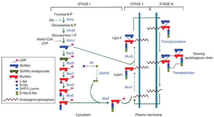
Peptidoglycan synthesis is a complex multistep process (Figure 4.2), which is divided in three stages (Rogers HJ 1980).
4.1.1 Stage I of PG biosynthesis
Stage I reactions occur in the cytoplasm by ten enzymes located in this compartment. The six Mur enzymes (named Mur A to Mur F; Figure 4.2) are responsible for the formation of uridine diphosphate MurNAc from UDP-GlcNAc precursor (Mur A and Mur B) and subsequently adding five amino acids onto the UDP- MurNAc (Mur C to Mur F). The peptide sequence may vary in different species but the two last amino acids should be D-Ala-D-Ala moiety (Macheboeuf et al. 2006), whose ligation is catalyzed by the Mur F ligase. This soluble UDP-MurNAc-pentapeptide becomes associated to the bacterial membrane in the next stage.
Figure 4.2. Schematic representation of the bacterial peptidoglycan biosynthesis pathway ( Taken from (Gautam et al. 2011)).
4.1.2 Stage II of PG biosynthesis
In the second stage, UDP-MurNAc-pentapeptide molecule is attached to a membrane- bound lipid carrier molecule, named undecaprenol phosphate, through the action of phosphor-N-acetylmuramoyl-pentapeptide transferase, MraY (van Heijenoort 2001; Bouhss et al. 2004). The first lipid intermediate, undecaprenyl-pyrophosphoryl-Mur-NAc-pentapeptide formed in this stage is called lipid I. In the next step upon action of Mur G, an N-acetylglucosaminyl transferase, a soluble UDP-GlcNAc group is added to lipid I and leads to the formation of a second lipid intermediate, undecaprenyl-pyrophosphoryl-Mur-NAc-pentapeptide-GlcNAc (lipid II), which becomes the final monomer unit for PG polymer (van Heijenoort 2007; Bouhss et al. 2008). Lipid II is still in the cytoplasmic compartment and must be “flipped” over to the exterior of the membrane, which occur in the third step.
4.1.3 Stage III of PG biosynthesis
Lipid II is “flipped” over to the outer surface of cytoplasmic membrane by a “flippase” activity, which could involve either one or both membrane proteins FtsW and RodA (Holtje 1998; Nanninga 1998). Once in the outer side of the membrane, polymerisation of monomer units and binding of the newly made material to the pre-existing cell wall happens. This last stage of PG biosynthesis is catalysed by the membrane-associated enzymes with transpeptidase (TP) and glycosyltransferase (GT) activity which are known as penicillin-binding proteins (PBPs). Glycosyltransferases catalyze the formation of linear glycan chain (van Heijenoort 2001b) and transpeptidases catalyze the cross-linking of the adjacent stem peptide (van Heijenoort 2001b)
4.2 Penicillin-binding proteins (PBPs)
Penicillin-binding proteins (PBPs), the membrane-associated enzymes, with transpeptidase (TP) and glycosyltransferase (GT) activity play essential roles in peptidoglycan biosynthesis. PBPs have been divided in two main categories: high
molecular mass (HMM) PBPs and low molecular mass (LMM) PBPs (Goffin and Ghuysen 1998; Macheboeuf et al. 2006). The HMM PBPs, consist of several proteins that can be classified into two main categories class A and class B depending on the number of reactions a single polypeptide is able to catalyze. The C-terminal domain of both classes has transpeptidase activity and ß-lactam antibiotics bind to its catalytic site. The N-terminal domain of class A is responsible for glycosyltransferase activity, while that of class B seems to play a role in cell morphogenesis (Marrec-Fairley et al. 2000). Both catalytic sites of bi-functional class A enzymes (TP and GT) are supposed to function independently (Di Guilmi et al. 2003).
Structural studies revealed that the TP active site of all PBPs harbors three conserved active motifs: SXXK with catalytic nucleophilic serine residue crucial for catalysis, S/YXN and a K/H(S/T)G (Hakenbeck et al. 2012). Transpeptidation reaction requires an enzyme to recognize the terminal D-Ala-D-Ala moiety of the stem peptide and catalyze the attack of carboxyl group of the penultimate D-Ala by the lateral amino group at position 3 of an adjacent chain. In the first step, transpeptidase enzyme binds to PGN stem peptide non-covalently followed by attack of the active site serine residue on D-Ala-D-Ala peptide bond, resulting in the formation of an acyl-enzyme intermediate, leading to the release of C-terminal D-Ala (acylation). The last step (deacetylation) consists of either a cross-link formation with the other PGN stem peptide (transpeptidation) or hydrolysis, with the release of tetra peptide (carboxypeptidation) (Sauvage et al., 2008). In S. pneumoniae, the acceptor involved in deacetylation is the amino group of a lysine residue. In transpeptidation reaction, the acceptor molecule can also be a water molecule, which will thus terminate a D-Ala-D-Ala carboxypeptidation reaction, which is catalyzed by low-molecular mass (LMM) PBPs, leading to the release of D-Ala and formation of tetrapeptide, preventing further reticulation of peptidoglycan (Goffin and Ghuysen 2002; Morlot et al. 2004).
4.3 ß- lactam mode of action
For many years transpeptidases (TP) have been the principal target for ß-lactam antibiotics (e.g., penicillin and cephalosporins). The activity of ß-lactam antibiotics is
due to their structural similarity with D-Ala-D-Ala moiety of PGN (Figure 4.3). Upon binding to the active serine residue of TP domain, ß-lactams form stable covalent complex that results in acyl-enzymes that can only be hydrolyzed at a very low rate, thus preventing the cross–linking of stem-peptides and weakening of the PGN structure (Gautam et al. 2011; Hakenbeck et al. 2012). This powerful mechanism, which has made ß-lactam antibiotics the most widely used dug for any streptococcal infections for the past 70 years, has been challenged by the rapid dissemination of penicillin resistant
S. pneumoniae.
Figure 4.3. Chemical structures of ß-lactam drugs (penicillins and cephalosporins). The inhibitory effect of ß-lactams comes from the structural similarity with D-Ala-D-Ala moiety of PGN chain. R1, R2, X and Z reflect different moieties which are associated to the central structure order to generate distinct antibiotics or second- or third-generation molecules.(adapted from (Macheboeuf et al. 2006))