Science Arts & Métiers (SAM)
is an open access repository that collects the work of Arts et Métiers Institute of Technology researchers and makes it freely available over the web where possible.
This is an author-deposited version published in: https://sam.ensam.eu Handle ID: .http://hdl.handle.net/10985/19542
This document is available under CC BY license
To cite this version :
Britt-Isabelle BERG, Aurélien LAVILLE, Delphine S. COURVOISIER, Philippe ROUCH, Thomas SCHOUMAN - Experiences with a new biplanar low-dose X-ray device for imaging the facial skeleton: A feasibility study - PLoS One - Vol. 7, p.15 - 2020
Any correspondence concerning this service should be sent to the repository Administrator : [email protected]
Experiences with a new biplanar low-dose
X-ray device for imaging the facial skeleton: A
feasibility study
Britt-Isabelle BergID1*, Aure´lien Laville2, Delphine S. Courvoisier3, Philippe Rouch2, Thomas Schouman4
1 Department of Cranio-Maxillofacial Surgery, University Hospital Basel and University Basel, Basel, Switzerland, 2 Laboratoire de Biome´canique, Institut de Biome´canique Humaine Georges Charpak, Arts et Me´tiers ParisTech, Paris, France, 3 CRC & Division of Clinical Epidemiology, Faculty of Medicine, University of Geneva & University Hospitals of Geneva, Geneva, Switzerland, 4 Department of Maxillofacial Surgery, APHP—Groupe Hospitalier Pitie-Salpetriere, Universite Paris 6—UPMC, Paris, France
Abstract
Background
This study aimed to evaluate the feasibility of a new biplanar low-dose X-ray device for facial skeletal imaging.
Methods
We evaluated 48 biplanar radiographs from 12 patients (posteroanterior/lateral), originally taken for a scoliosis examination with a biplanar low-dose X-ray device. For this study, the images were further evaluated for the perceptibility of 38 facial skeleton landmarks. To determine the reliability and reproducibility of perceptibility, two independent observers determined the landmarks twice, during a time interval of at least two weeks.
Results
Both interoperator and intraoperator reliability were excellent for all landmarks [intraclass correlation coefficient (ICC)>0.92].
Conclusions
The biplanar low-dose X-ray device demonstrated good feasibility for precisely assessing the anatomical landmarks of the facial skeleton. Given its excellent precision, the biplanar low-dose X-ray device data sets should be forwarded from the treating orthopedic surgeon or neurosurgeon to the orthodontist or dentist for further assessment in their field.
Data Availability Statement: All relevant data are within the manuscript and its Supporting Information files.
Funding: For this study, no author has received any funding. During the time this retrospective study took place, the institute/laboratory in which Prof. P. Rouch works and Dr. A. Laville worked received funding from the EOS-Imaging company for other EOS studies. The funders had no role in study design, data collection and analysis, decision to publish, or preparation of the manuscript.
Introduction
In 1992, the Nobel Prize in Physics was awarded to Georges Charpak “for his invention and development of particle detectors, in particular the multiwire proportional chamber” [1]. This technology was used to develop a new biplanar X-ray imaging system. Ille´s & Somoskeo¨y published the detailed technology behind the system, stating that the method is highly sensitive, and could potentially detect even one photon. In addition, it is less affected by scattered radiation [2]. Until now, the biplanar low-dose X-ray imaging system was used primarily in orthopedics to image the spine, hip, shoulder, joints, and extremities [3,4]; furthermore, its role in the detection of bone lesions has been assessed [5]. Since the biplanar X-ray imaging system produces head-to-foot images, while the patient maintains a standing position, it frequently captures the facial skeleton [6–8], even when it is of no relevance for an initial patient-related assessment. Moreover, use of these images to assess dentofacial structures could enhance safety by avoiding additional exposure to radiation and reduce healthcare costs. The imaging system simultaneously acquires posteroanterior and lateral images. Both views are of great interest for dentists, orthodontists, maxillofacial surgeons, and pediatric surgeons, due to the use in cephalometric analyses. Cephalometric analyses based on landmarks allow orthodontists and maxillofacial surgeons to assess the severity and cause of dentofacial deformities, growth patterns of the facial skeleton, the ability to set a treatment plan, and eventually assess the efficacy of the treatment and its stability. Facial asymmetry can include a distortion of the entire face, if there appears to be a relationship between facial asymmetry and adolescent idiopathic scoliosis [9]. Pan-oramic radiographs and cephalograms, rather than standard X-rays, are routine diagnostic tools in these cases [10]. The analysis is based on a number of specific landmarks on fron-tal and lateral cephalograms. Thus, patients (mainly children or young adults) will be sub-ject to repeated radiation exposure of the head over a couple of years for orthodontic or orthognathic treatment.
Keeping radiation exposure as low as possible is necessary, as radiation accumulates over an individual’s lifetime. An association between dental X-rays and the risk of intra-cranial meningioma has been reported by Claus et al. [11]: they found that a history of exposure from dental X-rays during an earlier period (when radiation exposure was greater than now) appears to be associated with an increased risk of intracranial menin-gioma. A plain dental X-ray on average has a similar radiation dosage [12] as X-rays cap-tured for cephalometric measurements. Therefore, it is sensible to use X-rays that are already available, even if they are not captured for the purpose of cephalometric analyses. Until now, no other research group assessed the feasibility of cephalometric analysis based on this biplanar imaging or any other biplanar low-dose X-ray system that cap-tured both views of the face at the same time for precise depiction of the facial skeleton. One publication assessed cephalograms in posteroanterior and lateral views with a dry skull, but the X-rays were not captured at the same time [13]. Therefore, we investigated whether this low-dose biplanar X-ray imaging system would be a useful tool for facial skeletal assessments. Our hypothesis was that biplanar X-ray imaging quality was accept-able for sufficient facial landmark placement, as used in orthodontic planning and orthognathic surgery. Our aim was determine if the biplanar low-dose X-ray imaging of the facial skeleton was sufficient for landmark assessment, even if artifacts occur (as in the images we assessed for patients who had their hands in front of the face). We decided to review the images of the same patients, once with hands in front of the face and once without hands in front of the face (hands on the shoulders).
Competing interests: I have read the journal’s policy and the authors of this manuscript have the following competing interests: Prof. P. Rouch and Dr. A. Laville have to declare a conflict of interest with the EOS-Imaging company, as the laboratory has received EOS-Imaging funding associated to anterior studies. All other authors have no conflicts of interest to daclare. Herewith, we confirm that this does not alter our adherence to PLOS ONE policies on sharing data and materials.
Materials and methods
Volunteers
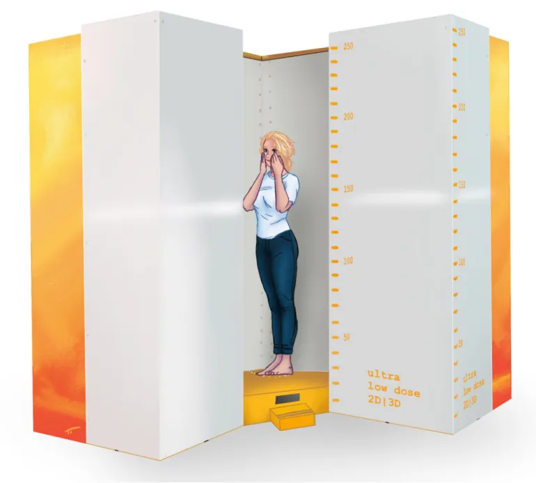
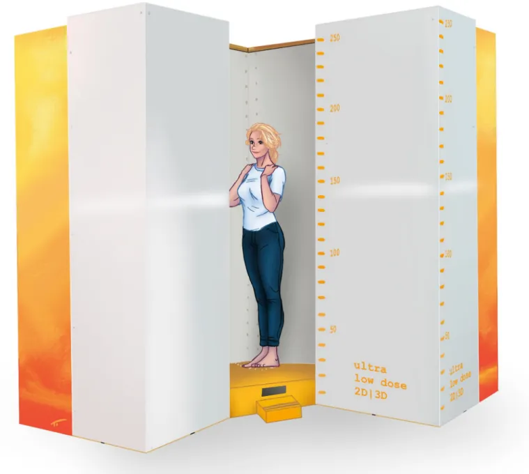
Ethical approval was received from the local ethical committee "Comite´s de protection des per-sonnes en Ile-de-France" (CPP IDF VI, No 90–06). Patients had provided written informed consent for the original study. We used a total of 48 biplanar radiographs from 12 patients, which were acquired with a biplanar low-dose X-ray imaging system, initially for spinal assess-ments with various upper extremity positions (patients provided informed consent for the original study). The images included 24 data sets (12 posteroanterior and 12 lateral) from patients in a standing position with hands in front of their face (Group A), and 24 data sets (12 posteroanterior and 12 lateral data sets) from the same patients without hands in front of their face (Group B). Depicted in Figs1and2is the exact position of the patient in the biplanar low-dose X-ray device.Fig 1shows the patient with hands in front of the face (Group A), andFig 2
shows the same patient with hands on the shoulders (Group B).
The biplanar low-dose X-ray imaging system
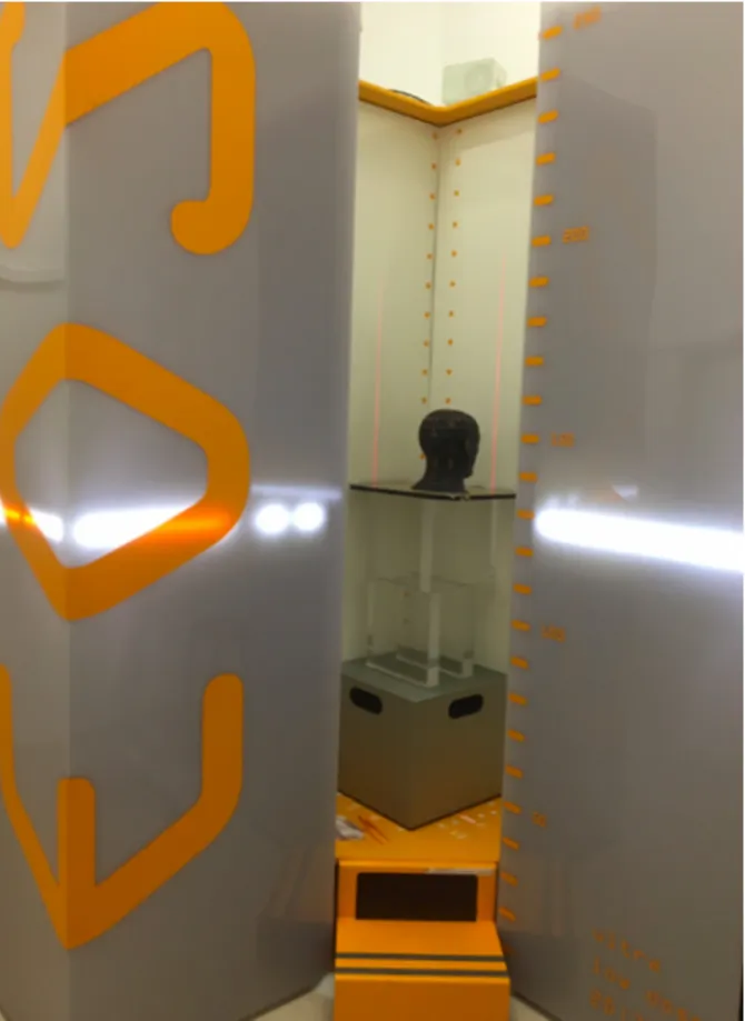
The device is the EOS1 system (EOS1, EOS Imaging SA, Paris, France). All patients were standing. The biplanar X-ray imaging system used two perpendicular X-ray beams that were collimated in two extremely thin, horizontal, fan-shaped beams with two detectors. Since this was a retrospective study, we used X-rays of a phantom head during data acquisition to illus-trate the installation (seeFig 3).
Effective resolution was reduced to 193μm × 185 μm for the frontal view and
179μm × 185 μm for the lateral view [14]. The reference settings were chosen as follows: Fron-tal: tube voltage 90 kV, current-time product 50 mAs, DAP 716 mGy cm2; Lateral: tube voltage 105 kV, current-time product 320 mAs, DAP 1082 mGy cm2. The morphology was adult, the scanned length was 76 cm, scanning speed was 5 s, and acquisition time was 12.5 s.
Measurements
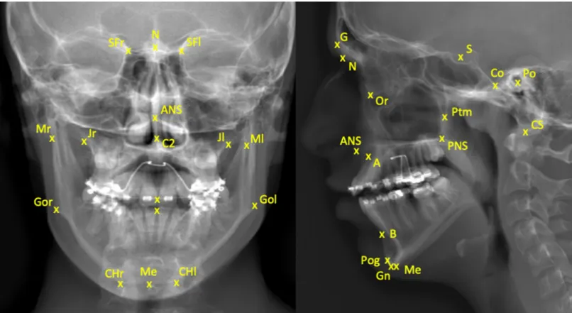
To evaluate the feasibility of the biplanar X-ray imaging system for pictorial depiction of the facial skeleton, two craniomaxillofacial surgeons (experienced in orthognathic surgery and dentomaxillofacial radiology) placed 38 landmarks on each image. The landmarks for assess-ment of the frontal and lateral images were placed according to the description provided in “Essentials of Orthognathic Surgery” [15]. For both views, landmark C2 was added to repre-sent the odontoid process.Table 1gives detailed descriptions of the 38 anatomical regions of the landmarks as seen inFig 4.
Two independent raters were instructed to place the landmarks twice, i.e., on two separate occasions, at an interval of two weeks to assess consistency of the landmark placement at two time points, given consistency of the estimates of placement (interoperator reproducibility). The landmarks were digitally placed (computer mouse on a high-resolution monitor) with image-treatment software (Idefix, an imaging processing software based on C++, developed at the Laboratoire de Biome´canique/Institut de Biome´canique Humaine Georges Charpak, Arts et Me´tiers ParisTech, Paris, France) to provide coordinates of each landmark in a two-dimen-sional reference system. The stereo-corresponding landmarks (visible in both views: N, ANS, Orl, Orr, Ml, Mr, Gol, Gor, Me, seeTable 1) were selected in one view, as the level (y-axis) of the same landmark was indicated in the other view by an epipolar line. Values in the x- and y-axis were transferred to an Excel spreadsheet for further calculation.
Analysis
Interoperator and intraoperator reliability were evaluated for each landmark as recommended by the International Organization for Standardization [16]. One-way random effects, absolute agreement, and single rater (or single measurement) intraclass correlation coefficients [Ref. to (ICC (1,1), according to the Shrout and Fleiss convention [17]], were calculated for each land-mark and presented according to group membership (Group A vs. Group B). The ICC expressed the proportion of true variance and the proportion of total variability due to differ-ences among the participants. ICC values > 0.91, 0.71–0.91, 0.51–0.70, or < 0.51 were consid-ered to represent excellent, good, moderate, or poor agreement, respectively [18]. The R
v.3.0.1 software package (R Development Core Team, R Foundation for Statistical Computing, Vienna, Austria) was used to perform all statistical analyses. We used the moment distance to report the difference between the two observers in millimeters. Further information on this method can be found in the publication by S.D. Springate [19].
Results
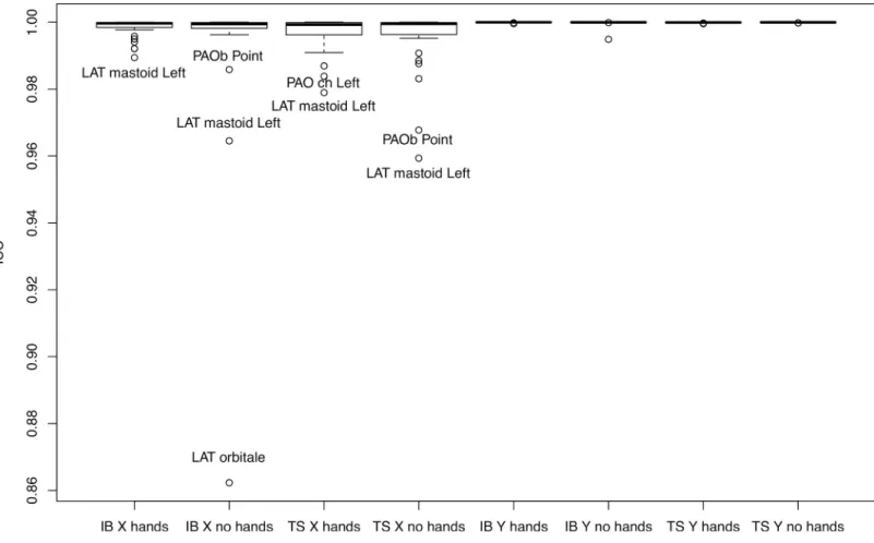
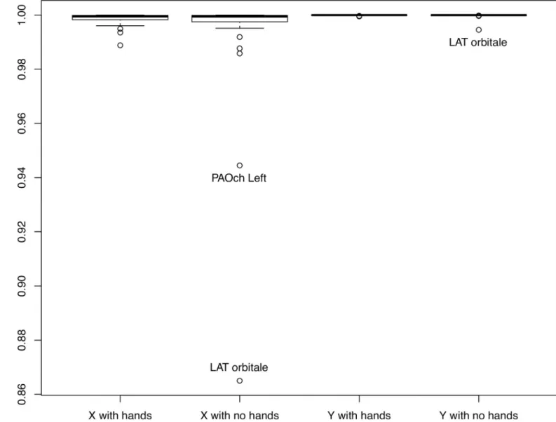
The average age of the 12 volunteers was 46 (range, 17–71 years).Fig 5(Group A, with hands) andFig 6(Group B, without hands) contain ICC values, averaged for x- and y-axis values, as well as for intraoperator and interoperator reliability for the perceptibility of the 38 facial skele-ton landmarks on the 12 biplanar radiographs.
Fig 3. For better illustration, an Alderson head phantom was placed in the biplanar low-dose X-ray imaging system. The light outside is a laser beam, showing the height in which the image is captured at this moment. A certain distance (in cm) can be set in the system.
Both interoperator and intraoperator reliability were excellent for all landmarks (ICC > 0.92). The ICCs of Group A were not significantly different from those of Group B (interoperator:t = 1.39; p = 0.17; d = 0.32; intraoperator rater 1: t = 1.13; p = 0.26; d = 0.26; intraoperator rater 2: t = 0.69; p = 0.49; d = 0.17). Detailed ICC values are inS1
andS2Tables.
The greatest difference in millimeters between the two observers, for the x and y values (in Groups A and B) was found for the Orbital (Or) landmark in the lateral view; this is likely due to the fact that one value was aberrant. Most likely, the rater did not push the button on the computer mouse strongly enough, although the overall values were still excellent. The detailed millimeter differences for each landmark are shown inS3 Table.
Table 1. Landmarks according to descriptions provided in “Essentials of Orthognathic Surgery” [16]. For both views, landmark “C2” was added to represent the odontoid process.
Posterior view
(SF) Where the smaller wing of the sphenoid bone crosses the medial orbital ridge (ANS) Anterior nasal spine: the center at the base of the nose
(J) Jugular: the most superior and medial point of the zygomatic buttress (M) Mastoid: the most inferior point of the mastoid point
(A) The contact area between the maxillary incisors (B) The contact area between the mandibular incisors
(Go) Gonion: the most inferior posterior point at the angle of the mandible (Me) Menton: the most inferior posterior point at the anterior mandibular area (CH) The most inferior posterior point on the anterior inferior border of the mandible (C2) Upper border of the odontoid process
Lateral view
(G) Glabella: the most anterior point of the frontal bone
(N) Nasion: the most anterior point of the frontal nasal suture in the midsagittal plane (Or) Orbital: the lowest point on the inferior orbital rim
(S) Sella: the center of the sella turcica on the lateral cephalogram, which is located by inspection
(Ptm) Pterygomaxillary: the apex of the teardrop-shaped pterygomaxillary fissure (lowest point of the opening) (ANS) Anterior nasal spine: anterior tip of the nasal spine
(PNS) Posterior nasal spine: the most posterior aspect of the palate bone
(A)- Point or subspinal: the most posterior midline point in the concavity where the lower anterior adage of the anterior nasal spine meets the alveolar bone, overlying the mandible incisors (infradentale) and the pogonion
(B)- Point or supra-mental: the most posterior midline point in the concavity of the mandible between the alveolar bone overlying the mandible incisors (infradentale) and the pogonion
(Pog) Pogonion: the most anterior point of the chin
(Go) Gonion: the point defined by using two lines, with one tangential to the inferior border of the ramus; this can be assessed by bisecting the angle formed by the two lines and extending the bisectors through the curvature of the mandible
(Gn) Gnathion: the lowest, most anterior midline point on the symphysis of the mandible in the midline (Me) Menton: the most inferior point on the symphysis of the mandible in the midline
(Po) Porion: the most superior point of the external auditory meatus; the machine porion is the uppermost point on the outline of the rods of the cephalometer
(Co) Condylion: the most posterosuperior point on the head of the condyle (C2) Upper border of the odontoid process
Discussion
Our study was the first to investigate the feasibility of a biplanar low-dose X-ray imaging sys-tem for the facial skeleton. Image quality and excellent reproducibility/repeatability of the facial landmarks are promising. No significant differences are found between the two groups in terms of ICC values; therefore, the landmarks were precisely detectable even if patients had their hands in front of the face. This indicates the potential use of this system, not only in the field of orthopedics, but for imaging of craniofacial bony structures.
The patients in our retrospective study had been diagnosed with scoliosis. Scoliosis requires continuous follow-up with imaging. In cases of congenital scoliosis, for which surgery is indi-cated, annual follow-up imaging is also necessary until age 20 [20]. Additional radiation doses can be prevented with extended spinal biplanar X-ray imaging. Previously, these images were not used to assess facial asymmetries. Our assessment showed that these images were of ade-quate quality for evaluation of the facial skeleton, despite evidence of a correlation between facial asymmetry and scoliosis. A previous publication reported that children with scoliosis were more likely to have malocclusions [9]. Another study compared the occlusal parameters of 103 children with idiopathic scoliosis in a random group and found more asymmetric fea-tures of malocclusion relative to the control group [21]. Scoliosis patients are not the only group in which assessment of the captured faces might have demonstrated this. Proffit et al. reported that 57% to 59% of the US population is in need of some degree of orthodontic treat-ment [22]; this indicates that imaging for cephalometric assessment is necessary, which would expose a substantial part of the population to radiation. Given our results, biplanar X-ray
Fig 4. Posteroanterior and lateral images of a patient without hands in front of the face, using the biplanar low-dose X-ray imaging system. The 38 landmarks are placed to illustrate the anatomical regions.
imaging would be acceptable for an initial radiological assessment and follow-up when ortho-dontic/ maxillofacial treatment becomes necessary.
A comparison of our results with other studies using the same landmarks, but other X-ray devices, reveals that our interoperater and intraoperater reproducibility was comparable or better, even with an aberrant value; this could have occurred when the person performing the digital marking did not push the button on the computer mouse strongly enough. The rater might have thought that the mouse was correctly positioned and clicked in a certain way, but in fact, the “click” was delayed. The authors of a study on the reliability of locating landmarks of the porion (Po) and condyle (Co) compared two groups of patients, one with open mouth and one with teeth in centric occlusion. This showed that the “average reproducibility of all measurements for observers I and II was 93% and 81%” [23]. Superimposing both data sets on special foils, the set with the mouth open and the set in the centric position, were assessed in millimeters [23]. Manual setting of landmarks is still common, although landmarks are mostly digitally set. In a study conducted by Durão et al. [24], the accuracy of two-dimensional cepha-lometric analysis was evaluated. They used the same landmarks as in our study (though not all of them: N–Nasion; Me–Menton; ANS–Anterior Nasal Spine; Co–Condylion; Gn–Gnathion; A–Point A; B–Point B; Pog–Pogonion; Po–Porion; Or–Orbitale; Go–Gonion). They com-pared it to measurements of skulls, the gold standard. They found intraoperator consistency ICC above 0.9, except for one landmark in which the ICC was 0.79 [24]. Interoperator reliabil-ity with ICCs between 0.67–0.99 depend on the landmark. In their study, two observers
Fig 5. ICC values for intraoperator reliability for perceptibility of the 38 facial skeleton landmarks on the 12 biplanar radiographs of patients with hands/without hands in front of the face. IB and TS are the raters’ initials; x and y are the shortening of the x- and y-axis.
performed the measurements. The second measurement was done after a one-month interval. Differences of < 1 mm were accepted as within one standard deviation (SD) and were consid-ered clinically acceptable in this study [24]. Landmarks were digitally placed on the monitor [24]. Paixão et al. [25]. found no statistically significant differences (p > 0.05) when comparing cephalometric measurements of digital cephalometric tracing, with manual placement in lat-eral cephalometric radiographs. Comparing our results with conventional digital latlat-eral cepha-logram radiographs, reproducibility measurements in that study were slightly more precise. Comparing our results with those of Durão et al. [24], in some landmarks, our differences were > 1mm. Using the middle of the sella as an example (“S-point”), measurements pub-lished by Greiner et al. [26] yielded a mean “x”/”y” value quality range of 0.28 mm/0.21 mm. In our measurements, sella “S-point” values of 0.42 mm/ 0.42 mm were found with respect to inter-reproducibility (in Group B). An accuracy of less than 0.5 mm is precise and will be more than sufficient for the assessment used in clinical routine. The landmarks that exhibit greater differences than 1 mm are, in general, subject to interpretation in placement and are
Fig 6. ICC values for interoperator reliability for the perceptibility of 38 facial skeleton landmarks on the 12 biplanar radiographs of patients with hands/without hands in front of the face. IB and TS are the rater’s initials; x and y are the shortening of the x- and y-axis.
therefore more difficult to position, which might explain greater interoperator differences. The detailed verification of all landmarks of the face guarantees that the biplanar X-ray can be used for most interventions in the dentofacial area. Other studies described difficulties in identify-ing the “Go” and “Me” landmarks [27–29], which proved to not be the case in our study with the biplanar X-ray imaging system tool. When comparing reproducibility of landmarks/ana-tomical points of our results with other studies, using the biplanar low-dose X-ray imaging sys-tem, e.g., reproducibility of vertebrae angle measurements, we reported ICC values of 0.97– 1.00 [18]. A study of scapular landmarks found maximum interoperator differences of 1.48– 13.24 mm and average intraoperator differences of 0.25–2.53 mm [30]. Images in our study were not originally captured for detection of facial skeletal structures, but to clarify if hands in front of the face had an impact on spinal assessment. Positioning of the tongue, the Frankfurter Horizontal, a line to orient the head or the chin, were not considered. The patient was not per-fectly placed in the device, yet it was possible to achieve very good image quality.
Even with hands in front of the face, these captured images could also be used by maxillofa-cial surgeons or orthodontists in deciding between conservative treatment and orthognathic surgery. The lateral view could be assessed to obtain the growth pattern, and the posteroanter-ior images could be used to assess facial asymmetries.
Since this study was retrospective, dosage measurements of the face were not performed. It should be left to future investigation whether (in regard to radiation dosage), the biplanar low-dose X-ray imaging system is superior to conventional cephalometric devices. However, the results of the present study, e.g., the high quality of landmarks assessment, promote the use of images captured with the biplanar low-dose X-ray imaging system for the same measurements, usually performed by conventional cephalometric devices, when available.
Conclusion
The biplanar low-dose X-ray device showed good feasibility for precisely assessing the anatom-ical landmarks of the facial skeleton. Given such excellent precision, the biplanar low-dose X-ray device data sets should be forwarded from the treating orthopedic surgeon or neuro-surgeon to the orthodontist or dentist for further assessment in their field.
Supporting information
S1 Table. ICC values for intraoperator and interoperator reliability for the perceptibility of 38 facial skeleton landmarks on the 12 biplanar radiographs of patients with their hands in front of the face.
(DOCX)
S2 Table. ICC values for intraoperator and interoperator reliability for the perceptibility of 38 facial skeleton landmarks on the 12 biplanar radiographs of patients without their hands in front of the face.
(DOCX)
S3 Table. The millimeter differences for interoperator reliability of 38 facial skeleton land-marks on the 12 biplanar radiographs of patients with their hands in front of the face (X. H.- x-value of the patient group with hands in front of the face), X.N.H.- x-value of patient group with no hands in front of the face, Y.H.- y-value of patient group with hands in front of the face, Y.N.H.- y-value of patient group with no hands in front of the face).
Acknowledgments
We greatly thank Prof. Jean-Marc Vital, Hospital Tripode, CHU de Bordeaux, France for pro-viding us with the biplanar low-dose X-ray imaging data sets.
Author Contributions
Conceptualization: Britt-Isabelle Berg, Aure´lien Laville, Delphine S. Courvoisier, Philippe
Rouch, Thomas Schouman.
Data curation: Britt-Isabelle Berg.
Formal analysis: Britt-Isabelle Berg, Delphine S. Courvoisier. Investigation: Britt-Isabelle Berg.
Methodology: Britt-Isabelle Berg, Aure´lien Laville. Writing – original draft: Britt-Isabelle Berg.
Writing – review & editing: Britt-Isabelle Berg, Aure´lien Laville, Delphine S. Courvoisier,
Phi-lippe Rouch, Thomas Schouman.
References
1. Nobel No, AB M. The Nobel Prize in Physics 1992:http://www.nobelprize.org/nobel_prizes/physics/ laureates/1992/index.html; 214 Available from:http://www.nobelprize.org/nobel_prizes/physics/ laureates/1992/index.html.
2. Ille´s T, Somoskeo¨ y S. The EOS™imaging system and its uses in daily orthopaedic practice. Int Orthop. 2012; 36: 1325–1331.https://doi.org/10.1007/s00264-012-1512-yPMID:22371113
3. Gheno R, Nectoux E, Herbaux B, Baldisserotto M, Glock L, Cotten A, et al. Three-dimensional mea-surements of the lower extremity in children and adolescents using a low-dose biplanar X-ray device. Eur Radiol. 2012; 22: 765–771.https://doi.org/10.1007/s00330-011-2308-yPMID:22011904
4. Gaume´ tou E, Quijano S, Ilharreborde B, Presedo A, Thoreux P, Mazda K, et al. EOS analysis of lower extremity segmental torsion in children and young adults. Orthop Traumatol Surg Res. 2014; 100: 147– 151.https://doi.org/10.1016/j.otsr.2013.09.010PMID:24439563
5. Boutry N, Dutouquet B, Leleu X, Vieillard MH, Duhamel A, Cotten A. Low-dose biplanar skeletal survey versus digital skeletal survey in multiple myeloma. Eur Radiol. 2013; 23: 2236–2245.https://doi.org/10. 1007/s00330-013-2812-3PMID:23512194
6. Dietrich TJ, Pfirrmann CW, Schwab A, Pankalla K, Buck FM. Comparison of radiation dose, workflow, patient comfort and financial break-even of standard digital radiography and a novel biplanar low-dose X-ray system for upright full-length lower limb and whole spine radiography. Skeletal Radiol. 2013; 42: 959–967.https://doi.org/10.1007/s00256-013-1600-0PMID:23536038
7. Jolivet E, Sandoz B, Laporte S, Mitton D, Skalli W. Fast 3D reconstruction of the rib cage from biplanar radiographs. Med Biol Eng Comput. 2010; 48: 821–828.https://doi.org/10.1007/s11517-010-0610-5
PMID:20414812
8. Ille´s T, Tunyogi-Csapo´ M, Somoskeo¨ y S. Breakthrough in three-dimensional scoliosis diagnosis: signifi-cance of horizontal plane view and vertebra vectors. Eur Spine J. 2011; 20: 135–143.https://doi.org/10. 1007/s00586-010-1566-8PMID:20821027
9. Hong JY, Suh SW, Modi HN, Yang JH, Hwang YC, Lee DY, et al. Correlation between facial asymmetry, shoulder imbalance, and adolescent idiopathic scoliosis. Orthopedics. 2011; 34: 187.https://doi.org/10. 3928/01477447-20110427-14PMID:21667906
10. Mu¨ssig E, Wo¨rtche R, Lux CJ. Indications for digital volume tomography in orthodontics. J Orofac Orthop. 2005; 66: 241–249.https://doi.org/10.1007/s00056-005-0444-1PMID:15959637
11. Claus EB, Calvocoressi L, Bondy ML, Schildkraut JM, Wiemels JL, Wrensch M. Dental x-rays and risk of meningioma. Cancer. 2012; 118: 4530–4537.https://doi.org/10.1002/cncr.26625PMID:22492363
12. European guidelines on radiation protection in dental radiology, Issue N˚ 136, 2004https://ec.europa. eu/energy/sites/ener/files/documents/136.pdf, accessed 23.07.2019
13. Wu MC, Cheng KS, Chen YT, Liu JK, Ting WH. Three-dimensional analysis of biplanar cephalograms. Eur J Orthod. 2010; 32: 627–32.https://doi.org/10.1093/ejo/cjq026PMID:20660132
14. Deschênes S, Charron G, Beaudoin G, Labelle H, Dubois J, Miron MC, et al. Diagnostic imaging of spi-nal deformities: reducing patients radiation dose with a new slot-scanning X-ray imager. Spine (Phila Pa). 2010; 35: 989–994.
15. Reyneke JP. Essentials of Orthognathic Surgery. Hanover Park, IL: Quintessence Publishing; 2003. 16. Standardization IOf. ISO 5725–2:1994 Accuracy (trueness and precision) of measurement methods
and results-Part 2: Basic method for the determination of repeatability and reproducibility of a standard measurement method. Geneva, Switzerland; 1994.
17. Koo TK, Li MY. A Guideline of Selecting and Reporting Intraclass Correlation Coefficients for Reliability Research. J Chiropr Med. 2016; 15:155–63. Erratum in: J Chiropr Med. 2017;16: 346.https://doi.org/ 10.1016/j.jcm.2016.02.012PMID:27330520
18. Ilharreborde B, Steffen JS, Nectoux E, Vital JM, Mazda K, Skalli W, et al. Angle measurement reproduc-ibility using EOS three-dimensional reconstructions in adolescent idiopathic scoliosis treated by poste-rior instrumentation. Spine (Philadelphia, Pa). 2011; 36: E1306–E 1313.
19. Springate SD. The effect of sample size and bias on the reliability of estimates of error: a comparative study of Dahlberg’s formula. Eur J Orthod. 2012; 34: 158–63.https://doi.org/10.1093/ejo/cjr010PMID:
21447784
20. Faria R, McKenna C, Wade R, Yang H, Woolacott N, Sculpher M. The EOS 2D/3D X-ray imaging sys-tem: a cost-effectiveness analysis quantifying the health benefits from reduced radiation exposure. Eur J Radiol. 2013; 82: e342–e349.https://doi.org/10.1016/j.ejrad.2013.02.015PMID:23473735
21. Damet J, Fournier P, Monnin P, Sans-Merce M, Ceroni D, Zand T, et al. Occupational and patient expo-sure as well as image quality for full spine examinations with the EOS imaging system. Med Phys. 2014; 41: 063901.https://doi.org/10.1118/1.4873333PMID:24877841
22. Proffit WR, Fields HW Jr, Moray LJ. Prevalence of malocclusion and orthodontic treatment need in the United States: estimates from the NHANES III survey. Int J Adult Orthodon Orthognath Surg. 1998; 13: 97–106. PMID:9743642
23. Adenwalla ST, Kronman JH, Attarzadeh F. Porion and condyle as cephalometric landmarks—an error study. Am J Orthod Dentofacial Orthop. 1988; 94: 411–415.https://doi.org/10.1016/0889-5406(88) 90130-8PMID:3189243
24. Durão AR, Bolstadb N, Pittayapatc P, Lambrichtse I, Ferreiraf AP, Jacobs R. Accuracy and reliability of 2D cephalometric analysis in orthodontics. Revista Portuguesa de Estomatologia, Medicina Denta´ ria e Cirurgia Maxilofacial. 2014; 55: 135–141.
25. Paixão MB, Sobral MC, Vogel CJ, de Araujo TM. Comparative study between manual and digital cepha-lometric tracing using Dolphin Imaging software with lateral radiographs. Dental Press J Orthod. 201015: 123–130.
26. Greiner M, Greiner A, Hirschfelder U. Variance of landmarks in digital evaluations: comparison between CT-based and conventional digital lateral cephalometric radiographs. J Orofac Orthop. 2007; 68: 290– 298.https://doi.org/10.1007/s00056-007-0710-5PMID:17639277
27. Chien PC, Parks ET, Eraso F, Hartsfield JK, Roberts WE, Ofner S. Comparison of reliability in anatomi-cal landmark identification using two-dimensional digital cephalometrics and three-dimensional cone beam computed tomography in vivo. Dentomaxillofac Radiol. 2009; 38: 262–273.https://doi.org/10. 1259/dmfr/81889955PMID:19474253
28. Polat-Ozsoy O, Gokcelik A, Toygar Memikoglu TU. Differences in cephalometric measurements: a comparison of digital versus hand-tracing methods. Eur J Orthod. 2009; 31: 254–259.https://doi.org/ 10.1093/ejo/cjn121PMID:19349417
29. Santoro M, Jarjoura K, Cangialosi TJ. Accuracy of digital and analogue cephalometric measurements assessed with the sandwich technique. Am J Orthod Dentofacial Orthop. 2006; 129: 345–351.https:// doi.org/10.1016/j.ajodo.2005.12.010PMID:16527629
30. Ohl X, Stanchina C, Billuart F, Skalli W. Shoulder bony landmarks location using the EOS low-dose stereoradiography system: a reproducibility study. Surg Radiol Anat. 2010; 32: 153–158.https://doi.org/ 10.1007/s00276-009-0566-zPMID:19806289
![Table 1. Landmarks according to descriptions provided in “Essentials of Orthognathic Surgery” [16]](https://thumb-eu.123doks.com/thumbv2/123doknet/7407196.217944/8.918.302.864.154.787/table-landmarks-according-descriptions-provided-essentials-orthognathic-surgery.webp)