Understanding the interactions between Wnt and BMP signalling pathways
in human Periosteum Derived Cells
M. Germain
*1,2, J. Bolander
2,3, L. Geris1,21 Biomechanics Research Unit, GIGA In Silico Medicine, Université de Liège, Belgium 2 Prometheus, Division of Skeletal Tissue Engineering, K.U. Leuven, Belgium
3 Skeletal Biology and Engineering Research Center, KU Leuven, Belgium
* Correspondence:[email protected], Allée de la Découverte 9, 4000 Liège, Belgium. 1. Introduction
Bone Morphogenetic Proteins (BMP) and Wnt are key elements in the regulation of the bone formation process. Crosstalks between these two pathways highly depend on the cellular context [1]. A detailed understanding of their mechanisms will enable us to develop efficient and robust tissue engineering products. In this study, we develop a mathematical model of these crosstalks, focusing on the canonical BMP and Wnt pathways and their interactions as they are proposed in the literature.
2. Materials and Methods
Figure 1 shows a schematic representation of the pathways modelled [2,3].
Figure 1: Schematic representation of the pathways
modelled.
This model includes a detailed BMP and Wnt signalling pathway and different crosstalks between both.
The obtained model is a system of ordinary differential equations, built on the principles of the law of mass action and rate kinetics and has been implemented in MATLAB. Parameters values are initially derived from literature.
In order to validate the model, optimize the parameters and have more clues of the actual crosstalks between both pathways, experiments
on human Periosteum Derived Cells have been performed (hPDCs). Cells were stimulated with BMP2 and/or Wnt3a and were analysed through western blots and QPCR at multiple time points.
3. Results
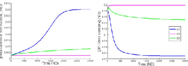
Figure 2 shows the evolution of the end points of Wnt (β-catenin) and BMP (P-Smad) pathways for three different conditions: presence of Wnt only (C1), BMP only (C2) and both (C3) and for one particular crosstalk configuration modeled.
The comparison of the results between these simulations for different crosstalk configurations and the experimental results allows us to identify and better understand the actual interactions present in hPDCs.
Figure 2: Evolution of relative quantities of
β-catenin and Psmad for three conditions.
4. Discussion and Conclusions
Additional simulations and experimental work are being carried out to expand these preliminary results.
5. References
1. Itasaki N and Hoppler S., Developmental Dynamic; 2010; 239(1): 16-33.
2. Goldbeter A. and Pourquié O., Journal of theoretical biology, 2008, 252(3) : 574-585. 3. Clarke D. Et al., Syst. Bio., 2006, 153(6): 412.
Acknowledgements:
This study was supported by the Belgian National Fund for Scientific Research (FNRS) and the European Research Council. This work is part of Prometheus, the Leuven R&D Division of Skeletal Tissue Engineering of the KU Leuven.