Fe3O4 nanoparticles are the most widely used metal oxide nanoparticles especially, in biomedical appli-cations. Although, nanoparticles can enter to the dif-ferent organs, little is known so far on the neurotoxic potential and oxidative stress of Fe3O4. Here the un-derstanding of the effect of Fe3O4 nanoparticles on the general Redox state of rabbit brain and the effect on mitochondrial swelling and respiration were as-sessed. Fe3O4 resulted in increase of brain markers, lipid peroxidation, protein and ROS formation. Mito-chondrial enzymes and swelling were elevated with decreased respiration level. Caspase 3 activity and TNF-α level were also increased. Finally, our study suggested that the mitochondrial disease and dys-function with elevated oxidative stress in rabbit brain treated with 200 and 300μg/kg per Os is the original of neurotoxicity and maybe the original cause of neurodegenerative disease.
Keywords: Fe3O4, Oryctolagus cuniculus, Neurotoxicity, Mitochondrial swelling, Oxidative stress, Respiration, Ner-vous system, TNF-α, Caspase-3
Introduction
Iron oxide nanoparticles are very interesting; they are used in cell labeling1, drug targeting2, gene
deliv-ery3, biosensors, hyperthermia therapy and as contrast
agents in magnetic resonance imaging4-6. However,
their biological reactivity can be enhanced and can lead to potential toxic interactions. Thus, (Zhu et al., 2010)7 dem onstrated that superparamagnetic iron oxide
nanoparticles(SPION) generate injuries in endothelial cells and might play a key role in downstream cardio-vascular diseases such as atherosclerosis, hypertension and myocardial infarction. Owing to their ultra-fine sizes, ION can elicit a spectrum of tissue responses including cell activation, generation of reactive oxy-gen species(ROS), and cell death8,9. Remyaa et al.
(2014)10 found that iron oxide NPs affected
hematolo-gy and iono-regulation and gill K+/Na+ ATPase
activi-ty; all this leads us to think about the effect of nano-particles on nervous system as a possible target to their toxicity.
Neurodegenerative diseases are very dominant illness-es; it is linked directly to the environment quality. The most common form of cell death in neurodegeneration is through the intrinsic mitochondrial apoptotic path-way. This pathway controls the activation of caspase-9 by regulating the release of cytochrome c from the mito-chondrial intermembrane space(IMS)11. There is strong
evidence that mitochondrial dysfunction and oxidative stress play a causal role in neurodegenerative disease pathogenesis, including in four of the more well-known diseases Alzheimer’s, Parkinson’s, Huntington’s, and Amyotrophic lateral sclerosis12.
Based on these finding, we hypothesized that Fe3O4
-NPs induced neurotoxicity. Therefore, the aim of this work was to investigate the potential toxicity effect of Fe3O4-NPs in rabbit and to show the link between
molecular mechanisms in terms of oxidative stress and apoptotic mediators.
Taib Chahinez1, Rouabhi Rachid1, Gasmi Salim1, Bakhouche Lamia1, Zarai Ghozala1,
Toualbia Nadjiba1, Salmi Aya1, Henine Sara1, Chenikher Hajer1, Boussekine Samira1, Kebieche Mohamed2, Aouimeur Mariem3 & Djabri Belgacem1
1Applied Biology department, Tebessa University, 12000, Tebessa,
Algeria
2Laboratory of pharmacology and phyotochemestry, University of
Jijel, Algeria
3Molecular and cellular biology department, Eloued University,
39000, Algeria
Correspondence and requests for materials should be addressed to R. Rouabhi(r_rouabhi@yahoo.fr)
Received 25 September 2016 / Received in revised form 1 November 2016 Accepted 7 November 2016
DOI 10.1007/s13530-016-0296-7
©The Korean Society of Environmental Risk Assessment and Health Science and Springer 2016
pISSN : 2005-9752 / eISSN : 2233-7784
Toxicol. Environ. Health. Sci. Vol. 8(5), 349-355, 2016
Abstract
Stromal Enzymes and Mitochondrial Respiration and
Swelling of Oryctolagus cuniculus Brain Cortex
Results
Assessment of GSH and MDA
Treatment of rabbits with 200 and 300μg/kg/day of the Fe3O4-NPs during 14 days, results a highly
signifi-cant(p≤0.01) decrease of the glutathione content in the
brain cortex mitochondria(0.055±0.0001 and 0.035± 0.0001μM/mg) compared to the control group(0.09± 0.002μM/mg)(Figure 1).
The Figure 2 shows the impact of Fe3O4-NPs in two
doses 200 and 300μg/kg/day on the lipidic peroxida-tion in brain cortex cells by the estimaperoxida-tion of the MDA level. These two doses cause a very significant(p<
0.001) increase in MDA level (83.05±0.9μM/mg) and (68.11±0.5μM/mg) against (61.49±0.9μM/mg) in controls.
Assessment of Stromal Oxidative Stress Enzymes
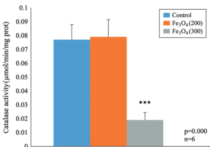
Treatment by 200 and 300μg/kg/day of Fe3O4-NPs
causes a high significant decrease in enzymatic activity of GST, CAT and GPx and SOD respectively, according to the group control(Figures 3, 4, 5 and 6).
trol and treated rabbits brain cortex after 14 days of treatment (p<0.01).
Figure 3. Effect of Fe3O4-NPs on the variation of stromal
GST(μM/mg) after 14 days of per os administration(p<0.01).
Figure 2. Variation of stromal MDA(μM/mg) level in brain
cortex after 14 days of treatment with 200 and 300μg/kg of Fe3O4-NPs(p<0.001).
Figure 4. Effect of Fe3O4-NPs on the variation of stromal CAT
(μM/mg) after 14 days of per os administration(p<0.001).
Figure 5. Effect of Fe3O4-NPs treatments on the variation of
stromal GPx(μM/mg) after 14 days of per os administration (p<0.001).
Effect of Fe3O4-NPs on TNF-α, Caspase-3 of Brain Cortex
Impact of Fe3O4-NPs on TNF-α and caspase-3 of
brain cortex homogenates is shown in the Figures 7 and 8, it is clear that the two doses of iron nanoparticle increase the level of these biomarkers significantly (p<0.01).
Mitochondrial Assays
Effects of Fe3O4-NPs on brain cortex mitochondrial
swelling, permeability and respiration are shown in the Figures 9, 10 and 11 it is clear that the two doses of iron nanoparticle increase the mitochondrial swelling by the increase of its permeability significantly(p<
0.01).
Mitochondrial Respiration was also influenced in dose-dependence manner; it shows a clear decrease of this parameter(Figure 11).
Figure 9. Effect of Fe3O4 at 200 and 300μg/kg on brain
cor-tex mitochondria swelling at OD=540nm after 14 days of per os administration(p<0.001).
Figure 10. Effect of Fe3O4 at 200 and 300μg/kg on brain
cor-tex mitochondria permeability after 14 days of per os adminis-tration(p<0.001). 0.04 0.03 0.02 0.01 0 Addition of Ca++ Control Fe3O4(300) Fe3O4(200) OD. (540 nm) Time(second) -50 0 50 100 150 200
Figure 6. Effect of Fe3O4 at 200 and 300μg/kg on stromal
superoxide dismutase(SOD) activity(U/mg prot) after 14 days of per os administration(p<0.001).
Figure 7. Effect of Fe3O4 at 200 and 300μg/kg on brain
corti-cal TNF-α level(pg/mL) after 14 days of per os administration (p<0.001).
Figure 8. Effect of Fe3O4 at 200 and 300μg/kg on brain
corti-cal caspase-3 activity(OD at 405nm) after 14 days of per os administration(p<0.001).
Mitochondrial Swelling
See the Figure 9.
Mitochondrial Permeability
See the Figure 10.
Mitochondrial Respiration
See the Figure 11.
Discussion and Conclusions
Iron oxide nanoparticles are very interesting; they are used in cell labeling1, drug targeting2, gene delivery3,
biosensors, hyperthermia therapy and as contrast agents in magnetic resonance imaging4-6. However, their
bio-logical reactivity can be enhanced and can lead to po-tential toxic interactions. In this study, the administra-tion per-os of two doses(200 and 300μg/kg) of Fe3O4
-NPs to the male rabbits caused very large perturbation on enzymatic, redox state and mitochondrial parame-ters.
Treatment by these two doses decreased significantly the amount of stromal GSH concordant to the results of13,14 and increased the MDA level according to the
controls, this due by the increased utilization of GSH to scavenge the ROS and by consequence increase the oxidative stress and increase the MDA level by the enhancement of lipid degradation, these results are in concord with the results of15 Rouabhi et al.(2015).
Garcia et al.(2011)16 found that the Iron oxide
nano-particles inhibit the development of some laboratory test models by the influence on general metabolism
and an increased oxidative stress state.
Caspase-3 is a caspase protein that interacts with cas-pase-8 and caspase-9. It is encoded by the CASP3 gene.
CASP3 orthologs have been identified in numerous
mammals for which complete genome data are avail-able. The CASP3 protein is a member of the cysteine- aspartic acid protease(caspase) family19,20. Sequential
activation of caspases plays a central role in the execu-tion-phase of cell apoptosis. Caspases exist as inactive proenzymes that undergo proteolytic processing at con-served aspartic residues to produce two subunits, large and small, that dimerize to form the active enzyme. This protein cleaves and activates caspases 6 and 7; and the protein itself is processed and activated by caspases 8, 9, and 10. It is the predominant caspase involved in the cleavage of amyloid-beta 4A precursor protein, which is associated with neuronal death in Alzheimer’s disease. Caspase-3 is activated in the apoptotic cell both by extrinsic(death ligand) and intrinsic(mitochondrial) pathways21,22. The zymogen feature of caspase-3 is
ne-cessary because if unregulated, caspase activity would kill cells indiscriminately23.
Increasing level of ROS induces a significant eleva-tion of caspase-3 level which induced an apoptotic state in neurons, this results is confirmed by the elevated amount of TNF-α that stimulate the activation of cas-pase-3 and the apoptotic pathway, this is confirmed by the work of Zhao et al.(2001)24 and Utaisincharoen et
al.(2000)25.
TNF-α with TNFR stimulate and activate NF-κB by the inhibition of a protein, IκBα, that normally binds to NF-κB and inhibits its translocation, is phosphorylated by IKK and subsequently degraded, releasing NF-κB. NF-κB is a heterodimeric transcription factor that trans-locate to the nucleus and mediates the transcription of a vast array of proteins involved in inflammatory re-sponse and pro-apoptotic. Our results confirmed that the treatment by 200 and 300μg/kg of Fe3O4-NPs
in-duce an elevated oxidative stress state followed by an apoptosis confirmed by a high mitochondrial swelling and permeability, that make us thought about the exo- diffusion of the cytochrome B to the neuronal cytoplasm because of the high permeability, following by the
ex-Figure 11. Effect of Fe3O4 at 200 and 300μg/kg on brain
cor-tex mitochondria respiration(nmoles) after 14 days of per os administration(p<0.001).
ecution of the apoptotic phenomenon, these results are in concord with the results of Yang et al.(2006)26 and
Zhang et al.(2011)27. This inflammation and cell death
conducted to the neurodegenerative disease like Alz-heimer.
Fe3O4 with two doses and 14 days of treatment caused
a decrease in the mitochondrial respiration probably by the direct effect on the mitochondria that confirm our results about the swelling and the permeability increase. The dysfunction of mitochondria results a decrease amount of oxygen a substrate of monooxygenases and influence directly on the activity of oxidative stress enzymes this finding is in concordance with those of Baratli et al.(2014)28.
In conclusion: IONPs are very used in biomedical field, this work showed a toxic effect of these nanopar-ticles on mitochondrial enzymes of brain cortex and induced many effects conducting to the death of cells (neurons) and cause a neurodegenerative disease as Alzheimer.
Materials and Methods
Chemicals
Fe3O4-NPs nanopowder(<50nm average particle
size, characterized by TEM), bovine serum albumin (BSA), Bradford reagent, collagenase type I, Dulbecco’s modified eagle’s medium(DMEM) and Fetal bovine sera(FBS), 1-Chloro-2,4-dinitrobenzene(CDNB), di-methyl sulphoxide(DMSO), 5,50-dithiobis-(2-nitro-benzoic acid) [DTNB, (Ellman’s reagent)], ethylene diamine tetra acetic acid(EDTA), 20,70-dichlorofluo-rescein diacetate, glacial acetic acid, trichloroacetic acid(TCA), hydrogen peroxide(H2O2),
N-ethylmalei-mide(NEM), nicotinamide adenine dinucleotide re-duced disodium salt(NADH), nitro blue tetrazolium chloride(NBT), phenazine 116 methosulphate(PMT), potassium dihydrogen phosphate 117(KH2PO4),
re-duced glutathione(GSH) and thiobarbituric acid(TBA), were provided from toxicology laboratory, Tebessa University, Algeria.
Animals
Male rabbits strain Oryctolagus cuniculus body weight 1.5-1.9kg was used in this study. They have been housed individually in stainless metal cages dur-ing an adaptation period of 14 days in a controlled tem-perature/humidity/photoperiod room(22±2°C; 50%; 12h dark/light cycle). The feeding of rabbits was based on specific artificial diet for rabbits, and they accessed water freely.
Treatment Protocol
Rabbits were divided in 03 lots of 06 individuals each: Controls(no treatments), treated with Fe3O4-NPs at
200μg/kg/day, treated with Fe3O4-NPs at 300μg/kg/day.
All treatments are per os(p. o.) way, for 14 days and carried out with conformity to the international guide-lines for the care and use of laboratory animals.
After 14 days of treatment, the rabbits were sacrificed and the brains were picked up, (liver and kidney were recovered, weighed and preserved for other assays) and mitochondrial extraction were proceeding. Some biochemical metabolites(proteins, carbohydrates and fats), and some parameters of oxidative stress in the brain cortex mitochondria(GSH, GPx, CAT, MDA, GST, SOD) was investigated crowned by the estima-tion of swelling and respiraestima-tion parameters.
Assay of Antioxidant Enzymes
For this study, SOD activity was measured by follow-ing the method of Kakkar et al.(1984)29. Briefly, cell
lysate containing 5mg protein was mixed with 0.52M sodium pyrophosphate buffer, PMS(186lM), NBT (300lM). The reaction was started by the addition of NADH. The absorbance of the chromogen formed was measured at 560nm. The enzymatic activity of GPx was measured by the method of Flohe and Günzler (1984)30, using H
2O2 as substrate. The
spectrophoto-metric assay of catalase(CAT) activity was performed according to the method of Cakmak and Horst(1991)31.
The decrease of absorbance is recorded for three min-utes by a spectrophotometer at a wavelength of 240nm and an extinction coefficient ε=39400L·μM-1·cm-1.
The activity of glutathione S-transferase(GST) was determined according to the method of Habig et al. (1974)32. It is based on the conjugation reaction
be-tween GST and a substrate, the CDNB(1-Chloro2, 4 dinitrobenzene) as a cofactor of glutathione(GST), the conjugation results in the formation of a new molecule: 1-S-glutathionyl 2-4 Di nitrobenzene to measure the activity of GST.
Assay of Glutathione and Lipid Peroxidation
Glutathione(GSH) level was determined according to the method of Weckbeker and Cory(1988)33. This
assay is based on measuring the absorbance of the 2- nitro-5-mercapturic. The latter results from the reduc-tion of the acide5,5′-dithiobis-2-nitrobenzoic acid (re-agent Elleman) by groups(-SH) of glutathione. Once prepared, must undergo homogenate deproteinization (by 0.25% sulfosalicylic acid) to protect the SH-groups of glutathione.
MDA can be detected by a colorimetric reaction with thiobarbituric acid(TBA). Detecting MDA after degra-dation of polyunsaturated fatty acids 3 or 4 Double
per-The micro ELISA plate provided in this kit has been pre-coated with an antibody specific to Rabbit TNF- alpha. Standards or samples are added to the appropri-ate micro ELISA plappropri-ate wells and bound by the specific antibody. Then a biotinylated detection antibody spe-cific for Rabbit TNF-α and Avidin-Horseradish Perox-idase(HRP) conjugate is added to each micro plate well successively and incubated. Free components are washed away. The substrate solution is added to each well. Only those wells that contain Rabbit TNF-α, biotinylated detection antibody and Avidin-HRP con-jugate will appear blue in color. The enzyme-substrate reaction is terminated by the addition of a sulphuric acid solution and the color turns yellow. The optical density(OD) is measured spectrophotometrically at a wavelength of 450nm±2nm. The OD value is propor-tional to the concentration of Rabbit TNF-α in pg/mL. You can calculate the concentration of Rabbit TNF- alpha in the samples by comparing the OD of the sam-ples to the standard curve.
Assay of Caspase-3 Activity Level
Determination of caspases-3 activity in rabbit brain cortex was performed using commercial kit(Caspase 3 Assay Kit(Colorimetric)(ab39401)) from Abcam tech according to manufacturer guidelines.
Extraction of Mitochondria
All operations were carried on ice. A piece of brain tissue was placed into buffer A containing 50mM tris, 1mM EGTA, 70mM Sucrose, 210mM Mannitol, pH 7.40 at +4°C. Tissues were finely minced with scis-sors, placed in buffer A and homogenized with a Potter- Elvehjem. Then, the homogenate was centrifuged at 1300g for 3min, 4°C. The supernatant was centrifuged at 10,000g for 10min, 4°C to sediment mitochondria. Finally, the mitochondrial pellet was washed twice and then suspended in 50mM Tris, 70mM sucrose, 210 mM mannitol, pH 7.4 at +4°C. Protein content was routinely assayed with a bradford assay using bovine serum albumin as a standard35. Mitochondria were
kept on ice and used within 4h.
Statistical Analysis
The numerical and graphical results are presented as mean 6 standard error(SE). The significance of the Difference between two treatment groups was verified by the Student’s t-test. The degree of statistical signifi-cance was set at a level of p<0.05. Statistical
calcula-tions were carried out using Minitab 17.1 statistical package and the Excel 16.0(Microsoft, Inc.).
Acknowledgements
First, my thanks are addressed to DGRDT of Algeria and Prof. Said Fekra the rector of Tebessa university for their support. This work was achieved in the labo-ratory of toxicology at applied biology department, Tebessa university which I address my gratitude to all personals; my appreciation goes also to the personals of Sfax biotechnology center, Tunisia for the help in TNF-α and caspases quantification.
References
1. Chen, C. L. et al. New Nano-sized Iron Oxide Particle with High Sensitivity for Cellular Magnetic Resonance Imaging. Mol. Imag. Biol. 13, 825-839(2010).
2. Alexiou, C., Tietze, R., Schreiber, E. & Lyer, S. Nano-medicine: Magnetic nanoparticles for drug delivery and hyperthermia-new chances for cancer therapy.
Bundes-gesundheitsblatt Gesundheitsforschung, Gesundheitss-chutz 53, 839-845(2005).
3. McBain, S. C., Yiu, H. H. P. & Dobson, J. Magnetic nanoparticles for gene and drug delivery. Int. J.
Nano-medicine. 3, 169-180(2008).
4. Puppi, J. et al. Use of a Clinically Approved Iron Oxide MRI Contrast Agent to Label Human Hepatocytes.
Cell Transplant. 20, 963-975(2011).
5. Rümenapp, C., Gleich, B. & Haase, A. Magnetic Nano-particles in Magnetic Resonance Imaging and Diagnos-tics. Pharm. Res. 29, 1165-1179(2012).
6. Cochran, D. B. et al. Suppressing iron oxide nanoparti-cle toxicity by vascular targeted antioxidant polymer nanoparticles. Biomaterials 34, 9615-9622(2013).
7. Zhu, M. T. et al. Oxidative stress and apoptosis induced by iron oxide nanoparticles in cultured human umbilical endothelial cells. J. Nanosci. Nanotechnol. 10, 8584-8590(2010).
8. Wilson, M. R., Lightbody, J. H., Donaldson, K., Sales, J. & Stone, V. Interactions between ultrafine particles and transition metals in vivo and in vitro. Toxicol. Appl.
Pharmacol. 184, 172-179(2002).
9. Zhu, M. T. et al. Endothelial dysfunction and inflam-mation induced by iron oxide nanoparticle exposure: risk factors for early atherosclerosis. Toxicol. Lett. 203, 162-171(2011).
10. Remyaa, A. S. et al. Iron oxide nanoparticles to an Indian major carp, Labeo rohita: Impacts on hematolo-gy, iono-regulation and gill Na+/K+ ATPase activity. J. King Saud Univ. Sci. 27, 151-160(2015).
11. DiMauro, S. & Schon, E. A. Mitochondrial disorders in the nervous system. Ann. Rev. Neurosci. 31, 91-123 (2008).
12. Lin, M. T. & Beal, M. F. Mitochondrial dysfunction and oxidative stress in neurodegenerative diseases. Nat. 443, 787-795(2006).
13. Ashley, R. M. et al. Oxidative Stress and Dermal Tox-icity of Iron Oxide Nanoparticles in Vitro. Cell Biochem.
Biophys. 67, 461-476(2013).
14. Henine, S. et al. Oxidative stress status, caspase-3, stromal enzymes and mitochondrial respiration and swelling of Paramecium caudatum in responding to the toxicity of Fe3O4 nanoparticles. Toxicol. Environ. Health. Sci. 8, 161-167(2016).
15. Rouabhi, R., Gasmi, S., Boussekine, S. & Kebieche, M. Hepatic Oxidative Stress Induced by Zinc and Opposite Effect of Selenium in Oryctolagus cuniculus.
J. Environ. Anal. Toxicol. 5, 289(2015).
16. García, A. et al. Acute toxicity of cerium oxide, titanium oxide and iron oxide nanoparticles using standardized tests. Desalination 269, 136-141(2011).
17. Rouabhi, R., Djebar-Berrebbah, H. & Djebar, M. R. Impact of Flufenoxuron, an IGR pesticide on Gallus
domesticus embryonic development in ovo. J. Cell Anim. Biol. 2, 87-91(2008).
18. Lakroun, Z. et al. Oxidative stress and brain mitochon-dria swelling induced by endosulfan and protective role of quercetin in Rat. Environ. Sci. Pollut. Res. Int. 22, 7776-7781(2014).
19. Rouabhi, R., Djebar-Berrebbah, H. & Djebar, M. R. Toxic Effect of a Pesticide, Diflubenzuron on Fresh-water Macroinvertebrate(Tetrahymena pyriformis).
Chinese J. Appl. Environ. Biol. 12, 514-517(2006). 20. Alnemri, E. S. et al. Human ICE/CED-3 protease
no-menclature. Cell 87, 171(1996).
21. Salvesen, G. S. Caspases: opening the boxes and inter-preting the arrows. Cell Death Differ. 9, 3-5(2002). 22. Ghavami, S. et al. Apoptosis and cancer: mutations
within caspase genes. J. Med. Genet. 46, 497-510 (2009).
23. Boatright, K. M. & Salvesen, G. S. Mechanisms of caspase activation. Curr. Opin. Cell Biol. 15, 725-731 (2003).
24. Zhao, X. et al. TNF-alpha stimulates caspase-3 activa-tion and apoptotic cell death in primary septo-hippo-campal cultures. J. Neurosci. Res. 64, 121-131(2001). 25. Utaisincharoen, P., Tangthawornchaikul, N., Ubol, S.,
Chaisuriya, P. & Sirisinha, S. TNF-alpha induces cas-pase 3(CPP 32) dependent apoptosis in human cholan-giocarcinoma cell line. Southeast Asian J. Trop. Med.
Public Health. 31(Suppl. 1), 167-170(2000).
26. Yang, S., Thor, A. D., Edgerton, S. & Yang, X. Caspase- 3 mediated feedback activation of apical caspases in doxorubicin and TNF-alpha induced apoptosis.
Apop-tosis 11, 1987-1997(2006).
27. Zhang, G. et al. Hydroxycamptothecin-Loaded Fe3O4 Nanoparticles Induce Human Lung Cancer Cell Apop-tosis through Caspase-8 Pathway Activation and Disrupt Tight Junctions. Cancer Sci. 102, 1216-1222(2011). 28. Baratli, Y. et al. Age Modulates Fe3O4 Nanoparticles
Liver Toxicity: Dose-Dependent Decrease in Mito-chondrial Respiratory Chain Complexes Activities and Coupling in Middle-Aged as Compared to Young Rats.
BioMed Res. Int. DOI:10.1155/2014/474081(2014). 29. Kakkar, P., Das, B. & Viswanathan, P. A modified
spec-trophotometric assay of superoxide dismutase. Indian
J. Biochem. Biophys. 21, 130-132(1984).
30. Flohe, L. & Gunzler, W. A. Assays of glutathione per-oxidase. Methods Enzymol. 105, 114-121(1984). 31. Cakmak, I. & Horst, W. J. Effect of aluminum on lipid
peroxidation, superoxide dismutase, catalase, and per-oxidase activities in root tips of soybean(Glycine max).
Physiol. Plantarum. 83, 463-468(1991).
32. Habig, H., Pabst, M. J. & Jokoby, W. B. Glutathione- S-transferase: the first enzymatic step in mercapturic acid formation. J. Biol. Chem. 249, 7130-7139(1974). 33. Weckbker, G. & Cory, J. G. Ribonucleotide reductase
activity and growth of Glutathoine-depleted mouse leukemia L1210 cells in vitro. Cancer Lett. 40, 257-264(1988).
34. Esterbaer, H., Gebicki, J., Puhl, H. & Jungens, G. The role of lipid peroxidation and antioxidants in oxidative modification of LDL. Free Radic. Biol. Med. 13, 341 (1992).
35. Bradford, M. A rapid and sensitive method for the quan-tities of microgram quanquan-tities of protein utilizing the principle of protein binding. Anal. Biochem. 72, 248-254(1976)
36. Kristal, B. S., Park, B. K. & Yu, B. P. 4-hydroxynonénal est un puissant inducteur de la transition de perméabilité mitochondriale. J. Biol. Chem. 271, 6033-6038(1996). 37. Rouabhi, R., Djebar, H. & Djebar, M. R. Toxic Effects
of Combined Molecule from Novaluron and Difluben-zuron on Paramecium caudatum. Am-Euras. J. Toxicol.