Phosphorylation of NF-kB and IkB
proteins: implications in cancer and
inflammation
Patrick Viatour, Marie-Paule Merville, Vincent Bours and Alain Chariot
Laboratory of Medical Chemistry and Human Genetics, CHU, Sart-Tilman, Center for Biomedical Integrated Genoproteomics, University of Liege, Belgium
Nuclear factor-kB (NF-kB) is a transcription factor that has crucial roles in inflammation, immunity, cell pro-liferation and apoptosis. Activation of NF-kB mainly occurs via IkB kinase (IKK)-mediated phosphorylation of inhibitory molecules, including IkBa. Optimal induction of NF-kB target genes also requires phosphorylation of NF-kB proteins, such as p65, within their transactivation domain by a variety of kinases in response to distinct stimuli. Whether, and how, phosphorylation modulates the function of other NF-kB and IkB proteins, such as B-cell lymphoma 3, remains unclear. The identification and characterization of all the kinases known to phosphorylate NF-kB and IkB proteins are described here. Because deregulation of NF-kB and IkB phos-phorylations is a hallmark of chronic inflammatory diseases and cancer, newly designed drugs targeting these constitutively activated signalling pathways represent promising therapeutic tools.
Cellular responses to bacterial or viral infections and to stress require rapid and accurate transmission of signals from cell-surface receptors to the nucleus [1]. These signalling pathways rely on protein phosphorylation and, ultimately, lead to the activation of specific transcrip-tion factors that induce the expression of appropriate target genes. Among the activated transcription factors, the nuclear factor-kB (NF-kB) family proteins are essen-tial for inflammation, immunity, cell proliferation and apoptosis. NF-kB exists in a latent state in the cytoplasm and requires a signalling pathway for activation. Such NF-kB-activating pathways are triggered by a variety of extracellular stimuli and lead to the phosphorylation and subsequent proteasome-mediated degradation of inhibi-tory molecules, the inhibitor of NF-kB (IkB) proteins[2]. Activated NF-kB migrates into the nucleus to regulate the expression of multiple target genes. The NF-kB–IkB complex can also shuttle between the cytoplasm and the nucleus in unstimulated cells, but the nuclear export is more efficient and, therefore, the NF-kB–IkB complex is mainly cytoplasmic in resting cells.
Here, we tentatively demonstrate the key role of protein phosphorylation in NF-kB activation. We
summarize the current knowledge in this field, namely all the kinases known to phosphorylate the NF-kB and IkB proteins, including recent data regarding B-cell lym-phoma 3 (BCL-3) phosphorylation. In addition, we illustrate how deregulation of NF-kB and IkB phosphoryl-ation is crucial in inflammphosphoryl-ation and cancer, and suggest potential targets for the design of new and specific therapeutic agents.
Three NF-kB-activating pathways
Three distinct NF-kB-activating pathways have emerged, and all of them rely on sequentially activated kinases (Figure 1). The first pathway –the classical pathway – is triggered by pro-inflammatory cytokines such as tumour necrosis factor (TNF)-a and leads to the sequential recruitment of various adaptors including TNF-associated death domain protein (TRADD), receptor-interacting protein (RIP) and TNF-receptor-associated factor 2 (TRAF2) to the cytoplasmic membrane [3]. This is followed by the recruitment and activation of the classical IkB-kinase (IKK) complex [4], which includes the scaffold protein NF-kB essential modulator (NEMO; also named IKKg)[5], IKKa and IKKb kinases[6]. Once activated, the IKK complex phosphorylates IkBa on Ser32 and Ser36, and is subsequently ubiquitinated and degraded via the proteasome pathway. A second pathway –the alternative pathway – is NEMO-independent and is triggered by cytokines such as lymphotoxin b [7], B-cell activating factor (BAFF) [8] or the CD40 ligand [9] (Figure 1) and by viruses such as human T-cell leukaemia virus[10]and the Epstein–Barr virus[11]. This signalling pathway relies on the recruitment of TRAF proteins to the membrane and on the NF-kB-inducing kinase (NIK)[12], which activates an IKKa homodimer – the substrate of which is the ankyrin-containing and inhibitory molecule p100[13]. Once phosphorylated by IKKa on specific serine residues located in both the N- and C-terminal regions [14], p100 is ubiquitinated and cleaved to generate the NF-kB protein p52, which moves as heterodimer with RelB into the nucleus. In both cases, phosphorylation of the inhibitory molecules is essential for the activation of NF-kB, and this has been demonstrated by the inability of cells expressing an IkBa mutant that does not become phosphorylated to activate NF-kB[15].
Corresponding author: Chariot, A. (Alain.chariot@ulg.ac.be). Available online 7 December 2004
Atypical IkBa phosphorylations for NF-kB activation Most stimuli activate NF-kB by IKK-mediated IkBa phosphorylation on N-terminal serine residues. The third signalling pathway is classified as atypical because it is independent of IKK, still requires the proteasome and is triggered by DNA damage such as UV [16] or doxorubicin [17] (Figure 1). UV radiation induces IkBa degradation via the proteasome, but the targeted serine residues are located within a C-terminal cluster, which is recognized by the p38-activated casein kinase 2 (CK2) [16]. Oxidative stress also leads to NF-kB activation via IkBa tyrosine phosphorylation[18]. The N-terminal Tyr42 residue is crucial for this pathway [19], and the Syk
protein tyrosine kinase seems to be required for H2O2
-mediated NF-kB activation [20]. All of these phosphoryl-ation events are signal-induced. However, IkBa is also constitutively phosphorylated on Ser293 within its C-terminal Pro-Glu-Ser-Thr sequence by CK2, and this phosphorylation is required for rapid proteolysis of IkBa [21–23].
Phosphorylations of other IkB proteins
All these reports convincingly demonstrate that phos-phorylation of IkB proteins such as IkBa and p100 is essential for NF-kB activation. The inhibitory molecule IkBb is also targeted for phosphorylation on Ser19 and
IKKα IKKβ RelB TiBS Nucleus κB site SODD TRADD RIP TRAF2 Ub Ub Ub Ub Ub Ub TRAF2 NEMO Hsp90 P P Hsp90 NIK P IKKα P P p100 Ub Ub Ub Ub Ub Ub P P P IκBα P p65 P NEMO TRAF2 TRADD p65 p50 p50 IκBα p65 p50 p50 P P IκBα p65 p50 P P p65 P p50 p52 P RelB p52 p65 P P P IKKα RelB P Cdc37 Cdc37 P P TRAF6 TRAF3 TNF CD154 TNFR1 P p38 CK2 DNA damage CD40 ELKS P
Figure 1. The classical (blue arrows), alternative (green arrows) and atypical (purple arrows) NF-kB-activating pathways as illustrated by the TNF-a-mediated, CD40-mediated and DNA-damage-mediated NF-kB activation pathways, respectively. In the classical NF-kB-activating pathway, upon binding of TNFa to TNFR1, SODD is released from the receptor and triggers the sequential recruitment of the adaptors TRADD, RIP and TRAF2 to the membrane. Then, TRAF2 mediates the recruitment of the IKK complex – composed of IKKa, IKKb and NEMO – to the TNFR1 signalling complex. Hsp90 and Cdc37 are also part of the IKK complex and are required for the TNFa-induced IKK activation and shuttling of the IKK complex from the cytoplasm to the membrane, and ELKS connects IkBa to the IKK complex[83]. Activation of the IKK complex leads to the phosphorylation of IkBa at specific residues, ubiquitination through binding of ubiquitin proteins and degradation of this inhibitory molecule via the proteasome pathway. Then, the heterodimer p50–p65 is released and migrates to the nucleus where it binds to specific kB sites and activates a variety of NF-kB target genes, including IL-8, IL-6, TNFa and many more. The alternative pathway is triggered by binding of the CD40 ligand to its receptor, leading to recruitment of TRAF proteins and the sequential activation of NIK and IKKa, which then induces the processing of the inhibitory protein p100. p100 proteolysis releases p52 which forms heterodimers with RelB. This pathway is NEMO-independent and relies on IKKa homodimers. The atypical pathway, which is triggered by DNA damage such as UV, relies on sequential p38 and CK2 activations, and involves phosphorylation and subsequent IkBa degradation via an IKK-independent pathway. Subsequently, free NF-kB moves into the nucleus to activate its target genes. Note that the DNA-damaging agent doxorubicin also triggers p65 phosphorylation via a p53- and RSK1-dependent pathway (not shown). Phosphorylation of the signalling molecules in addition to NF-kB and IkB proteins are illustrated. Abbreviations: CK2, casein kinase 2; ELKS, Glu-Leu-Lys-Ser; Hsp90, heat shock protein 90; IkB, inhibitor of NF-kB; IKK, IkB kinase; NEMO, NF-kB essential modulator; NF-kB, nuclear factor-kB; NIK, NF-kB-inducing kinase; RIP, receptor-interacting protein; RSK1, ribosomal S6 kinase 1; SODD, silencer of death domains; TNF-a, tumour necrosis factor a; TNFR1, TNF receptor 1; TRADD, TNF-receptor-associated death domain protein; TRAF, TNF-receptor-associated factor; Ub, ubiquitin.
Ser23 by the IKK complex[24]and this phosphorylation triggers IkBb degradation – similar to IKK-mediated IkB3 phosphorylation of N-terminal serine residues[25].
The ankyrin-containing and inhibitory molecule p105 is also subjected to TNFa and IKK-mediated phosphoryl-ation on Ser927 and Ser932 [26] (Figure 2). This phosphorylation is required for subsequent p105 ubiqui-tination and processing into the NF-kB protein p50[26]. In addition, glycogen synthase kinase 3b (GSK3b) phosphor-ylates p105 on Ser903 and Ser907 and stabilizes p105 by preventing its degradation in unstimulated cells [27]. However, GSK3b-mediated p105 phosphorylation is required to prime IKK-mediated p105 phosphorylation and subsequent degradation upon TNFa treatment[27]. Therefore, GSK3b exerts a dual role towards p105,
depending on whether or not the cells are stimulated [27]. The mitogen-activated kinase kinase kinase (MAP3K) Tpl2 (also named Cot) is another kinase that regulates p105 phosphorylation and subsequent degra-dation [28]. Although Tpl2 fails to phosphorylate p105 in vitro, it might affect p105 phosphorylation via a downstream kinase [28]. Interestingly, p105 interacts and governs Tpl2 stability in the MAP kinase signalling pathway [29]. Lipopolysaccharide (LPS)-mediated Tpl2 activation involves the dissociation of Tpl2 from p105, which requires IKKb but not IKKa [30], and leads to subsequent Tpl2 degradation. Therefore, p105 phos-phorylation is a key event in the LPS-stimulated mito-gen-activated kinase (MAPK)/extracellular signal-related kinase (ERK) kinase (MEK) and ERK signalling cascade.
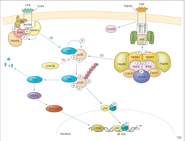
TiBS SODD IRAK p105 Nucleus κB site p50 p65 P CREB p105 P P P P P P P LPS TLR4 MyD88 IRAK4 P P TRAF6 MEK1 ERK1/2 Tollip IKKα IKKβ TRADD RIP TRAF2 TRAF2 NEMO Hsp90 P P Hsp90 NEMO TRADD Cdc37 Cdc37 P P TNF TNFR1 P ELKS P p65 P p50 Ub Ub Ub Ub Ub Ub Tpl2 Tpl2 Tpl2 GSK3β P (1) (2) (3)
Figure 2. The p105-dependent pathways. The p105 inhibitory molecule is a phosphoprotein involved in three signalling pathways. (1) The first (purple arrows) relies on GSK3, which phosphorylates and stabilizes p105 in resting cells. This primo-phosphorylation also triggers p105 processing upon IKK-mediated phosphorylation in stimulated cells. (2) The second (blue arrows) occurs upon binding of TNFa to the TNFR1, which activates the IKK complex by the sequential recruitment of TRADD, RIP and TRAF2 to the membrane. IKKb-mediated phosphorylation of p105 triggers its processing into p50; p50, in turn, moves as a heterodimer with p65 into the nucleus. (3) The third pathway (green arrows) is Tpl2-dependent and occurs through the TLR4 in LPS-stimulated cells. This treatment triggers the phosphorylation of the kinase IRAK by IRAK4 through a Tollip and a MyD88-dependent pathway, and leads to the activation of Tpl2, which phosphorylates its interacting partner p105. Activated Tpl2 activates ERK1/2 through a MEK1 pathway, leading to the binding of the transcriptional factor CREB to the regulatory sequences of its target genes. Tpl2 is quickly degraded once activated. Abbreviations: CREB, cAMP response element-binding; ELKS, Glu-Leu-Lys-Ser; ERK, extracellular signal-related kinase; GSK3, glycogen synthase kinase 3; Hsp90, heat shock protein 90; IkB, inhibitor of NF-kB; IKK, I-kB kinase; IRAK, interleukin-1-receptor-associated kinase; LPS, lipopolysaccharide; MEK, mitogen-activated kinase/ERK kinase; NEMO, NF-kB essential modulator; NF-kB, nuclear factor-kB; RIP, receptor-interacting protein; SODD, silencer of death domains; TLR, Toll-like receptor; TNF, tumour necrosis factor; TNFR1, TNF receptor 1; Tpl2, tumour progression locus-2; TRADD, TNF-receptor-associated death domain protein; TRAF, TNF-receptor-associated factor; Ub, ubiquitin.
Other IkB proteins include BCL-3 and IkBz[31], they harbour ankyrin repeats but are constitutively nuclear, and are described as transcription factors. IkBz is required for the expression of a subset of genes that are activated in Toll-like receptor (TLR) and interleukin (IL)-1 receptor signalling pathways, as judged by the severe impairment of IL-6 production in response to a variety of TLR ligands and to IL-1b in IkBz-deficient cells[32]. Both BCL-3 and IkBz proteins are phosphorylated in vivo, but potential kinases have not yet been characterized. Moreover, it is not clear whether, and how, these phosphorylation events modulate NF-kB activation. However, recent data demon-strate that BCL-3 is constitutively phosphorylated by
GSK3 on two C-terminal residues [33]. GSK3-mediated BCL-3 phosphorylation targets the degradation of BCL-3 via the proteasome pathway and, therefore, limits its expression. This BCL-3 phosphorylation inhibits its oncogenicity and modulates its ability to regulate a subset of target genes [33]. Therefore, phosphorylation is a key mechanism for the regulation of BCL-3 activity.
Optimal NF-kB activation by phosphorylation of p65 Besides the phosphorylation and subsequent degradation of inhibitory molecules, protein kinases are also required for optimal NF-kB activation by targeting functional domains of NF-kB proteins themselves. These additional
PKA PKA PKA IKKα IKKβ NEMO NEMO TNF TNFR1 RNA pol II TFIIE TATA TFIIB TAFs TFIIH TFIIF Initiation complex ERK1 CBP/p300 p38 GSK3 PKCζ K1 CK2 DNA-damaging agents RSK1 p53 TB Ub Ub Ub Ub Ub Ub IκB-α p65 p50 P P IκBα p65 p50 P P IKKα PI3K Akt p65 p50 P MSK1/2 MSK1/2 p65 p50 P TFIIA TBP Nucleus Cytosol P P P TF P TF P TF P TiBS (1) (2) (3) (4) (5) (6)
Figure 3. Modulation of the transcriptional activity of p65 by phosphorylation. Upon stimulation by TNFa, a pro-inflammatory cytokine, the IKK complex is activated (1) and phosphorylates p65 at Ser536. Because IKKa also moves into the nucleus to modulate NF-kB activity[84,85], it is currently unclear whether IKK-mediated p65 phosphorylation occurs in the cytoplasm and/or in the nucleus. TNFa also triggers MSK1-mediated phosphorylation of p65 a Ser276 in the nucleus via an ERK-dependent pathway (2). The kinases TBK1, GSK3 and PKCz also phosphorylate p65 (3) as shown by the defects of TNFa-mediated NF-kB activation in their respective knock-out mice. CK2-mediated p65 phosphorylation also occurs upon TNFa stimulation (4). Finally, treatment with this cytokine also activates the PI3K–Akt signalling pathway, which phosphorylates p65 through a p38 and IKKb-dependent, or an IKKa-dependent, mechanism (5). The DNA-damaging drugs trigger RSK1 activation, the substrates of which include p65 (6). All these p65 phosphorylations enhance its transactivation potential by positively regulating p65 interactions with co-activators such as CBP and p300. NF-kB target genes are subsequently induced following the recruitment of the initiation complex that includes the TATA-binding protein TBP, TFIIA, -B,-E,-F,-H, the TAFs in addition to RNA pol II on the promoter. Note that lymphotoxin b also triggers p65 phosphorylation through a NIK- and IKKa-dependent pathway that is not illustrated in this figure. Arrows indicating the pathways are: brown, pathway 1; dark blue, pathway 2; purple, pathway 3; green, pathway 4; light blue, pathway 5; orange, pathway 6. Abbreviations: CK2, casein kinase 2; CBP, CREB-binding protein; CREB, cAMP response element-binding; ERK, extracellular signal-related kinase; GSK3, glycogen synthase kinase 3; IkB, inhibitor of NF-kB; IKK, I-kB kinase; MSK1, mitogen- and stress-activated protein kinase-1; NEMO, NF-kB essential modulator; NF-kB, nuclear factor-kB; PI3K, phosphatidylinositol 3-kinase; PKA, protein kinase A; PKC, protein kinase C; RNA pol II, RNA polymerase II; RSK1, ribosomal S6 kinase 1; TAF, TBP-associated factor; TBK1, TRAF-associated NF-kB activator (TANK)-binding kinase 1; TNF, tumour necrosis factor; TF, transcription factor; TNFR1, TNF receptor 1; TRAF, TNF-receptor-associated factor; Ub, ubiquitin.
pathways explain why cells lacking kinases such as GSK3b and TRAF-associated NF-kB activator (TANK)--binding kinase 1 (TBK1; also named T2K or NAK) have defects in NF-kB activation despite an unaltered profile of IkBa phosphorylation and degradation in response to TNFa[34,35]. Therefore, it is assumed that both GSK3b and TBK1 kinases target p65 for phosphorylation and enhance its transactivation potential. Indeed, this hypoth-esis has been confirmed experimentally, at least, in vitro [36,37,38]. In the case of GSK3b, it is worth mentioning that the GSK3b-mediated p65 phosphorylation awaits validation by in vivo studies. If such data are confirmed in vivo, this observation indicates that GSK3b is import-ant for the modulation of NF-kB and IkB phosphorylations because at least three proteins of this family (p65, p105 and BCL-3) are phosphorylated by this kinase. Therefore, GSK3b inhibits BCL-3 function by targeting its degra-dation via the proteasome pathway but, by contrast, participates in the functional activation of p65. In other words, GSK3b could favour a rapid NF-kB activation wave by targeting a TNFa- and p65-dependent pathway and limit NF-kB activation in unidentified BCL-3-dependent pathways.
Numerous other studies have reported the ability of various kinases to phosphorylate p65. These p65 phos-phorylation events occur in the cytoplasm or in the nucleus and are stimuli-specific and, probably, cell-type-specific (Figure 3). In the cytoplasm, the protein kinase PKAc is maintained in an inactive form by binding to IkBa. After stimulus-induced IkBa-degradation, activated PKAc phosphorylates p65 on Ser276 [39]. This phos-phorylation of p65 enhances its ability to recruit histone acetyltransferases such as cAMP response element-bind-ing (CREB)-bindelement-bind-ing protein (CBP) and p300 [40]and to displace p50–histone deacetylase (HDAC)-1 complexes from DNA [41]. Therefore, PKAc-mediated phosphoryl-ation positively regulates the transactivphosphoryl-ation potential of p65[40,41]. Ser276 of p65 is also phosphorylated by the mitogen- and stress-activated protein kinase-1 (MSK1) in the nucleus, and this phosphorylation is required for an optimal TNFa-mediated NF-kB activation [42]. Thus, a single p65 residue is targeted by two kinases in distinct cellular compartments.
Ser311 is another residue within the N-terminal Rel homology domain (RHD) domain that is targeted for phosphorylation by another kinase – protein kinase C (PKC)-z – in TNFa-stimulated cells [43]. Similar to the mechanism described for PKAc, PKCz-mediated phos-phorylation of p65 enhances its interaction with CBP and its recruitment with RNA polymerase II on the IL-6 promoter [43]. As a result, this phosphorylation causes optimal p65 transactivation potential as demonstrated by the defect of NF-kB activation in TNFa-stimulated mouse embryonic fibroblast (MEF) PKCzK/K cells, despite unaltered IKK activation[44].
IL-1b stimulation also triggers p65 phosphorylation within minutes in the cytoplasm and CK2 activity associated with this NF-kB protein has been reported [45]. This cytoplasmic CKII activity is also associated with p65 upon TNFa stimulation and targets Ser529 of this substrate [46]. Moreover, this phosphorylation requires
IkBa degradation and is prevented by p65 binding to IkBa in unstimulated cells[46].
In addition, p65 is phosphorylated at Ser536 by a variety of kinases via various signalling pathways. In most cases, these phosphorylations enhance p65 transac-tivation potential. Upon stimulation by TNFa[47]or the human T-cell lymphotropic virus type 1 Tax protein[48], activation of the IKK complex leads to phosphorylation of p65 at Ser536. Interestingly, whereas TNFa-mediated p65 phosphorylation requires IKKb, the oncoprotein Tax relies on IKKa[48]. IKKa also phosphorylates p65 at Ser536 in lymphotoxin-b-stimulated cells, and this pathway requires the MAP3K NIK[49]. This mechanism, combined with altered p100 processing into p52, accounts for the defect of lymphotoxin-b receptor-induced NF-kB acti-vation in NIK-deficient mice [50]. The IKKb-mediated phosphorylation of p65 also occurs at Ser536 upon T-cell co-stimulation by the T-cell receptor. This requires the upstream kinases Tpl2 and PKCq, but not the phospha-tidylinositol 3-kinase (PI3K)–Akt pathway, and regulates p65 nuclear import [51]. The PI3K-dependent pathway, which involves Akt (also known as protein kinase B), also targets the transactivating domain of p65 upon IL-1 stimulation [52–54]. The identity of the downstream effectors of PI3K and Akt required for this pathway is controversial because both IKKb and p38 have been suggested to be involved[52]. However, the use of IKKa-and/or IKKb-deficient MEF cells suggests that IKKa is the only kinase required downstream of PI3K and Akt for p65 phosphorylation[54]. Most of these p65 phosphorylations occur upon stimulation by pro-inflammatory cytokines, but other molecules such as DNA-damaging agents, in addition to their ability to target IkBa degradation via an IKK-independent pathway [16], also lead to p65 phos-phorylation [55]. Indeed, drugs such as doxorubicin or etoposide activate NF-kB via a p53-dependent pathway that relies on a ribosomal S6 kinase 1-mediated p65 phosphorylation at Ser536[55]. As a result, the affinity of p65 for IkBa and, consequently, the IkBa-mediated nuclear export of NF-kB are reduced[55].
It is likely that other, as yet unidentified, p65 kinases are involved, such as one that phosphorylates p65 at Ser468 upon T-cell co-stimulation [56]. Nevertheless, all the reports to date illustrate a crucial role for p65 phosphorylation in NF-kB activation. It is worth mention-ing, however, that these p65 phosphorylation data were obtained using different cell lines and it is unclear whether all these p65 phosphorylations occur simul-taneously in vivo across different cell types. Moreover, it remains to be experimentally demonstrated whether or not p65 phosphorylation on all these sites is required for optimal NF-kB activity. Careful phenotypic analysis of mice deficient for p65 kinases has indicated that there might be several redundancies in kinase action. Recon-stitution of p65-deficient cells with various p65 proteins harbouring point mutations at their phosphorylable residues is an elegant way to identify the targeted amino acid that is required for the p65 transactivation potential. This experimental strategy has been conducted, and Ser276 of p65 has been identified as the most important
phosphorylated residue for NF-kB-mediated gene acti-vation in TNFa-stimulated MEF cells[57].
Interestingly, although it is now accepted that NF-kB harbours anti-apoptotic properties in most cases, some stimuli such as UV light and chemotherapeutic drugs paradoxically repress NF-kB anti-apoptotic target gene transcription by enhancing the association of p65 with the HDAC proteins[58]. However, this observation cannot be generalized to every cell type and might be cell-type specific. So, how can the same protein (i.e. p65) sometimes activate transcription of a gene but, in other circum-stances, repress transcription of the same gene? The
explanation might come from differential p65 phosphoryl-ations triggered by chemotherapeutic drugs, as compared with the ones triggered by pro-inflammatory cytokines [58], even if Ser536 is targeted in both pathways (as described). Again, precise mapping of the targeted p65 residues will help to better understand how protein phosphorylation can modulate the ability of p65 to activate or repress anti-apoptotic gene expression by recruiting histone acetylases or deacetylases, respectively. Furthermore, phosphorylation of other NF-kB proteins might affect the ability of NF-kB to activate or repress these genes, as demonstrated by the fact that, similarly to
Table 1. Kinases known to phosphorylate the NF-kB and IkB proteinsa,b
Substrates Kinases Residues Location Function Biological stimuli
Refs IkBa IKKb Ser32, Ser36 N-terminal domain Proteasome-mediated degradation TNFa, IL-1b [6]
CK2 Ser293 PEST domain Destabilization Constitutive [21–23]
Syk Tyr42 N-terminal domain H2O2 [20]
CK2 Ser283–Thr299 PEST domain Degradation UV light [16]
IkBb IKKb Ser19, Ser23 N-terminal domain Proteasome-mediated degradation TNFa, IL-1b [24]
IkB3 IKKb Ser18, Ser22 N-terminal domain Proteasome-mediated degradation TNFa, IL-1b [25]
p100 IKKa Ser108, Ser115, Ser123, Ser872
N- and C-terminal domains
Processing into p52 CD40, BAFF, lymphotoxin b
[7–9]
p105 IKKb Ser927, Ser932 PEST domain Processing into p50 TNFa [26]
Tpl2 Indirect phosphorylation
Processing into p50? Unknown [28]
GSK3b Ser903, Ser907 Stabilization Constitutive [27]
BCL-3 GSK3 Ser394, Ser398 C-terminal domain Proteasome-mediated degradation Constitutive [33]
RelA/p65 PKAc Ser276 Rel homology domain Regulation of DNA-binding and oligomerization
LPS [39]
MSK1/2 Ser276 Rel homology domain Enhanced transactivation potential TNFa [42]
PKCz Ser311 Rel homology domain Enhanced transactivation potential TNFa [43]
Unknown Ser468 Unknown T-cell co-stimulation (CD3/CD28)
[51]
CK2 Ser529 C-terminal TAD Enhanced transactivation potential TNFa, IL-1b [45.46]
IKKa Ser536 C-terminal TAD Enhanced transactivation potential HTLV-1 infection, lymphotoxin b
[48.49]
IKKb Ser536 C-terminal TAD Enhanced transactivation potential TNFa, T-cell co-stimulation (CD3/CD28)
[47]
Akt Ser536 C-terminal TAD Enhanced transactivation potential IL-1b [52.53]
RSK1 Ser536 C-terminal TAD Decreased affinity to IkBa DNA-damaging agents
[55]
GSK3b Four sites within amino acids 354–551
C-terminal TAD Enhanced transactivation potential Constitutive [36]
TBK1 Ser536 C-terminal TAD Enhanced transactivation potential IL-1b [37.38]
IKK3 Ser536 C-terminal TAD Enhanced transactivation potential IL-1b [38]
RelB Unknown Ser368 Rel homology domain Dimerization and p100 stabilization [60]
Unknown Thr84, Ser552 Degradation T-cell co-stimulation (CD3/CD28), TPA/ ionomycin
[61]
Crel PI3K/PKC Ser471 C-terminal TAD Enhanced transactivation potential TNFa [64]
PKA-Cb Unknown C-terminal TAD Enhanced transactivation potential Unknown [66]
Unknown Unknown tyrosine
G-CSF [67]
p50 PKAc S337 Rel homology domain Enhanced binding to DNA [69]
aAbbreviations: BAFF, B-cell activating factor; CK2, casein kinase 2; G-CSF, granulocyte-colony stimulating factor; GSK3, glycogen synthase kinase 3; HTLV-1, human T-cell
leukaemia virus type-1; IkB, inhibitor of NF-kB; IKK, I-kB kinase; IL-1b, interleukin-1b; LPS, lipopolysaccharide; MSK, mitogen- and stress-activated protein kinase; NF-kB, nuclear factor-kB; PEST, Pro-Glu-Ser-Thr; PI3K, phosphatidylinositol 3-kinase; PKA, protein kinase A; PKC, protein kinase C; RSK1, ribosomal S6 kinase 1; TBK1, TRAF-associated NF-kB activator (TANK)-binding kinase 1; TNFa, tumour necrosis factor a; TPA, 12-O-tetradecanoylphorbol 13-acetate; Tpl2, tumour progression locus-2; TRAF, TNF-receptor-associated factor.
p65, p52 undergoes the same transcriptional switch from activator to repressor upon induction of endogenous p53 in response to UV light[59].
The crucial role of phosphorylation in the regulation of RelB and c-Rel activity
Multiple residues of the NF-kB protein RelB are phos-phorylated. Ser368 seems to be essential for RelB dimerization and p100 stabilization, but not for RelB nuclear import[60]. However, the kinase that targets this site has not been identified. Phosphorylation of RelB at Thr84 and Ser552 in cells stimulated by 12-O-tetradeca-noylphorbol 13-acetate and ionomycin triggers degra-dation of RelB via the proteasome pathway but, again, the identity of the kinase that phosphorylates RelB at these residues is unknown at present[61]. What are the consequences of these RelB phosphorylations on NF-kB activation via the IKKa-dependent alternative pathway? Which NF-kB target genes are regulated by RelB phosphorylation? Such issues are currently unclear and certainly deserve further investigation.
The role of protein phosphorylation in the regulation of c-Rel activity has been demonstrated by the inability of a v-Rel protein with Ser/Ala substitutions within its C-terminal domain to transform cells[62]. This mutation also abolishes the transactivating and anti-apoptotic abilities of this protein, suggesting that phosphorylation of these serine residues might be required for v-Rel function[63].
In Jurkat cells, a mutation at Ser471 within the cRel transactivating domain abolishes the ability of these cells to respond to TNFa-mediated NF-kB activation [64]. Ser471 seems to be targeted for phosphorylation by a PI3K- and PKC-dependent pathway [64]. Interestingly, additional downstream residues might also be targeted after phorbol myristate acetate and ionomycin stimulation [65]. Moreover, the kinase PKA-Cb enhances c-Rel transactivation potential by direct phosphorylation, but the targeted amino acid remains to be identified [66]. Finally, stimulation of neutrophils with granulocyte-colony-stimulating factor (G-CSF) leads to tyrosine phos-phorylation of c-Rel and might increase its ability to bind DNA, but both the kinase and the phosphorylated residue are unknown[67]. Taken together, these reports demon-strate that protein phosphorylation is crucial for the regulation of the biological properties of c-Rel, namely its transactivation and oncogenic potential. It is, however, not known which of the anti-apoptotic genes that are induced by c-Rel are sensitive to phosphorylation of this protein. p50 and p52 NF-kB subunits are phosphoproteins Although much has been reported regarding the phos-phorylation of p65, and to a lesser extend RelB and c-Rel, there is little information about the phosphorylation of other NF-kB proteins, despite the fact that it has been known for many years that members of the NF-kB family (e.g. p50) are phosphorylated upon cell stimulation[68]. Because p50 lacks a transactivating domain, protein phosphorylation regulates its DNA-binding properties. Indeed, PKA-mediated phosphorylation at Ser337, which is located within the Rel homology domain, enhances the
p50 DNA-binding abilities [64]. So far, this is the only reporting of a potential p50 kinase, although the sequence surrounding the targeted residue also fits the profile of several other kinases such as calmodulin II, CK2 and protein kinase G [69]. Thus, additional experiments are required to address the physiological relevance of these putative p50 phosphorylations. All the kinases known to phosphorylate the NF-kB and IkB proteins are summar-ized inTable 1.
Implications of NF-kB and IkB phosphorylation in inflammation and cancer
Because NF-kB is activated by pro-inflammatory cyto-kines, induces cell proliferation and anti-apoptotic gene expression, and also enhances angiogenesis via vascular endothelial growth factor expression, it is not surprising that aberrant NF-kB activity is a hallmark of cancer and chronic inflammatory diseases. Altered NF-kB activation is caused by deregulated, and often constitutive, NF-kB and IkB phosphorylations, which are major contributors to these diseases. Indeed, constitutive IKK activity and consequently enhanced levels of nuclear p65 have been described in inflammatory diseases[70]and in a variety of solid tumours [71–74]. It is, however, unclear as to whether constitutive p65 phosphorylation is also observed in human cancer cells. Nevertheless, phosphorylation of NF-kB proteins is required for their oncogenicity – as demonstrated for v-Rel[63]– and also for p65 phosphoryl-ation at Ser536 in a model of TNFa-induced transform-ation of mouse epidermal cells [75]. Recently, the role of NF-kB activation in tumour development has been demonstrated using various animal models, including a mouse model of colitis-associated cancer[76]. Deletion of IKKb in intestinal epithelial cells leads to a dramatic decrease in tumour incidence in such models, whereas deletion of IKKb in myeloid cells in these mice results in decreased tumour size by diminished expression of pro-inflammatory cytokines that act as tumour growth factors [76]. These results highlight the ability of IKKb to link inflammation and cancer [76] and provide additional evidence for specific inactivation of NF-kB as a promising tool to attenuate the formation of inflammation-associated tumours. A similar conclusion was based on a mouse model for hepatitis-associated cancer[77]. Mdr2K/Kmice, which spontaneously develop hepatitis and subsequently hepatocellular carcinoma, still develop hepatitis but rarely cancer when NF-kB is specifically inactivated using an IkBa super-repressor transgene[77]. Therefore, IKK-dependent constitutive NF-kB activation is required for tumour development. However, phosphorylation can also negatively regulate the oncogenicity of IkB proteins. Indeed, GSK3-mediated BCL-3 phosphorylation attenu-ates its oncogenic potential by triggering its degradation via the proteasome pathway [33]. Moreover, GSK3 phosphorylation of this oncoprotein affects the ability of BCL-3 to induce a subset of its cancer-relevant target genes such as secretory leucocyte protease inhibitor[33].
Therefore, these aberrant and often constitutive NF-kB and IkB phosphorylations represent promising targets for the treatment of chronic inflammatory diseases and cancer. Selective IKK inhibitors, such as BMS-345541,
have been generated and have shown anti-inflammatory activities in vivo that make them, potentially, efficient drugs for rheumatoid arthritis[78]. Regarding the treat-ment of cancer, effective drugs for acute leukaemia, such as the pyrimidine analogue cytosine arabinoside, induces apoptosis in treated cells, and the underlying mechanism involves the activation of the protein phosphatases 2A and 2B-A and the subsequent p65 dephosphorylation[79,80]. Preventing NF-kB and IkB phosphorylation is also possible with the administration of a cell-permeable peptide that disrupts the interaction between a kinase and its scaffold protein, making the kinase non functional [81]. Such a peptide that targets the interaction between IKKb and the scaffold protein NEMO inhibits cytokine-induced NF-kB activation and shows promising effects in two models of acute inflammation, namely the phorbol-12-myristate-13-acetate-induced ear edaema and the zymo-san-induced peritonitis[81]. Because of the important role of NF-kB in osteoclast differentiation, blocking the activation of this transcription factor is also a good strategy for prevention of inflammatory bone resorption. Therefore, the cell-permeable peptide has also been tested in models of chronic inflammatory diseases involving bone resorption and it does, indeed, inhibit RANKL (receptor activator of NF-kB ligand)-stimulated NF-kB activation and osteoclastogenesis in vivo[82]. Moreover, this peptide abrogates joint swelling and reduces destruction of bone and cartilage by lowering levels of TNFa and IL-1b in the same experimental model[82]. Although other strategies such as proteasome inhibition have been developed to block NF-kB activation and have already demonstrated their efficiency in clinical trials, the inhibition of NF-kB-activating kinases might be more specific and, therefore, could generate fewer side effects. However, deciphering the correct biological and pathophysiological roles of each kinase is required to test these novel drugs in the most appropriate settings and, therefore, to reduce the risk of trial failures.
Future directions
Although many phosphorylation sites on NF-kB proteins have been characterized, it is still unclear how these phosphorylations regulate the ability of such proteins to induce or to repress defined target genes. The answers might come from the use of knock-in experiments in which a mouse expressing mutant NF-kB proteins that lack key phosphorylation sites is generated. Phenotypic analysis of these mice would provide a powerful biological model to address the regulation of NF-kB protein activities by phosphorylation in vivo.
Acknowledgements
A.C. and M-P.M. are Research Associates at the Belgian National Funds for Research. A.C. is supported by a grant from the Belgian Federation against Cancer and TELEVIE. The members of the laboratory are also supported by grants from the Belgian Funds for Research (FNRS) and the Centre anti-cance´reux. We thank E. Dejardin for helpful discussions.
References
1 Karin, M. and Hunter, T. (1995) Transcripional control by protein phosphorylation: signal transmission from the cell surface to the nucleus. Curr. Biol. 5, 747–757
2 Karin, M. and Ben-neriah, Y. (2000) Phosphorylation meets ubiqui-tination: the control of NF-kB activity. Annu. Rev. Immunol. 18, 621–663
3 Hsu, H. et al. (1995) The TNF receptor 1-associated protein TRADD signals cell death and NF-kB activation. Cell 81, 495–504
4 Devin, A. et al. (2000) The distinct roles of TRAF2 and RIP in IKK activation by TNF-R1: TRAF2 recruits IKK to TNF-R1 while RIP mediates IKK activation. Immunity 12, 419–429
5 Yamaoka, S. et al. (1998) Complementation cloning of NEMO, a component of the I kB kinase complex essential for NF-kB activation. Cell 93, 1231–1240
6 Zandi, E. et al. (1997) The IkB kinase complex (IKK) contains two kinase subunits, IKKa and IKKb, necessary for IkB phosphorylation and NF-kB activation. Cell 91, 243–252
7 Dejardin, E. et al. (2002) The lymphotoxin-b receptor induces different patterns of gene expression via two NF-kB pathways. Immunity 17, 525–535
8 Claudio, E. et al. (2002) BAFF-induced NEMO-independent proces-sing of NF-kB2 in maturing B cells. Nat. Immunol. 3, 958–965 9 Coope, H.J. et al. (2002) CD40 regulates the processing of NF-kB2
p100 to p52. EMBO J. 21, 5375–5385
10 Xiao, G. et al. (2001) Retroviral oncoprotein Tax induces processing of NF-kB2/p100 in T cells: evidence for the involvement of IKKa. EMBO J. 20, 6805–6815
11 Eliopoulos, A.G. et al. (2003) Epstein-Barr virus-encoded latent infection membrane protein 1 regulates the processing of p100 NF-kB2 to p52 via an IKKg/NEMO-independent signalling path-way. Oncogene 22, 7557–7569
12 Xiao, G. et al. (2001) NF-kB-inducing kinase regulates the processing of NF-kB2 p100. Mol. Cell 7, 401–409
13 Senftleben, U. et al. (2001) Activation by IKKa of a second, evolutionary conserved, NF-kB signaling pathway. Science 293, 1495–1499
14 Xiao, G. et al. (2004) Induction of p100 processing by NF-kB-inducing kinase involves docking IkB kinase a (IKKa) to p100 and IKKa-mediated phosphorylation. J. Biol. Chem. 279, 30099–30105 15 Brown, K. et al. (1995) Control of IkB-ampha proteolysis by
site-specific, signal induced phopshorylation. Science 267, 1485–1488 16 Kato, T. et al. (2003) CK2 is a C-terminal IkB kinase responsible for
NF-kB activation during the UV response. Mol. Cell 12, 829–839 17 Tergaonkar, V. et al. (2003) IkB kinase-independent IkBa degradation
pathway: functional NF-kB activity and implications for cancer therapy. Mol. Cell. Biol. 23, 8070–8083
18 Imbert, V. et al. (1996) Tyrosine phosphorylation of IkB-a activates NF-kB without proteolytic degradation of IkB-a. Cell 86, 787–798 19 Schoonbroodt, S. et al. (2000) Crucial role of the amino-terminal
tyrosine residue 42 and the carboxyl-terminal PEST domain of IkBa in NF-kB activation by an oxidative stress. J. Immunol. 164, 4292–4300 20 Takada, Y. et al. (2003) Hydrogen peroxide activates NF-kB through tyrosine phosphorylation of IkBa and serine phosphorylation of p65: evidence for the involvement of IkBa kinase and Syk protein-tyrosine kinase. J. Biol. Chem. 278, 24233–24241
21 Lin, R. et al. (1996) Phosphorylation of IkBa in the C-terminal PEST domain by casein kinase II affects intrinsic protein stability. Mol. Cell. Biol. 16, 1401–1409
22 McElhinny, J.A. et al. (1996) Casein kinase II phosphorylates IkBa at S-283, S-289, S-293, and T-291 and is required for its degradation. Mol. Cell. Biol. 16, 899–906
23 Schwarz, E.M. et al. (1996) Constitutive phosphorylation of IkBa by casein kinase II occurs preferentially at serine 293: requirement for degradation of free IkBa. Mol. Cell. Biol. 16, 3554–3559
24 Wu, C. and Ghosh, S. (2003) Differential phosphorylation of the signal-responsive domain of IkBa and IkBb by IkB kinases. J. Biol. Chem. 278, 31980–31987
25 Whiteside, S.T. et al. (1997) IkB3, a novel member of the IkB family controls RelA and cRel NF-kB activity. EMBO J. 16, 1413–1426 26 Lang, V. et al. (2003) bTrCP-mediated proteolysis of NF-kB1 p105
requires phosphorylation of p105 serines 927 and 932. Mol. Cell. Biol. 23, 402–413
27 Demarchi, F. et al. (2003) Glycogen synthase kinase-3b regulates NF-kB1/p105 stability. J. Biol. Chem. 278, 39583–39590
28 Belich, M.P. et al. (1999) Tpl-2 kinase regulates the proteolysis of the NF-kB-inhibitory protein NF-kB1 p105. Nature 397, 363–368
29 Waterfield, M.R. et al. (2003) NF-kB1/p105 regulates lipopolysacchar-ide-stimulated MAP kinase signaling by governing the stability and function of the Tpl2 kinase. Mol. Cell 11, 685–694
30 Waterfield, M. et al. (2004) IkB kinase is an essential component of the Tpl2 signaling pathway. Mol. Cell. Biol. 24, 6040–6048
31 Yamazaki, S. et al. (2001) A novel IkB protein, IkB-z, induced by proinflammatory stimuli negatively regulates nuclear factor-kB in the nuclei. J. Biol. Chem. 276, 27657–27662
32 Yamamoto, M. et al. (2004) Regulation of Toll/IL-1-receptor-mediated gene expression by the inducible nuclear protein IkBz. Nature 430, 218–222
33 Viatour, P. et al. (2004) GSK3-mediated BCL-3 phosphorylation modulates its degradation and its oncogenicity. Mol. Cell 16, 35–45 34 Bonnard, M. et al. (2000) Deficiency of T2K leads to apoptotic liver
degeneration and impaired NF-kB-dependent gene transcription. EMBO J. 19, 4976–4985
35 Hoeflich, K.P. et al. (2000) Requirement for glycogen synthase kinase-3b in cell survival and NF-kB activation. Nature 406, 86–90 36 Schwabe, R.F. and Brenner, D.A. (2002) Role of glycogen synthase-3
kinase in TNF-a-induced NF-kB activation and apoptosis in hepato-cytes. Am. J. Physiol. Gastrointest. Liver Physiol. 283, G204–G211 37 Fujita, F. et al. (2003) Identification of NAP1, a regulatory subunit of
IkB kinase-related kinases that potentiates NF-kB signalling. Mol. Cell. Biol. 23, 7780–7793
38 Buss, H. et al. Constitutive and IL-1-inducible phosphorylation of p65 NF-kB at serine 536 is mediated by multiple protein kinases including IKKa, IKKb, IKK3, TBK1 and an unknown kinase and couples p65 to TAFII31-mediated IL-8 transcription. J. Biol. Chem. (in press) 39 Zhong, H. et al. (1997) The transcriptional activity of NF-kB is
regulated by the IkB-associated PKAc subunit through a cyclic AMP-independent mechanism. Cell 89, 413–424
40 Zhong, H. et al. (1998) Phosphorylation of NF-kB p65 by PKA stimulates transcriptional activity by promoting a novel bivalent interaction with the coactivator CBP/p300. Mol. Cell 1, 661–671 41 Zhong, H. et al. (2002) The phosphorylation status of nuclear NF-kB
determines its association with CBP/p300 or HDAC-1. Mol. Cell 9, 625–636
42 Vermeulen, L. et al. (2003) Transcriptional activation of the NF-kB p65 subunit by mitogen- and stress-activated protein kinase-1. EMBO J. 22, 1313–1324
43 Duran, A. et al. (2003) Essential role of RelA Ser311 phosphorylation by zetaPKC in NF-kB transcriptional activation. EMBO J. 22, 3910–3918
44 Leitges, M. et al. (2001) Targeted disruption of the PKCz gene results in the impairment of the NF-kB pathway. Mol. Cell 8, 771–780 45 Bird, T.A. et al. (1997) Activation of nuclear transcription factor
NF-kB by interleukin-1 is accompanied by casein kinase II-mediated phosphorylation of the p65 subunit. J. Biol. Chem. 272, 32606–32612 46 Wang, D. et al. (2000) Tumor necrosis factor a-induced phosphoryl-ation of RelA/p65 on Ser529 is controlled by casein kinase II. J. Biol. Chem. 275, 32592–32597
47 Sakurai, H. et al. (1999) IkB kinases phosphorylate NF-kB p65 subunit on serine 536 in the transactivation domain. J. Biol. Chem. 274, 30353–30356
48 O’Mahony, A.M. et al. (2004) Human T-cell lymphotropic virus type 1 tax induction of biologically Active NF-kB requires IkB kinase-1-mediated phosphorylation of RelA/p65. J. Biol. Chem. 279, 18137–18145
49 Jiang, X. et al. (2003) The NF-kB activation in lymphotoxin b receptor signalling depends on the phosphorylation of p65 at serine 536. J. Biol. Chem. 278, 919–926
50 Yin, L. et al. (2001) Defective lymphotoxin-b receptor-induced NF-kB transcriptional activity in NIK-deficient mice. Science 291, 2162–2165 51 Mattioli, I. et al. (2004) Transient and selective NF-kB p65 serine 536 phosphorylation induced by T cell costimulation is mediated by IkB kinase b and controls the kinetics of p65 Nuclear Import. J. Immunol. 172, 6336–6344
52 Madrid, L.V. et al. (2001) Akt stimulates the transactivation potential of the RelA/p65 Subunit of NF-kB through utilization of the IkB kinase and activation of the mitogen-activated protein kinase p38. J. Biol. Chem. 276, 18934–18940
53 Sizemore, N. et al. (1999) Activation of phosphatidylinositol 3-kinase in response to interleukin-1 leads to phosphorylation and activation of the NF-kB p65/RelA subunit. Mol. Cell. Biol. 19, 4798–4805 54 Sizemore, N. et al. (2002) Distinct roles of the IkB kinase a and b
subunits in liberating nuclear factor kB (NF-kB) from IkB and in phosphorylating the p65 subunit of NF-kB. J. Biol. Chem. 277, 3863–3869
55 Bohuslav, J. et al. (2004) p53 induces NF-kB activation by an IkB kinase-independent mechanism involving phosphorylation of p65 by ribosomal S6 kinase 1. J. Biol. Chem. 279, 26115–26125
56 Schmitz, M.L. et al. (2004) A comparative analysis of T cell costimulation and CD43 activation reveals novel signaling pathways and target genes. Blood 104, 3302–3304
57 Okazaki, T. et al. (2003) Phosphorylation of serine 276 is essential for p65 NF-kB subunit-dependent cellular responses. Biochem. Biophys. Res. Commun. 300, 807–812
58 Campbell, K.J. et al. (2004) Active repression of anti-apoptotic gene expression by RelA(p65) NF-kB. Mol. Cell 13, 853–865
59 Rocha, S. et al. (2003) p53 represses Cyclin D1 transcription through down regulation of Bcl-3 and inducing increased association of the p52 NF-kB subunit with histone deacetylase 1. Mol. Cell. Biol. 23, 4713–4727
60 Maier, H.J. et al. (2003) Critical role of RelB serine 368 for dimerization and p100 stabilization. J. Biol. Chem. 278, 39242–39250 61 Marienfeld, R. et al. (2001) Signal-specific and phosphorylation-dependent RelB degradation: a potential mechanism of NF-kB control. Oncogene 20, 8142–8147
62 Chen, C. et al. (1999) Mapping of a serine-rich domain essential for the transcriptional, antiapoptotic, and transforming activities of the v-Rel oncoprotein. Mol. Cell. Biol. 19, 307–316
63 Rayet, B. et al. (2003) Mutations in the v-Rel transactivation domain indicate altered phosphorylation and identify a subset of NF-kB-regulated cell death inhibitors important for v-Rel transforming activity. Mol. Cell. Biol. 23, 1520–1533
64 Martin, A.G. and Fresno, M. (2000) Tumor necrosis factor-a activation of NF-kB requires the phosphorylation of Ser-471 in the transactiva-tion domain of c-Rel. J. Biol. Chem. 275, 24383–24391
65 Martin, A.G. et al. (2001) Regulation of nuclear factor kB transactiva-tion. Implication of phosphatidylinositol 3-kinase and protein kinase Cz in c-Rel activation by tumor necrosis factor a. J. Biol. Chem. 276, 15840–15849
66 Yu, S.H. et al. (2004) Stimulation of c-Rel transcriptional activity by PKA catalytic subunit b. J. Mol. Med. 82, 621–628
67 Druker, B.J. et al. (1994) Rel is rapidly tyrosine-phosphorylated following granulocyte-colony stimulating factor treatment of human neutrophils. J. Biol. Chem. 269, 5387–5390
68 Hayashi, T. et al. (1993) Identification of a new serine kinase that activates NF-kB by direct phosphorylation. J. Biol. Chem. 268, 26790–26795
69 Hou, S. et al. (2003) Phosphorylation of serine 337 of NF-kB p50 is critical for DNA binding. J. Biol. Chem. 278, 45994–45998
70 Marok, R. et al. (1996) Activation of the transcription factor nuclear factor-kB in human inflamed synovial tissue. Arthritis Rheum. 39, 583–591
71 Romieu-Mourez, R. et al. (2001) Roles of IKK kinases and protein kinase CK2 in activation of nuclear factor-kB in breast cancer. Cancer Res. 61, 3810–3818
72 Biswas, D.K. et al. (2004) NF-kB activation in human breast cancer specimens and its role in cell proliferation and apoptosis. Proc. Natl. Acad. Sci. U. S. A. 101, 10137–10142
73 Gasparian, A.V. et al. (2002) The role of IKK in constitutive activation of NF-kB transcription factor in prostate carcinoma cells. J. Cell Sci. 115, 141–151
74 Robe, P.A. et al. (2004) In vitro and in vivo activity of the nuclear factor-kB inhibitor sulfasalazine in human glioblastomas. Clin. Cancer Res. 10, 5595–5603
75 Hu, J. et al. (2004) Insufficient p65 phosphorylation at S536 specifically contributes to the lack of NF-kB activation and transformation in resistant JB6 cells. Carcinogenesis 25, 1991–2003
76 Greten, F.R. et al. (2004) IKKb links inflammation and tumorigenesis in a mouse model of colitis-associated cancer. Cell 118, 285–296
77 Pikarsky, E. et al. (2004) NF-kB functions as a tumor promoter in inflammation-associated cancer. Nature 431, 461–466
78 McIntyre, K.W. et al. (2003) A highly selective inhibitor of IkB kinase, BMS-345541, blocks both joint inflammation and destruc-tion in collagen-induced arthritis in mice. Arthritis Rheum. 48, 2652–2659
79 Sreenivasan, Y. et al. (2003) Mechanism of cytosine arabinoside-mediated apoptosis: role of Rel A (p65) dephosphorylation. Oncogene 22, 4356–4369
80 Yang, J. et al. (2001) Protein phosphatase 2A interacts with and directly dephosphorylates RelA. J. Biol. Chem. 276, 47828–47833 81 May, M.J. et al. (2000) Selective inhibition of NF-kB activation by a
peptide that blocks the interaction of NEMO with the IkB kinase complex. Science 289, 1550–1553
82 Jimi, E. et al. (2004) Selective inhibition of NF-kB blocks osteoclas-togenesis and prevents inflammatory destruction in vivo. Nat. Med. 10, 617–624
83 Ducut Sigala, J.L. et al. (2004) Activation of transcription factor NF-kB requires ELKS, an INF-kB kinase regulatory subunit. Science 304, 1963–1967
84 Anest, V. et al. (2003) A nucleosomal function for IkB kinase-a in NF-kB-dependent gene expression. Nature 423, 659–663
85 Yamamoto, Y. et al. (2003) Histone H3 phosphorylation by IKK-a is critical for cytokine-induced gene expression. Nature 423, 655–659
Endeavour
the quarterly magazine for the history and philosophy of science
You can access Endeavour online via ScienceDirect, where you’ll find a collection of beautifully illustrated articles on the history of science, book
reviews and editorial comment.
Featuring
Sverre Petterssen and the Contentious (and Momentous) Weather Forecasts for D-Day, 6 June 1944 by J.R. Fleming Food of Paradise: Tahitian breadfruit and the Autocritique of European Consumption by P. White and E.C. Spary
Two Approaches to Etiology: The Debate Over Smoking and Lung Cancer in the 1950s by M. Parascandola Sicily, or sea of tranquility? Mapping and naming the moon by J. Vertesi
The Prehistory of the Periodic Table by D. Rouvray Two portraits of Edmond Halley by P. Fara
and coming soon
Fighting the ‘microbe of sporting mania’: Australian science and Antarctic exploration in the early twentieth century by P. Roberts
Learning from Education to Communicate Science as a Good Story by A. Negrete and C. Lartigue The Traffic and Display of Body Parts in the Early-19th Century by S. Alberti and S. Chaplin
The Rise, Fall and Resurrection of Group Selection by M. Borrello Pomet’s great ‘‘Compleat History of Drugs’’ by S. Sherman
Sherlock Holmes: scientific detective by L. Snyder The Future of Electricity in 1892 by G.J.N. Gooday
The First Personal Computer by J. November Baloonmania: news in the air by M.G. Kim
and much, much more . . .